DEVELOPMENT OF A COLORIMETRIC METHOD FOR THE
DOSAGE OF OI
-ANIONS AND I
2IN AQUEOUS MEDIA
F. BAFORT*, J.P. BARTHELEMY**, O. PARISI*, J.P. PERRAUDIN***,
H. M. JIJAKLI*
* Plant Pathology Laboratory, Liège University, Gembloux Agro-Bio Tech, Passage des Déportés 2, 5030 Gembloux, Belgium
** Analytical Chemistry Laboratory, Liège University, Gembloux Agro-Bio Tech, Passage des Déportés 2, 5030 Gembloux, Belgium
*** Taradon Laboratory, Avenue Léon Champagne 2, 1480 Tubize, Belgium
ABSTRACT
Lactoperoxidase catalyzes the oxidation of thiocyanate (SCN-) and iodide (I-) in presence of hydrogen peroxide in hypothiocyanite (OSCN-) ions and, depending on the pH, in hypoiodite (OI-) ions or in iode (I2). Oxidized SCN -and I- are part of the lactoperoxidase system, which is a natural biological protection in cow milk, and are described as having inhibitory properties against pathogenic human bacteria, fungi and viruses. We have developed an aqueous solution containing only OSCN- and OI- ions (without the enzyme) and we tested it successfully against plant pathogens. In order to characterize this new soft chemical control against plant pathogens we had to determine the concentration of OSCN- and OI- ions. The dosage of OSCN -consists in a well referenced colorimetric method but no procedure is described for the determination of OI- ions. We have thus developed an easy method, based on the oxidation of the amine moiety of 3,3′,5,5′-tetramethylbenzidine (TMB) by OI- or I2 in a strongly absorbing blue product for the detection and dosage of both molecules. Interestingly the OSCN- ions are not able to oxidize TMB and render this method specific to enzymatic oxidized iodide. We have calculated its sensitivity, repeatability and linearity. This method could also be used for the determination of OCl- and OBr- ions produced during the enzymatic oxidation of chloride and bromide by mammalian’s peroxidases.
INTRODUCTION
Peroxidases are oxidoreductase enzymes that are part of the non-immune natural defense system described in milk and exocrine secretions such as saliva, tears, airway mucosa or intestinal secretions [1]. Peroxidases have no antibacterial activity by themselves, but exerted their activity in presence of cofactors which are hydrogen peroxide H202, and a second ion, being the pseudohalogen thiocyanate ion (SCN-), or a halogen chloride ion (CI-), bromide ion (Br-), or iodide ion (I-) depending on the peroxidase [2]. Lactoperoxidase, present in cow milk, catalyzes the oxidation of SCN- and I -in presence of H202 in hypothiocyanite (OSCN-) ions and, depending on the pH, in hypoiodite (OI-) ions or in iode (I2) [2, 3]. These molecules are described as having inhibitory properties against pathogenic human bacteria, fungi and viruses [2]. As we demonstrated the efficacy of OSCN -and OI- against plant bacteria and fungi, we needed to characterize the two molecules for agricultural applications and homologation procedures. The dosage method for OSCN- is known but no one is described for the determination of OI-.
3,3′,5,5′-tetramethylbenzidine (TMB) and dimethyl sulfoxide (DMSO) were purchased from Sigma-Aldrich. Hydrogen peroxide 35%, iode 0.5 M, sodium acetate, acetic acid, potassium thiocyanate and potassium iodide were purchased from VWR. Aluminium polychlorosulfate (Pac Sachtoklar) was purchased from Brenntag. Green clay and lactoperoxidase were provided by Taradon Laboratory. Absorbances were recorded with an UV-visible spectrophotometer SmartSpecTM 3000 from Biorad. The 5-thio-2-nitrobenzoic acid (Nbs) method was used for the determination of OSCN -concentrations [4].
Enzymatic production of OI- and/or OSCN- ions
In 1 L tap water containing lactoperoxidase (50 mg) and clay (2 g), 0.78 mM KI, 0.34 mM KSCN and 1,2 mM H2O2 were added while shaking. After 1 minute of incubation, 150 µl of Pac Sachtoklar was added and shaking was stopped. After 5 minutes, the clear water was used for testing the OI- ions. Same method was followed with 0.34 mM KSCN and 0.34 mM H2O2.
The TMB method
Stock solutions of TMB at 1 mg/ml DMSO were kept at -20°C. Working solutions of TMB were freshly prepared by diluting 1 ml of stock solution in 9 ml 50 mM sodium acetate buffer, pH 5.4. In one ml of TMB at 0.1 mg/ml, 50 µl of sample is added and mixed. After 180 minutes, absorbance is read at 655 nm.
Standard curve was constructed with titred 0.5 M iode solution from diluting initial solution with distilled water to obtain a calibration curve ranging from 0.1 mM to 1.4 mM iode.
Ions solutions samples were tested undiluted and diluted with distilled water.
Protocol for the validation of an analytical chemistry method
The limit of detection (LD), the limit of quantification (LQ), the repeatability and the linearity of the TMB method were calculated following the protocols described in [5]. LD and LQ were calculated with the I2 calibration curve. The LD is the standard deviation (s) multiply by 3. The LQ is s multiply by 10. The concentration chosen for the calculation of LD and LQ has to fit two conditions: i) to be between (LD x 5) and (LD x 7) and ii) having a ratio R (mean/LD) between 4 and 10. Repeatability was calculated with the following equation . √; where t(0,975;n-1) is the T-student value with a 95% bi-interval confidence with n=9.
RESULTS
Calibration curve
Absorbance is proportional to the concentration in iode. Iode oxidized -NH2 groups of TMB in a blue product measured at 655 nm. Results are showed in Figure 1.
A655= 1,0476 [I2] + 0,0232 R² = 0,9988 0 0,2 0,4 0,6 0,8 1 1,2 1,4 1,6 0 0,1 0,2 0,3 0,4 0,5 0,6 0,7 0,8 0,9 1 1,1 1,2 1,3 1,4 1,5 O x id a ti o n o f T M B ( A6 5 5 ) [I2] (mM)
Figure1: Calibration curve. Linear regression and correlation coefficient were calculated on the mean of nine replicates (three replicates on three days).
The limit of detection and quantification were calculated on a total of nine replicates (3 replicates on 3 days). They correspond respectively to 19 µM [I2] and 65 µM [I2]. As show in Table 1 the concentration chosen for the calculation of LD and LQ was 0.1 mM.
Table 1: Limit of Detection (LD) and Limit of Quantification (LQ) of the TMB method for the dosage of I2.
I2 0,01 mM I2 0,05 mM I2 0,1 mM Mean concentration 0,01582 0,05083 0,08829 Standard deviation (s) 0,00772 0,00582 0,00650 Limit of detection (LD) 0,02315 0,01746 0,01951 LD x 5 0,11577 0,08731 0,09755 LD x 7 0,13656 0,01mM < 0,1157 0,05mM < 0,0873 0,097 < 0,1mM < 0,13 Reject of concentration Reject of concentration Selected concentration Ratio R (mean/LD) 4,5254 Limit of Detection 0,019 mM Limit of quantification 0,065 mM
The linearity is fulfilled when the coefficient of correlation is above 0,995 and the limit of linearity of the TMB method has been determined as 1.4 mM [I2] (figure 2).
A655= 1,0308x [I2]+ 0,0403 R² = 0,9938 0 0,2 0,4 0,6 0,8 1 1,2 1,4 1,6 1,8 0,0 0,1 0,2 0,3 0,4 0,5 0,6 0,7 0,8 0,9 1,0 1,1 1,2 1,3 1,4 1,5 1,6 O xi d a ti o n o f T M B ( A6 5 5 ) [I2] (mM)
Figure 2: Linearity of the TMB method with [I2] up to 1,6 mM. Linear regression and correlation coefficient were calculated on the mean of 10 replicates.
The repeatability was calculated on nine replicates (3 replicates on three days) with the same analyst and same spectrophotometer at several concentrations comprised between the limit of quantification and the limit of linearity (Table 2).
Table 2: Repeatability of the TMB method.
Dosage of enzymatic production of OI- anions
Absorbance of 50 µl OI- samples from enzymatic oxidation of iodide were read after 180 minutes of incubation with TMB, which is the time needed to obtain the maximal absorbance. OI- mean concentration expressed in I2 concentration through the calibration curve is 186 µM. Five times dilution of the initial solution gives non reproducible results due to a too low absorbance (Table 3).
Table 3: Mean absorbance, standard deviation and mean difference between undiluted sample and diluted samples of 9 replicates (3 replicates/ 3 independent ions solutions).
Dosage of enzymatic production of OSCN- anions
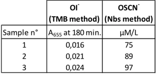
Absorbance of 50 µl OSCN- samples from enzymatic oxidation of SCN- were read after 180 minutes of incubation with TMB and concentration measured with Nbs method (Table 4).
0,1 mM 0,5 mM 0,7 mM 1 mM 1,2 mM 1,4 mM Mean concentration 0,08828662 0,502319692 0,714922641 1,010630147 1,223765137 1,364861989 Standard deviation (s) 0,00650308 0,083009658 0,090555343 0,120533456 0,112182589 0,120613746 Repeatability 0,004998701 0,063806757 0,069606874 0,09265005 0,086231017 0,092711766 Iode concentration (0,78 mM KI + 0,34 mM KSCN + 1,2 mM H2O2) (0,78 mM KI + 0,34 mM KSCN + 1,2 mM H2O2) 1/ 3 (0,78 mM KI + 0,34 mM KSCN + 1,2 mM H2O2) 1/ 5 Difference in absorbance between undiluted sample and 1/3 diluted sample Difference in absorbance between undiluted sample and 1/5 diluted sample Mean Absorbance 0,218 0,084 0,062 15,20% 42,69% Standard deviation 0,0944 0,0353 0,0264
Table 4: Mean absorbance at 655 nm with the TMB method and mean concentration with the Nbs method (3 replicates/ 3 independent ions solutions).
DISCUSSIONS
The lactoperoxidase system is an antimicrobial system largely described in literature. Attention has been mainly focused on OSCN- as SCN- is the most oxidized peroxidase’s substrate in in vivo fluids [6]. OSCN- reacts specifically through oxidation of –SH moiety and its concentration is measured by oxidation of thiol groups of Nbs [7]. HOI, the conjugate base OI- and I2 are able to oxidized aromatic cycles, thiols, thioethers and very slowly amines [8, 9]. The Nbs method is not specific and in presence of OI- and OSCN- it will measure the total thiol oxidant activity [10].
TMB is a substrate for peroxidases and, in presence of H2O2, undergoes an enzymatic oxidation into a blue product largely described for the measurement of peroxidase activity [11]. Enhanced TMB activity has been recorded with added iodide and bromide but not with chloride and thiocyanate [12, 13]. It has also been described for the indirect measurement of enzymatic OCl- and OBr- production by trapping them as taurine haloamines [14, 15]. Oxidation of TMB by chemically produced OCl-, OBr-, OI- or I2 has been described [13]. OSCN- has been reported as a poor TMB oxidant [13]. This was confirmed by our results.
We have developed a direct OI- or I2 quantification method after retrieval of the enzyme by decantation of the clay. Alternatively, 1) the reaction can be stopped by the addition of catalase or 2) the enzyme can be removed by ultrafiltration. This latter method cannot be used when I2 is formed because it is absorbed on the 30 kDa membrane (data not shown). Aqueous oxidized iodide solutions contains a mixture of several species, depending mainly on the pH and iodide concentrations, such as HOI, OI-, I2, I3- [9, 16, 17]. At pH 6 to 9, which is in the pH range of our enzymatic solution, the main active oxidized I- species is believed to be HOI [9]. Indeed the enzymatic solution is colorless, not yellowish colored and ultrafiltration didn’t diminish Nbs concentrations (data not shown) so we assume that the main oxidized I -species is HOI and not I2. Calibrated solutions of HOI didn’t exist and calibrated I2 solutions were used to construct a standard curve. We expressed the OI- concentration in I2 equivalent. It is assuming that one I2 or one HOI oxidized one TMB molecule [13] which make it worthwhile to divide by two concentrations calculated from the I2 standard curve for the HOI solutions. The concentration of [HOI] measured in the enzymatic solution with the TMB method was 93 µM/L. This value is in accordance with the Nbs method for which the [HOI + OSCN-] measured concentration was 138 µM/L.
HOI and I2 are described as poor oxidant of amine groups [9]. The long incubation needed before stabilization of absorbance could be explained by the weak oxidant capacity of HOI. Nevertheless this was not observed with I2
OI- (TMB method) OSCN- (Nbs method) Sample n° A655 at 180 min. µM/L 1 0,016 75 2 0,021 89 3 0,024 97
which oxidized TMB within 30 minutes in a stable absorbance up to 180 minutes (data not shown) [13].
This method could also be used for direct measurement, after retrieval of the enzyme, of OCL- and OBr- with probably shortening TMB-incubation time since both are more reactive oxidants than HOI.
BIBLIOGRAPHY
1. O'Brien, P.J., Peroxidases. Chemico-Biological Interactions, 2000. 129(1-2): p. 113-139.
2. Pruitt, K.M., ed. The lactoperoxidase system: chemistry and biological
significance. immunology series. Vol. 27. 1985, Marcel Dekker: New
York.
3. Kohler, H. and H. Jenzer, Interaction of lactoperoxidase with
hydrogen peroxide. Formation of enzyme intermediates and
generation of free radicals. Free Radic Biol Med, 1989. 6(3): p.
323-39.
4. Thomas, E.L., K.P. Bates, and M.M. Jefferson, Hypothiocyanite ion:
detection of the antimicrobial agent in human saliva. Journal of
Dental Research, 1980. 59(9): p. 1466-1472.
5. Protocole pour la validation d'une méthode d'analyse en chimie. 2009,
Centre d'expertise en analyse environnementale du Québec. 6. Furtmüller, P.G., et al., Active site structure and catalytic
mechanisms of human peroxidases. Archives of Biochemistry and
Biophysics, 2006. 445(2): p. 199-213.
7. Aune, T.M. and E.L. Thomas, Accumulation of hypothiocyanite ion
during peroxidase-catalyzed oxidation of thiocyanate ion. European
Journal of Biochemistry, 1977. 80(1): p. 209-214.
8. Davies, M.J., et al., Mammalian heme peroxidases: From molecular
mechanisms to health implications. Antioxidants and Redox
Signaling, 2008. 10(7): p. 1199-1234.
9. Prütz, W.A., et al., On the irreversible destruction of reduced
nicotinamide nucleotides by hypohalous acids. Archives of
Biochemistry and Biophysics, 2000. 380(1): p. 181-191.
10. Bosch, E.H., H. van Doorne, and S. de Vries, The lactoperoxidase
system: the influence of iodide and the chemical and antimicrobial
stability over the period of about 18 months. J Appl Microbiol, 2000.
89(2): p. 215-24.
11. Josephy, P.D., T. Eling, and R.P. Mason, The horseradish
peroxidase-catalyzed oxidation of 3,5,3',5'-tetramethylbenzidine. Free
radical and charge-transfer complex intermediates. Journal of
Biological Chemistry, 1982. 257(7): p. 3669-3675.
12. Tatzber, F., et al., Dual method for the determination of peroxidase
activity and total peroxides-iodide leads to a significant increase of
peroxidase activity in human sera. Analytical Biochemistry, 2003.
316(2): p. 147-153.
13. Bozeman, P.M., D.B. Learn, and E.L. Thomas, Assay of the human
leukocyte enzymes myeloperoxidase and eosinophil peroxidase.
Journal of Immunological Methods, 1990. 126(1): p. 125-133. 14. Dypbukt, J.M., et al., A sensitive and selective assay for chloramine
production by myeloperoxidase. Free Radical Biology and Medicine,
2005. 39(11): p. 1468-1477.
15. Bhave, G., et al., Peroxidasin forms sulfilimine chemical bonds using
hypohalous acids in tissue genesis. Nature Chemical Biology, 2012.
8(9): p. 784-790.
16. Ashby, M.T., Chapter 8 - Hypothiocyanite, in Advances in Inorganic
Chemistry, E. Rudi van and I.-B. Ivana, Editors. 2012, Academic
Press. p. 263-303.
17. Bichsel, Y. and U. Von Gunten, Oxidation of iodide and hypoiodous
acid in the disinfection of natural waters. Environmental Science and

![Figure 2: Linearity of the TMB method with [I2] up to 1,6 mM. Linear regression and correlation coefficient were calculated on the mean of 10 replicates](https://thumb-eu.123doks.com/thumbv2/123doknet/6661363.182326/4.892.194.697.109.395/figure-linearity-linear-regression-correlation-coefficient-calculated-replicates.webp)