HAL Id: hal-02301340
https://hal.archives-ouvertes.fr/hal-02301340
Submitted on 15 Jan 2021HAL is a multi-disciplinary open access archive for the deposit and dissemination of sci-entific research documents, whether they are pub-lished or not. The documents may come from teaching and research institutions in France or abroad, or from public or private research centers.
L’archive ouverte pluridisciplinaire HAL, est destinée au dépôt et à la diffusion de documents scientifiques de niveau recherche, publiés ou non, émanant des établissements d’enseignement et de recherche français ou étrangers, des laboratoires publics ou privés.
Radionuclide candidates for β+γ coincidence PET: An
overview
Mateusz Sitarz, Jean-Pierre Cussonneau, Tomasz Matulewicz, Ferid Haddad
To cite this version:
Mateusz Sitarz, Jean-Pierre Cussonneau, Tomasz Matulewicz, Ferid Haddad. Radionuclide candi-dates for β+γ coincidence PET: An overview. Applied Radiation and Isotopes, Elsevier, 2020, 155, pp.108898. �10.1016/j.apradiso.2019.108898�. �hal-02301340�
Radionuclide candidates for β+γ coincidence PET: An overview
Mateusz Sitarz1,2,3,*, Jean-Pierre Cussonneau4, Tomasz Matulewicz2,Férid Haddad1,41 Groupement d’Intérêt Public ARRONAX, 44817 Saint-Herblain Cedex, France 2 Faculty of Physics, University of Warsaw, 02-093 Warszawa, Poland
3 Heavy Ion Laboratory, University of Warsaw, 02-093 Warszawa, Poland
4 Subatech, CNRS/IN2P3, IMT Atlantique, Université de Nantes, CS 20722 44307 Nantes cedex, France
* Correspondence: mateusz.sitarz@fuw.edu.pl
Keywords
● PET
● Production of medical radionuclides ● Cyclotrons
● β+γ coincidence Highlights
This paper summarizes the properties, applications and feasible production of all known radioisotopes that can be used in the novel β+γ coincidence PET technique. These radioisotopes include: 10C, 14O, 22Na, 34mCl, 44gSc, 48V, 52mMn, 55Co, 60Cu, 66Ga, 69Ge, 72As, 76Br, 82gRb, 86gY, 94mTc, 110mIn and 124I.
Abstract
Following the advancement of the Positron Emission Tomography (PET), a novel technique emerged which takes advantage of the gamma quanta emitted, in some cases, after the β+ decay. While 44gSc is commonly agreed the best choice to validate this new modality, other radioisotopes must also be considered in the future. We present them in this paper, along with their possible applications, properties and the optimal production routes with the use of the accelerators.
Introduction
During the recent years, we have witnessed the birth and the steady development of a new approach in Positron Emission Tomography (PET) for the nuclear imaging, namely a β+γ coincidence technique (Martin et al., 1995; Pentlow et al., 2000; Herzog et al., 2002; Lubberink et al., 2002a; Beattie et al., 2003; Buchholz et al., 2003; Sandström et al., 2004; Grignon et al., 2007; Lang et al., 2013; Thirolf et al., 2015). Compared to the classical PET, it offers potentially better spatial resolution (Sandström et al., 2004; Grignon et al., 2007; Duval et al., 2009) or, alternatively, provides the same image quality with less radioactivity (hence reducing the dose to the patient). It also reduces the exposure time that may translate to the increased number of consultations in hospitals per day.
Today, the conventional PET is used worldwide and has been acknowledged as the gold standard in oncology imaging when used with FDG. Its principle and numerous applications were summarized recently by The International Atomic Energy Agency (IAEA, 2008) and The European Association of Nuclear Medicine (EANM 2010, 2011). In general, PET is a technique commonly used in medicine for diagnostic purposes as it allows quantitative in vivo measurements of the distribution of administered positron-emitting radioisotopes in the human body. In short, the radioactive tracer (which have been defined to accumulate in some specific cells) is injected in the patient, decays and produces the positron (β+) that travels few millimetres (depending on the energy) from the decay vertex before annihilating with an electron. Positron-electron annihilation converts them into two 511 keV energy photons (with over 99% probability), which are emitted in opposite directions (in positron-electron center of mass system) and can escape the human body. These photons are then detected in parallel rings of scintillation crystals surrounding the patient. Two detected photons of the right energy allow to assume that the positron emitter is located somewhere in between, at the Line of Response (LoR). In the first approximation, the intersection of multiple LoR provides the distribution of the tracer (although in practice many additional corrections are applied).
The β+γ coincidence is one of the possible extensions of PET modality and its key element is the use of a β+ source that emits additional γ quanta (sometimes called “third γ”). To prevent the overlap of the signals, the typical constraint in the acquisition systems is that the third γ should be emitted from the radioisotope few ps after the β+ decay allowing coincidence measurements (Grignon et al., 2007). Assuming that the β+ particle and the third γ are emitted at the same location, the position of the radioisotope is then obtained by the intersection of the arrival direction of the third γ with the conventional LoR (Fig. 1). This technique allows to localize the emission point on the event-by-event basis (Martin et al., 1995; Pentlow et al., 2000; Grignon et al., 2007; Duval et al., 2009). The localization via direction cone implies the Compton scattering of the third γ and the detection of the scattered photon in the photoelectric interaction. This favours a third γ of high energy, around 1 MeV (Grignon et al., 2007). While it would give additional radiation dose to the patient, the precision of the β+γ PET is supposed to allow reducing the administered activity, significantly reducing the overall dose (Lang et al., 2013).
In this article, we review the medical radioisotopes suitable for β+γ coincidence PET. The most important criteria for the candidates are the β+ decay followed by a γ quanta of around 1 MeV emitted few ps later. However, there are many more requirements related to the practical aspects of medical radioisotopes. In particular, β+ branching ratio as well as the intensity of third γ should be as high as possible (preferably 100% for both) to assure the highest probability for the signal detection in the PET scanner. At the same time,
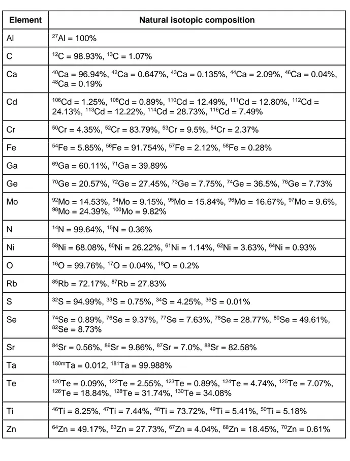
radioisotope of interest should emit a limited number of additional γ lines (which would only introduce the noise to the imaging and dose to the patient). Finally, the radioisotope should have feasible production route and convenient half-life (from few hours to few days), suitable for the clinical practice. Obviously, no radioisotope fulfils all these criteria. However, some are very close and could be potentially investigated in β+γ coincidence PET, depending on the parameters of available scanner and the aim of the research or imaging. The most promising radioisotopes are presented in this work, including their applications and production routes investigated so far. The physical properties of all candidates are listed in Table 1 and the natural abundance of the targets discussed for their production is shown in Table 2 (although in certain cases the discussed production route and its yield requires the use of an enriched target with modified composition discussed in the text).
Fig. 1. Principle of the β+γ coincidence PET (Grignon et al., 2007).
Finally, it is worth mentioning that with the development of new PET scanners, further advancement in β+γ coincidence technique can be expected. Already mentioned XEMIS and XEMIS2 machines offer a sub-millimetre position resolution, a good energy resolution, and a possibility to measure third γ of up to 5 MeV (Grignon et al., 2007; Duval et al., 2009; Cussonneau et al., 2017; Gallego Manzano et al., 2018). At the same time, the first PET scanner build from plastic scintillators, J-PET, offers cost-effective whole-body scans with the feasibility of ortho-positronium imaging (Gajos et al., 2016; Kamińska et al., 2016;
Moskal et al., 2019). Recently constructed EXPLORER might be also of interest as it has proved its high-sensitive total-body scan (Badawi et al., 2019).
As a side remark, it is possible to imagine an approach in which LoR is replaced with two (or more) γ-rays emitted from the nucleus, forcing the reconstruction based on three complete Compton events. Some radioisotopes satisfy this criterion, with three high-intensity γ lines and low β+ branching ratio: 94gTc (T1/2 = 4.9 h), 96Tc (T1/2 = 4.3 d), 108In (T1/2 = 58 min), 110gIn (T1/2 = 4.9 h) and 206Bi (T1/2 = 6.2 d). However, the imaging with this method would require more activity compared to the β+γ coincidence counterpart and is not discussed further in this paper.
There is also a similar PET technique which utilizes a rare three γ quanta emission from the ortho-positronium annihilation (Kacperski et al., 2004; Kacperski and Spyrou, 2005;
Abuelhia et al., 2007; Gajos et al., 2016; Kamińska et al., 2016; Moskal et al., 2019). This method can be applied with all β+ radionuclides and is not discussed further here.
34mCl
Compared to other radioisotopes from this overview, 34mCl is less popular in the nuclear medicine field. However, it was recognized as potential PET radionuclide (Qaim and Stöcklin, 1983; Helus et al., 1985) and has already been used to label dopamine D1 agonists (DeJesus et al., 2007; Murali et al., 2011). The first factor limiting its popularity is the number of high-intensity γ lines increasing the dose without providing the relevant diagnostic data. The γ line of the highest intensity could be used in β+γ coincidence imaging although other radioisotopes discussed here offer better physical properties for this purpose.
The second limiting factor is the difficult production of 34mCl. The practical no-carrier-added production uses natS(⍺ ,x) reaction. According to the cross-section data (Hintz and
Ramsey, 1952; Umbarger et al., 1970; Nagatsu et al., 2008), the required beam energy is around 65 MeV. The saturation of the thick target irradiation results in radio-contaminant-free 1500 MBq/μA 34mCl (Zatolokin et al., 1976; Takei et al., 2007; Nagatsu et al., 2008). A feasible production chain was also developed, consisting of the 80% effective separation using heated water and HPLC pump (Takei et al., 2007). Even though this production
requires high energy ⍺ beam. Alternative methods face even more difficult problems.
44gSc
The 44gSc radioisotope is one of the most promising β+γ candidate (mentioned as such by Lang et al., 2013 and Thirolf et al., 2015) and the perfect one to set-up the proof of concept of this new imaging modality. It has a convenient half-life (T1/2 = 3.9 h) and emits only one, high-intensity γ-line with a desirable energy (1157 keV, 99%). It has been already used in the study of β+γ coincidence PET with the use of XEMIS and XEMIS2 detectors (Grignon et al., 2007; Duval et al., 2009; Cussonneau et al., 2017; Gallego Manzano et al., 2018). The general interest of 44gSc was also stressed by Huclier-Markai et al., 2018. It is not only related to its convenient physical properties, but also to the feasible chemistry (reported mainly for the labelling of DOTA-peptides) supported with the stability and biodistribution studies (Koumarianou et al., 2011; Cydzik et al., 2012; Krajewski et al., 2012, 2013; Pruszyǹski et al., 2012; Roesch, 2012; Severin et al., 2012; Müller et al., 2013; Huclier-Markai et al., 2014; Alliot et al., 2015a, 2015b; Valdovinos et al., 2015; van der Meulen et al., 2015; Domnanich et al., 2016; Kilian et al., 2018). Additionally, its metastable state, 44mSc (T1/2 = 58.6 h), can be used as long-lived in vivo generator (Huclier-Markai et al., 2014; Alliot et al., 2015a, 2015b; Duchemin et al., 2015) as it decays mainly by a low energy transition to the ground state. Finally, 44gSc can be used as counterpart of therapeutic partners. It forms a theranostic pair with 47Sc (T1/2 = 3.35 d), which is a low-energy β- emitter for targeted radiotherapy, allowing the application of the theranostic approach (Müller et al., 2014, 2018). It can also be used with the therapeutic 177Lu as they share similar chemistry (Umbricht et
al., 2017).
The most interesting production route is via 44Ca(p,n) reaction with the CaCO3 or CaO, (Khandaker et al., 2009; Krajewski et al.,2012, 2013; Severin et al., 2012; Müller et al., 2013, 2014; Hernandez et al., 2014; Hoehr et al., 2014; Valdovinos et al., 2015; van der Meulen et al., 2015; Singh et al., 2015; Domnanich et al., 2016; Carzaniga et al., 2017;
Sitarz et al., 2018) which can be later easily dissolved for the chemical separation (Cydzik et al., 2012; Krajewski et al.,2012, 2013; Pruszyǹski et al., 2012; Severin et al., 2012; Müller et
al., 2013; Huclier-Markai et al., 2014; Alliot et al., 2015b; Valdovinos et al., 2015; van der Meulen et al., 2015; Domnanich et al., 2016; Kilian et al., 2018). A standard 16 MeV proton beam of 1 μA can produce up to 20 MBq (CaCO3) or 35 MBq (CaO) after 1 h of irradiation of natural targets and 40-50 times more if the commercially available >90% enrichment is used. In case of natural target, 43Sc (T1/2 = 3.89 h) is present at the level of 3% while the use of the enriched target eliminates the radioactive impurities almost completely (Sitarz et al., 2018). Alternatively, 44gSc can be obtained from 44Ti/44gSc generator (T1/2 = 59.1 y) as pointed out by
Filosofov et al., 2010; Pruszyǹski et al., 2010, 2012;Roesch, 2012 and summarized recently by Hassan et al., 2017. Currently, such generator is available in Mainz, with 185 MBq 44Ti and the possibility to extract 97% 44gSc in 20 mL solution (Filosofov et al., 2010).
Still, the most accessible optimal production route is the irradiation of >90% enriched 44CaCO3 or 44CaO with proton beam, yielding up to 1000 MBq/μAh or 1700 MBq/μAh respectively and less than 1% of radioactive contaminants.
48V
48V radioisotope is well-known only as a monitor for the beam current measurements (IAEA, 2017). It emits two high-intensity γ lines and has a β+ branching ratio of only 50%. However, it has the longest half-life (16 d) among all discussed β+γ candidates, suitable for the studies of slow metabolic processes and for the labelling of organic compounds (Qaim, 2011). The prospects of 48V were recently reminded by Usman et al., 2017 as it is already finding applications as a tracer in biological actions in plants (Xuam Tham et al., 2001), in material science (Rorat et al., 2005) or in the renal artery brachytherapy (Arbabi et al., 2009). As suggested by Martin et al., 1995 it is also a promising candidate for the coincidence PET. However, it should be noted that the second high-intensity γ line from 48V would introduce additional dose for the patient and noise in the imaging without providing the relevant diagnostic data.
As summarized in IAEA, 2017, the cross-sections for different production of 48V are well measured and suggest the route via natTi(p,x) reaction (Smith et al., 2011). The major contribution comes from 48Ti(p,n) reaction suitable for the energy of the commonly available cyclotrons and the target nuclide has high natural abundance. With 16 MeV beam and 1 μAh irradiation of 90% enriched 48Ti target, over 20 MBq of 48V can be produced, with less than 0.5% of radioactive impurity of 49V (T1/2 = 330 d).
52mMn
The recent advancements in hybrid MRI induced the interest in 52gMn radioisotope. It was suggested as the tracer of Mn+2 ions (Lewis et al., 2015) that serve as the T1 MRI relaxation agent in the Manganese-Enhanced Magnetic Resonance Imaging (MEMRI) (Koretsky and Silva, 2004; Silva et al., 2004; Wadghiri et al., 2004; Silva and Bock, 2008; Massaad and Pautler, 2011; Cacace et al., 2014). However, in the light of the reported risk regarding the use of the bulk manganese (Crossgrove and Zheng, 2004), the use of 52gMn in conventional PET/MRI was suggested (Graves et al., 2015) to obtain analogous data with lower biological toxicity. Meanwhile, its metastable level, 52mMn, has very high β+ branching ratio and emits only one, high-energy and high-intensity γ line. Since the β+γ coincidence imaging requires less activity, the toxicity of magnesium in PET/MRI could be further
reduced with the use of 52mMn (already suggested for β+γ coincidence PET by Martin et al.,
1995). However, it should be noted that significant modifications might be in order for PET hardware to permit the acquisition in the presence of a strong magnetic field and radiofrequency pulses (Disselhorst et al., 2014).
The best access to 52mMn is through a generator, 52gFe/52mMn, since 52gFe decays in 100% to 52mMn. The co-produced 52mFe (T1/2 = 46 s) decays in 100% to 52gMn though so it should be taken into account by managing the post-irradiation separation, rapidly removing early 52gMn and later waiting for 52mMn to be formed from 52gFe. The possible production routes for 52gFe/52mMn generator have been summarized by Atcher et al., 1980 and Steyn et
al., 1990. According to the literature, a reasonable option is to irradiate thick natMn target with protons of energy from 40-50 to 60-75 MeV. This procedure is routinely used in Brookhaven Linac Isotope Producer. Up to 22 MBq/μAh of 52gFe/52mMn was reached, with no more than 1% of 55Fe contaminant.
55Co
55Co has been acknowledged as “the emerging PET radionuclide” by Amjed et al.,
2016, as it features high β+ branching ratio, half-life favouring the studies of slow biological
process and feasible labelling with different complexes as well as satisfactory biodistribution (Srivastava, et al., 1994; Thisgaard et al., 2011; Mastren et al., 2015; Dam et al., 2016; Garousi et al., 2017). The mentioned paper summarized numerous applications of 55Co which include: the lung cancer detection (Nieweg et al., 1982), the renal imaging (Goethals et al., 2000) and the neuro-imaging (Jansen et al. 1994, 1996, 1997; Stevens et al.,1999; Reuck et al., 2004). These procedures can be also performed with β+γ coincidence PET (suggested by Martin et al., 1995) since 55Co emits one high-intensity high-energy γ line. Additionally, there is a theranostic matched pair with Auger-emitting 58mCo. However, it is worth mentioning that two additional low-intensity γ lines from 55Co would introduce additional dose to the patient and noise to the imaging.
The production routes of 55Co have been thoroughly studied via different nuclear reactions, for which the cross-section and thick target yield data are very well reported. The most promising one is 54Fe(d,n) reaction (Sharma et al., 1986; Zaman and Qaim, 1996;
Hermanne et al., 2000; Zaman et al., 2003; Nakao et al., 2006; Király et al., 2009; Thisgaard et al., 2011; Závorka et al., 2011; Khandaker et al., 2013; Avrigeanu et al., 2014; Valdovinos et al., 2017) which requires enriched target to produce sufficient quantities as well as avoid the co-production of long-lived radioactive impurities of 56Co and 57Co as they emit high intensity γ lines which unnecessarily increase the dose. The commercially available enrichment exceeds 95% in the form of metal (54Fe) or oxide (54Fe2O3). For example, with the metal target and 1 μAh irradiation of 8 or 15 MeV deuteron beam, up to 20 MBq or 40 MBq of 55Co respectively can be produced, with negligible amount of 56Co and 57Co (Sharma
et al., 1986).
60Cu
60Cu is a short-lived β+ emitter with an additional γ line making it suitable for β+γ coincidence PET (Martin et al., 1995). So far, it has only been used in standard PET technique along with the labelling studies of 60Cu-ATSM for tumor hypoxia imaging (Blower
2008; Lewis et al., 2008). It also has a theranostic matched pair, 67Cu, the β- emitter for targeted radionuclide therapy.
The most commonly used production route of 60Cu is via 60Ni(p,n) or natNi(p,x) reactions (used in above-mentioned papers). The corresponding cross-sections have been already well reported (Blosser and Handley, 1955; Tanaka et al., 1972; Barrandon et al., 1975; Levkovskij, 1991; Singh et al., 2006; Al Saleh et al., 2007; Amjed et al., 2014). Irradiations of thick 60Ni targets (with commercially available 99% enrichment) with proton beam of 16 MeV and 1 μA for 20 minutes, followed by 1 h post-irradiation processing time, are sufficient to achieve up to 400 MBq of 60Cu.
66Ga
The radioisotope of 66Ga already has multiple applications and it has been widely used as PET radioisotope. Labelled with albumin colloids from commercially available kits designed for 99mTc, 66Ga was successfully used in the imaging of the lymphatic transport (Goethals et al., 1988). The feasible 66Ga labelling and purification was also reported for DOTA-peptides 66Ga-DOTA-Tyr3-octreotide and 66Ga-DOTA-biotin (Lewis et al., 2002), the blood cells (Ellis and Sharma 1999; Jalilian et al. 2003) and 66Ga-deferoxamine-folate for in vivo and in vitro imaging (Ke et al., 2003, 2004; Mathias et al., 2003). Additionally, the most abundant β+ emitted by 66Ga has a uniquely high energy, which allowed the use of 66 Ga-DOTATOC for both PET imaging and radiotherapy (Ugur et al., 2002). Finally, 66Ga emits high-energy γ line making it appropriate for β+γ coincidence PET. However, it also emits many less intense but high-energy γ lines that will introduce noise in the imaging and dose to the patient.
The method of choice to produce 66Ga is via 66Zn(p,n) reaction. In this case, several cross-section data (Hille et al., 1972; Little and Lagunas-Solar, 1983; Kopeckỳ, 1990; Tárkányi et al., 1990; Levkovskijj, 1991; Hermanne et al., 1991; Nortier et al., 1991; Szelecsényi et al., 1998, 2005) and thick target yield data (Barrandon et al., 1975; Intrator et al., 1981; Dmitriev, 1986; Kopeckỳ, 1990; Tárkányi et al., 1990; Nortier et al., 1991; Lewis et al., 2002; Rowshanfarzad et al., 2004) were investigated. This reaction was also summarized and re-evaluated with ALICE/ASH 0.1 and TALYS-1.2 by Sadeghi et al., 2010. The natural abundance of 66Zn is quite low but a feasible method was reported for the preparation and recovery of the enriched target (Rowshanfarzad et al., 2004). Around 200 mg/cm2 of 66Zn material is enough for the optimal production of 66Ga with the 15 MeV proton beam. The reported yield for 99% enriched target was around 500 MBq/μAh of radionuclidically pure 66Ga. The optimal post-irradiation processing of Zn targets and 66Ga separation was found to be a cation-exchange chromatography and/or liquid-liquid extraction method (Lewis et al., 2002; Rowshanfarzad et al., 2004).
69Ge
The unravelled potential of 69Ge (Mirzadeh and Lambrecht, 1996) is mainly related to the common interest shifted on a different germanium isotope (namely, the 68Ge/68Ga generator, summarized by Rösch, 2013 and Velikyan, 2015). However, its physical properties make it a possible β+γ coincidence PET agent, yet with an inconveniently low β+ branching ratio, similarly low intensity of 1 MeV γ line, and the presence of additional γ lines. On the other hand, its chemistry is already well developed for the purpose of the mentioned generator. So far, it was only used to label nanoparticles for successful in vivo PET/MRI imaging (Chakravarty et al., 2014). Due to its long half-life, it can be also considered for
immunoPET studies and antibody labelling.
The production of the applicable amounts of 69Ge was not yet investigated but the cross-section for 69,natGa(p,n) reaction (Levkovskij, 1991; Porile et al., 1963; Johnson et al.,
1964; Adam-Rebeles et al., 2013; Hermanne et al., 2015) seems the most cost-efficient and available for small cyclotrons. The 16 MeV beam interacting on a thick natGa target would produce around 110 MBq/μAh. However, no cross-section data is available to estimate the co-production of radioactive impurity of 71Ge. The measurement is challenging due to no γ emission but important as 71Ge is the long-lived Auger-emitter which would contribute to the dose.
72As
In the nuclear medicine, another popular PET radioisotope, 72As, can be considered in the β+ɣ coincidence PET thanks to its additional γ line. It has proven its favourable physical and chemical properties in labelling and preclinical studies (Hosain et al., 1982; Emran et al., 1984; Ballard et al., 2012; Ellison et al., 2016). Furthermore, it has a therapeutic matched pair in the form of the β- emitting 77As. Both radioisotopes have been studied in preclinical and clinical research (Nayak and Brechbiel, 2009; Ellison et al., 2016). Arsenic itself, in the form of the arsenic trioxide, is a popular anticancer drug (Ravandi, 2004), successfully used recently in the clinical treatment of the acute promyelocytic leukemia (Miller et al., 2002; Lu et al., 2007).
There are many methods for the direct production of 72As, however much more attention is paid to the generator 72Se/72As due to its convenient half-life of 8.4 d. Many practical extraction methods for this generator have already been reported (Al-Kouraishi and Boswell, 1978; Phillips et al., 1991; Jennewein et al., 2004, 2005; Ballard et al., 2012, 2012b; Chajduk et al., 2012; Wycoff et al., 2014; Feng et al., 2019). According to the literature, the best method for the production of 72Se/72As leads via 70Ge(⍺ ,2n) reaction (Amiel, 1959;
Al-Kouraishi and Boswell, 1978; Calboreanu et al., 1987; Mushtaq and Qaim, 1990; Levkovskij, 1991; Jennewein et al., 2005; Szkliniarz et al., 2015; Takács et al., 2016; Feng et al., 2019). To reduce the formation of radioactive impurity 75Se that decays to stable 75As, the enriched 70Ge target is recommended (commercially available enrichment is about 96%). The 6 h irradiation with 20 μA and the energy range of 47→0 MeV produces around 250 MBq 72Se (Feng et al., 2019) from which around 70 MBq 72As can be extracted each day during the following week. The reported radioactive impurities of arsenic can be removed during the preparation of the generator.
76Br
The radioisotope of 76Br has a large number of accompanying γ rays, from which the most intensive makes 76Br a possible β+γ candidate (as suggested by Lubberink et al.,
2002a; Sandström et al., 2004; Lang et al., 2013; Thirolf et al., 2015) whose coincidence PET imaging and γ cascade correction have already been investigated (Lubberink et al., 2002a). There are two factors limiting the possible interest in 76Br: the γ line used for the coincidence has quite low energy and other γ lines contribute to the dose. However, the labelling chemistry of bromine is similar to that of iodine, which is relatively well investigated (Maziere and Loc'h, 1985) and might render 76Br worth considering. So far, it has been successfully used to study the dopamine receptors associated with the diagnosis of schizophrenia (Martinot et al., 1991; 1994), as the amino acid tracer (Hanaoka et al., 2015),
as the monitor for corticotropin-releasing hormone (Jagoda et al., 2011), as the bromo analogue marker to diagnose heart disease (Loc'h et al., 1994), for the labelling of mouse epidermal growth factor (Scott-Robson et al., 1991) and to study the tumor angiogenesis by labelling a human antibody (Rossin et al., 2007). Additionally, 76Br was used to verify the thymidine analogue, BUdR, as the tumor cell proliferation imaging agent (Gardelle et al., 2001).
As summarized by Hassan et al., 2004 and Sadeghi et al., 2010, the most feasible method for production of 76Br is the direct route via 76Se(p,n) reaction for which the cross-section data (Kovàcs et al., 1985, Levkovskij, 1991, Hassan et al., 2004; El-Azony et al., 2009) and experimental yields (Janssen et al., 1980; Tolmachev et al., 1998) are well measured. However, the favourable cross-section requires an enriched target due to the low abundance of 76Se. The literature indicates that around 360 MBq/μA of 76Br can be produced by the irradiation of the commercially available 97% enriched metal target with 15→8 MeV proton beam. The radioactive impurity of 77Br was observed at the level below 2% and originated from 77Se impurity in the target.
82gRb
An interesting case, 82gRb (T1/2 = 1.3 min), emits β+ radiation followed by only one low-intensity γ line. Still, it was introduced as the β+γ PET candidate (Lang et al., 2013; Thirolf et al., 2015).
The radioisotope of 82Sr (T1/2 = 25.4 d) that decays to 82gRb is the only reasonable method to acquire 82gRb. The generator 82Sr/82gRb has already gained incredible popularity and is widely used to diagnose the cardiovascular disease (a leading cause of death in modern industrialized countries) in myocardial perfusion imaging (Yano et al., 1977; Kensett et al., 1987; Go et al., 1990; Saha et al., 1990; Di Carli et al., 2007; Klein et al., 2007; Merhige et al., 2007; Yoshinaga et al., 2010; Dhar and Ananthasubramaniam, 2011; Scholtens and Barneveld, 2017). It provides significantly better precision compared to 201Tl (Go et al., 1990) and presents less radiation exposure for patients compared to 99mTc scan (Yoshinaga et al., 2010). Many studies have also been performed on the elution system and the optimized chemical separation (Grant et al., 1975; Yano et al., 1977; Kensett et al., 1987; Mausner et al., 1987; Saha et al., 1990; Cackette et al., 1993; Bilewicz et al., 2005; Klein et al., 2007; Yoshinaga et al., 2010).
The method of choice for 82Sr/82gRb generator production is the (p,4n) reaction on 85Rb which benefits from high natural abundance. This method was studied multiple times and is most often employed for the large-scale production (Mausner et al., 1987; Huszár et al., 1989; Deptula et al., 1990; Lagunas-Solar, 1992; Cackette et al., 1993; Gilabert et al., 1998; Ido et al., 2002; Buthelezi et al., 2006), although, as summarized by Takács et al., 2003, significant discrepancies still exist. For the proton energy of 70-60 MeV (with 40 MeV exiting from the thick Rb or RbCl target), the 82Sr/82gRb production yields of 8-13 MBq/μAh were reported. The observed long-lived radioactive impurity of 85Sr was below 1%. As the typical generator activity used for clinical studies reaches 4 GBq (Saha et al., 1990), the typical irradiation lasts for few days and requires high beam current.
86gY
The potential of 86gY lies within its theranostic matched pair (Lopci et al., 2011; Rösch
et al., 2017; Bandara et al., 2018), the β- emitter 90Y available from the long-lived 90Sr/90Y generator system, which is a versatile therapy agent (as reviewed by Goffredo et al., 2011). However, the emission of multiple intensive, dose-contributing γ rays from 86gY and the recent development of the 90Y imaging with the bremsstrahlung photons (summarized by
Wright et al., 2015) might render the matched pair obsolete. Still, many radiochemicals and in vivo PET imaging studies were performed with 86gY (summarized by Nayak and Brechbiel,
2011) and its dominating γ line was recognized for the β+ɣ coincidence PET (Pentlow et al., 2000; Beattie et al., 2003; Buchholz et al., 2003; Sandström et al., 2004; Lang et al., 2013; Thirolf et al., 2015). In fact, the quantitative coincidence imaging for this radioisotope have been already investigated (Pentlow et al., 2000; Beattie et al., 2003; Buchholz et al., 2003).
Several methods of 86gY production were investigated (in each, the co-produced 86mY decays with T1/2 = 47.4 min to 86gY). As reviewed by Schmitz, 2011, the most commonly used is the direct production via 86Sr(p,n) reaction, for which the excitation function has been also re-evaluated with nuclear codes by Sadeghi et al., 2010. It requires about 14 MeV as higher energies increase the percentage of the contaminants. The reported irradiation of around 200 mg/cm2 of the commercially available 95% enriched 86SrCO3 target yields about 150 MBq/μAh with less than 3% of radioactive impurities (Rösch et al., 1993a,1993b; Kettern et al., 2002; Yoo et al., 2005, Avila-Rodriguez et al., 2008, Lukić et al., 2009; Elbinawi et al., 2018). For comparison, around 10 MBq of 86gY is enough for in vivo mice studies (Lövqvist et
al., 2001; McQuade et al., 2005).
94mTc
Despite the clear dominance of 99mTc in nuclear medicine, other technetium radioisotope, 94mTc, is of a potential interest as a PET quantification of 99m Tc-radiopharmaceuticals (Bigott et al., 2001; Qaim, 2012) due to the same chemistry of both isotopes. It decays completely to 94Mo and emits one high-intensity high-energy ɣ line making it suitable for β+γ coincidence imaging (Martin et al., 1995; Lang et al., 2013; Thirolf et al., 2015). The feasibility of standard in vivo PET studies has been already reported (Nickles et al., 1993; Stone et al., 1994; Luyt et al., 2003).
The method of choice for the 94mTc production is the bombardment of 94Mo with medium energy proton beam (Rösch and Qaim, 1993c; Rösch et al., 1994; Bigott et al., 2001, 2006; Uddin et al., 2004; Kakavand et al., 2013). The commercially available enrichment of about 95% is available in MoO3 powder form. The irradiation with the optimal energy range of 13→8 MeV produces about 2 GBq/μAh 94mTc with about 8% of 94gTc impurity. A 1 h irradiation with 4 μA beam followed by 0.5 h thermochromatographic separation with 90% efficiency results in 1300 MBq 94mTcO4-, ready for medical application (Rösch et al., 1994). Other purification and target recovery methods were also investigated (summarized in Bigott et al., 2006).
110mIn
So far, the radioisotope of 110mIn has not drawn a lot of attention in the nuclear medicine field. However, as confirmed by in vivo clinical studies, it provides 3 times better resolution than the typical 111In SPECT (Lubberink et al., 2002b) which opens the possibilities for the detection of small tumors with indium-labelled radiopharmaceuticals. This might be important in the light of an emerging radioisotope 114mIn (IAEA, 2007), an Auger emitter with almost instant β- emissions (from its short-lived daughter), whose therapeutic properties are expected (Tolmachev et al., 2000). Additionally, 110mIn emits medium-energy but high-intensity γ line which could be potentially interesting for β+γ coincidence PET.
Several cross-sections measurements are available, suggesting the potential 110mIn direct and indirect production options (summarized by Tárkányi et al., 2015). The direct production of 110mIn (Jπ = 2+) always leads to the co-formation of the radioactive impurity 110gIn (Jπ = 7+) but the higher isomeric ratio can be achieved with lower projectile energies. The recommended reaction, 110Cd(p,n) (Otozai et al., 1966;Abramovich et al., 1975; Skakun
2006, 2015; Al-Saleh, 2008; Khandaker et al., 2008; Büyükuslu et al., 2010), at 15 MeV energy (available in the commonly used machines) and with electroplated natCd target (Kakavand et al., 2015b) yields 160 MBq/uAh of 110mIn with reported 3% of 110gIn and 6% of 111gIn radioactive impurities (Mukhammedov et al., 1984; Kakavand et al., 2015b).
124I
A textbook medical radionuclide 124I has relatively low β+ branching ratio and the medium-intensive medium-energy γ line but was still suggested as β+γ PET candidate (Martin et al., 1995; Herzog et al., 2002; Sandström et al., 2004; Lang et al., 2013; Thirolf et al., 2015) and the quantitative coincidence imaging have also been investigated (Herzog et al., 2002). This is mainly because it is the only isotope of iodine suitable for PET that can be paired with the strategic therapeutical 131I (Lopci et al., 2011) commonly used for the treatment of hyperthyroidism and thyroid cancer (overviewed by de Klerk, 2000 and Higashi et al., 2012). Furthermore, 124I itself has already been used for the imaging of tissue proliferation (Blasberg et al., 1996; 2000; Roelcke et al., 2002) and for multiple in vivo cancer imaging studies (Langen et al., 1990; Snook et al., 1990; Wilson et al., 1991), including thyroid (Frey et al., 1986; Phan et al., 2008; Capoccetti et al., 2009; Kharazi et al., 2011; Plyku et al., 2017). It is also considered as a potential Auger-emitter for the radiotherapy (Stepanek et al., 1996).
The methods for production of 124I were thoroughly studied by many groups with the use of protons, deuterons and ⍺ -particles (summarized by Schmitz, 2011 and Azizakram et al., 2016). As one of the “emerging isotopes”, its production cross-sections are also collected in IAEA database (IAEA, 2007). The most typical 124I production method is the 124Te(p,n) reaction (Kondo et al., 1977; Scholten et al., 1995; Qaim et al., 2003; Sajjad et al., 2006; Nye et al., 2007; Aslam et al., 2010) suitable for popular small cyclotrons but requiring the enriched target material (which is crucial to increase the yield and to reduce the radioactive impurities). Recent thick target yield measurements (Sajjad et al., 2006) indicate that the irradiation of the commercially available 124TeO2 targets (99.9% 124Te) with the energy range of 14→7 MeV produces around 21 MBq/μAh of 124I with around 0.03% of radioactive impurities of 125I and 126I, followed by the dry distillation. Basically, a few hours run with around 20 μA is enough to produce several 50 MBq batches used for the imaging (Plyku et al., 2017).
Online monitors (10C, 14O)
There are two very short-lived β+γ PET radionuclide candidates mentioned in the literature (Martin et al., 1995; Lang et al., 2013; Thirolf et al., 2015), namely: 10C (T1/2 = 19.3 s) and 14O (T1/2 = 70.6 s). They are formed via the fragmentation of the high energy heavy ions or different nuclear reactions induced by the high-energy projectiles on 12C, 14N, 16O nuclides which are immensely abundant in the organic compounds. The beams used in the ion therapy, apart from the delivery of the radiation dose, produce 10C and 14O allowing the visualization of the treatment with the online acquisition system.
Online PET scans with 10C and 14O have already been reported (Litzenberg et al.,
1999; Enghardt et al., 2004; Cambraia Lopes et al., 2016; D’Ascenzo et al., 2018). Still, this technique poses a challenge from the point of the time resolution (Oelfke et al., 1996; Beebe-Wang et al., 2003; Cambraia Lopes et al., 2016). The method with β+ɣ coincidence PET can be potentially considered in cases where the lower statistic is expected.
22Na is a common calibration source, used even in the recent PET research (D’Ascenzo et al., 2018). It has a very long half-life of 2.6 years and high β+ branching ratio, making it a convenient β+ emitter for the repetitive calibrations. 22Na also emits 1274.5 keV gamma-line that was used as a trigger during the calibration of liquid argon detector (Amaudruz et al., 2016). Being already recognized as the β+γ PET candidate (Martin et al., 1995; Lang et al., 2013; Thirolf et al., 2015), 22Na can be therefore considered as a calibration source for β+γ coincidence PET.
22Na sources can be bought from the different suppliers or produced on-site with the proton beam of low and medium energy (as summarized by Takács et al., 1996). Today, the only large-scale production is performed with the use of 70 MeV proton cyclotron by iThemba LABS in South Africa.
Summary
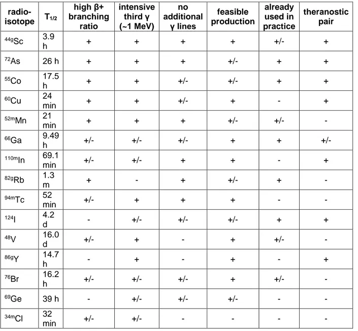
In this paper, we summarized the properties and availability of radioisotopes suitable for the β+γ coincidence PET imaging. The 44gSc radioisotope is undoubtedly most useful for the proof of concept. However, there are many other attractive candidates, in particular those already popular in nuclear medicine: 66Ga, 72As, 82Sr/82gRb and 124I. The coincidence imaging can also be employed in PET/MRI technique with the use of 52mMn and 69Ge or with many 99mTc-radiopharmaceuticals with the use of 94mTc. The summary of β+γ PET radioisotopes is shown in Table 3. Despite discussed drawbacks, the presented radioisotopes can be useful in the β+γ coincidence PET depending on the available scanner and the nature of the study.
Acknowledgements
The cyclotron Arronax is supported by CNRS, Inserm, INCa, the Nantes University, the Regional Council of Pays de la Loire, local authorities, the French government and the European Union. This work has been, in part, supported by a grant from the French National Agency for Research called "Investissements d'Avenir", Equipex ArronaxPlus no. ANR-11-EQPX-0004, Labex IRON no. ANR-11-LABX-18-01 and ISITE NEXT no. ANR-16-IDEX-0007. Part of this work was performed within the framework of EU Horizon 2020 project RIA-ENSAR2 (654 002). The PhD cotutelle scholarship from French Government for Mateusz Sitarz is also acknowledged.
Table 1. Physical properties of the possible β+γ PET radioisotopes (IAEA, 2019)
Isotope T1/2 Decay (%) Main gamma lines [keV] (intensity; delay)
10C 19.3 s EC (0.1), β+ (99.9) 718.4 (100%; 0.71 ns) 14O 70.6 s EC (0.1), β+ (99.9) 2312.6 (99.4%; 68 fs) 22Na 2.6 y EC (10), β+ (90) 1274.5 (99.9%; 3.6 ps) 34mCl 32 min EC (1.1), β+ (54.3), IT (44.6) 146.4 (40.5%; instant), 1176.6 (14.1%; 136 fs), 2127.5 (42.8%; 318 fs), 3304.0 (12.3%; 136 fs) 44gSc 3.9 h EC (5.7), β+ (94.3) 1157.0 (99.9%; 2.61 ps) 48V 16.0 d EC (50.1), β+ (49.9) 983.5 (100%; 4.04 ps), 1312.1 (97.5%; 0.76 ps) 52mMn 21 min EC (1), β+ (97), IT (2) 1434.1 (98.3%; 0.783 ps) 55Co 17.5 h EC (24), β+ (76) 477.2 (20.2%; 37.9 ps), 931.1 (75%; 8 ps), 1408.4 (16.9%; 37.9 ps) 60Cu 24 min EC (7), β+ (93) 826.1 (22%; 0.59 ps), 1332.5 (88%; 0.735 ps) 66Ga 9.49 h EC (44), β+ (56) 1039.2 (37%; 1.68 ps), 2752 (23%; not found) 69Ge 39 h EC (76), β+ (24) 574.2 (13.3%; 1.7 ps), 872.1 (11.9%; 0.25 ps), 1107 (36%; 0.222 ps) 72As 26 h EC (14), β+ (86) 834.0 (80%; 3.35 ps) 76Br 16.2 h EC (44.4), β+ (55.6) 559.1 (74.0%; 12.3 ps), 657.0 (15.9%; 11 ps), 1853.7 (14.7%; not found) 82gRb 1.3 m EC (5), β+ (95) 776.5 (13%; 4.45 ps) 86gY 14.7 h EC (68.1), β+ (31.9) 443.1 (16.9%; 5 ns), 627.7 (32.6%; 0.9 ps), 703.3 (15.4%; 5 ns), 777.4 (22.4%; 0.386 ps), 1076.6 (82.5%; 1.46 ps), 1153.1 (30.5%; 1.73 ps), 1920.7 (20.8%; not found) 94mTc 52 min EC (28), β+ (72) 871.1 (94%; 2.77 ps) 110mIn 69.1 min EC (38.8), β+ (61.2) 657.8 (97.7%; 5.42 ps) 124I 4.2 d EC (77.3), β+ (22.7) 602.7 (62.9%, 6.2 ps), 722.7 (10.4%; 1.04 ps), 1691.0 (11.2%; 0.17 ps)
Table 2. Natural abundance of the targets discussed in the text (IAEA, 2019).
Element Natural isotopic composition
Al 27Al = 100% C 12C = 98.93%, 13C = 1.07% Ca 40Ca = 96.94%, 42Ca = 0.647%, 43Ca = 0.135%, 44Ca = 2.09%, 46Ca = 0.04%, 48Ca = 0.19% Cd 106Cd = 1.25%, 108Cd = 0.89%, 110Cd = 12.49%, 111Cd = 12.80%, 112Cd = 24.13%, 113Cd = 12.22%, 114Cd = 28.73%, 116Cd = 7.49% Cr 50Cr = 4.35%, 52Cr = 83.79%, 53Cr = 9.5%, 54Cr = 2.37% Fe 54Fe = 5.85%, 56Fe = 91.754%, 57Fe = 2.12%, 58Fe = 0.28% Ga 69Ga = 60.11%, 71Ga = 39.89% Ge 70Ge = 20.57%, 72Ge = 27.45%, 73Ge = 7.75%, 74Ge = 36.5%, 76Ge = 7.73% Mo 92Mo = 14.53%, 94Mo = 9.15%, 95Mo = 15.84%, 96Mo = 16.67%, 97Mo = 9.6%, 98Mo = 24.39%, 100Mo = 9.82% N 14N = 99.64%, 15N = 0.36% Ni 58Ni = 68.08%, 60Ni = 26.22%, 61Ni = 1.14%, 62Ni = 3.63%, 64Ni = 0.93% O 16O = 99.76%, 17O = 0.04%, 18O = 0.2% Rb 85Rb = 72.17%, 87Rb = 27.83% S 32S = 94.99%, 33S = 0.75%, 34S = 4.25%, 36S = 0.01% Se 74Se = 0.89%, 76Se = 9.37%, 77Se = 7.63%, 78Se = 28.77%, 80Se = 49.61%, 82Se = 8.73% Sr 84Sr = 0.56%, 86Sr = 9.86%, 87Sr = 7.0%, 88Sr = 82.58% Ta 180mTa = 0.012, 181Ta = 99.988% Te 120Te = 0.09%, 122Te = 2.55%, 123Te = 0.89%, 124Te = 4.74%, 125Te = 7.07%, 126Te = 18.84%, 128Te = 31.74%, 130Te = 34.08% Ti 46Ti = 8.25%, 47Ti = 7.44%, 48Ti = 73.72%, 49Ti = 5.41%, 50Ti = 5.18% Zn 64Zn = 49.17%, 63Zn = 27.73%, 67Zn = 4.04%, 68Zn = 18.45%, 70Zn = 0.61%
Table 3. The arbitrary summary of β+γ PET radioisotopes discussed in the text. “Plus” sign indicates favourable characteristic, “minus” stands for unfavourable, and “plus/minus” marks moderate. The order of the radioisotopes in the table corresponds to their overall score.
radio-isotope T1/2 high β+ branching ratio intensive third γ (~1 MeV) no additional γ lines feasible production already used in practice theranostic pair 44gSc 3.9 h + + + + +/- + 72As 26 h + + + +/- + + 55Co 17.5 h + + +/- +/- + + 60Cu 24 min + + +/- + - + 52mMn 21 min + + + +/- +/- - 66Ga 9.49 h +/- +/- +/- + + +/- 110mIn 69.1 min +/- +/- + + - + 82gRb 1.3 m + - + +/- + - 94mTc 52 min +/- + + + - - 124I 4.2 d - +/- +/- +/- + + 48V 16.0 d +/- + - + +/- - 86gY 14.7 h - + - + - + 76Br 16.2 h +/- +/- +/- + +/- - 69Ge 39 h - +/- +/- +/- - - 34mCl 32 min +/- +/- - - - -
References
Abramovich, S.N., Guzhovskij, B.Ja., Zvenigorodskij, A.G., Trusillo, S.V., 1975. Isobaric Analog Resonances Appearing during Elastic Scattering of Protons and in the (p,n) Reaction on 110,112,114,116Cd. Izv. Ross. Akad. Nauk, Ser. Fiz. 39, p. 1688.
Abuelhia, E., Kacperski, K., Spyrou, N.M., 2007. Three-photon annihilation in PET: 2D imaging experiments. J. Radioanal. Nucl. Chem. 271(2), p. 489.
Adam-Rebeles, R., Hermanne, A., Van Den Winkel, P., De Vis, L., Waegeneer, R., Tárkányi, F., Takács, S., Takács, M.P., 2013. 68Ge/68Ga production revisited: excitation curves, target preparation and chemical separation - purification. Radiochim. Acta 101(8), p. 481.
Al Saleh, F.S., Al Mugren, K.S., Azzam, A., 2007. Excitation functions of (p,x) reactions on natural nickel between proton energies of 2.7 and 27.5 MeV. Appl. Radiat. Isot. 65, p. 104. Al-Saleh, F.S., 2008. Cross sections of proton induced nuclear reactions on natural cadmium leading to the formation of radionuclides of indium. Radiochim. Acta 96, p. 461.
Al-Kourashi, S.H., Boswell, G.G.J., 1978. An isotope generator for 72As. Appl. Radiat. Isot. 29, p. 607.
Alliot, C., Audouin, N., Barbet, J., Bonraisin, A.C., Bossé, V., Bourdeau, C., Bourgeois, M., Duchemin, C., Guertin, A., Haddad, F., Huclier-Markai, S., Kerdjoudj, R., Laizé, J., Métivier, V., Michel, N., Mokili, M., Pageau, M., Vidal, A., 2015a. Is there an interest to use deuteron beams to produce non-conventional radionuclides? Rev. Med. 2, p. 31.
Alliot, C., Kerdjoudj, R., Michel, N., Haddad, F., Huclier-Markai, S., 2015b. Cyclotron production of high purity 44m,44Sc with deuterons from 44CaCO
3 targets. Nucl. Med. Biol. 42, p. 524.
Amaudruz, P.-A., Batygov, M., Beltran, B., Bonatt, J., Boudjemline, K., Boulay, M.G., Broerman, B., Bueno, J.F., Butcher, A., Cai, B., Caldwell, T., Chen, M., Chouinard, R., Cleveland, B.T., Cranshaw, D., Dering, K., Duncan, F., Fatemighomi, N., Ford, R., Gagnon, R., Giampa, P., Giuliani, F., Gold, M., Golovko, V.V., Gorel, P., Grace, E., Graham, K., Grant, D.R., Hakobyan, R., Hallin, A.L., Hamstra, M., Harvey, P., Hearns, C., Hofgartner, J., Jillings, C.J., Kuźniak, M., Lawson, I., La Zia, F., Li, O., Lidgard, J.J., Liimatainen, P., Lippincott, W.H., Mathew, R., McDonald, A.B., McElroy, T., McFarlane, K., McKinsey, D.N., Mehdiyev, R., Monroe, J., Muir, A., Nantais, C., Nicolics, K., Nikkel, J., Noble, A.J., O’Dwyer, E., Olsen, K., Ouellet, C., Pasuthip, P., Peeters, S.J.M., Pollmann, T., Rau, W., Retière, F., Ronquest, M., Seeburn, N., Skensved, P., Smith, B., Sonley, T., Tang, J., Vázquez-Jáuregui, E., Veloce, L., Walding, J., Ward, M., 2016. Measurement of the scintillation time spectra and pulse-shape discrimination of low-energy β and nuclear recoils in liquid argon with DEAP-1. Astropart. Phys. 85, p. 1.
Amiel, S., 1959. Reactions of Alpha Particles with Germanium-70 and Zinc-70. Phys. Rev. 116, p. 415.
Amjed, N., Tárkányi, F., Hermanne, A., Ditrói, F., Takács, S., Hussain, M., 2014. Activation cross-sections of proton induced reactions on natural Ni up to 65 MeV. Appli. Radiat. Isot. 92, p. 73.
Amjed, N., Hussain, M., Aslam, M.N., Tárkányi, F., Qaim, S.M., 2016. Evaluation of nuclear reaction cross sections for optimization of production of the emerging diagnostic radionuclide 55Co. Appl. Radiat. Isot. 108, p. 38.
Arbabi, A., Sadeghi, M., Joharifard, M., 2009. Irradiation and dosimetry of Nitinol stent for renal artery brachytherapy. Appl. Radiat. Isot. 67, p. 129.
Aslam, M.N., Sudár, S., Hussain, M., Malik, A.A., Shah, H.A., Qaim, S.M., 2010. Evaluation of excitation functions of proton and deuteron induced reactions on enriched tellurium isotopes with special relevance to the production of iodine-124. Appl. Radiat. Isot. 68, p. 1760.
Atcher, R. W., Friedman, A.M., Huizenga, J.R., 1980. Production of 52Fe for Use in a Radionuclide Generator System. Int. J. Nucl. Med. Biol. 7, p. 15.
Avrigeanu, M., Avrigeanu, V., Bém, P., Fischer, U., Honusek, M., Katovský, K., Mănăilescu, C., Bojowald, J., Machner, H., Nann, H., Oelert, W., Rogge, M., Turek, P., 2014. Elastic deuteron scattering and optical model parameters at energies up to 100 MeV. Phys. Rev. C 38, p. 1153.
Avila-Rodrigueza, M.A., Nyeb, J.A., Nickles, R.J., 2008. Production and separation of non-carrier-added 86Y from enriched 86Sr targets. Appl. Radiat. Isot. 66, p. 9.
Azizakram, H., Sadeghi, M., Ashtari, P., Zolfagharpour, F., 2016. An overview of 124I production at a medical cyclotron by ALICE/ASH, EMPIRE-3.2.2 and TALYS-1.6 codes. Appl. Radiat. Isot. 112, p. 147.
Badawi, R.D., Shi, H., Hu, P., Chen, S., Xu, T., Price, P.M., Ding, Y., Spencer, B.A., Nardo, L., Liu, W., Bao, J., Jones, T., Li, H., Cherry, S.R., 2019. First Human Imaging Studies with the EXPLORER Total-Body PET Scanner. J. Nucl. Med. 60(3), p. 299.
Ballard, B., Nortier, F.M., Birnbaum, E.R., John, K.D., Phillips, D.R., Fassbender, M.E., 2012. Radioarsenic from a portable 72Se/72As generator: a current perspective. Curr. Radiopharm. 5, p. 264.
Bandara, N., Stott Reynolds, T.J., Schehr, R., Bandari, R.P., Diebolder, P.J., Krieger, S., Xu, J., Miao, Y., Rogers, B.E., Smith, C.J., 2018. Matched-pair, 86Y/90Y-labeled, bivalent RGD/bombesin antagonist, [RGD-Glu-[DO3A]-6-Ahx-RM2], as a potential theranostic agent for prostate cancer. Nucl. Med. Biol. 62–63, p. 71.
Barrandon, J.N., Debrun, J.L., Kohn, A., Spear, R.H., 1975. Étude du dosage de Ti, V, Cr, Fe, Ni, Cu et Zn par activation avec des protons d'énergie limitée a 20 MeV. Nucl. Instr. Meth. 127, p. 269.
Beattie, B.J., Finn, R.D., Rowland, D.J., Pentlow, K.S., 2003. Quantitative imaging of bromine-76 and yttrium-86 with PET: a method for the removal of spurious activity introduced by cascade gamma rays. Med. Phys. 30, p. 2410.
Beebe-Wang, J., Vaska, P., Dilmanian, F.A., Peggs, S.G., Schlyer, D.J., 2003. Simulation of Proton Therapy Treatment Verification via PET Imaging of Induced Positron-Emitters. IEEE Cat. No.03CH37515 (Nuclear Science Symposium. Conference Record), p. 2496.
Bigott, H.M., Mccarthy, D.W., Wüst, F.R., Dahlheimer, J.L., Piwnica‐W orm s, D.R., W elch, M.J., 2001. Production, processing and uses of 94mTc. J. Label. Compd. Radiopharm. 50 (Suppl. 1), p. S119.
Bigott, H.M., Laforest, R., Liu, X., Ruangma, A., Wuest, F., Welch, M.J., 2006. Advances in the production, processing and microPET image quality of technetium-94m. Nucl. Med. Biol. 33(7), p. 923.
Bilewicz, A., Barto, B., Misiak, R., Petelenz, B., 2005. Separation of 82Sr from rubidium target for preparation of 82Sr/82Rb generator. J. Radioanal. Nucl. Chem. 268(3), p. 485.
Blasberg, R.G., Roelcke, U., Weinreich, R., 1996. [124I]-iododeoxyuridine imaging tumor proliferation. J. Nucl. Med. 37 (Suppl. 5).
Blasberg, R.G., Roelcke, U., Weinreich, R., Beattie, B., von Ammon, K., Yonekawa, Y., Landolt, H., Guenther, I., Crompton, N.E.A., Vontobel, P., Missimer, J., Maguire, R.P., Koziorowski, J., Joachim Knust, E., Finn, R.D., Leenders, K.L., 2000. Imaging Brain Tumor Proliferative Activity with [124I]Iododeoxyuridine. Cancer Res. 60, p. 624.
Blosser, H.G., Handley, T.H., 1955. Survey of (p, n) Reactions at 12 MeV. Phys. Rev. 100(5), p. 1340.
Blower, P.J., Lewis, J.S., Zweit, J., 1996. Copper radionuclides and radiopharmaceuticals in nuclear medicine. Nucl Med Biol. 23, p. 957.
Buchholz, H.G., Herzog, H., Forster, G.J., Reber, H., Nickel, O., Rösch, F., Bartenstein, P., 2003. PET imaging with yttrium-86: comparison of phantom measurements acquired with different PET scanners before and after applying background subtraction. Eur. J. Nucl. Med. Mol. Imaging 30, p. 716.
Buthelezi, E.Z., Nortierb, F.M., Schroeder, I.W., 2006. Excitation functions for the production of 82Sr by proton bombardment of natRb at energies up to 100 MeV. Appl. Radiat. Isot. 64, p. 915.
Büyükuslu, H., Kaplan, A., Yildirim, G., Aydin, A., Tel, E., Bölükdemir, M.H., 2010. Production cross sections of medical 110,111In radionuclides. Kerntechnik 75(3), p. 103.
Cacace, A.T., Brozoski, T., Berkowitz, B., Bauer, C., Odintsov, B., Bergkvist, M., Castracane, J., Zhang, J., Holt, A.G., 2014. Manganese enhanced magnetic resonance imaging (MEMRI): a powerful new imaging method to study tinnitus. Hear. Res. 311, p. 49.
Cackette, M.R., Ruth, T.J., Vincent, J.S., 1993. 82Sr Production from Metallic Rb Targets and Development of an 82Rb Generator System. Appl. Radiat. Isot. 44(6), p. 917.
Calboreanu, A., Salagean, O., Pencea, C., Zimmer, K.W., Ciocanel, A., 1987. Formation and Decay of the Compound Nucleus in Alpha Induced Reaction on 70Ge. Rev. Roum. Phys. 32, p. 725
Cambraia Lopes, P., Bauer, J., Salomon, A., Rinaldi, I., Tabacchini, V., Tessonnier, T., Crespo, P., Parodi, K., Schaart, D.R., 2016. First in situ TOF-PET study using digital photon counters for proton range verification. Phys. Med. Biol. 61, p. 6203.
Capoccetti, F., Criscuoli, B., Rossi, G., Ferretti, F., Manni, C., Brianzoni, E., 2009. The effectiveness of 124I PET/CT in patients with differentiated thyroid cancer. J. Nucl. Med. Mol. Imaging 53(5), p. 536.
Carzaniga, T.S., Auger, M., Braccini, S., Bunka, M., Ereditato, A., Nesteruk, K.P., Scampoli, P., Türler, A., van der Meulen, N., 2017. Measurement of 43Sc and 44Sc production cross-section with an 18 MeV medical PET cyclotron. Appl. Radiat. Isot. 129, p. 96.
Chakravarty, R., Valdovinos, H.F., Chen, F., Lewis, C.M., Ellison, P.A., Luo, H., Meyerand, M.E., Nickles, R.J., Cai, W., 2015. Intrinsically Germanium-69 Labeled Iron Oxide Nanoparticle: Synthesis and In Vivo Dual-modality PET/MR Imaging. Adv. Mater. 26(30), p. 5119.
Chao, K.S., Bosch, W.R., Mutic, S., Lewis, J.S., Dehdashti, F., Mintun, M.A., Dempsey, J.F., Perez, C.A., Purdy, J.A., Welch, M.J., 2001. A novel approach to overcome hypoxic tumor resistance: Cu-ATSM-guided intensity-modulated radiation therapy. Int. J. Radiat. Oncol. Biol. Phys. 49, p. 1171.
Chajduk, E., Doner, K., Polkowska-Motrenko, H., Bilewicz, A., 2012. Novel radiochemical separation of arsenic from selenium for 72Se/72As generator. Appl. Radiat. Isot. 70(5), p. 819. Crossgrove, J., Zheng, W., 2004. Manganese toxicity upon overexposure. NMR Biomed. 17(8), p. 544.
Cussonneau, J.P., Abaline, J.M., Acounis, S., Beaupère, N., Beney, J.L., Bert, J., Bouvier, S., Briend, P., Butterworth, J., Carlier, T., Chanal, H., Cherel, M., Dahoumane, M., Diglio, S., Gallego-Manzano, L., Giovagnoli, D., Idier, J., Kraeber-Bodere, F., Lefebvre, F., Lemaire, O., Le Ray, P., Manen, S., Masbou, J., Mathez, H., Morteau, E., Pillet, N., Royer, L., Staempflin, M., Stutzmann, J.S., Vandaele, R., Virone, L., Visvikis, D., Xing, Y., Zhu, Y., Thers, D., 2017. 3γ Medical Imaging with a Liquid Xenon Compton Camera and 44Sc Radionuclide. Acta Phys. Pol. B 48(10), 1661.
Cydzik, I., Seweryn, K., Abbas, K., Simonell, F., Bulgheroni, A., Kasperek, A., 2012. Labelling of DOTATATE with cyclotron produced 44Sc and 43Sc. Q. J. Nucl. Med. Mol. Imaging 56, p. 33.
Dam, J.H., Olsen, B.B., Baun, C., Hoilund-Carlsen, P.F., Thisgaard, H., 2016. In Vivo Evaluation of a bombesin analogue labeled with Ga-68 and Co-55/57. Mol. Imaging Biol. 18, p. 368.
D’Ascenzo, N., Gao, M., Antonecchia, E., Gnudi, P., Chen, H.-H., Chen, F.-H., Hong, J.-H., Hsiao, I.-T., Yen, T.-C., Wang, W., Xi, D., Zhang, B., Xie, Q., 2018. New Digital Plug and Imaging Sensor for a Proton Therapy Monitoring System Based on Positron Emission Tomography. Sensors 18, 3006.
de Klerk, J.M.H., 2000. 131I Therapy: Inpatient or Outpatient? J. Nucl. Med. 41(11), p. 1876.
DeJesus, O.T., Converse, A.K., Nickles, R.J., 2007. Development of 34mCl-labeled dopamine D1 agonists as PET imaging agents. J. Label Comp. Radiopharm. 50, p. S339.
Dehdashti, F., Mintun, M.A., Lewis, J.S., Bradley, J., Govindan, R., Laforest, R., Welch, M.J., Siegel, B.A., 2003. In vivo assessment of tumor hypoxia in lung cancer with 60Cu-ATSM. Eur. J. Nucl. Med. Mol. Imaging 30, p. 844.
Deptula, C., Khalkin, V.A., Kim Sen Han, Knotek, O., Konov, V.A., Mikecz, P., Poponenkova, L.M., Rurarz, E., Zaitseva, N.G., 1990. Excitation functions and yields for medically generator Sr82-Rb82, Xe123-I123 and Bi201-Pb201-Tl201 obtained with 100 MeV protons. Nukleonika 35, p.3.
Dhar, R., Ananthasubramaniam, K., 2011. Rubidium-82 Cardiac Positron Emission Tomography Imaging: An Overview for the General Cardiologist. Cardiol. Rev 19(5), p. 255. Di Carli, M.F., Dorbala, S., Meserve, J., El Fakhri, G., Sitek, A., Moore, S.C., 2007. Clinical Myocardial Perfusion PET/CT. J. Nucl. Med. 48(5), p. 783.
Dietz D.V., Dehdashti, F., Grigsby, P.W., Malyapa, R.S., Myerson, R.J., Picus, J., Ritter, J., Lewis, J.S., Welch, M.J., Siegel, B.A., 2008. Tumor hypoxia detected by positron emission tomography with 60Cu-ATSM as a predictor of response and survival in patients undergoing neoadjuvant chemoradiotherapy for rectal carcinoma: a pilot study. Dis. Colon. Rectum. 51, p. 1641.
Disselhorst, J.A., Bezrukov, I., Kolb, A., Parl, C., Pichler, B.J., 2014. Principles of PET/MR Imaging. J. Nucl. Med. 55 (Suppl. 2), p. 2S.
Dmitriev, P.P., 1986. Radionuclide yield in reaction with protons, deuterons, alpha particles and helium-3 (hand book). IAEA, Vienna, Austria.
Domnanich, K.A., Müller, C., Farkas, R., Schmid, R.M., Ponsard, B., Schibli, R., Türler, A., van der Meulen, N.P., 2016. 44Sc for labeling of DOTA- and NODAGA-functionalized peptides: preclinical in vitro and in vivo investigations. EJNMMI Radiopharm. Chem. 1, p. 8. Duval, S., Breskin, A., Carduner, H., Cussonneau, J.-P., Lamblin, J., Le Ray, P., Morteau, E., Oger, T., Stutzmann, J.-S., Thers, D., 2009. MPGDs in Compton imaging with liquid-xenon. J. Instr. 4, P12008.
EANM, 2010. Principles and Practice of PET/CT Part 1 (Editors: P. Hogg, G. Testanera). In: A Technologist‘s Guide. Vienna, Austria.
EANM, 2011. Principles and Practice of PET/CT Part 2 (Editors: G. Testanera, W.J.M. van den Broek). In: A Technologist‘s Guide. Vienna, Austria.
El-Azony, K.M., Suzuki, K., Fukumura, T., Szelecsényi, F., Kovács, Z., 2009. Excitation functions of proton induced reactions on natural selenium up to 62 MeV. Radiochim. Acta 97, p. 71.
Elbinawi, A., Al-abyad, M., Bashter, I., Seddik, U., Ditrói, F., 2018. Excitation function of proton induced nuclear reaction on strontium: Special relevance to the production of 88Y. Appl. Radiat. Isot. 140, p. 272.
Ellis, B.L., Sharma, H.L., 1999. Co, Fe and Ga chelates for cell labelling: a potential use in PET imaging. Nucl. Med. Commun. 20, p. 1017.
Ellison, P.A., Barnhart, T.E., Chen, F., Hong, H., Zhang, Y., Theuer, C.P., Cai, W., Nickles, R.J., DeJesus, O.T., 2016. High Yield Production and Radiochemical Isolation of Isotopically Pure Arsenic-72 and Novel Radioarsenic Labeling Strategies for the Development of Theranostic Radiopharmaceuticals. Bioconjugate Chem. 27, p. 179.
Emran, A., Hosain, F., Spencer, R.P., Kolstad, K.S., 1984. Synthesis and biodistribution of radioarsenic labeled dimethylarsinothiols: Derivatives of penicillamine and mercaptoethanol. Nucl. Med. Biol. 11(3-4), p. 259.
Enghardt, W., Crespo, P., Fiedler, F., Hinz, R., Parodi, K., Pawelke, J., Ponisch, F., 2004. Charged hadron tumour therapy monitoring by means of PET. Nucl. Instr. Meth. A 525, p. 284.
Feng, Y., Phipps, M.D., Phelps, T.E., Okoye, N.C., Baumeister, J.E., Wycoff, D.E., Dorman, E.F., Lake Wooten, A., Vlasenko, V., Berendzen, A.F., Wilbur, D.S., Hoffman, T.J., Cutler, C.S., Ketring, A.R., Jurisson, S.S., 2019. Evaluation of 72Se/72As generator and production of 72Se for supplying 72As as a potential PET imaging radionuclide. Appl. Radiat. Isot. 143, p. 113.
Filosofov, D.V., Loktionova, N.S., Rösch, F., 2010. A 44Ti/44Sc radionuclide generator for potential nuclear-medical application of 44Sc-based PET-radiopharmaceuticals. Radiochim. Acta 98, p. 149.
Frey, P., Townsend, D., Flattet, A., De Gautard, R., Widgren, S., Jeavons, A., Christin, A., Smith, A., Long, A., Donath, A., 1986. Tomographic imaging of the human thyroid using 124I. J. Clin. Endocrinol. Metab. 63(4), p. 918.
Gajos, A., Kamińska, D., Czerwiński, E., Alfs, D., Bednarski, T., Białas, P., Głowacz, B., Gorgol, M., Jasińska, B., Kapłon, Ł., Korcyl, G., Kowalski, P., Kozik, T., Krzemień, W., Kubicz, E., Mohammed, M., Niedźwiecki, Sz., Pałka, M., Pawlik-Niedźwiecka, M., Raczyński,
L., Rudy, Z., Rundel, O., Sharma, N.G., Silarski, M., Słomski, A., Strzelecki, A., Wieczorek, A., Wiślicki, W., Zgardzińska, B., Zieliński, M., Moskal, P., 2016. Trilateration-based reconstruction of ortho-positronium decays into three photons with the J-PET detector. Nucl. Instr. Meth. Phys. A 819, p. 54.
Gallego Manzano, L., Abaline, J.M., Acounis, S., Beaupère, N., Beney, J.L., Bert, J., Bouvier, S., Briend, P., Butterworth, J., Carlier, T., Chanal, H., Cherel, M., Cussonneau, J.P., Dahoumane, M., Diglio, S., Giovagnoli, D., Idier, J., Kraeber-Bodere, F., Lefevre, F., Lemaire, O., Le Ray, P., Manen, S., Masbou, J., Mathez, H., Morteau, E., Pillet, N., Royer, L., Staempflin, M., Stutzmann, J.S., Vandaele, R., Virone, L., Visvikis, D., Xing, Y., Zhu, Y., Thers, D., 2018. XEMIS2: A liquid xenon detector for small animal medical imaging. Nucl. Instr. Meth. Phys. A 912, p. 329.
Gardelle, O., Roelcke, U., Vontobel, P., Crompton, N.E.A., Guenther, I., Blӓuenstein, P., Schubiger, A.P., Blattmann, H., Ryser, J.E., Leenders, K.L., Kaser-Hotz, B., 2001. [76Br]Bromodeoxyuridine PET in tumor-bearing animals. Nucl. Med. Biol. 28, p. 51.
Garousi, J., Andersson, K.G., Dam, J.H., Olsen, B.B., Mitran, B., Orlova, A., Buijs, J., Ståhl, S., Löfblom, J., Thisgaard, H., Tolmachev, V., 2017. The use of radiocobalt as a label improves imaging of EGFR using DOTA- conjugated Affibody molecule. Sci. Rep. 7, 5961. Gilabert, E., Lavielle, B., Neumann, S., Gloris, M., Michel, R., Schiekel, Th., Sudbrock, F., Herpers, U., 1998. Cross sections for the proton-induced production of krypton isotopes from Rb, Sr, Y, and Zr for energies up to 1600 MeV. Nucl. Instr. Meth. Phys. B 145, p. 293.
Go, R.T., Marwick, T.H., Maclntyre, W.J., Saha, G.B., Neumann, D.R., Underwood, D.A., Simpfendorfer, C.C., 1990. A Prospective Comparison of Rubidium-82 PET and Thallium-201 SPECT Myocardial Perfusion Imaging Utilizing a Single Dipyridamole Stress in the Diagnosis of Coronary Artery Disease. J. Nucl. Med. 31(12), p. 1899.
Goethals, P., Coene, M., Slegers, G., Agon, P., Deman, J., Schelstraete, K., 1988. Cyclotron production of carrier-free 66Ga as a positron emitting label of albumin colloids for clinical use. Eur. J. Nucl. Med. 14, p.152.
Goethals, P., Volkaert, A., Vandewielle, C., Dierckx, R., Lameire, N., 2000. 55Co-EDTA for renal imaging using positron emission tomography (PET): A feasibility study. Nucl. Med. Biol. 27(1), p. 77.
Goffredo, V., Paradiso, A., Ranieri, G., Gadaleta, C.D., 2011. Yttrium-90 (90Y) in the principal radionuclide therapies: An efficacy correlation between peptide receptor radionuclide therapy, radioimmunotherapy and transarterial radioembolization therapy. Ten years of experience (1999–2009). Crit. Rev. Onc./Hem. 80, p. 393.
Grant, P.M., Erdal, B.R., O'Brien, H.A.Jr., 1975. A 82Sr-82Rb isotope generator for use in nuclear medicine. J. Nucl. Med. 16(4), p. 300.
Graves, S.A., Hernandez, R., Fonslet, J., England, C.G., Valdovinos, H.F., Ellison, P.A., Barnhart, T.E., Elema, D.R., Theuer, C.P., Cai, W., Nickles, R.J., Severin, G.W., 2015. Novel Preparation Methods of 52Mn for ImmunoPET Imaging. Bioconjug. Chem. 26(10), p. 2118.
Grignon, C., Barbet, J., Bardiès, M., Carlier, T., Chatal, J.F., Couturier, O., Cussonneau, J.P., Faivre, A., Ferrer, L., Girault, S., Haruyama, T., Le Ray, P., Luqiun, L., Lupone, S., Métivier, V., Morteau, E., Servagent, N., Thers, D., 2007. Nuclear medical imaging using β+ γ coincidence from 44Sc radio-nuclide with liquid xenon as detection medium. Nucl. Instr. Meth. A 571, p. 142.
Hanaoka, H., Ohshima, Y., Suzuki, Y., Yamaguchi, A., Watanabe, S., Uehara, T., Nagamori, S., Kanai, Y., Ishioka, N.S., Tsushima, Y., Endo, K., Arano, Y., 2015. Development of a
Widely Usable Amino Acid Tracer: 76Br-⍺-Methyl-Phenylalanine for Tumor PET Imaging. J.
Nucl. Med. 56(5), p. 791.
Hassan, H.E., Qaim, S.M., Shubin, Yu., Azzam, A., Morsy, M., Coenen, H.H., 2004. Experimental studies and nuclear model calculations on proton-induced reactions on natSe, 76Se and 77Se with particular reference to the production of the medically interesting radionuclides 76Br and 77Br. Appl. Radiat. Isot. 60, p. 899.
Hassan, H.E., Alabyad, M., Mohamed., G.Y., 2017. Production of 44Ti⍺44Sc Generator in Comparison with Direct Routes by Cyclotrons: Cross Section Evaluation Using Nuclear Models Codes. Arab J. Nucl. Sc. Appl. 51(1), p. 57.
Helus, F., Gasper, H., Rettig, W., Maier-Borst, W., 1985. Cyclotron production of 34mCl for biomedical use. J. Radioanal. Nucl. Chem. Lett. 94, p. 149.
Hermanne, A., Walravens, N., Cicchelli, O., 1991. Optimization of isotope production by cross-section determination. In: Proc. of the Int. Conf. Nuclear Data for Science and Technology, May 1991, Jülich, Germany (Editor: Qaim S.M.), Springer-Verlag, Berlin, p. 616.
Hermanne, A., Sonck, M., Takács, S., Tárkányi, F., 2000. Experimental study of excitation functions for some reactions induced by deuterons (10-50 Mev) on natural Fe and Ti. Nucl. Instr. Meth. Phys. B 161-163, p. 178.
Hermanne, A., Adam-Rebeles, R., Tárkányi, F., Takács, S., Ditrói, F., 2015. Proton and deuteron induced reactions on natGa: Experimental and calculated excitation functions. Nucl. Instrum. Meth. Phys. B 359, p. 145.
Hernandez, R., Valdovinos, H.F., Yang, Y., Chakravarty, R., Hong, H., Barnhart, T.E., Cai, W., 2014. 44Sc: an attractive isotope for peptide-based PET imaging. Mol. Pharm. 11, p. 2954.
Herzog, H., Tellmann, L., Qaim, S.M., Spellerberg, S., Schmid, A., Coenen, H.H., 2002. PET quantitation and imaging of the non-pure positron-emitting iodine isotope 124I. Appl. Radiat. Isot. 56, p. 673.
Higashi, T., Kudo, T., Kinuya, S., 2012. Radioactive iodine (131I) therapy for differentiated thyroid cancer in Japan: current issues with historical review and future perspective. Ann. Nucl. Med. 26, p. 99.