i
Université de Montréal
Neuron-Derived Semaphorin 3A is an Early Inducer of
Vascular Permeability in Diabetic Retinopathy
par Agustin Cerani
Département de biochimie Faculté de médecine
Mémoire présenté à la Faculté des études supérieures en vue de l’obtention du grade de maître ès science (M.Sc.) en biochimie
Décembre, 2012
ii
Université de Montréal Faculté des études supérieures
Ce mémoire intitulé
Neuron-Derived Semaphorin 3A is an Early Inducer of
Vascular Permeability in Diabetic Retinopathy
Présenté par : Agustin Cerani
a été évalué par un jury composé des personnes suivantes :
Nathalie Grandvaux Président-rapporteur Przemyslaw Sapieha Directeur de recherche Kenneth Hastings Membre du jury
iii Résumé
La détérioration de la barrière hémato rétinienne et l'œdème maculaire consécutif est une manifestation cardinale de la rétinopathie diabétique (RD) et la caractéristique clinique la plus étroitement associée à la perte de la vue. Alors que l'œdème maculaire affecte plus de 25% des patients souffrant de diabète, les modalités de traitement actuellement disponibles tels que les corticostéroïdes administrés localement et les thérapies anti-VEGF récemment approuvés présentent plusieurs inconvénients. Bien que le lien entre une rupture de l’unité neuro-vasculaire et la pathogénèse de la RD ait récemment été établi, l’influence de la signalisation neuro-vasculaire sur la vasculopathie oculaire diabetique a jusqu’à présent reçu peu d’attention. Ici, à l’aide d’ètudes humaines et animales, nous fournissons la première preuve du rôle essentiel de la molécule de guidage neuronale classique Sémaphorine 3A dans l’instigation de la perméabilité vasculaire maculaire pathologique dans le diabète de type 1. L’étude de la dynamique d’expression de Sémaphorine 3A révèle que cette dernière est induite dans les phases précoces hyperglycèmiques du diabète dans la rétine neuronale et participe à la rupture initiale de la fonction de barrière endothéliale. En utilisant le modèle de souris streptozotocine pour simuler la rétinopathie diabétique humaine, nous avons démontré par une série d’approches analogue que la neutralisation de Sémaphorine 3A empêche de façon efficace une fuite vasculaire rétinienne. Nos résultats identifient une nouvelle cible thérapeutique pour l’œdème maculaire diabétique en plus de fournir d’autres preuves de communication neuro-vasculaire dans la pathogènese de la RD.
iv Mots-clés Sémaphorine 3A, œdème, diabète, rétinopathie diabétique,
v Abstract
The deterioration of the blood retinal barrier and consequent macular edema is a cardinal manifestation of diabetic retinopathy (DR) and the clinical feature most closely associated with loss of sight. While macular edema affects over 25% of patients suffering from diabetes, currently available treatment modalities such as locally administered corticosteroids and recently approved anti-VEGF therapies, present several drawbacks. Although recent insight on the pathogenesis of DR points to a breakdown in the neurovascular unit, neurovascular cross-talk and its influence on diabetic ocular vasculopathy has thus far received limited attention. Here we provide the first evidence from both human and animal studies for the critical role of the classical neuronal guidance cue Semaphorin3A in instigating pathological macular vascular permeability in type I diabetes. Investigation of the dynamics of expression reveal that Semaphorin3A is induced in the early hyperglycemic phases of diabetes within the neuronal retina and precipitates initial breakdown of endothelial barrier function. Using the streptozotocin mouse model as a proxy for human diabetic retinopathy, we demonstrate by a series of orthogonal approaches (gene silencing or treatment with soluble Neuropilin-1 employed as a Semaphorin3A trap), that neutralization of Semaphorin3A efficiently prevents retinal vascular leakage. Our findings identify a new therapeutic target for DME and provide further evidence for neurovascular cross-talk in pathogenesis of DR.
vi Key Words Semaphorin3A, Edema, Diabetes, Diabetic Retinopathy, Retinal Ganglion Cells
vii Table of Contents Résumé……….…iii Mots-clés…………...………...…iv Abstract……….…………..…….………v Key words………....vi Table of contents………..………..vii List of figures………...……….…...ix List of abbreviations………..………...….x Acknowledgments………...……….………...xi Chapter 1: Introduction………...……….………1 1.1 Diabetes………2
1.1.1 A major public health issue...……….2
1.1.2 Complications related to diabetes………..3
1.1.3 Diabetic microvascular pathology in the retina………...5
1.2 The Vasculature..………7
1.2.1 The Endothelium………..…...………...7
1.2.2 Across the endothelium: paracellular and transcellular transport..9
1.2.3 The Pericytes………..….……12
1.3 Nerves and Vessels: united by guidance………...………14
viii
1.3.2 The Semaphorins……….17
1.4 The Retina……….18
1.5 Hypothesis and Objectives……….20
Chapter 2: Article: Neuron-Derived Semaphorin 3A is an Early Inducer of Vascular Permeability in Diabetic Retinopathy………..22
2.0 List of contributions by figure………...………23
2.1 Title page………...25
2.2 Summary………..………..……26
2.3 Introduction………27
2.4 Results………29
2.5 Discussion………..34
2.6 Materials and Methods………...37
2.7 References………..42
2.8 Figure Legends………...46
2.9 Figures………51
Chapter 3: Discussion ……….………..59
ix List of Figures
Chapter 1 and Chapter 3
Figure 1: Diabetes: mortality and complications.
Figure 2: Examples of tight and adherens junctions found in the endothelium. Figure 3: Types of endothelia and their associated components.
Figure 4: Endothelial-pericyte interaction in the microvasculature.
Figure 5: Nerves and vessels follow a stereotyped trajectories and share similar cellular structural features.
Figure 6: The neural and vascular retina: cross-section.
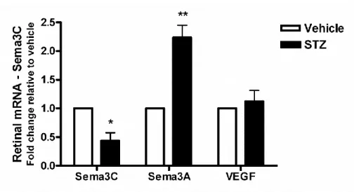
Figure 7: Retinal Semaphorin 3C expression decreases early on in STZ-induced diabetes.
Chapter 2: Article (Accepted for revision in a peer-reviewed journal)
Figure 1: Sema3A is elevated in the vitreous of human diabetic patients suffering from diabetic retinopathy and in retinal neurons in the early phases of STZ-induced diabetes. Figure 2: Retinal barrier function is compromised by Sema3A.
Figure 3: Targeted silencing of neuron-derived Sema3A or intravitreal neutralization of Sema3A efficiently reduces diabetes-induced retinal vascular permeability.
Figure 4: Conditional knockout of Nrp-1 prevents Sema3A-induced retinal barrier function breakdown.
x List of Abbreviations
Adherens junction (AJ); age-related macular degeneration (AMD) basement membrane (BM); basic fibroblast growth factor (bFGF); blood-brain barrier (BBB); blood-nerve barrier (BNB); blood- retinal barrier (BRB); central nervous system (CNS); diabetic macular edema (DME); diabetic retinopathy (DR); diabetic nephropathy (DNeph); diabetic neuropathy (DN); Evans blue (EB); epiretinal membrane (ERM); focal adhesion kinase (FAK); glomerular filtration barrier (GFB); ganglion cell layer (GCL); Hôpital Maisonneuve-Rosemont (HMR); human retinal microvascular endothelial cells (HRMEC); human umbilical vein vascular endothelial cells (HUVEC); inner nuclear layer (INL); macular hole (MH); neuropilin 1 (Nrp-1); NG2 proteoglycan (NG2); lentivirus (Lv); outer nuclear layer (ONL); pericyte (PC); placental growth factor (PlGF); plasmalemma vesicle associated protein (Plvap); platelet derived growth factor receptor (PDGFR); peripheral nervous system (PNS); proliferative diabetic retinopathy (PDR); Public Health Agency of Canada (PHA); retinal ganglion cells (RGC); semaphorin 3A (Sema3A); small hairpin RNA (shRNA); smooth muscle actin (SMA); spectral-domain optical coherence tomography (SD-OCT); stomatal diaphragms (SD); streptozotocin (STZ); whole-animal tamoxifen-inducible Cre mouse (TamCre-Esr1); tight junction (TJ); Type 1 diabetes mellitus (T1DM); Type 2 diabetes mellitus (T2DM); vascular endothelial cadherin (VE-cadherin); vascular endothelial growth factor (VEGF); VEGF Receptor-2 (VEGFR-2); vascular permeability (VP); vesiculo-vacuolar organelles (VVOs).
xi Acknowledgements
First of all, I would like to sincerely express my gratitude to my supervisor, Dr. Przemyslaw Sapieha, for the constant support and direction he provided me during this odyssey of research that we both embarked on over two years ago. I consider myself fortunate to have joined Dr. Sapieha’s laboratory at its genesis for I have acquired invaluable professional, academic and personal experience in a milieu of creative minds and high standards that I believe would have been difficult to come by elsewhere.
I would also like to thank the members of the Thesis Jury, Dr. Nathalie Granvaux and Dr. Kenneth Hastings, for partaking in the review of this thesis and for providing the feedback that helped to improve it.
In addition, I want to thank my colleagues, Eric Lapalme, Nicolas Tetreault, Catherine Menard, Nicholas Sitaras, Agnieszka Dejda, Dominique Leboeuf, Chintan Patel, François Binet, Flavio Rezende, for the great and invaluable work that led to our article. Thank you very much, as well, to Jida El Hajjar and the personnel from the Animal Centre at HRM for the help provided.
I would like to give special thanks to Biology professor Ana Maiorana, who inspired me to follow an education in Biological Sciences. Moreover, I also want to show my appreciation to the other educators at Colegio Nacional de Buenos Aires for the priceless teachings I received from them that fully prepared me to face an education anywhere in the world.
On another front, I want to thank de corazón my very good friends here in Canada Courtenay, Mauricio, Glyn, Chantelle, Norine, Maeve, Naureen, Jason, Eimear and Douglas who gave me their unconditional support throughout these two years of hard and rewarding work. The same goes to SdV, my friends and brothers, from Argentina.
Last but not least, thank you very much to my family for their love: my siblings Federico, Lucia and Sebastian; my grandparents, Alfredo, Dolly, Mario and Mirta; my awesome aunt Paula and uncle Pablo; my cousins and my step-parents Claudio and Maria Laura. But above all, I want to offer my most wholehearted gratitude to my parents Celina and Jorge for the love and support they give me every day, for the hardships they endured to ensure my happiness: for everything.
1
Chapter1:
Background
2 1.1 Diabetes
1.1.1 A major public health issue
Diabetes encompasses a group of diseases characterized by hyperglycemia and glucose intolerance as a result of insulin deficiency and/or impaired sensitivity to insulin. This condition is classified into two groups: Type 1 diabetes mellitus (T1DM), which is characterized by an autoimmune destruction of pancreatic beta cells, and Type 2 diabetes mellitus (T2DM), which occurs in 90% of cases, has a more diverse etiology and manifests itself later in life (1).
Diabetes worldwide has reached epidemic proportions, affecting both developed and developing countries. Globally, the prevalence of diabetes in the age group from of 20 to 79—number of diabetics over total population—was estimated at 6.4% in 2010 (285 million adults) and is expected to reach 7.7% by 2030 (439 million adults) (2). Specifically in North America, the prevalence of diabetes was reported to be 10.2% in 2010 and represented the highest in the world.
As of 2009 there were approximately 2.4 million Canadians (6.8%) living with diabetes. The Public Health Agency of Canada (PHA) has estimated that for the 11-year period between 1998/99 to 2008/09 the age-standardize prevalence rate of diabetes increased by 70% (3). Additionally, assuming incidence rates continue to rise in the context of the 2008/09 mortality rates, the PHA projects that by 2018/19 there will be 3.7 million Canadians with diabetes. It is therefore clear that diabetes represents a major public health issue that will continue to significantly burden healthcare systems given the annual per capita health care cost for diabetics is approximately four times that of non-diabetics.
3
It is therefore crucial to gain further pathophysiological insight to this condition and consequently develop more cost-effective and efficient treatments for this condition (3-5).
In terms of the impact of diabetes on mortality rates,Vital Statistics data in Canada underestimate the association between diabetes and mortality because diabetes is rarely recorded as cause of death on death certificates (3, 6). For instance, in 2007 3.1% of all deaths were attributed to diabetes as a primary cause, although 29.9% of the total diseased had been diagnosed with diabetes (6). In addition, in 2008/09, Canadians aged 20 to 39 and 40 to 74 years showed all-cause mortality rates of 4.2 to 5.8 and 3.6 to 2 times higher, respectively, in diabetics versus non-diabetics (Figure 1a). It is therefore clear that people affected by diabetes are more likely to die prematurely in every age group, and that it is a life-threatening disease.
1.1.2 Complications Related to Diabetes
The underestimated relation between diabetes and death comes from the fact that numerous diabetes- related deaths are reported to arise from the complications associated with diabetes and not from the disease itself (Figure 1b). Diabetes-related comorbidities also contribute significantly to the total burden of the disease on the healthcare system since the medical- and mortality-associated costs of diabetes increase by 3.6 fold (1.024 to 3.701 billion US dollars) when the cost of complications are included (5). Therefore, it is necessary to further understand the genesis of diabetes-related comorbidities in order to prolong the life expectancy of patients suffering from this condition.
4
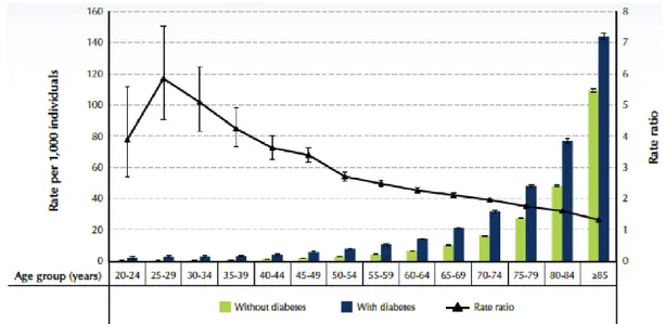
Figure 1. Diabetes: mortality and complications. (a) All-cause mortality rates and rate ratios among individuals aged 20 years and older, by diabetes status, Canada, 2008/09 (b) Prevalence rate ratios, standardized to 1991 population, of complications among hospitalized individuals aged 20 years and older, Canada, 2008/09. Source: Public Health Agency of Canada (August 2011); using 2008/09 data from the Canadian Chronic Disease Surveillance System (Public Health Agency of Canada). Modified from (3).
5
Complications secondary to diabetes can be divided in short- and long-term. Short-term complications are associated with: 1) poor glycemic control and hyperglycemia that can lead to infection/slow wound healing, as well as diabetic ketoacidosis (DKA) and hyperglycemic hyperosmolar nonketotic syndrome (HHNS); and 2) inadequate utilization of glycemic control treatments, e.g. insulin, which can trigger hypoglycemic episodes. Nevertheless, the most health-threatening complications that have a more direct impact on quality of life and life-expectancy are those that arise due to long-term exposure to hyperglycemia. These include, but are not limited to cardiovascular, ophthalmic (e.g. retinopathy, glaucoma, cataracts) and renal disease (nephropathy), as well as nerve damage (central and peripheral neuropathy), lower extremity lesions (peripheral vascular pathology, which can lead to amputation), gingivitis/periodontis and depression (3, 7-11).
Interestingly, however, the effects of hyperglycemia on the vasculature are the main source of morbidity and mortality in Type 1 and 2 diabetes (12). At the macrovascular level, complications arise in the form of coronary artery disease, peripheral arterial disease and stroke, whereas microvascular complications originate mainly in the retina, brain and kidneys (see Discussion for more information on diabetic microvascular pathology on brain and kidney).
1.1.3 Diabetic microvascular pathology in the retina
Diabetic retinopathy (DR) is the most common complication associated with diabetes (12). DR also represents the leading cause of blindness in working age adults, affecting approximately 50 % of diabetics (13). This form of microvascular diabetic pathology is characterized by a decrease in the barrier function of the endothelium that leads to increased extravasation of plasma components into the underlying interstitium and
6
tissue. In the retina, as well as in other tissues, diabetes-linked vascular deterioration of the retinal capillaries and endothelial barrier breakdown— blood- retinal barrier (BRB) in this case—have been associated with an overall state of hypoxia, which represents the first stage of diabetic retinopathy. DR generally manifests itself through an initial microvascular degeneration that can induce a destructive and excessive vascularization, during its proliferative stage (Proliferative DR, PDR), in an attempt to reinstate metabolic equilibrium in the hypoxic retina. This shortage in oxygen is believed to be triggered by hemoglobin glycation (i.e. non-enzyme mediated glycosylation), which increases the pigment’s oxygen affinity thus hindering efficient delivery of O2 (14). Hypoxia is further enhanced in the diabetic retina as a result of increased resistance to blood flow that reduces capillary circulation to the already stressed neural retina due to: a thickening of the basement membrane (BM; thicker BM reduces the rate O2 diffusion into the irrigated tissue and decreases capillary elasticity); and an abnormally high blood viscosity, which is most salient in diabetics with retinopathy (15).
In addition, PDR is characterized by two prominent pathological features: 1) a decrease in the barrier function of retinal vessels that leads to a detrimental increase in vascular permeability, which leads to vasogenic oedema, retinal thickening and a subsequent loss of central vision; 2) an abnormal and misdirected growth of these leaky vessels towards the vitreous, which can ultimately cause retinal detachment.
However, increased retinal vascular permeability is not only seen during the proliferative stage of DR, which is the most common cause of vision loss in Type 1 diabetics. Vascular leakage can also arise independently, leading to diabetic macular edema (DME), which represents the main cause of loss of visual acuity in Type 2 diabetics (16). In DME, the retina secretes vascular endothelial growth factor (VEGF) as a result of
7
hypoxic conditions, which decreases inter endothelial cells (EC) adherens junction interactions, increases endothelial fenestrations, thus promoting vascular permeability, which leads to retinal thickening and visual complications. As a result, there is a new approach in the treatment of DME that involves the use of anti-VEGF agents that appear to be more efficient and less invasive in mitigating vascular leakage, and therefore edema, than previous treatments such as corticosteroids and photocoagulation (16).
Notably, the effects of hyperglycemia on the macro- and microvasculature are the main source of illness and death in diabetes (12). As the term indicates, diabetic microvascular pathology involves the microvessels of the organism, which include the arterioles, metarterioles, capillaries and venules. Unlike the macrovessels, e.g. arteries, that are composed of endothelium, internal elastic lamina, basement membrane, external elastic lamina and adventitia, the microscopic vessels of the vasculature are structurally characterized by a much simpler tissue make up. As such, capillaries are primarily composed of endothelium, basement membrane and pericytes. However, the capillary endothelium exhibits a high degree of structural variation depending of the nature and requirements of the tissue it irrigates.
1.2 The vasculature
1.2.1 The endothelium
In vertebrates, blood circulates in a closed system from the heart to every tissue in the organism through the arteries, arterioles, capillary beds. Once the tissue is perfused, venules and veins return the blood to the heart, thus completing the circuit. The collective of these structures form the circulatory system, which is in charge of delivering oxygen
8
and nutrients to the various tissues, and collecting wastes and carbon dioxide from them for their disposal. The common denominator to the different components of the vasculature is the endothelium, formed by a layer of cells (endothelial cells) that lines the lumen through which the blood circulates.
The endothelium exhibits a wealth of phenotypes as determined by the relative expression of numerous junctional and adherence proteins, specialized structures for transport (e.g. channels, transporters, etc.), as well as the coverage of the basement membrane. Different combinations of these components allow the endothelium to adapt to the numerous physiological requirements imposed by the several tissues that the vasculature irrigates. Such heterogeneity of the endothelium can be already observed in hagfish, whose ancestor is the last common ancestor of all modern vertebrates, thus suggesting that heterogeneity evolve as an early and necessary characteristic of ECs (17).
During development, ECs arise from mesoderm through the differentiation of hemangioblast/angioblast, yet other cell lineages, such as adipose and neural stem cells, have the ability to transdifferentiate into ECs as well (18-20). In addition, ECs have in common very few specific protein or mRNA markers, of which the most uniformly expressed, though not unique to them, are vascular endothelial cadherin (VE-cadherin), platelet/endothelial cell adhesion molecule 1 (PECAM-1) and thrombomodulin (20). The varied ontogenical origins of ECs and the difficulty to find characteristic molecular markers reflect, once again, the phenotypic variability that characterizes the endothelium.
From a functional perspective, the endothelium serves several roles including the regulation of vasomotor tone, angiogenesis, innate and acquired immunity, leukocyte trafficking and hemostasis (20). Throughout the range of different physiological functions
9
that the endothelium must perform, it is key that vascular integrity is not compromised for prolonged periods of time. Controlled vascular permeability is crucial for proper transport and delivery of water and solutes between the blood and the underlying interstitium. Conversely, breakdown of vascular barrier function is the hallmark of several pathologies.
1.2.2 Across the endothelium: paracellular and transcellular routes
The molecular exchange across the vasculature occurs mostly at the level of capillaries either via the paracellular route, i.e. in between ECs, or via the transcellular route, through the cell. The former is determined and regulated by two major types of intercellular junctions: a) tight junctions (TJ) composed of occludins and claudins; and b) adherens junctions (AJ) formed by cadherins. Both TJs and AJs not only connect ECs and limit the movement of macromolecules across the endothelium while allowing smaller solutes and water to diffuse, but also contribute to setting up their polarity by establishing the border between luminal and abluminal sides. Some examples of EC-specific junctional proteins include VE-cadherin and claudin-5 (21, 22) (Figure 2).
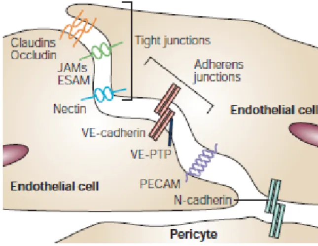
Figure 2. Examples of tight and adherens junctions found in the endothelium. The movement of blood components via the paracellular route is determined and regulated by two major types of intercellular junctions: tight junctions composed of occludins and
10
claudins; and adherens junctions formed by cadherins. Both TJs and AJs determine the border between luminal and abluminal aspects, thus helping establish proper endothelial cell polarity. Some examples of EC-specific junctional proteins are VE-cadherin and claudin-5. Modified from (23).
In addition to the paracellular transport that occurs through the intercellular cleft, molecules can permeate the endothelium directly through ECs via the transcellular route following three different paths. First, transmembrane transporters differentially positioned and distributed along the opposite ends of the ECs, carry water and small molecules vectorially. Second, macromolecules move across the epithelium in membrane-bound carriers in a process denominated transcytosis. This system operates through several components: 1) caveolae (or plasmalemmal vesicles) are plasma membrane spherical invaginations of regular size (70 nm) that occur individually or in grape-like clusters on the luminal and abluminal aspects of ECs (24, 25), and whose openings or stomata can contain selectively permeable stomatal diaphragms (SDs) (26); 2) vesiculo-vacuolar organelles (VVOs) are conglomerates of interconnected vesicles that span from luminal to abluminal ends across the cytoplasm of ECs and are separated by SDs (27). Even though, caveolae and VVOs share a high morphological resemblance, EM data from caveolin 1-knockout mice lacking caveolae (but still containing VVO-like organelles) suggest that they are functionally distinct structures (28).
Third, molecules can also diffuse transcellularly through pores or channels that may be selective or not. Examples of such structures include: 1) fenestrae (Latin for “window”), which are regular circular openings that cover the entire length of the EC and run individually or in groups to form a “sieve plate”. Most fenestrae carry fenestral diaphragms (FDs) on the luminal side and, unlike SDs, contain protein tufts that limit their permeability; 2) transendothelial channels (TEC) are pores that span across ECs, arise from
11
the interconnection of two to four luminal and abluminal caveolae and contain two SDs on each aspect of the EC (29). The relative abundance and localization of transporters, caveolae, VVOs, fenestrae, TECs and their related structures determine the phenotypic and, thus, functional heterogeneity of the endothelium (Figure 3).
Another important structure associated with the endothelium is the basement membrane (BM), which is a layer on which the EC monolayer lies. It is made of secreted extracellular proteins that include elastin, collagen IV, enactin/nidogen, heparan sulfate proteoglycans and laminin (30). Based on the degree of coverage (i.e. continuous/discontinuous) and organization of the BM, as well as by the presence or absence of fenestrae, the endothelium can be classified into three types: continuous fenestrated, continuous non-fenestrated, and discontinuous/sinusoidal (Figure 3). Continuous non-fenestrated endothelium is found in the capillaries of the brain, retina, lung and skin, as well as in veins and arteries and is characterized by low and highly selective permeability (20). In turn, fenestrated continuous is present in tissues where transendothelial transport and filtration are crucial, such as endocrine glands, choroid capillaries, glomeruli and gastric and enteric mucosae. Finally, discontinuous or sinusoidal epithelium is highly permeable and is characteristic of liver vascular beds. Fenestrations in the liver are larger (100-200 vs. 70 nm in diameter), contain gaps within individual cells and lack diaphragms when compared to continuous fenestrated tissue. The different BM arrangements and degrees of fenestrations allow the endothelium to regulate its permeability accordingly and, thus, adapt to the numerous and specific physiological requirements of the various tissues that are irrigated the given vasculature.
12
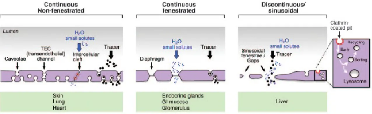
Figure 3. Types of endothelia and their associated components. Continuous non-fenestrated endothelium is found in the capillaries of the brain, retina, lung and skin, as well as in veins and arteries and is characterized by a low and highly selective permeability. Fenestrated continuous endothelium can be encountered in tissues where transendothelial transport and filtration are crucial, such as endocrine glands, choroid capillaries, glomeruli and gastric and enteric mucosa. Discontinuous or sinusoidal endothelium is highly permeable and is characteristic of liver vascular beds. Modified from (20) .
1.2.3 The Pericytes
Embedded in the BM, resides another important component of the vasculature: the pericytes (PC). These cells play a unique role as evidenced in their association with the BM and special contacts shared with the ECs (31). PCs are found in precapillary arterioles, capillary beds, postcapillary venules and collecting venules (32), and have been shown to develop from various embryonic tissues including trunk vessels in the axial and the lateral plate mesenchyme (33), neural crest cells in the brain (34) and epicardial cells in the splanchnic mesoderm (35).
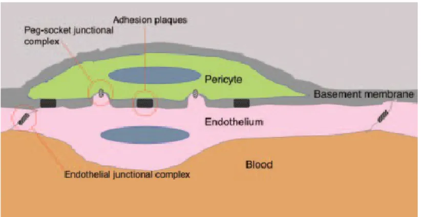
In most vascular beds, pericytes are found embedded in the BM, which separates them from the ECs, and contribute to vascular structure. PCs thus interact with ECs indirectly via fibronectin-rich adhesion plaques (36). However, in areas of the endothelium
13
where the BM is not present or is very thin, pericytes interconnect with ECs through membrane invaginations called peg-socket contacts that possess an array of junctional complexes that include tight-, gap- and adherens junctions, such as N-cadherin (Figure 4) (31, 37). Interestingly, one pericyte is usually associated with several ECs, thus, suggesting a role in the coordination and integration of cellular communication among neighboring ECs.
PCs provide a plethora of physiological functions. Pericytes show the highest rate of microvascular coverage within the central nervous system (CNS) where they actively partake in the formation of the blood-brain barrier (BBB) during embryogenesis and provide neuroprotection (38, 39). During this process, increased pericyte coverage strengthens barrier function and decreases vascular permeability of the developing brain vasculature by: inducing tight junction formation, hindering EC expression of molecules that enhance transcytosis such as Plvap, and limiting immune cell infiltration (38).
Similarly, in the retina, pericytes interact with the underlying continuous non-fenestrated endothelium and regulate regional blood flow by contraction through the action of contractile proteins such as actin, myosin and tropomyosin (37, 40). They contract in hyperoxic conditions (41), as well as in the presence of ATP, and relax when exposed to nitric oxide and CO2 (42, 43). Pericytes have also been involved in the control of capillary structure, inhibition of endothelial cell proliferation and angiogenesis (14). Moreover, PCs play an important role in maintaining the integrity of the internal blood-retinal barrier (iBRB), thus controlling the vascular barrier function of the retinal capillaries. This is achieved through EC-pericyte-induced of the tight junctional proteins occludin and zona occludens 1 (ZO-1) during normoxia (44).
14
The maintenance of blood-barriers, BBB and BRB, in the CNS is crucial for the proper functioning of the brain and retina, respectively, and their breakdown is associated with the etiology of several pathologies, including diabetic neuropathy and retinopathy, amyotrophic lateral sclerosis and stroke (14, 45-47).
Figure 4. Endothelial-pericyte interaction in the microvasculature. The pericytes are found embedded in the BM in most vascular beds. This layout separates the PC from the ECs .PCs interact with ECs indirectly via fibronectin-containing adhesion plaques. However, in those areas where the BM is not present, pericytes contact ECs through membrane invaginations called peg-socket contacts that posses an array of junctional complexes that include tight-, gap- and adherens junctions such as N-cadherin. One pericyte is typically associated with several ECs, suggesting a role in the coordination and integration of cellular communication among neighboring ECs. Modified from (31).
1.3 Nerves and vessels
1.3.1 Common guidance
Since the appearance of metazoans, there has been evolutionary pressure to develop sensory and motor capabilities to allow a more efficient interaction between the organism and its environment. The advent of nervous tissue and subsequent development of a central
15
nervous system allowed for such a sensory-motor processing. As organisms became larger with more complex body plans, they required a more elaborate nutrient and oxygen delivery system. This was achieved with the development of a convection system such as the circulatory system.
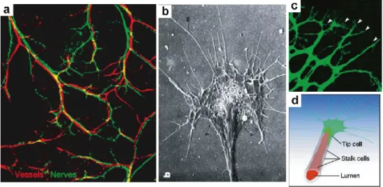
In order for the nervous and circulatory systems to innervate and irrigate a given tissue, vessels and the axons of neurons have to successfully extend and navigate through the developing organism. Interestingly, vessels and nerves follow a common stereotyped path through the body, even though macroscopically they seem to develop distinctly (48). Microscopically, nerves and vessels share an analogous structure at their growing fronts: the growth cone of the neuron’s axon and the tip cell of the developing/branching endothelium (Figure 5). These specialized structures are highly chemosensitive, malleable and dynamic, constantly projecting and retracting filopodia as a result of attractive and repulsive cues found in the surrounding interstitium. These cues help nerves and vessels find their targets and are determined by the relative concentration of guidance molecules present in their microenvironment, as well as by the differential expression and distribution of the corresponding receptors for these guidance cues.
Beyond anatomical similarities, the molecules and receptors involved in nerve pathfinding have also been found to guide vessels. Examples include netrins and their Unc5 and DCC receptor families (49-58); slits and their robo receptors (59-66); ephrins and their eph receptors (67-73); and semaphorins and their neuropilin and plexin receptors (49-51, 74-79).
16
Figure 5. Nerves and vessels follow stereotyped trajectories and share similar cellular structural features. (a) Nerves and vessels follow the same path; murine skin sensory nerves assist in proper arteriogenesis (80). (b) Axon and growth cone containing several filipodia (scanning electron image). (c) Proliferating vascular network, showing a growing front containing tip cells that project several filipodia (white arrowheads). (d) Schematic of extending stalk cell attached to the leading filipodia-rich tip cell. Figured modified from (48, 80).
Interestingly, however, semaphorins serve other roles beyond axon and vessel guidance. They are expressed in endocrine, gastrointestinal, hepatic, immune, musculoskeletal, renal, reproductive and respiratory systems, where they serve distinct functions (79, 81-94). The numerous biological functions of semaphorins arise from their shared ability to affect cytoskeleton dynamics. By altering actin filaments and the microtubule network, semaphorins are able to influence cellular morphology, attachment, migration, polarization and growth; this is a characteristic that makes them an attractive target for study (92).
17
1.3.2 The Semaphorins
While the biological functions of semaphorins have been studied for the past 20 years, novel physiological roles continue to emerge (74). There are eight main classes of semaphorins: class 1 and 2 are found in invertebrates, 3 to 7 are found in vertebrates, in addition to the V class that is only found in viruses (95). Semaphorins can be secreted, diffusible and act long-distance, or be membrane-bound and act short-distance , but all of them have in common a 500 amino acid sequence, the semaphorin domain (49-51, 74, 92, 96). This is the most highly conserved domain across all the classes and is responsible for mediating their effects (97-99). Semaphorins are ubiquitously expressed in the organism, but they were originally characterized as axonal guidance molecules in the development of nervous system (100).
The first dicovered semaphorin of the secreted family was Semaphorin3A (Sema3A), which was initially discovered to promote axonal collapse (74). Sema3A signals to target cells through a direct interaction with neuropilin 1 (Nrp-1) receptor, which is also a co-receptor for vascular endothelial growth factor (VEGF) (101, 102). Sema3A also acts indirectly via the coreceptor plexinD1 (92). In the nervous system, Sema3A generally affects growth cone morphology by destabilizing the peripheral cytoskeleton; it promotes the depolymerization and hinders the repolymerization of F-actin, also trumping microtubule dynamics, thus promoting partial or total cellular structural collapse (51). Moreover, Sema3A can induce apoptosis as evidenced: in vitro, by exposing primary neurons to this protein; and in vivo by the protective effect of its neutralization on retinal ganglion cells (RGCs) following optic nerve axotomy, which is a phenomenon known to lead to RGCs apoptosis (85, 103-105).
18
More recently, Sema3A and its receptors were found to be involved in cardiovascular development (106). Endothelial cells not only express Nrp1 and plexinD1, but in the presence of Sema3A, EC proliferation is impaired thus hindering vessel branching (78, 101). Interestingly, one study found Sema3A to affect mature vessels by destabilizing inter endothelial cell junctions and thereby promoting increased vascular permeability, in addition to inhibiting VEGF-induced proliferation (93).
From a pathological point of view, Sema3A has been found to participate in nervous system pathologies, such as schizophrenia and Alzheimer’s disease (107, 108). In addition, Sema3A has been shown to block tumor growth and normalize tumor vasculature, as well as blocking and misdirecting physiological revascularization in a model of retinopathy of prematurity (86, 89).
It is not surprising that Sema3A serves an important role during development, homeostasis and even pathology, considering its effects on cell survival, proliferation and cytoskeletal dynamics. Even though Sema3A has been mostly characterized in the nervous and circulatory systems, the evidence indicating its involvement in other tissues continues grow.
1.4 The retina
The retina contains the photosensitive tissue of the eye. It is part of the central nervous system (CNS) and is highly organized in three layers of neurally derived cells (the neural retina) that are closely associated with the retinal vasculature (vascular retina), which feeds this highly metabolic tissue. The neural retina, from the outside to the inside of the eye, is composed of: the retinal pigmented epithelium (RPE), the photoreceptor layer (rods and
19
cones), the outer nuclear layer (ONL, cell bodies of rods and cones), the outer plexiform layer (synapses between photoreceptors and bipolar cells), the inner nuclear layer (INL, nuclei and cell bodies of the bipolar cells), inner plexiform layer (synapses between bipolar cells), the ganglion cell layer (ganglion neuron bodies and nuclei) and the nerve fiber layer (axons from the ganglion neurons) (Figure 6). When light enters the eye, it stimulates the photoreceptors resulting in a chemoelectrical impulse that travels via the afore-mentioned cellular path. From the ganglion neurons’ nerve fibers the transduction signal is relayed to the primary visual cortex where it is further processed to produce the phenomenon of vision. Because of the high energy demand required for phototransduction and its related metabolic pathways, the retina consumes oxygen at a higher rate than in any other tissue (14, 109). The highest oxidative enzymatic activity of the retina has been localized to the RPE and inner segment of photoreceptors, where there is a constant, arduous synthesis and daily replacement, respectively, of phototransduction-specific cell components (i.e. pigments) (14, 110-112). High metabolic activity in these tissues can also be evidenced by the large density of mitochondria present in them, which indicates a strong reliance on aerobic metabolism (14, 113-115).
20
Figure 6. The neural and vascular retina: cross-section. (a) Schematic of the retina and (b) immunofluorescent staining of a retina cryosection dyed for nuclei (blue, DAPI) and retinal vessels (red, Lectin) illustrate the close association between the neural and vascular retinae. GCL, ganglion cell layer; IPL, inner plexiform layer; INL, inner nuclear layer; OPL, outer plexiform layer; ONL, outer nuclear layer; RPE, retinal pigmented epithelium. (a) Figure modified from (14).
In order to maintain a high supply of oxygen, the adult mammalian retina is fed by two vascular systems: 1) the choroidal vessels that lie outside the retina, are highly fenestrated and, thus, permeable and provide support to the RPE and photoreceptors; 2) the retinal vessels irrigate the remaining neural components of the retina via an outer plexus that lies between the INL and outer plexiform layer, and a second plexus located in the proximity of the GCL and nerve fibers (116). The retinal vasculature posses a similar barrier to that found in the rest of the CNS vasculature termed blood-retina barrier, which makes the retinal endothelium very selectively permeable. In some clinical conditions such as central retinal vein occlusion, retinopathy of prematurity and diabetic retinopathy, the resulting hypoxia that affects the retina leads to a degradations of the BRB, yielding increased and pathological vascular permeability (14).
1.5 Hypothesis and Objectives
The pathological barrier-breakdown observed in DR has received considerably less attention than the pathological pre-retinal vascularisation (i.e. neovascularization) that is characteristic of the advanced stages of DR (117-119). As a result, the current standards of care present side-effects that cannot be ignored. These include increased cataract formation and a detrimental rise in intraocular pressure with intravitreal use of corticosteroid (117). Comparably, anti-VEGF therapies, which are generally effective, may be associated with
21
increased thromboembolic events, possible neuronal toxicity and geographic atrophy when used frequently as long term regimens (120-122). In addition, even though panretinal
photocoagulation and grid/focal laser are the most widely used forms of treatment for PDR and DME, respectively, they destroy hypoxic retinal tissue, which inadvertently leads to reduced visual field and promotes secretion of pro-angiogenic factors. As a result, these therapeutic limitations emphasize the need for novel pharmacological approaches.
In addition, from a physiological perspective, the presence of VEGF in the diabetic retina does not explain other prominent pathological features of DR, such as the initial vascular decay or the misdirected nature of the pathological neovascularization observed in PDR. While several other factors such as hyperglycemia and oxidative stress have been linked to vascular decay in DR, the mechanisms that precipitate vessel breakdown remain largely ill-defined.
Here we focused on the neurovascular guidance cue semaphorin 3A (Sema3A) and its potential role in mediating barrier function compromise in diabetic retinopathy. Our lab has previously shown that Sema3A participates in vascular degeneration and later blocks physiological vascularization in a different model of proliferative retinopathy that shares common features with PDR and is used to study retinal pathology in the premature newborn retina (86). At the same time, Sema3A has been shown to be a strong inducer of vascular permeability (93). In this context, we hypothesize that Semaphorin 3A provokes vascular permeability in Diabetic Retinopathy. Thus, we set out:
1) To elucidate the dynamics of semaphorin 3A induction in diabetic retinopathy
2) To evaluate the role of semaphorin 3A in the barrier function of the retinal vasculature 3) To elucidate the mechanism through which semaphorin 3A acts on barrier function
22
Chapter 2: Article
(
Accepted for revision in a peer-reviewed journal)
Neuronal-Derived Semaphorin 3A is an Early Inducer of
Vascular Permeability in Diabetic Retinopathy
23 Contributions by Figure:
Candidate’s name: Agustin Cerani (AC)
1. Figure 1: Sema3A is elevated in the vitreous of human diabetic patients suffering from
diabetic retinopathy and in retinal neurons in the early phases of STZ-induced diabetes.
Preparation and set up of the entire Figure 1 was performed by AC.
A-D: OCT and 3D retinal map data were obtained from Dr. Rezende (ophthalmologist). Images were selected by AC.
E: Human vitreous samples were obtained and provided by Dr. Rezende; western blot was perfomed by Catherine Menard (CM); picture was prepared by AC. F: Patient data was organized by AC
G: drawing modified by AC from Wei Li’s design.
H-J: STZ diabetes induction protocol was carried out by AC. Tissue extraction and preparation was performed by AC. Data analysis, statistics and graph preparation were performed by AC.
K-N: Retinas were extracted and prepared by AC (except for some stainings and imaging).
O: Retinas were extracted and prepared by AC; laser capture microscopy (i.e. collection of micro-cuts) was perfomed by AC.
2. Figure 2: Retinal barrier function is compromised by Sema3A.
A: microsurgery/intravitreal injections and Evans Blue Permeation assay (technique adapted and optimized by AC) were carried out by AC. Data analysis, statistics and graph preparation done by AC.
B: microsurgery/intravitreal injections of Evans Blue performed by AC. Extractions of eyes and sample preparation done by AC.
C: Part of cell culture of HUVECs, part of statistics and graph preparation by AC. ECIS performed by Chintan Patel.
D: Drawing by Dr. Sapieha. E: Drawing done by AC.
F-H: Part of HRMEC culture and cell treatment by AC. Blots and quantification by CM. Picture and graph preparation for figure by AC.
I: HRMEC culture by AC. The rest performed by Dr. Nicolas Tetreault (NT). J: HRMEC culture and plan by AC. Treatment and Blot by Dr. Agnieszka Dejda
24
3.
Figure 3: Targeted silencing of neuron-derived Sema3A or intravitreal neutralizationof Sema3A efficiently reduces diabetes-induced retinal vascular permeability.
A: STZ diabetes induction protocol was carried out by AC. Tissue extraction and preparation was performed by AC. Immunofluorescence, imaging and panel preparation by NT.
B, E, G: microsurgery/intravitreal injections and Evans Blue Permeation assay (technique adapted and optimized by AC) were carried out by AC. Data analysis, statistics and graph preparation done by AC.
C: Perfomed by Nicholas Sitaras (NS).
D: Microsurgery/intravitreal injections of Lv.shGFP and Lv.shSema3A by AC; STZ diabetes induction protocol was carried out by AC. Tissue extraction and preparation was performed by AC. RT-qPCR by NT. Data analysis, statistics and graph by AC.
F: Drawing by Wei Li
4. Figure 4: Conditional knockout of Nrp-1 prevents Sema3A-induced retinal barrier function breakdown.
A: Drawing by Ac.
B-D: Planning, Tamoxifen protocol adaptation and optimization by AC. Samples were extracted and prepared by AC; blot and RT-qPCR by CM. Data analysis, statistics and panel preparation for figure by AC. Immunofluorescence by AD. E-F: Planning, Tamoxifen protocol adaptation and optimization by AC.
microsurgery/intravitreal injections and Evans Blue Permeation assay (technique adapted and optimized by AC) were carried out by AC. Data analysis, statistics and graph preparation done by AC.
G-I: Part of HRMEC culture of AC. Blot, quantification and statistics by CM. Panel preparation by CM and AC.
J: Drawing by Dr. Sapieha.
Note: For Evans Blue (EB) Permeation, Dominique Leboeuf assisted with intravenous injections of EB.
25
Title: Neuron-Derived Semaphorin 3A is an Early Inducer of Vascular
Permeability in Diabetic Retinopathy
Authors: Agustin Cerani1,2,4,Nicolas Tetreault3,4, Catherine Menard2,4, Eric Lapalme1, Chintan Patel1,Nick Sitaras1, Dominique Leboeuf2, Vincent De Guire2,François Binet1, Agnieszka Dejda2, Flavio A Rezende1, Przemyslaw Sapieha*1,2,3
Affiliations:
1Department of Ophthalmology, Maisonneuve-Rosemont Hospital Research Centre, University of Montreal, Montreal, Quebec, H1T 2M4, Canada;
2Department of Biochemistry, Maisonneuve-Rosemont Hospital Research Centre, University of Montreal, Montreal, Quebec, H1T 2M4, Canada;
3Department of Neurology-Neurosurgery, McGill University, Montreal, Quebec, H3A 2B4 Canada.
4These authors contributed equally.
*Correspondence: Przemyslaw (Mike) Sapieha, Ph.D.
Maisonneuve-Rosemont Hospital Research Centre
Key words: Semaphorin3A, Edema, Diabetes, Diabetic Macular Edema, Diabetic Retinopathy, Retinal Ganglion Cells, Vascular Permeability.
One Sentence Summary: The neuro-vascular guidance cue Semaphorin3A is produced by retinal neurons early in diabetes and provokes vascular leakage.
26 SUMMARY:
The deterioration of the inner blood retinal barrier and consequent macular edema is a cardinal manifestation of diabetic retinopathy and the clinical feature most closely associated with loss of sight. Currently available treatments such as locally administered corticosteroids and anti-VEGF therapies, present several drawbacks. Here we provide the first evidence from both human and animal studies for the critical role of the classical neuronal guidance cue, Semaphorin3A, in instigating pathological vascular permeability in diabetes. We reveal that Semaphorin3A is induced in the early hyperglycemic phases of diabetes within the neuronal retina and precipitates initial breakdown of endothelial barrier function. We demonstrate by a series of orthogonal approaches that neutralization of Semaphorin3A or conditional knockout of its receptor Neuropilin-1 in TamCre-Esr1/Nrp1flox/flox mice efficiently prevents diabetes-induced retinal vascular leakage. Our findings identify a new therapeutic target for macular edema and provide further evidence for neurovascular cross-talk in the pathogenesis of DR.
HIGHLIGHTS
-The classical neuronal guidance cue Semaphorin3A is induced in the early hyperglycemic phases of diabetes within the neuronal retina.
- Semaphorin3A instigates pathological retinal vascular permeability in type I diabetes. - Neutralization of Semaphorin3A or its receptor Neuropilin-1 efficiently prevents retinal vascular leakage secondary to diabetes.
27 INTRODUCTION
Diabetic retinopathy (DR) is the most prominent complication of diabetes and the leading cause of blindness in working age individuals (Kempen et al., 2004). It is characterized by an initial microvascular degeneration followed by a compensatory but pathological hyper-vascularization mounted by the hypoxic retina in an attempt to reinstate metabolic equilibrium (Cheung, 2008; Sapieha, 2012). Although often initially asymptomatic, loss of sight is provoked primarily by diabetic macular edema (DME), vitreous hemorrhages and in advanced cases, pre-retinal neovascularization and tractional retinal detachment (Antonetti et al., 2012; Wang et al., 2012). Of these, DME is the most common cause of central vision loss in diabetics affecting over 25% of patients suffering from diabetes (Moss et al., 1998). It is triggered secondary to the deterioration of the blood-retinal barrier (BRB) and the consequent increase in extravasation of fluids and plasma components into the vitreous cavity. Ultimately, the decrease in retinal vascular barrier function leads to vasogenic edema and pathological retinal thickening.
Although significant effort has been invested in elucidating the mechanisms that govern destructive pre-retinal neovascularization in DR (Silva et al., 2010; Stahl et al., 2010; Wang et al., 2012), considerably less is known about the cellular processes that lead to increased retinal vascular permeability. Consequently, the current standards of care present non-negligible side-effects. These include increased cataract formation and a harmful rise in intraocular pressure with intravitreal use of corticosteroid (Silva et al., 2010). Similarly, anti-VEGF therapies, which in general exhibit respectable safety profiles, may be associated with increased thromboembolic events (Stewart, 2012), possible neuronal toxicity (Robinson et al., 2001) and geographic atrophy (Comparison of Age-related Macular Degeneration Treatments Trials Research et al., 2012; Group et al., 2011) when used as frequent long term regiments. Moreover, the first and most widely used form of treatment is panretinal photocoagulation, either for proliferative diabetic retinopathy PDR or grid/focal laser for DME. Laser-based photocoagulation approaches destroy hypoxic retinal tissue secreting pro-angiogenic factors and inadvertently lead to reduced visual field or central or paracentral scotomas. These therapeutic limitations highlight the need for novel pharmacological interventions.
28
Current investigations into the molecular mechanisms that cause DME have largely focused on VEGF. This may in part be attributed to the fact that the prominent clinical features of DR have led to the general inference that it is entirely of a microvascular nature. Yet evidence points to early changes in the neural retina (Kern and Engerman, 1996) (Barber et al., 2005; Barber et al., 1998; Gastinger et al., 2008). While there is irrefutable evidence for a neurovascular link in the progression of DR (Antonetti et al., 2012), neurovascular cross-talk has received limited attention in the context of DR pathogenesis. Consistent with a breakdown in neurovascular cross-talk in ischemic retinopathies, we have recently shown that Semaphorin 3A (Sema3A), a classic neuronal guidance cue that also affects endothelial cell behaviour is produced by stressed retinal ganglion neurons (RGCs) and partakes in deviating neo-vessels towards physiologically avascular regions of the eye (Joyal et al., 2011).
In neurons, binding of Sema3A to its cognate receptor Neuropilin-1 (Nrp-1) provokes cytoskeletal collapse (Takahashi et al., 1999); the transduction mechanism in endothelial cells remains ill-defined (Gelfand et al., 2009). Neuropilin-1 has the particular ability to bind two structurally dissimilar ligands via distinct sites on its extracellular domain (Gluzman-Poltorak et al., 2001; Lee et al., 2002; Mamluk et al., 2002). It binds Sema3A (Klagsbrun and Eichmann, 2005; Miao et al., 1999) provoking cytoskeletal collapse and VEGF165 (Gluzman-Poltorak et al., 2001; Klagsbrun and Eichmann, 2005; Klagsbrun et al., 2002; Mamluk et al., 2002)enhancing binding to VEGFR2 and thus increasing its angiogenic potential (Soker et al., 2002). Crystallographic evidence revealed that VEGF165 and Sema3A do not directly compete for Nrp-1 but rather can simultaneously bind to Nrp-1 at distinct, non-overlapping sites (Appleton et al., 2007). Moreover, genetic studies show that Nrp-1 distinctly regulates the effects of VEGF and Sema3A on neuronal and vascular development (Vieira et al., 2007). Of note, it was proposed that similar to VEGF, Sema3A may itself promote vascular permeability (Acevedo et al., 2008); a counter-intuitive observation given the divergent biological roles of VEGF and Sema3A. However, the role of Sema3A in mediating the breakdown of barrier function such as that observed in diabetic retinopathy had to date not been explored.
Here we provide the first evidence for the role of Sema3A in disrupting retinal barrier function in diabetic retinopathy. We demonstrate in both human patients and animal
29
models that ocular Sema3A is robustly induced in the early stages of diabetes and mediates via Nrp-1, the breakdown of the inner blood retinal barrier. Neutralizing Sema3A may represent an attractive alternative therapeutic strategy to counter pathologic vascular permeability in DR.
RESULTS
Sema3A is elevated in the vitreous of human patients suffering from diabetic retinopathy.
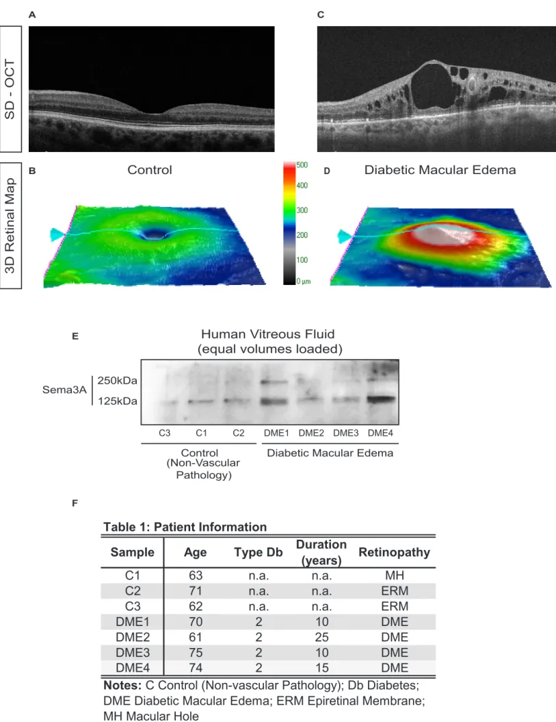
In order to evaluate the potential role of Sema3A in mediating the edematous phenotype observed in DR, we first sought to determine the presence of this guidance cue in the vitreous of patients suffering from DME. Vitreous was recovered during standard vitroretinal surgery from 7 patients. Four samples were obtained from patients suffering from DME and 3 from control patients (non-vascular pathology) undergoing surgery for macular hole (MH) or epiretinal membrane (ERM). Spectral-domain optical coherence tomography (SD-OCT) was performed and 3D retinal maps were generated to evaluate the extent of retinal damage and edema. In contrast to controls, sampled DME patients showed significant retinal swelling, specifically in the macular and peri-macular zones (Fig 1a-d).
Consistent with a prospective role in DME, Western blot analysis of patient vitreous revealed that Sema3A (125kDa) was robustly induced in most patients affected by DME (Fig 1e). In addition, a second heavier Sema3A band (>200 kDa) was detected in DME patients and corresponds to a reported functional Sema3A dimmer (Koppel and Raper, 1998). Detailed patient characteristics are presented in Figure 1f. These data on human subjects provide the rational to explore the role of Sema3A in the context of diabetes-induced retinal vasculopathy.
Neuronal Sema3A is upregulated in the early phases of streptozotocin-induced diabetes.
Given the elevated levels of Sema3A in the vitreous of DME and PDR patients, we sought to elucidate the dynamics and pattern of Sema3A expression in a mouse model of T1DM. Streptozotocin (STZ) was administered to ~6 week-old C57BL/6J mice and
30
glycemia monitored according to the scheme depicted in Figure 1g. Mice were considered diabetic if their non-fasted glycemia was higher than 17 mM (300 mg/dL). As early as 4 weeks after induction of diabetes, retinal levels of Sema3A where over 2-fold higher in STZ treated mice when compared to vehicle injected controls (P=0.0045, n=5) (Fig. 1h). Significantly higher retinal levels of Sema3A persisted at 8 weeks (P=0.0011, n=8). Importantly, throughout these early time points of diabetes, VEGF levels in STZ-treated mice remained at similar levels to that observed in vehicle treated congener mice as has been previously described (Mima et al., 2012). As expected, at all analyzed time-points, STZ-treated mice showed pathologically elevated blood glucose levels of ~30mM (p<0.0001 for both 4 and 8 weeks of diabetes) (Fig 1i).
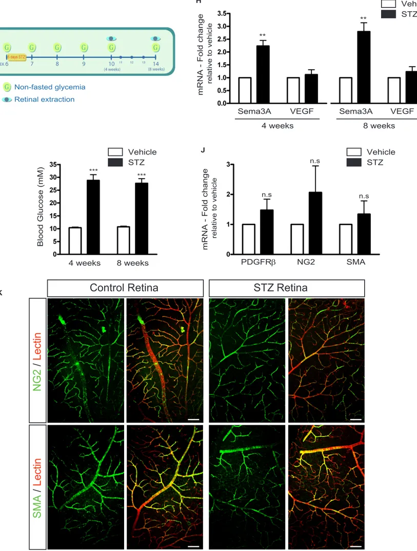
Importantly, the rise in Sema3A expression was an early event in pathogenesis as it preceded pericyte loss as both STZ and vehicle-treated mice showed no significant difference in transcript levels for pericyte markers platelet derived growth factor-receptor PDGFR- (P=0.219, n , NG2 proteoglycan (NG2) (P=0.316, n=4), and smooth muscle actin (SMA) (P=0.494, n=4) (Fig 1j). Similarly, immunohistochemistry on retinal flatmounts from control and STZ animals confirmed similar vascular coverage by NG2 and SMA-expressing pericytes (Fig 1k).
We next investigated the cellular source of Sema3A in the diabetic retina. Immunohistochemistry on retinal cryosections revealed that Sema3A was strongly expressed by retinal neurons of the ganglion cell layer (GCL) (Fig 1l&m). Co-localization with the RGC marker III-tubulin confirmed that retinal ganglion cells (RGCs) were an important source of Sema3A within the diabetic retina (Fig 1m; inset). Consistent with this retinal immuno-localization, laser-capture micro-dissection of the retinal ganglion cell layer from normal and diabetic mice followed by quantitative RT-PCR revealed a 5-fold increase of Sema3A transcript in RGCs from STZ retinas (P=0.014, n=3) (Fig 1n&o). These data provide evidence for the local production of Sema3A by diabetic neurons in close proximity to the retinal vascular plexus and agree with a role for Sema3A in mediating the vascular phenotype associated with DME and PDR.
31
Retinal barrier function is compromised by Sema3A
Given the increase in retinal Sema3A levels observed in the vitreous samples of patients with DME and mouse retinas in the early stages of diabetes (Fig 1), we proceeded to investigate the propensity of Sema3A to disrupt vascular barrier function. A single intravitreal injection of Sema3A (100 g/ml) into adult mouse retinas resulted in a significant ~2-fold increase (Fig 2a; P<0.01, n=3 distinct experiments with a total of 9 mice) in retinal vascular permeability as determined by Evans Blue (EB) permeation. This increase was similar to that observed with intravtireal administration of VEGF (50 g/ml) (Fig 2a; P<0.05, n=3 distinct experiments with a total of 9 mice) or a combination of both Sema3A and VEGF (Fig 2a; P<0.001, n=3 distinct experiments with a total of 9 mice). The propensity of Sema3A to induce vascular leakage was corroborated by confocal imaging of retinal sagittal sections where increased EB permeation throughout the retina (red) signifies elevated plasma albumin extravasation and translates into increased retinal edema (Fig 2b).
Further evidence for the ability of Sema3A to compromise endothelial barrier function, was obtained from real-time analysis of trans-endothelial electric resistance (Fig 2d). Treatment of an intact monolayer of endothelial cells with Sema3A reduced endothelial monolayer impedance (interval from 3.26h to 6h = 0.048>P>0.009; n=3) and hence a drop in barrier function in the first 6 hours by a magnitude similar to, yet lower than VEGF (interval from 1.12h to 6h = 0.045>P>0.001; n=4)(Fig 2c).
We next proceeded to determine if Sema3A activated classical signaling pathways that have reported roles in promoting vascular permeability. In this respect we investigated, by Western blot analysis, the activation profiles of Src and focal adhesion kinase (FAK) that are known to transduce extracellular signals that provoke the loosening of endothelial cell tight junctions(Acevedo et al., 2008; Eliceiri et al., 1999; Scheppke et al., 2008) (Fig 2e). Stimulation of Human Retinal Microvascular Endothelial Cells (HRMECs) by either Sema3A (100ng/ml) or VEGF (50ng/ml) lead to robust phosphorylation of Src at Tyr416 in the activation loop of the kinase domain which is reported to enhance enzyme activity (Hunter, 1987) (Fig 2f). In turn, FAK was phosphorylated on Tyr576 and 577 (sites for
32
Src-kinases) (Fig 2g). Ultimately, the tight junction protein VE-cadherin became phosphorylated on tyrosine-731, which is a posttranslational modification associated with increased vascular permeability (Potter et al., 2005; Schlaepfer et al., 1994) (Fig 2h). Consistent with the above data on retinal permeability (Fig 2a,b), we did not observe an additive or synergistic effect when simulation of HRMECs was performed with a combination of Sema3A and VEGF suggesting a potential eventual convergence of signaling pathways for both factors.
The ability of an endothelial cell to maintain intact intra-cellular junctions dictates the quality of barrier function. Consistent with a role in inducing vascular permeability, confocal microscopy of Sema3A-treated HRMECs revealed pronounced formation of vascular retraction fibers as determined by VE-cadherin and phalloidin staining (white arrows; Fig 2i). The retraction was similar to that observed with VEGF alone or with a combination of VEGF and Sema3A. Importantly, at the doses employed in our study (100-200ng/ml), Sema3A did not induce cell death or apoptosis as determined by assessment of activation (cleavage) of caspase-3 (Fig 2j). These data support role of Sema3A in mediating the breakdown of endothelial cell barrier function and further substantiate the involvement of Sema3A in diabetes-induced retinal vascular permeability.
Inhibition of neuron-derived Sema3A efficiently reduces pathological vascular permeability in T1DM.
To investigate the therapeutic potential of blocking Sema3A in diabetic retinopathy, we proceeded to inhibit Sema3A using 2 distinct approaches, namely virally delivered interference RNA or a Sema3A trap. The magnitude of retinal vascular leakage was assessed 8 weeks after administration of STZ in adult mice. At this time-point, flat-mount retinas from STZ mice show elevated expression of phosphorylated VE-cadherin in lectin-stained retinal endothelial cells (Fig 3a) and animals have a significant ~57% increase in retinal vascular leakage (Fig 3b) (P=0.027; n=4 distinct experiments with a total of 12 mice).
Recent evidence suggests that retinal neurons exert an important influence on the blood vessels that perfuse them (Antonetti et al., 2012; Binet et al., 2013; Fukushima et al.,
![Figure 4 - Part 2 G H I *** **** Lv.sh.GFPLv.sh.Nrp1Lv.sh.GFPLv.sh.Nrp1Sema3ASrcpSrcFAKpFAK VE-cadherin[Y731] pVE-cadherin pSrc / Src RatiopFAK/FAKRatio pVE-cad/VE-cad Ratio](https://thumb-eu.123doks.com/thumbv2/123doknet/2072826.6676/69.918.251.607.91.497/figure-gfplv-gfplv-asrcpsrcfakpfak-cadherin-cadherin-ratiopfak-fakratio.webp)