HAL Id: pastel-01058915
https://pastel.archives-ouvertes.fr/pastel-01058915
Submitted on 28 Aug 2014HAL is a multi-disciplinary open access
archive for the deposit and dissemination of sci-entific research documents, whether they are pub-lished or not. The documents may come from teaching and research institutions in France or abroad, or from public or private research centers.
L’archive ouverte pluridisciplinaire HAL, est destinée au dépôt et à la diffusion de documents scientifiques de niveau recherche, publiés ou non, émanant des établissements d’enseignement et de recherche français ou étrangers, des laboratoires publics ou privés.
mechanism and physical properties
Lili Lu
To cite this version:
Lili Lu. Thin films based on Prussian blue analogues: growth mechanism and physical properties. Material chemistry. Ecole Polytechnique X, 2014. English. �pastel-01058915�
Thèse présentée pour obtenir le grade de
DOCTEUR DE L’ÉCOLE POLYTECHNIQUE
Spécialité : Chimie des matériaux
Par
Lili LU
Thin films based on Prussian blue analogues:
growth mechanism and physical properties
Thèse soutenue le 21 Mai 2014 devant le jury composé de :
Thierry GACOIN LPMC, Ecole Polytechnique Président
Anne BLEUZEN ICMMO, Université Paris-Sud 11 Rapporteur
Yves DUMONT
GEMAC, Université de VersaillesRapporteur
Jean-François LÉTARD
ICMCB, Université de Bordeaux ExaminateurPhilippe ALLONGUE LPMC, Ecole Polytechnique Directeur de thèse
Isabelle MAURIN LPMC, Ecole Polytechnique Co-directeur de thèse
I Chem is try.
This work was performed in the Laboratory of Physique of Condensed Matter (PMC), thanks to a China Scholarship Council (CSC) fellowship. I would like to show my gratitude to Francois OZANAM, director of PMC, who let me work in such a great laboratory.
I would like to express my sincere gratitude to the members of the jury. In particular, I am indebted to Anne BLEUZEN and Yves DUMONT, who acted as referees and wrote a report. I am also very grateful to Francois LETARD and Thierry GACOIN examined my work. I really appreciated that all the members of my jury had carefully read and corrected my manuscript. I also enjoyed the lively discussion during the thesis defense and the brilliant ideas they suggested.
I would like to thank Philippe ALLONGUE and Isabelle MAURIN, my supervisors, who offered me the opportunity to work on PBA. Philippe ALLONGUE showed me the real attitude for science and Isabelle MAURIN provided me her continuous enthusiasm on giving me invaluable advices, on facing and solving all the problems with me. I would like to say one sentence more to Isabelle, thanks for listening, for sharing, for always be there support me.
I also wish to thank other key team members, in particular Catherine HENRY de VILLENEUVE, for teaching me the surface chemistry and showing me the secrets of “micro”-wonderland of Si substrate and Thierry GACOIN, for the fruitful discussions and suggestions. Last but not least, I am deeply indebted to Robert CORTES, for putting so much of his time and interest in helping me performing the X ray characterizations, and explaining me the physics behind.
There are also other people in this lab who gave me suggestions and ever inspired me: thanks Fouad for always being so helpful (comme Encyclopédie), for the music that cheer me up; thanks Morgane, the first PhD student who picked up the wonderful subject of Prussian blue analogue in this lab, the experiments that we did together open the door of PBA for me; thanks Charlotte, my dearest previous officemate, for always share with me good and “free“ scientific books, as well as the discussion that we had together about the spins, the orbitals, the anisotropy…
This research required various characterization techniques, which brought the help from outside of the lab: thank Emrick BRIAND and Ian. VICKRIDGE for the RBS measurement;
II
enthusiasm on this PBA subject and providing plenty of interesting and useful ideas.
It is really a pleasure to be a member of PMC, as there are so many lovely people here, I want to thank Anne-Marie DUJARDIN, who is always very warm-hearted to help me with the administrative documents; Patrice, for the “bisou” every morning; the “thésa!”: Morgane, the girls’ party, shopping together, the speleology…thanks for sharing so many beautiful moments with me, they are unforgettable! Charlotte, those tea breaks, accompanied by sharing chocolates, snacks, as well as those little gossips, made days very interesting and vivid; Xiaoxin, the first Chinese I met in France, who helped me and made the first days in France not so difficult; Long, who can make the most delicious nem that I ever tasted, thanks for being so nice even though sometimes we have to “fight” for AFM; Nikoletta & Manon, the cutest French speaking group (including me), thanks for those post-it, the symbols (on the phone) and the chocolates, which contain so many courage and support; Fabien, the greatest magician during coffee break; Joseph, for sharing ur music and jokes during those difficult but happy days doing experiments in Grenoble, Lucie, Nicolas, Maxime, Jie, Maria, Tapajyoti……Thank you all.
Finally, I would like to thank the people who mean most to me, those who are always there with unconditional support, keep faith in me, and listen to me: my parents, my friend Jin, especially my boyfriend Bei. Thanks you all for being so patient and thoughtful, for all the efforts that you have been done to make me feel beloved all the time.
V
Chapter 1: Introduction ... 1
1.1 Crystal structure and properties of Prussian blue analogues ... 1
1.1.1 Description of the crystal structure ... 1
1.1.2 Main properties of Prussian blue analogues ... 2
1.2 Thin film deposition of Prussian blue analogues... 3
1.2.1 Techniques used in previous works ... 3
1.2.2 Multilayer and core-shell heterostructures ... 6
1.2.3 Morphology of Prussian blue analogue films ... 7
1.3 An alternative strategy for film growth ... 8
1.3.1 The choice of the substrate ... 9
1.3.2 The choice of the solution composition ... 10
1.4 Objectives and outline of the manuscript ... 13
1.5 References ... 14
Chapter 2: Functionalization of Si (111) surfaces ... 23
2.1 Introduction ... 23
2.2 Experimental protocols ... 24
2.2.1 Materials ... 24
2.2.2 Surface functionalization ... 25
2.3 Surface morphology ... 26
2.3.1 Atomic Force Microscopy (AFM) ... 26
2.3.2 X-ray reflectivity (XRR)... 28 2.4 Surface composition ... 29 2.4.1 Qualitative analysis ... 29 2.4.2 Quantitative analysis ... 31 2.4.3 Discussion ... 34 2.5 Conclusion ... 35 2.6 References ... 36
Chapter 3: Growth and magnetic properties of RbxNi[Cr(CN)6]y▪zH2O films ... 39
3.1 Introduction ... 39
3.2 Experimental section ... 40
VI
3.3 Results ... 41
3.3.1 X-ray reflectivity (XRR) measurements ... 41
3.3.2 Atomic Force Microscopy (AFM) characterizations ... 45
3.3.3 X-ray diffraction (XRD) ... 47
3.3.4 Rutherford backscattering (RBS) characterizations ... 50
3.3.5 Influence of sample rotation ... 52
3.3.6 Magnetic characterizations ... 54
3.4 Discussion ... 57
3.4.1 Chemical composition of the RbNiCr films ... 57
3.4.2 Nucleation and growth mechanism ... 57
3.4.3 Magnetic properties ... 61
3.5 Conclusion ... 65
3.6 References ... 67
Chapter 4: Growth and photo-switching properties of RbxCo[Fe(CN)6]y▪zH2O films ... 69
4.1 Introduction ... 69
4.2 Experimental section ... 70
4.2.1 Preparation of the solution of precursors ... 70
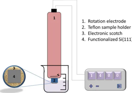
4.2.2 Set-up used for film deposition ... 71
4.3 Film nucleation and growth ... 72
4.3.1 Results ... 72 4.3.2 Discussion ... 81 4.4 Photo-switching properties ... 84 4.4.1 Results ... 84 4.4.2 Discussion ... 88 4.5 Conclusion ... 92 4.6 References ... 93
Chapter 5: Heterostructures made of RbxNi[Cr(CN)6]y▪zH2O and RbxCo[Fe(CN)6]y▪zH2O . 97 5.1 Introduction ... 97
5.2 Experimental section ... 98
5.2.1 Preparation of the solutions used for deposition ... 98
5.2.2 Set-up used for film deposition ... 99
5.3 Results and discussion ... 100
VII
5.4 Conclusion ... 115
5.5 References ... 116
General Summary and Conclusion ... 119
Appendix 1: Experiments and techniques ... 123
A.1.1 Structural and microstructural characterizations ... 123
A.1.1.1 Atomic force microscopy (AFM) ... 123
A.1.1.2 Scanning Electron Microscopy (SEM) ... 124
A.1.1.3 X-ray reflectivity (XRR) ... 124
A.1.1.4 X-ray diffraction (XRD)... 125
A.1.2 FTIR Spectroscopy ... 126
A.1.2.1 General method ... 126
A.1.2.2 Fitting procedure ... 127
A.1.3 Rutherford Backscattering Spectroscopy ... 129
A.1.3.1 General method ... 129
A.1.3.2 Calibration of the energy-channel curve7... 129
A.1.3.3 Conversion in absolute areal density values ... 130
A.1.4 Magnetic measurements ... 131
A.1.5 X-ray absorption spectroscopy measurements ... 132
A.1.5.1 General method ... 132
A.1.5.2 Sample preparation and measurement geometry ... 136
A.1.5.3 Data extraction, normalization and fitting ... 136
A.1.6 References ... 139
Appendix 2: Supplementary information on Chapter 2 ... 141
A.2.1 Solution preparation ... 141
A.2.2 Complementary ATR-FTIR spectra ... 142
Appendix 3: Supplementary Information for Chapter 3... 143
A.3.1 Synthesis and characterization of the RbNiCr reference particles ... 143
A.3.2 Scaning Electron Microscopy (SEM) imaging ... 144
A.3.3 RBS spectra for RbNiCr films with different deposition time. ... 145
A.3.4 Characterizations of RbNiCr films prepared with different rotation rates ... 146
A.3.4.1 AFM ... 146
A.3.4.2 XRR ... 147
VIII
A.3.5 Full M - H curves ... 150
A.3.6 Analysis of Tc and values from Field cooled magnetization curve ... 151
Appendix 4: Supplementary information for Chapter 4 ... 153
A.4.1 Effect of solution deoxygenation ... 153
A.4.2 Scan Electronic Microscopy (SEM) ... 154
Appendix 5: Supplementary information for Chapter 5 ... 155
A.5.1 Growth of Si (111)/RbNiCr layers monitored by FTIR measurements ... 155
A.5.2 Growth of Si (111)/RbCoFe layers monitored by FTIR measurements ... 158
Chapter 1
Introduction
Prussian blue analogues, with general formula AxM[M’(CN)6]y▪zH2O, where A is an alkali-metal cation, and M and M’ are divalent and trivalent transition-alkali-metal cations respectively, have attracted considerable interest in the last decades. In addition to magnetic1 and photo-magnetic properties2,3,4, the availability of degenerate or quasi-degenerate electronic states has made them ideal systems to explore cooperative switching phenomena, with potential applications for memory devices and magneto-optical switching. As some of these applications require their elaboration as thin layers or heterostructures, we shortly describe in this introduction the crystal structure and the main properties of Prussian blue analogues. In a second part, we survey the different deposition methods described in the literature and end with an introduction to the original deposition route that will be used in this work.
1.1 Crystal structure and properties of Prussian blue analogues
1.1.1 Description of the crystal structure
Prussian blue, Fe3+[Fe2+(CN)6]0.75. 3H2O, is a well-known mixed valence system, used as a deep blue pigment. Prussian blue analogues, AxM[M’(CN)6]y▪zH2O are isostructural compounds obtained by varying the nature of the M and M’ metal cations. For divalent M cation and trivalent M’(CN)6 cyanometallate, two types of crystal structure can be adopted: for y= 1, the Prussian blue derivative presents a regular face-centered cubic (fcc) structure, with two M and M’ fcc networks interconnected via C≡N bridges (see Figure 1.1b). To compensate the charge balance between the M and [M’(CN)6] species, alkali metal ions occupy half of the eight sub-octants of the lattice. For a description in terms of polyhedral units, the crystal structure may be represented as corner-shared [M’(CN)6] and [MN6] octahedra. For y < 1, Ludi et al.5 proposed a fractional occupancy of the [M’(CN)6] sites, with additional ligand water molecules filling the vacant sites and coordinated to the nearby M metal cations (see Figure 1.1a). y values between 0 and 1 can also be observed, with a fractional number of alkali ions for charge compensation. In the two latter cases, the space group is Fm3_m (No. 225). M ions occupy the apexes (position: 0, 0, 0) of the asymmetric unit and M’ ions are in the middle of the edges (position: 0.5, 0, 0). Different alkali ions, such as Na, K, Rb or Cs, can be incorporated in the crystal structure at Wyckoff position 8c (1/4, 1/4, 1/4). According to a previous neutron diffraction study,6,7 water molecules occupy three
independent positions: one is ligand water (H2OL), coordinated to Co atoms (24e site), while the two others are zeolitic water (H2OZ), with a large spatial distribution, either in the center of the empty sub-octants (H2Oz1, 8c site) or slightly off-centered at (x, x, x) due to hydrogen bonding with H2OL (H2Oz2, 32f site).
Figure 1.1. Scheme of the crystal structure of Prussian blue analogues: (a) M2+M’3+[(CN)6]2/3 ▪zH2O and (b) A+M2+M’3+[(CN)6] ▪zH2O.8
1.1.2 Main properties of Prussian blue analogues
Prussian blue analogues are commonly synthesized in aqueous solvents starting from M’(CN)6m- and M(OH2)6n+ soluble precursors; and the variety of M’(CN)6m- and metal ions M building blocks that can be assembled leads to multiple functionalities. Their porous host-guest structure is used in exchanging of alkali ions, such as 137Cs and Tl+;9 in chemical and electrochemical sensors10 and gas storage11,12. Electrochemical behaviors have been found in PBAs, associated with high proton conductivity in Co(II)-Cr(III) compounds, as well as Lithium incorporation for Li batteries in Cu(II)-Fe(III) PBAs.
In terms of magnetic exchange interaction between the two metal ions, superexchange coupling through the CN bridge led to a variety of behaviors up to relatively high ordering temperatures.1 Ferromagnetism has been reported for Ni-Cr PBA compound up to 90 K,13 ferrimagnetism for Co-Fe PBAs up to 20 K,14 and ferrimagnetism in V-Cr PBA.1 The magnetic ordering temperature typically depends on the M’/M ratio (number of magnetic neighbors), which is related to the alkali metal content because of charge compensation.15
Some Prussian blue analogues exhibit phase transitions induced by an electron transfer between the M and M’ metal sites that can be triggered by temperature16,17, light irradiation18, or pressure18. Among which the photo- or thermally-driven charge-transfer-induced spin transitions (CTIST) in cobalt hexacyanoferrates have been extensively studied.19,20,21,22,23 In these compounds, CTIST is accompanied by a large magnetization increase, due to the
transformation of Fe2+-CN-Co3+ diamagnetic pairs into Fe3+-CN-Co2+ magnetic pairs. Bleuzen
et al investigated in a systematic way the conditions to observe photo-magnetization changes24 and for the maximum photo-efficiency;25 the electronic configuration and the local structure of both the ground and excited states have been analyzed,26 as well as the influence of pressure and the relative stability of the low spin and high spin states27. They also found that the nature of the alkali metal ion can significantly affect the properties of the PBAs.24,28,29 Meisel et al found that the magnetization can either increase or decrease under visible light irradiation depending on Ni content in the ternary metal Prussian blue analogue, NaNi 1-xCox[Fe(CN)6] ▪zH2O.20
1.2 Thin film deposition of Prussian blue analogues
For applications in opto-electronics and memory devices, thin film deposition is essential. Preparing layers of photo-switchable AxCo[Fe(CN)6]y▪zH2O compounds with a thickness comparable or smaller than the depth of penetration of excitation light is also pivotal for a better understanding of the photo-switching properties of these systems. In addition, multiple properties or synergic effects30,31,32 could be expected in hybrid heterostructures due to surface and interface effects. In the following, we will present a short literature survey of the different deposition methods used to prepare films of Prussian blue analogues.
1.2.1 Techniques used in previous works
1.2.1.1 Electrochemical routes
Electrochemical deposition has been widely used for the growth of Prussian blue films and of its analogues.33,34,35 Sato et al.36 reported the electrochemical deposition of the high TC (270 K) Cr2.12Cr(CN)6▪zH2O compound onto SnO2 coated glass electrode, and found an electrochemically induced switching phenomenon between a ferromagnetic and a paramagnetic state. They also reported that the electronic structure of electrochemical deposited Na0.4Co1.3[Fe(CN)6]▪5H2O thin film can be switched from Fe3+-CN-Co2+(high spin) to Fe2+-CN-Co3+(low spin) by exchanging the alkali metal ion (from Na+ to K+).37
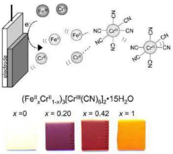
Hashimoto and co-workers prepared high TC (from 220 K to 300 K) magnetic films composed of mixed Vx2+/3+Cr2+(1-x)[Cr3+(CN6)]y▪zH2O, which shows various colors (from colorless to green and then blue) depending on the vanadium content.38 A similar method has been used to prepare (Fe2+xCr2+1-x)1.5[Cr3+(CN)6]▪zH2O magnetic films, as shown in Figure 1.2.39,40,41 Ohkoshi et al.41 investigated the magneto-optical properties of these (FexCr1-x)3[Cr(CN)6]2▪zH2O films, which display magnetization-induced second harmonic generation below their Curie temperature. A Faraday effect was also reported for
electrochemically deposited thin films of K0.31V2+0.49V3+0.51[Cr(CN)6]0.94▪6.5H2O and K0.61V2+0.97V3+0.03[CrIII(CN)6]0.88▪7.2H2O▪0.4C2H5OH.42
Figure 1.2. Scheme of the electrochemical deposition of magnetic films based on binary and
ternary metal Prussian blue analogues.41
1.2.1.2 Layer-by-Layer (LBL) deposition using sequential adsorption
Other wet chemistry methods have also been developed. The mostly used is based on sequential adsorption. This method, which consists of the alternate adsorption of polyanions and polycations on a charged surface, was developed in the late 90s43 and has known a significant throughput since then, due to its ease of implementation and applications of the as-obtained films in various fields (nonlinear optics, biomaterials, filtering, etc).
Culp et al44 first modified the surface of a Langmuir monolayer with a two dimensional Ni-Fe cyanide grid network, this surface layer can be considered as the starting layer of a PBA lattice. A film of Prussian blue was further obtained by sequential immersion of this substrate in two solutions of precursors, one containing the Fe(OH2)62+ reactant and the other one, Fe(CN)63- species as shown in Figure 1.3. The film thickness can then be controlled by increasing the number of immersion cycles. Similar work have been conducted for Fe-Co45,32,46, Fe-Mn45, and Ni-Cr31 cyanide grid networks and the corresponding thin films. Films of the photo-switchable RbxCo[Fe(CN)6]y▪zH2O compounds were prepared by such LbL assembly process and showed photomagnetic effects similar to those observed in the bulk material.47,32,48
Figure 1.3. Sequential deposition of cationic and anionic building blocks to form Prussian
blue onto a surface modified by a monolayer of a square grid template.44
Recently, Mallah et al. prepared magnetic films and multilayers made of NiCr and NiFe/NiCr Prussian blue analogues on functionalized Si (100) surfaces by sequential growth. The Si (100) surface was firstly functionalized by monolayers bearing bipyridine end groups that were further immersed in a solution of Ni2+
salt. The resultant films have a minimum thickness of 6 nm. However, AFM imaging shows isolated dots and a rather low coverage of the substrate.49
1.2.1.3 Formation of Langmuir mono and multi-layers
The Langmuir-Blodgett (LB) technique is based on the transfer of an assembled monolayer at the air/water interface onto a solid support. This method has been successfully used for the formation of the M-M’ cyanide grid networks described in the previous section.50 Mingotaud and coworkers also took use of it for the formation of Prussian blue-like mono and multilayers. They firstly dispersed a metal cation salt and the metallocyanide precursor (of diluted concentration, 10-6 mol/L) with positively charged dimethyldioctadecylammonium bromide, and transfer the as-formed Langmuir layer (see Figure 1.4) onto a solid substrate leading to LB monolayers (and multilayers by repeating the process) containing ferromagnetic entities.51 Thin films made of Prussian blue analogues and a ruthenium tris(bipyridine) complex52 (or a derivative of this complex53) have also be grown this way. Yamamoto et al.54,55,56 used a modified Langmuir monolayer, i.e. amphiphilic molecules and clay platelets, to play a template role for the formation of Co-Fe PBA films of different thickness. Titania nano-sheets were also used as a templates.57
Figure 1.4. Schematic organization of a LB monolayer made of a Fe3+-CN-Fe2+ grid.51
1.2.1.4 Other approaches for Prussian blue thin film deposition
Besides these widely used methods, other deposition techniques are described in the literature, such as composite films with a sol-gel matrix,58 using polyelectrolyte coated surfaces59,60 or tailor-made nanometer-scale patterns.61
1.2.2 Multilayer and core-shell heterostructures
The preparation of multifunctional heterostructures based on Prussian blue analogues has also drawn a considerable attention in the past years. The properties of these heterostuctures can derive from a simple addition of functionalities or from synergetic (or coupling) effects between functionalities. This coupling may have different origin: magnetic coupling (eg. exchange bias) or elastic coupling by combination of photo-strictive and piezomagnetic materials.62,63 A key point in this strategy is the control of the interface between the two materials.
In many works, core-shell heterostructures have been prepared.64,65,66 Catala et al64 designed core-shell CsFeCr@CoCr and CsNiCr@CsCoCr particles, as well as core-multishell CsCoCr@CsFeCr@CsNiCr particles to achieve multifunctionality at the nanoscale. The growth was carried out in aqueous solution and the size of the shell was well controlled. D.R. Talham and coworkers65 also synthesized several core-shell and core-multishell nanoparticles that associate a photo-strictive RbxCo[Fe(CN)6]y▪zH2O compound and a ferromagnetic KxNi[Cr(CN)6]y▪zH2O phase with a relatively high TC value of ca 70 K. The as-obtained core-shell particles exhibit persistent photo-induced magnetization change up to 70 K, which are absent from the individual components of the heterostructure. They interpret
their results by the transfer of strain through the interface between the two materials. They recently extend their investigations to core-shell particles for lithium battery made of a K0.1Cu[Fe(CN)6]0.7▪3.8H2O core and of a K0.1Ni[Fe(CN)6]0.7▪4.1H2O shell, to achieve higher capacity and enhanced cyclability, due to an increased stability of the crystal structure.67 However, ultrathin films and multilayers present distinct advantages as one may control their growth and multiply the number of interfaces to favor the synergetic properties of the hybrid structure. In addition, new effects such as interface anisotropy may be anticipated for this class of materials. Talham and coworkers62,63 were the first to report the preparation of multilayers composed of a photo-switchable Rb0.7Co4[Fe(CN)6]3▪zH2O film sandwiched between two ferromagnetic Rb0.8Ni4[Cr(CN)6]▪zH2O layers. The TEM cross section and the magnetic properties of this heterostructure is shown in Figure 1.5. The film exhibit a TC value of 70 K, and a decreased magnetization after light irradiation, which could be associated to the building up of external stresses applied to the RbNiCr layers due to the expansion of the Rb0.7Co4[Fe(CN)6]3▪zH2O film upon illumination.62 These effects are similar to those reported for the core-shell particles made of NiCr and CoFe PBAs.65
Figure 1.5. (left) Scheme of the NiCr/CoFe/NiCr heterostructure, and TEM micrograph
showing a cross-section image of the sample. (right) Photo-induced magnetization changes under 100 G with the magnetic field applied parallel to the surface.
1.2.3 Morphology of Prussian blue analogue films
For the deposition methods described in section 1.2, the film thickness typically range from few nanometers44,46,68,47 to hundreds of nanometers41,31,69 or micrometers.42,31 One of the main challenges is the control of the growth of ultrathin and continuous films in the perspective of designing devices for spintronic applications, and the improvement of the surface homogeneity to enhance interface effects.
The film morphology varies from one method to the other, and the surface and interface roughness is difficult to be well-controlled. Formation of a film at a Langmuir monolayer interface can produce very thin PBA films,54,55 as there is only one or few alternating Co-Fe metallate cyanide stacks on the solid support. This way, the thinnest film was estimated to be 0.44 nm thick, but the continuity and surface morphology of the film were not fully characterized.54 Layers and multilayers were also achieved down to ca. 6 nm by Layer-by-Layer (LbL) deposition using a functionalized Si (100) surface as a substrate,46 with for instance two 2.5 nm thick NiCr layers separated by a paramagnetic NiFe stack with a calculated thickness of 1.5 nm.49
Most of the films show a dependence of surface roughness on thickness.32,46,68 The roughness can range from 2.0 to 100 nm for thicknesses of 6.1 to 300 nm,46 and the film continuity is a common problem for all deposition methods.46,31 A recent work shows the possibility of getting smooth surfaces with a RMS roughness of 0.63 nm for 6.0 nm films by LbL deposition,49 while previous reports clearly show that a deposition cycle as described in
Figure 1.3 leads to the formation of the equivalent of several unit cells. The film growth does
not proceed layer by layer, maybe because of not optimized rinsing steps or redissolution/reprecipitation processes.
1.3 An alternative strategy for film growth
In this PhD work, we develop a simple deposition route of Prussian blue analogue thin films based on heterogeneous nucleation on surfaces functionalized by organic monolayers. The original method that we have proposed consists of promoting the selective immobilization of one precursor of the solid phase on the surface to initiate the film nucleation and its subsequent growth using controlled diffusion by contact with a solution containing all the required precursors.
By comparison to the extensively studied layer-by-layer deposition routes30,49, this “one-pot” or “one-bath” approach should allow for faster deposition rates but should also favor the formation of well-defined interfaces, both with the substrate and with a top layer in multilayer architectures. Indeed, this method allows avoiding many of the difficulties related to sequential deposition performed by dipping the sample in different baths that requires a controlled rinsing of the excess precursors and a drying stage often detrimental for the cleanliness of the surface and interfaces.
In this context, two main issues need to be considered, one is the choice of the substrate and of its functionalization, and the other one is the determination of the solution composition for deposition.
1.3.1 The choice of the substrate
To date, most of the thin films described in the literature were found to be very rough and polycrystalline. Obtaining individualized islands rather than continuous films is probably related to the difficulty in preparing a substrate suitable for an epitaxial growth of these coordination networks. The choice of the substrate is thus essential. Various materials have already been investigated as substrate because of their flatness, their surface chemistry or their surface charge, such as SnO2 and TiO2,33 clay mineral,54 Mylar,45,70 Melinex 535,46,31,30 Si (100),49 or Au(110)71. In this context, the originality of our approach is to use functionalized Si (111) surfaces as model substrates for the assembly of the films and supported heterostructures. These surfaces, which exhibit a well-defined structure at the atomic scale and well controlled chemical composition, open new interesting perspectives for the monitoring of the layer and multilayer growth. Indeed, they will allow the implementation of novel strategies for the characterization of the heterostructures based on in situ FTIR and AFM.
In addition, surface chemistry of semiconductors has drawn a great attention for several decades because of their huge potential in modern technology. Silicon and silicon based substrate are one of the most investigated substrates. Their surface passivation is well established, as well as their chemical and biochemical functionalization. Chabal and co-workers72,73 firstly found a way to prepare ideal hydrogen terminated Si (111) surfaces, and concluded that by using basic solution (pH=9-10) or buffered HF followed by etching in 40%NH4F solution can produce ideal hydrogen terminated Si (111) surfaces with ≡Si-H bonds normal to surface. Afterwards, many efforts have be made to get a stable coverage by a densely packed organic monolayer which is robustly linked to the silicon surface: Chidsey
et al.74,74b prepared close-packed alkyl-monolayer terminated Si (111) surfaces using free radicals. Many other methods have been studied, such as photochemical reactions75,76,77, mico-wave assisted reaction78 and Lewis acid-catalyzed hydrosilylation of alkenes79 etc. In the present work, the silicon surfaces will be prepared according to the methods developed by the Electrochemistry group of LPMC for (i) the preparation of H-Si(111) surfaces structured in atomically flat terraces, (ii) their functionalization by grafting of organic monolayers and (iii) quantitative analyses to determine the surface composition using in situ infra-red spectroscopy (FTIR) combined to AFM imaging80,81,82,83. The anchoring of monolayers of photochromic organic compounds has already been successfully achieved on these surfaces and their photo-switching evidenced by FTIR spectroscopy84. In the frame of this project, the Si surfaces were functionalized by pyridyl groups allowing the immobilization of the divalent metal cations, precursors of the Prussian blue analogue phases, by formation of dative metal-ligand bonds. Efficient immobilization of metal or organometallic precursors, as well as the self-assembly of gold nanoparticles on surfaces
based on the formation of pyridyl-metal complex have been already reported by several authors85,86,49.
1.3.2 The choice of the solution composition
1.3.2.1 Kinetics of solid formation in solution: Nucleation and growth
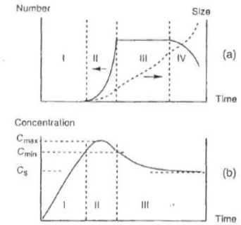
In order to investigate the deposition of a film on a substrate, it is important to recall the basics of the nucleation and growth processes based on LaMer theory.87 Figure 1.6(a) shows the evolution of the number and of the size of the particles formed during precipitation through the condensation of a soluble precursor. The condensation rate is first almost zero for undersaturated and slightly oversaturated solutions (regime I). When the concentration of the soluble precursor is beyond a critical concentration Cmin, condensation abruptly takes place and nuclei of the solid phase are formed in an explosive way (regime II). As the formation of a large number of nuclei will consume a lot of the soluble precursor, there will be a competition between the formation of the soluble precursor (for instance its addition in the reaction bath) and its consumption by condensation in new nuclei. If the rate of generation of the precursor is significantly smaller, nucleation will significantly decrease the precursor concentration and reduce the nucleation rate accordingly (concentration of Cmax in
Figure 1.6(b)). Once the concentration of precursor is less than Cmin, the nucleation process stops.
Figure 1.6. (a) Evolution of number and of the size of particles formed in solution. (b) Change
in the concentration C of the soluble precursor during precipitation. CS is the solubility of the solid phase, Cmin is the threshold concentration of soluble precursor to form nuclei, Cmax is the maximum concentration of the soluble precursor.
In regime II of Figure 1.6(a), the growth process takes place simultaneous with nucleation. When the precursor concentration drops below Cmin (regime III), the precursors only condense on the preformed nuclei, which result in the particle growth until the precursor concentration reaches the solubility limit of the solid phase.
We have now to consider the case of the formation of the solid in the presence of a second solid phase (preformed particles for core-shell synthesis or a surface for film deposition). Indeed, heterogeneous nucleation is thermodynamically favoured by the fact that the solid-solid surface tension is smaller than the solid-solid-solution surface tension. In the case of a shell grown onto core particles, or of a film grown on a surface, the strategy is to keep the precursor concentration below the threshold value for homogeneous nucleation in solution ( ), but higher than that of heterogeneous nucleation on the surface ( ). This will
avoid the nucleation and growth of particles in solution, that may be further adsorbed on the surface or simply decrease the precursor concentration and change the rate of shell/film growth.
1.3.2.2 Previous work on the growth of RbCoFe@RbNiCr core-shell particles
For deposition methods alternative to Layer-by-Layer (electrochemical or other solution routes), a key point is thus the determination of the solution composition, which should contain all necessary precursors and remain stable on the time scale of the deposition. Ideally, the concentration of the precursors should be kept below the threshold value for homogeneous nucleation, and above the value for heterogeneous nucleation. An example of this strategy is given by the work of Presle et al. for the synthesis of core-shell heterostructures made of Prussian blue analogues. This work focus on the growth of a Rb0.1NiCr(CN)6]0.7. zH2O (RbNiCr) shell onto preformed Rb0.5Co[Fe(CN)6]0.8. zH2O (RbCoFe) particles.88 This synthesis was carried out in water, by drop-wise addition of two solutions of reactants: the first solution contained the K3Cr(CN)6 reactant, and the second one the NiCl2 and RbCl precursors and the shell growth was monitored by dynamic light scattering (DLS) for various concentrations of the reactants, and keeping the [Cr]/[Ni] and [Rb]/[Ni] concentration ratios constant.
Figure 1.7. Average particle volume as a function of the nominal concentration of Ni2+ in the reaction medium. The addition rate used was 0.75 mL/min. The size of the nanoparticles was derived from DLS measurements, adapted from the PhD thesis of M. Presle88.
From Figure 1.7, we can see that for Ni2+ concentrations lower than ca. 8×10⁻⁵mol/L, no increase in particle size is detected. The precursors accumulate in solution but their concentrations do not exceed the threshold values necessary for the shell formation by heterogeneous nucleation. For Ni2+ concentrations between ca. 8×10⁻⁵mol/L and 5×10⁻⁴ mol/L, the increase of the mean particle volume reflects the growth of a shell onto the pre-existing RbCoFe particles, with a growth rate that gradually increases with the concentration of the NiCl2 solution. Eventually, for Ni2+ concentrations above 5×10⁻⁴ mol/L, the particle size after addition is similar to the dimensions of the core particles, suggesting the side formation of small RbNiCr particles by homogeneous nucleation in solution. These new particles are not accounted for by DLS as this technique tends to overestimate the contribution of the largest particles. All these conclusions were supported by direct observations using transmission electron microscopy. From this series of experiments, M. Presle determined critical concentrations: = 8×10⁻⁵ mol/L for heterogeneous nucleation, and = 5×10⁻⁴ mol/L for homogeneous nucleation. It should be noticed that these critical
values correspond to the experimental conditions used in this work. In particular, the stoichiometry of the shell phase does not match the ratio of the precursors, so that the threshold concentrations obtained for Ni2+ take into account the accumulation of [Cr(CN)6] 3-and Rb+ species in the reaction medium. It is also worth mentioning that the
value
drastically depends on the presence of RbCoFe particles in the reaction medium. This critical value should be lower when decreasing the number of RbCoFe particles.
1E-5 1E-4 1E-3
0.0 2.0x10⁵ 4.0x10⁵ 6.0x10⁵ 8.0x10⁵ 1.0x10⁶ 1.2x10⁶ 1.4x10⁶ 1.6x10⁶ 1.8x10⁶ 2.0x10⁶ 2.2x10⁶ 2.4x10⁶ 2.6x10⁶
C
het min M ean v olum e of t he part ic les (nm 3 )Concentration of Ni2+ in the reaction bath (mol.L-1)
[NiCl2] = 0.125 mM [NiCl 2] = 0.25 mM [NiCl 2] = 0.5 mM [NiCl 2] = 1 mM [NiCl2] = 2 mM [NiCl2] = 4 mM [NiCl2] = 8 mM
C
hom min1.4 Objectives and outline of the manuscript
The main objective of this PhD thesis is to investigate the deposition of Prussian blue analogues using a solution route based on heterogeneous nucleation on functionalized silicon surfaces. Special attention is paid to understanding the growth mode of ultra-thin films and bilayers, which is pivotal for a fine control of the morphology and of the interface quality in heterostructures. The structure of this dissertation is as follows:
Chapter 2 provides a protocol of surface functionalization ended with a pyridine termination, qualitative and quantitative analyses of the surface composition will be presented at each step. Chapter 3 discusses the growth of RbxNi[Cr(CN)6]y▪zH2O layers by combining different structural and microstructural characterization techniques. The magnetic properties of these layers will be further analysed based on the film morphology. In Chapter 4, the growth of RbxCo[Fe(CN)6]y▪zH2O thin films is presented, and their photo-switching properties are discussed on the basis of the chemical composition of the layers. At last, Chapter 5 presents preliminary results for the growth of bilayer structures of Si/RbxCo[Fe(CN)6]y▪zH2O/RbxNi[Cr(CN)6]y▪zH2O and Si/RbxNi[Cr(CN)6]y▪zH2O/RbxCo[Fe(CN)6]y▪zH2O.
1.5 References
1. S. Ferlay, T. Mallah, R. Ouahes, P. Veillet, M. Verdaguer, A room-temperature organometallic magnet based on prussian blue. Nature 1995, 378, 701-703.
2. O. Sato; T. lyoda; A. Fujishima; K. Hashimoto, Photoinduced magnetization of a cobalt-iron cyanide. Science 1996, 272, 704-705.
3. S. Ohkoshi; S. Yorozu; O. Sato; T. Iyoda; A. Fujishima; K. Hashimoto, Photoinduced magnetic pole inversion in a ferro–ferrimagnet: (Fe0.40IIMn0.60II)1.5CrIII(CN)6. Appl. Phys. Lett.
1997, 70, 1040-1042.
4. H. Tokoro; S. Ohkoshi; K. Hashimoto, One-shot-laser-pulse-induced demagnetization in rubidium manganese hexacyanoferrate. Appl. Phys. Lett. 2003, 82 (8), 1245-1247.
5. A. Ludi; H. U. Gudel, Structural chemistry of polynuclear transition metal cyanides.
Struct. Bonding. 1973, 14, 1-21.
6. F. Herren; P. Fischer; A. Ludi; W. Halg, Neutron diffraction study of Prussian blue, Fe4[Fe(CN)6]3.xH2O. Location of water molecules and long-range magnetic order. Inorg. Chem.
1980, 19, 956-959.
7. H. J. Buser; D. Schwarzenbach; W. Petter; A. Ludi, The crystal structure of Prussian blue: Fe4[Fe(CN)6]3.xH2O. Inorg. Chem. 1977, 16, 2704-2710.
8. H. Tokoro; S. Ohkoshi, Novel magnetic functionalities of Prussian blue analogs.
Dalton Transactions 2011, 40 (26), 6825-6833.
9. W. E. Prout; E. R. Russell; H. J. Groh, Ion exchange absorption of cesium by potassium hexacyanocobalt (II) ferrate (II). J. Inorg. Nucl. Chem. 1965, 27, 473-479.
10. A. A. Karyakin; E. E. Karyakina; L. Gorton, Amperometric biosensor for glutamate using Prussian blue-based “artificial peroxidase” as a transducer for hydrogen peroxide.
Anal. Chem 2000, 72, 1720-1723.
11. S. S. Kaye; J. R. Long, The role of vacancies in the hydrogen storage properties of Prussian blue analogues. Catalysis Today 2007, 120, 311–316.
12. S. S. Kaye; J. R. Long, Hydrogen storage in the dehydrated Prussian blue analogues M3[Co(CN)6]2 (M = Mn, Fe, Co, Ni, Cu, Zn). J. Am. Chem. Soc. 2005, 127, 6506-6507.
13. V. Gadet; T. Mallah; I. Castro; M. Verdaguer, High-Tc molecular-based magnets: a ferromagnetic bimetallic chromium(III)-nikel (II) cyanide with T=90K. J. Am. Chem. Soc. 1992,
114, 9213-9214.
14. O. Sato; Y. Einaga; T. Iyoda; A. Fujishima; K. Hashimoto, Reversible photoinduced magnetization J. Electrochem. Soc 1997, 144, L11-L13.
15. S. Ohkoshi; K. Hashimoto, Ferromagnetism of cobalt-chromium polycyanides. Chem.
Phys. Lett. 1999, 314 (3–4), 210-214.
16. T. Yokoyama; H. Tokoro; S. Ohkoshi; K. Hashimoto; K. Okamoto; T. Ohta, Photoinduced phase transition of RbMnFe(CN)6 studied by x-ray-absorption fine structure spectroscopy. Physical Review B 2002, 66 (18), 184111.
17. S. Cobo; R. Fernández; L. Salmon; G. Molnár; A. Bousseksou, Correlation between the stoichiometry and the bistability of electronic states in valence-tautomeric RbxMn[Fe(CN)6]y·zH2O complexes. Eur. J. Inorg. Chem. 2007, 2007 (11), 1549-1555.
18. Y. Moritomo; M.Hanawa; Y. Ohishi; K. Kato; M. Takata; A. Kuriki; E. Nishibori; M. Sakata; S. Ohkoshi; H. Tokoro; K. Hashimoto, Pressure- and photoinduced transformation into a metastable phase in RbMn[Fe(CN)6]. Physical Review B 2003, 68 (14), 144106.
19. D. Li; R. Clerac; O. Roubeau; E. Harte; C. Mathonie; R. Le Bris; S. M. Holmes, Magnetic and optical bistability driven by thermally and photoinduced intramolecular electron transfer in a molecular cobalt-iron Prussian blue analogue. J. Am. Chem. Soc. 2008,
130, 252-258.
20. D. M. Pajerowski; J. E. Gardner; D. R. Talham; M. W. Meisel, Tuning the sign of photoinduced changes in magnetization: spin transitions in the ternary metal Prussian blue analogue NaNi1-xCox[Fe(CN)6] ·nH2O. J. Am. Chem. Soc. 2009, 131, 12927-12936.
21. O. Sato; Y. Einaga; A. Fujishima; K. Hashimoto, Photoinduced long-range magnetic ordering of a cobalt-Iron cyanide. Inorg. Chem. 1999, 38, 4405-4412.
22. N. Shimamoto; S. Ohkoshi; O. Sato; K. Hashimoto, Control of charge-transfer-induced spin transition temperature on cobalt−iron Prussian blue analogues. Inorg. Chem. 2002, 41, 678-684.
23. A. Goujon; O. Roubeau; F. Varret; A. Dolbecq; A. Bleuzen; M. Verdaguer, Photo-excitation from dia- to ferri-magnetism in a Rb–Co–hexacyanoferrate Prussian blue analogue.
Eur. Phys. J. B 2000, 14, 115-124.
24. A. Bleuzen; C. Lomenech; V. Escax; F. Villain; F. Varret; C. C. dit Moulin; M. Verdaguer, Photoinduced ferrimagnetic systems in Prussian blue analogues CxICo4[Fe(CN)6]y (CI=Alkali Cation). 1. conditions to observe the phenomenon. J. Am. Chem. Soc. 2000, 122, 6648-6652.
25. A. Bleuzen; V. Escax; J.-P. Itié; P. Münsch; M. Verdaguer, Photomagnetism in CxCo[Fe(CN)6)](8+x)/3.nH2O Prussian blue analogues, looking for the maximum efficiency C. R.
26. C. C. dit Moulin; F. Villain; A. Bleuzen; M.-A. Arrio; P. Sainctavit; C. Lomenech; V. Escax; F. Baudelet; E. Dartyge; J.-J. Gallet; M. Verdaguer, Photoinduced ferrimagnetic systems in Prussian blue analogues CxICo4[Fe(CN)6]y (CI Alkali Cation). 2. X-ray absorption spectroscopy of the metastable state. J. Am. Chem. Soc. 2000, 122, 6653-6658.
27. J.-D. Cafun; J. Lejeune; F. Baudelet; P. Dumas; J.-P. Itie; A. Bleuzen, Room-temperature photoinduced electron transfer in a Prussian blue analogue under hydrostatic pressure. Angew. Chem. Int. Ed. 2012, 51, 9146-9148.
28. V. Escax; C. C. dit Moulin; F. Villain; G. Champion; J.-P. Itie; P. Münsch; M. Verdaguer; A. Bleuzen, Photo-induced electron transfer in ferrimagnetic Prussian-blue analogues XxICo4[Fe(CN)6]y (XI=alkali cation). C. R. Chimie 2003, 6, 1165-1173.
29. A. Bleuzen; V. Escax; A. Ferrier; F. Villain; M. Verdaguer; P. Munsch; J.-P. Itie, Thermally induced electron transfer in a CsCoFe Prussian blue derivative: the specific role of the alkali-metal ion. Angew. Chem. Int. Ed. 2004, 43, 3728-3731.
30. D. M. Pajerowski; J. E. Gardner; D. R. Talham; M. W. Meisel, Anisotropic magnetism in Prussian blue analogue films. New J. Chem. 2011, 35, 1320-1326.
31. D. M. Pajerowski; J. E. Gardner; M. J. Andrus; S. Datta; A. Gomez; S. W. Kycia; S. Hill; D. R. Talham; M. W. Meisel, Magnetic anisotropy in thin films of Prussian blue analogues.
Phys. Rev. B 2010, 82, 214405-1-214405-5.
32. F. A. Frye; D. M. Pajerowski; J.-H. Park; M. W. Meisel; D. R. Talham, Anisotropic photoinduced magnetism in thin films of the Prussian blue analogue AjCok[Fe(CN)6]l ·nH2O.
Chem. Mater. 2008, 20, 5706-5713.
33. K. Itaya; I. Uchida; V. D. Neff, Electrochemistry of polynuclear transition metal cyanides: Prussian blue and its analogues. Acc. Chem. Res. 1986, 19, 162-168.
34. V. D. Neff, Electrochemical oxidation and reduction of thin films of Prussian blue. J.
Electrochem. Soc 1978, 125, 886-887.
35. S. Sinha; B. D. Humphrey; A. B. Bocaesly, Reaction of nickel electrode surfaces with anionic metal-cyanide complexes: Formation of precipitated surfaces. Inorg. Chem. 1984, 23 (2), 203-212.
36. O. Sato; T. lyoda; A. Fujishima; K. Hashimoto, Electrochemically tunable magnetic phase transition in a high-Tc chromium cyanide thin film. Science 271, 49-51.
37. O. Sato; Y. Einaga; T. Iyoda; A. Fujishima; K. Hashimoto, Cation-driven electron transfer involving a spin transition at room temperature in a cobalt iron cyanide thin film. J.
38. M. Mizuno; S. Ohkochi; K. Hashimoto, Electrochemical synthesis of high-Tc, colored magnetic thin films composed of vanadium chromium of hexacyanochromate. Advanced
materials 2000, 12, 1955-1958.
39. S. I. Ohkoshi; A. Fujishima; K. Hashimoto, Transparent and colored magnetic thin films: (FexIICr1-xII)1.5[CrIII(CN)6]. J. Am. Chem. Soc. 1998, 120, 5349-5350.
40. S. Ohkoshi; K. Hashimoto, New magnetic functionalities presented by Prussian blue analogues. The Electrochemical Society Interface 2002, 34-38.
41. T. Nuida; T. Hozumi; W. Kosaka; S. Sakurai; S. Ikeda; T. Matsuda; H. Tokoro; K. Hashimoto; S. Ohkoshi, Colored magnetic films composed of cyano-bridged metal assemblies and magneto-optical functionalities. Polyhedron 2005, 24 (16-17), 2901-2905.
42. Ohkoshi, S.; M. Mizuno; G. Hung; K. Hashimoto, Magnetooptical effects of room temperature molecular-based magnetic films composed of vanadium hexacyanochromates. J.
Phys. Chem. B 2000, 104 (40), 9365-9367.
43. G. Decher, Fuzzy nanoassemblies: toward layered polymeric multicomposites. Science
1997, 277, 1232-1237.
44. J. T. Culp; J.-H. Park; I. O. Benitez; Y.-D. Huh; M. W. Meisel; D. R. Talham, Sequential assembly of homogeneous magnetic Prussian blue films on templated surfaces. Chem. Mater.
2003, 15, 3431-3436.
45. J. T. Culp; J.-H. Park; M. W. Meisel; D. R. Talham, Interface directed assembly of cyanide-bridged Fe–Co and Fe–Mn square grid networks. Polyhedron 2003, 22, 3059-3064. 46. F. A. Frye; D. M. Pajerowski; S. M. Lane; N. E. Anderson; J.-H. Park; M. W. Meisel; D. R. Talham, Effect of film thickness on the photoinduced decrease in magnetism for thin films of the cobalt iron Prussian blue analogue Rb0.7Co4[Fe(CN)6]3.0. Polyhedron 2007, 26, 2281-2286. 47. J.-H. Park; E. Cizmar; M. W. Meisel; Y. D. Huh; F. Frye; S. Lane; D. R. Talham, Anistropic photoinduced magnetism of a RbjCok[Fe(CN)6]l·H2O thin film. Appl. Phys. Lett.
2004, 85, 3798-3800.
48. J.-H. Park; Y.D. Huh; Cizmar., E.; S. G. Gamble; D.R. Talham; M. W. Meisel, Photoinduced magnetization in a thin Fe–CN–Co film. Journal of Magnetism and Magnetic
Materials 2004, 272, 1116-1117.
49. S. Tricard; C. Costa-Coquelard; S. Mazerat; E. Rivière; V. Huc; C. David; F. Miserque; P. Jegou; S. Palacind; T. Mallah, Cyanide-bridged NiCr and alternate NiFe–NiCr magnetic ultrathin films on functionalized Si(100) surface. Dalton Trans. 2012, 41, 4445-4450.
51. C. Mingotaud; C. Lafuente; J. Amiell; P. Delhaes, Ferromagnetic langmuir-blodgett film based on Prussian blue. Langmuir 1999, 15 (2), 289-292.
52. G. R. Torres; E. Dupart; C. Mingotaud; S. Ravaine, Electrochemical and photoelectrochemical properties of new hybrid langmuir-blodgett films containing Prussian blue and a tris(bipyridine) ruthenium derivative. J. Phys. Chem. B 2000, 104, 9487-9490.
53. G. R. Torres; B. Agricole; C. Mingotaud; S. Ravaine; P. Delhaes, Hybrid organic-inorganic langmuir-blodgett films starting from colloidal Prussian blue solution. Langmuir
2003, 19, 4688-4693.
54. T. Yamamoto; Y. Umemura; O. Sato; Y. Einaga, Photomagnetic Co–Fe Prussian blue thin films fabricated by the modified langmuir–blodgett technique. Chem. Lett. 2004, 33 (5), 500-501.
55. T. Yamamoto; Y. Umemura; O. Sato; Y. Einaga, Photoswitchable magnetic films: Prussian blue intercalated in langmuir-blodgett films consisting of an amphiphilic azobenzene and a clay mineral. Chem. Mater. 2004, 16, 1195-1201.
56. T. Yamamoto; Y. Umemura; O. Sato; Y. Einaga, Observation of the anisotropic photoinduced magnetization effect in Co-Fe Prussian blue thin films fabricated by using clay langmuir-blodgett films as a template. J. Am. Chem. Soc. 2005, 127, 16065-16073.
57. T. Yamamoto; N. Saso; Y. Umemura; Y. Einaga, Photoreduction of Prussian blue intercalated into titania nanosheet ultrathin films. J. Am. Chem. Soc. 2009, 131, 13196-13197. 58. Y. Guo; A. R. Guadalupe; O. Resto; L. F. Fonseca; S. Z. Weisz, Chemically derived Prussian blue sol-gel composite thin films. Chem. Mater. 1999, 11, 135-140.
59. M. Pyrasch; B.Tieke, Electro- and photoresponsive films of Prussian blue prepared upon multiple sequential adsorption. Langmuir 2001, 17, 7706-7709.
60. R. C. Millward; C. E. Madden; I. Sutherland; R. J. Mortimer; S. Fletcher; F. Marken, Directed assembly of multilayers—the case of Prussian Blue. Chem. Commun. 2001, 1994–1995. 61. S. Lepoutre; D. Grosso; C. Sanchez; G. Fornasieri; E. Rivière; A. Bleuzen, Tailor-made nanometer-scale patterns of photo-switchable Prussian blue analogues. Advanced Materials
2010, 22, 3992-3996.
62. D. M. Pajerowski; M. J. Andrus; J. E. Gardner; E. S. Knowles; M. W. Meisel; D. R. Talham, Persistent photoinduced magnetism in heterostructures of Prussian blue analogues.
J. Am. Chem. Soc. 2010, 132, 4058-4059.
63. D. M. Pajerowski; J. E. Gardner; E. S. Knowles; M. W. Meisel; D. R. Talham, Photoinduced magnetism in a series of Prussian blue analogue heterostructures. Chem. Mater.
64. L. Catala; D. Brinzei; Y. Prado; A. Gloter; O. Stephan; G. Rogez; T. Mallah, Core-multishell magnetic coordination nanoparticles: toward multifunctionality on the nanoscale.
Angew. Chem. Int. Ed. 2009, 48, 183-187.
65. M. F. Dumont; E. S. Knowles; A. Guiet; D. M. Pajerowski; A. Gomez; S. W. Kycia; M. W. Meisel; D. R. Talham, Photoinduced magnetism in core/shell Prussian blue analogue heterostructures of KjNik[Cr(CN)6]l.nH2O with RbaCob[Fe(CN)6]c.mH2O. Inorg. Chem. 2011, 50, 4295-4300.
66. M. Presle; J. Lemainque; J.-M. Guigner; E. Larquet; I. Maurin; J.-P. Boilot; T. Gacoin, Controlled growth of core and shell heterostructures based on Prussain blue analogue. New
Journal of Chemistry 2011, 35, 1296–1301.
67. D. Asakura; C. H. Li; Y. Mizuno; M. Okubo; H. Zhou; D. R. Talham, Bimetallic cyanide-bridged coordination polymers as lithium ion cathode materials: core@shell nanoparticles with enhanced cyclability. J. Am. Chem. Soc. 2013, 135, 2793-2799.
68. M. Clemente-Leon; E. Coronado; A. Lopez-Munoz; D. Repetto; C. Mingotaud; D. Brinzei; L. Catala; T. Malah, Magnetic langmuir-blodgett films of bimetallic coordination nanoparticles of Cs0.4Ni[Cr(CN)6]0.9. Chem. Mater. 2008, 20, 4642-4652.
69. S. Choudhury; N. Bagkar; G. K. Dey; H. Subramanian; J. V. Yakhmi, Crystallization of Prussian blue analogues at the air-water interface using an octadecylamine monolayer as a template. Langmuir 2002, 18, 7409-7414.
70. J. T. Culp; J-H. Park; D. Stratakis; M. W. Meisel; D. R. Talham, Supramolecular assembly at interfaces: formation of an extended two-dimensional coordinate covalent square grid network at the air-water interface. J. Am. Chem. Soc. 2002, 10083-10090.
71. S. Nakanishi; G. Lu; H. M. Kothari; E. W. Bohannan; J. A. Switzer, epitaxial electrodeposition of Prussian blue thin films on single-crystal Au (100). J. Am. Chem. Soc. 2003,
125, 14998-14999.
72. G. S. Higashi; Y. J. Chabal; G. W. Trucks; K. Raghavachari, Ideal hydrogen termination of the Si(111) surface. Appl. Phys. Lett. 1990, 56, 656-658.
73. P. Jakob; P. Dumas; Y. J. Chabal, Influence of silicon oxide on the morphology of HF etched Si(111) surfaces: Thermal versus chemical oxide. Appl. Phys. Lett. 1991, 59, 2968-2970. 74. (a) M. R. Linford; C. E. D. Chidsey, Alkyl monolayers covalently bonded to silicon surfaces. J. Am. Chem. Soc. 1993, 115, 12631-12632; (b) M. R. Linford; P. Fenter; P. M. Eisenberger; C. E. D. Chidsey, Alkyl monolayers on silicon prepared from 1-alkenes and hydrogen-terminated silicon. J. Am. Chem. Soc. 1995, 117, 3145-3155.
75. H. Asanuma; G. P. Lopinski; H. Yu, Kinetic control of the photochemical reactivity of hydrogen-terminated silicon with bifunctional molecules. Langmuir 2005, 21, 5013-5018. 76. L. A. Huck; J. M. Buriak, UV-Initiated hydrosilylation on hydrogen-terminated silicon (111): rate coefficient increase of two orders of magnitude in the presence of aromatic electron acceptors. Langmuir 2012, 28, 16285-16293.
77. R. Boukherroub; D. D. M. Wayner, Controlled functionalization and multistep chemical manipulation of covalently modified Si(111) surfaces J. Am. Chem. Soc. 1999, 121, 11513-11515.
78. R. Boukherroub; A. Petit; A. Loupy; J-N. Chazalviel; F. Ozanam, Microwave-assisted chemical functionalization of hydrogen-terminated porous silicon surfaces. J. Phys. Chem. B
2003, 107, 13459-13462.
79. R. Boukherroub; S. Morin; F. Bensebaa; D. D. M. Wayner, New synthetic routes to alkyl monolayers on the Si(111) surface. Langmuir 1999, 15, 3831-3835.
80. A. Moraillon; A. C. Gouget-Laemmel; F. Ozanam; J.-N. Chazalviel, Amidation of monolayers on silicon in physiological buffers: A quantitative IR study. J. Phys. Chem. C 2008,
112, 7158-7167.
81. S. Sam; L. Touahir; J. S. Andresa; P. Allongue; J.-N. Chazalviel; A. C. Gouget-Laemmel; C. Henry de Villeneuve; A. Moraillon; F. Ozanam; N. Gabouze; S. Djebbar, Semiquantitative study of the EDC/NHS activation of acid terminal groups at modified porous silicon surfaces. Langmuir 2010, 26 (2), 809-814.
82. D. Aureau; F. Ozanam; P. Allongue; J.-N. Chazalviel, The titration of carboxyl-terminated monolayers revisited: in situ calibrated fourier transform infrared study of well-defined monolayers on silicon. Langmuir 2008, 24 (17), 9440-9448.
83. A. Faucheux; A. C. Gouget-Laemmel; C. Henry de Villeneuve; R. Boukherroub; F. Ozanam; P. Allongue; J.-N. Chazalviel, Well-defined carboxyl-terminated alkyl monolayers grafted onto H-Si(111): packing density from a combined AFM and quantitative IR study.
Langmuir 2006, 22, 153-162.
84. C. Henry de Villeneuve; F. Michalik; J.- N. Chazalviel; K. Rück-Braun; P. Allongue, Photochromes: quantitative IR readout of fulgimide monolayer switching on Si(111) surfaces.
Advanced materials 2013, 25 (3), 416-421.
85. T. Zhu; X. Zhang; J. Wang; X. Fu; Z. Liu, Assembling colloidal Au nanoparticles with functionalized self-assembled monolayers. Thin Solid Films 1998, 327–329, 595–598.
86. B. Fleury; F. Volatron; L. Catala; D. Brinzei; E. Rivière; V. Huc; C. David; F. Miserque; G. Rogez; L. Baraton; S. Palacin; T. Mallah, Grafting a monolayer of superparamagnetic cyanide-bridged coordination nanoparticles on Si(100). Inorg. Chem. 2008, 47, 1898-1900. 87. J.-P. Jolivet, Metal oxide chemistry and synthesis: from solution to solid state. John Wiley & Sons, Inc.: 2000.
88. M. Presle. Synthèse et propriétés d'hétérostructure moléculaires de type multiferroique à base d'analogues du bleu de Prusse. PhD dissertation. Ecole Polytechnique, France, 2011.
Chapter 2
Functionalization of Si (111) surfaces
2.1 Introduction
The organic functionalization of oxide free Si surfaces has received strong interest during the past two decades because of its potential in many different fields of research. It is out of the scope of this thesis to review the many different processes that were developed and to explain the chemical mechanisms involved. The reader should consult recent reviews on the subject.1,2,3,4,5 It should be just recalled that one major interest of this approach is that it allows for the grafting of organic layers directly onto Si surfaces through robust covalent Si-C linkage. As a consequence of the large energy of the Si-C bonds (~4.5 eV) the grafting of organic monolayer onto oxide free silicon surfaces cannot be considered as a self-assembly process (the molecular chains are not mobile during the monolayer formation).6 The high energy of the Si-C bond and its weak electronic polarization confer however a superior chemical stability to organic monolayers on oxide free silicon surface. This is a key advantage in comparison with other systems, in particular thiol Self Assembled Monolayers (SAMs) on gold for which the monolayer stability may sometimes be a problem (Au-S bond energy of 3.5 eV).7 A second advantage of organic monolayers on oxide free silicon is the excellent long range morphology of the silicon substrate, with a very low roughness, especially the Si (111) surfaces which can be prepared flat at the atomic scale over long range.8
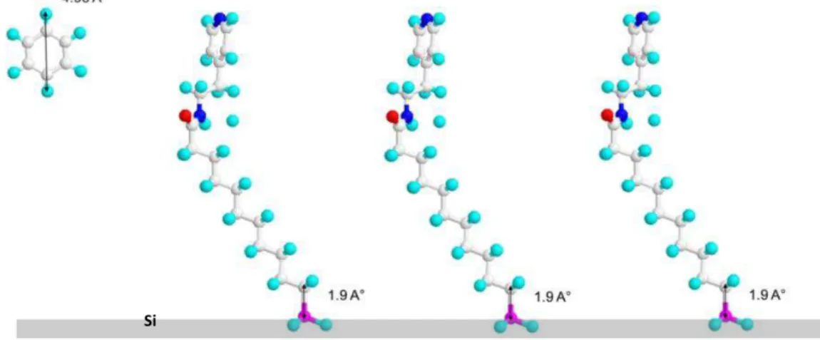
Considering the strong coordination between transition metal ions and the N from both the pyridine group and the cyanide group, we get inspired that by functionalizing the Si surface with pyridyl termination, films of Prussian blue analogue could be robustly coupled to with the surface through a coordination linkage, which could be easily found in the precursor solutions.
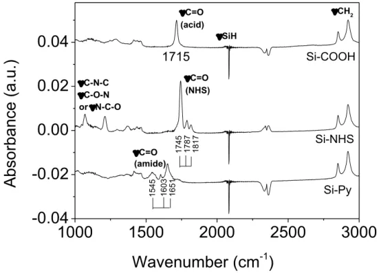
In this chapter, we will present the protocol used to functionalize Si (111) surfaces with a pyridine termination. The full protocol involves different steps: the grafting of undecylenic acid onto hydrogenated Silicon surfaces by photochemical (UV) hydrosilylation, the conversion of carboxylic acid end-groups into semi-stable succidimyl COOSuc-esters 9 and finally the coupling of aminoethyl pyridine. The whole schematic route is shown in Figure
2.1. We used atomically flat H-terminated Si (111) surfaces. The surface morphology, as well
Figure 2.1. Scheme of the multistep protocol used for the functionalization of H-Si (111)
surfaces by pyridyl end groups.
2.2 Experimental protocols
2.2.1 Materials
N-Ethyl-N’-(3-(dimethylamino) propyl) carbodiimide hydrochloride (EDC), N-hydroxysuccinimide (NHS, 98%), Undecylenic acid (99%), 4-(2-aminoethyl) pyridine (96%), ammonium sulfite monohydrate (92%), 2-(N-Morpholino) ethanesulfonic acid (MES, > 99.5%), and acetonitrile (> 99.9%) were purchased from Sigma-Aldrich. H2O2 (30%), H2SO4 (96%), acetic acid (100%) and etching (NH4F, 40%) reagents were VLSI grade and supplied by Merck. All reagents were used without post-treatment. Ultrapure water (UPW, Milli-Q, 18.2 MΩ cm) was used throughout the whole manipulations and rinsing steps.
500-550 µm thick Si (111) wafers one side-polished, with a miscut angle of 0.2° toward the [11 ̅] direction (Siltronix, France), were used as standard substrates. For IR spectroscopy measurements in Attenuated Total Reflectance (ATR) mode, we used Float Zone purified (FZ) and double-side polished wafers. Prior to use, the Silicon wafers were cut into squares of dimensions 10×10 mm2, and were cleaned by immersion into a Piranha solution. All the etching/hydrogenation reactions were carried out in Teflon pillbox.
2.2.2 Surface functionalization
Surface functionalization was carried out from H-terminated Si(111) surfaces (denoted Si-H) prepared by anisotropic etching in oxygen free 40% NH4F10. The Silicon samples and Teflon vials were first cleaned by immersion for 30 min in piranha solution (mixed aqueous solution of H2O2 30% and H2SO4 96% with a volume ratio 1:2) to remove organic contaminations.
Caution, Piranha solution reacts strongly with organic materials, it must be used under extreme care. After copious rinsing with Milli-Q water, the Silicon substrates were then immersed for 15 min into 40% NH4F aqueous solution in which ammonium sulfite ((NH4)2SO3▪H2O) was added to remove the dissolved oxygen. A quick rinse with Milli-Q water was then performed.
The first step of functionalization (denoted in Figure 2.1) consists in preparing acid terminated surfaces (Si-COOH). The grafting protocol that we used was established by Faucheux et al.11. The freshly hydrogenated surfaces were introduced into a schlenck tube containing a solution of undecylenic acid, previously heated at 90°C and outgassed under Argon for 30 min to remove traces of water and oxygen. After the introduction of the Si-H sample, the solution was further flushed with Ar for 15 min. Then, the schlenk tube was hermetically closed and transferred into a rayonnet reactor equipped with UV lamps (6 mW/cm2, 312 nm). After 3 hours irradiation, the schlenk tube was first kept at 80℃ in a water bath for 5 min with all openings closed, and then Ar gas was used to flush the solution for 5 min. In a further step, the grafting solution was exchanged with an acetic acid solution heated at 80℃ and outgassed for 10 min. Then, the Si surface was taken out and introduced into another schlenk tube filled with acetic acid at 80°C. Ar flushing was maintained for another 30 min, and this procedure was repeated twice. This rinsing protocol allows for efficiently remove all physisorbed acid chains that have tendency to strongly interact with the acid monolayer through hydrogen bonding.12 Finally the surface was carefully rinsed with water to make sure that there is no residual acetic acid remaining on the surface. Note that the Ar flushing was performed in order to avoid the oxidation of the surface.
The next step (step in Figure 2.1) is an activation step that consists in the transformation of carboxylic acids into succinimidyl ester (Si-COOSuc).13 The reaction was done by immersion of the Si-COOH surfaces into a freshly prepared solution containing 0.1 M N-hydrosuccinimide (NHS) and 0.1 M N-Ethyl-N’-(3-(dimethylamino) propyl) carbodiimide (EDC) in 0.1 M buffer solution at pH 5. The reaction was let to proceed for 1h at 15°C. The “activated” ester terminated surfaces (Si-COOSuc) were afterward rinsed successively into i) warm NaH2PO4 (0.1 M, pH = 5, 50℃) solution for 10 min, ii) diluted NaH2PO4 solution (NaH2PO4 0.1 M/H2O, v:v = 1:1, 50℃) and then iii) in UPW for another 10 min. After carefully drying under Ar flow, the Si-COOSuc surfaces were stored in acetonitrile solution.
The final step (step in Figure 2.1) consists in coupling pyridyl groups onto the surface by reacting the Si-COOSuc activated surface with aminethyl pyridine. The coupling reaction was done by immersion of the Si-COOSuc surfaces in 5 mM aminoethyl pyridine solution. After the introduction of the Si sample, the solution was outgassed with Ar for 5 min to remove O2. The reaction was let to proceed for 1h at 25°C in the closed schlenck. The resulting pyridyl-terminated surface Si-Py was copiously rinsed with acetonitrile and water, and finally blown dried under a stream of nitrogen gas.
2.3 Surface morphology
2.3.1 Atomic Force Microscopy (AFM)
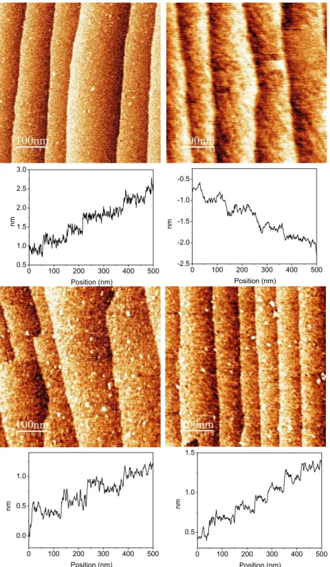
Representative AFM images of the surface at the different steps of the construction of the pyridine-terminated layer are displayed in Figure 2.2. The structure of the Si (111)-H surface prior grafting and the subsequent chemical modifications is shown on Figure 2.2(a). The surface exhibits a staircase structure with terraces, flat at the atomic scale and separated by steps of height 3.1 Å (corresponding to the distance between two Si (111) atomic planes). After the grafting, the surface activation and the coupling of pyridyl groups, no significant modification of the surface topography is detected. The surfaces look homogeneous and free of significant amount of contaminants or residues. The fact that the staircase structure is preserved, and the lack of significant increase of the roughness on the terraces, suggest at this stage a homogeneous coverage of the surface by a dense monolayer. For the pyridyl terminated surfaces, the staircase structure of the initial Si (111)-H surfaces is still observed, as well as the cleanness and flatness.
Figure 2.2. AFM images (500 × 500 nm2) of a Si (111) surface after each step of the functionalization: (a) hydrogenated Si-H surface; (b) acid terminated Si-COOH surface; (c) activated Si-COOSuc surface and (d) Si-Pyridine terminated surface. A line profile is given below each image.
0 100 200 300 400 500 0.5 1.0 1.5 Position (nm) nm 0 100 200 300 400 500 0.0 0.5 1.0 Position (nm) nm 0 100 200 300 400 500 -2.5 -2.0 -1.5 -1.0 -0.5 Position (nm) nm 0 100 200 300 400 500 0.5 1.0 1.5 2.0 2.5 3.0 Position (nm) nm