Université de liontréal
E ffect of Progesterone on the release of Prostaglandin F2 alpha
from uterine endometrial epithelial celis in ruminants
par
AHMAD ALI JAMSHIDI
Département de biomédecine vétérinaire
Faculté de médecine vétérinaire
Mémoire présenté à la Faculté des études supérieures
en vue de l’obtention du grade
Maître ès sciences (M.$c.)
en sciences vétérinaires
option reproduction
Octobre 2005
Direction des bibliothèques
AVIS
L’auteur a autorisé l’Université de Montréal à reproduire et diffuser, en totalité ou en partie, pat quelque moyen que ce soit et sur quelque support que ce soit, et exclusivement à des fins non lucratives d’enseignement et de recherche, des copies de ce mémoire ou de cette thèse.
L’auteur et les coauteurs le cas échéant conservent la propriété du droit d’auteur et des droits moraux qui protègent ce document. Ni la thèse ou le mémoire, ni des extraits substantiels de ce document, ne doivent être imprimés ou autrement reproduits sans l’autorisation de l’auteur.
Afin de se conformer à la Loi canadienne sur la protection des renseignements personnels, quelques formulaires secondaires, coordonnées ou signatures intégrées au texte ont pu être enlevés de ce document. Bien que cela ait pu affecter la pagination, il n’y a aucun contenu manquant.
NOTICE
The author of this thesis or dissertation has granted a nonexclusive license allowing Université de Montréal to reproduce and publish the document, in part or in whole, and in any format, solely for noncommercial educational and research purposes.
The author and co-authors ifapplicable retain copyright ownership and moral rights in this document. Neither the whole thesis or dissertation, nor substantial extracts from it, may be printed or otherwise reproduced without the author’s permission.
In compliance with the Canadian Privacy Act some supporting forms, contact information or signatures may have been removed from the document. While this may affect the document page count, it does not represent any Ioss of content from the document.
Ce mémoire intitulé
Effect
of Progesterone on
the release of Prostaglandin F2 alpha
from uterine endometrial epithelial
celis in ruminants
présenté par
Ahmad Ah Jamshidi
a été évalué par un jury composé des personnes suivantes
Christopher A Price, président-rapporteur
Alan K Goff, directeur de recherche
Résumé
La progestérone (P4) et l’oxytocine (OT) stimulent la sécrétion de la prostaglandine f2a (PGf2Œ) des cellules épithéliales de l’endomètre et il y a une réponse différentielle de ces cellules à OT pendant le cycle oestral des ruminants. Pour déterminer comment P4 influence cette réponse chez la vache, nous avons utilisé des utérus de bovin prélèvés au début du cycle (jour 1-3) afin d’effectuer une culture de cellules épithéliales de l’endomètre.
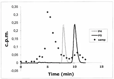
La chromatographie liquide sous haute pression et la spectrophotométrie de masse (HPLC) ont montré que les cellules en culture ont métabolisé P4 et que les métabolites étaient soit 5 u- soit 5
J3-
prégnanédione. Pour déterminer si ces métabolites peuvent modifier la synthèse de PGf2u, les cellules ont été incubé avec P4, 5 u- ou 5f3-
prégnanédione. Les résultats ont prouvé que seule P4 et non l’OT causent une augmentation significative de la synthèse de PGf2u. Les analyses de liaison de l’OT à son récepteur n’ont montré aucun effet significatif sur la concentration en récepteur doxytocine (OTR). L’augmentation de la synthèse de PGF2u au niveau des cellules traitées avec la P4 est probablement due à une augmentation des enzymes impliqtiées dans la synthèse de PGF2u. Comme RU 486, un anti-progestérone, diminue la synthèse de PGf2u et la concentration en OTR, nous avons traité nos cellules avec RU 486. L’anti-progestérone a augmenté significativement la synthèse de PGf2a (P<O.05) et l’effet maximal était à une concentration de 1 1iM tandis que la concentration des OTR était diminuée. Afin de comparer l’effet de RU 486 sur la synthèse des PG et sur la concentration des OTR avec celui d’un antagoniste pur, des cellules ont été traité avec ZK 137316 et des résultats semblables ont été obtenus. D’autres expériences ont montré que l’AMPc et le 5a-dihydrotestosterone (DHT) stimulent la sécrétion de PGF2u par les cellules épithéliales de l’endomètre de bovin via différentes voies. Ainsi, OT n’est pas le seul stimulant de la synthèse de PGf2u dans les cellules épithéliales de l’endomètre de bovin. Nous concluons qu’il existe d’autres facteurs capables de modifier la capacité de l’OT à augmenter la synthèse de PGF2u. Le traitement des cellules avec dePindométhacine, un inhibiteur non sélectif de COX, induit une diminution de la synthése de PGF2Œ ainsi que de la concentration des OTR. Nous suggérons que l’effet paracrine de PGf2Œ sur les cellules épithéliales de l’endométre serait de moduler le nombre d’OTR.
Mots clés
Bovin, utérus, cellules épithéliales de l’endomètre, progestérone, Mifepristone (RU 486), prostaglandine, androgène.
Surnm ary:
Progesterone (P4) and oxytocin (OT) stimulate secretion of prostaglandin F2a (PGF2Œ) from the endometrial epithelial ceils but there is differential responsiveness of these ceils to OT during the estrus cycle in ruminants. To determine how P4 influences this responsiveness in the cow, we used bovine uteri collected early in the cycle (daysl-3) for the culture of endometrial epithelial celis. HPLC and mass spectrophotometry showed that these ceils metabolized P4 and that the metabolites were either 5cc- or 5f3-pregnanedione. To determine if these metabolites of P4 were able to modify PGF2cc synthesis. celis were incubated with P4, 5cc- or 5f3-pregnanedione. Results showed that only P4 caused significant increase in PGF2a synthesis but not OT stimulation of PGf2cc synthesis. OT binding assays showed no significant effect on oxytocin receptor (OTR) concentration. The increase in PGF2u synthesis in P4 treated ceils is probably due to an increase in the enzymes involved in PG synthesis. Since RU 486, an antiprogesterone, has been shown to decrease PGf2a synthesis as weIl as OTR concentration, we treated our celis with RU 486. This antiprogesterone increased PGF2a synthesis significantly (P<O.05) and the maximal effect was at a concentration of I iM while OTR was down regulated. Tri order to compare the effect of RU 486 on PG synthesis and OTR concentration with that of a pure antagonist, celis were treated with ZK 137316 and the similar resuits were obtained. further experiments revealed that cAMP and 5cc-dihydrotestosterone (DHT) stirnulated PGF2a secretion from the bovine endometrial epithelial ceils via different pathways. Thus, OT is flot the only stimulant of PGF2cc synthesis in the bovine endometrial epithelial celis. We conclude that there are other factors that are able to modify the ability of OT to increase synthesis of PGF2cc. The treatment of the celis with indomethacin, a non selective COX-inhibitor, showed that the inhibitory effect of indomethacin on COX enzymes leads to decreased synthesis of PGF2cc and a decrease in OTR. We postulate that a paracrine effect of PGF2cc on the endometrial epithelial ceils may be able to modulate OTR number.
Keywords:
Bovine, utens, endometrial epithelial celis, progesterone, Mifepristone (RU 486), prostaglandin, androgen.
Table of contents
Summary (Résumés en Français)
iii
Summary (English)
yTable of contents
vii
List of figures
viii
List of abbreviatïons
xi
Thanks
xv
Dedication
xvi
Introduction
1
Hypothesis and Objectives
37
Materials and Methods
3$
Statistîcal analysis
42
Resuits
42
Discussion
59
Conclusion
67
List offiiziires
Figure 1
3
Concentration of 15-keto-13, 14-dihydroprostaglandin F2Πmetabolite (PGFM) in peripheral plasma of cyclic cow during luteolysis.
Figure2
5Location of the enzymes involved in the synthesis of prostaglandin F2a in the celis.
Figure 3
12
Mechanism of action ofoxytocin (OT).
Figure 4
14
Hypothetical model for TNF-a control of PGf2Πsynthesis in bovine endometrial celis during luteolysis.
Figure
519
Major metabolic pathways of the three principal steroids (Progesterone, Estradiol and Testosterone) secreted by the gonads.
Figure 6
23Steroid receptors’ (SR) genornic actions.
Figure
7
27$chematic model ofnongenornic inhibitory effects ofprogesterone.
Figure 8
30
Genomic and non-genomic actions of steroid hormones: a tentative interpretation.
Figure 9
43
Metabolism ofProgesterone by the bovine endometrial epithelial ceils.
FigurelO
44
Figure 11
.45
Appearance of two different metabolites of progesterone after changing the concentration ofAcN: water from 50:50 to 40:60.
Figure 12
46
Separation of free and conj ugated progesterone.
Figure 13
47The effect of P4 (100 iig rnF1) and its metabolites on the release ofOT-stirnulated PGf2Πfrom the bovine endometrial epithelial ceils.
Figure 14
4$
The effect ofprogesterone and its metabolites on the OTR concentrations.
Figure 15
49
The effect ofthe antiprogesterones RU 486 (5iM) and ZK 137316 (1tM) on the release ofPGF2a from the bovine endornetrial epithelial celis in the absence of P4.
Figure 16
50
The effects of RU 486 (5iM) on the synthesis of PGf2u with or without co treatrnent of P4 (100 ngmï1) on bovine endometrial epithelial celis.
Figure 17
51
The effect of P (lOOng mF1) and ZK 137316 (luM) on the synthesis ofPGF2Πfrom bovine endometrial epithelial ceils.
Figure 1$
52The effect
of ZK
137316 dose response on PGf2u concentration secreted by the bovine endometrial epithelial ce! !s.Fïgurel9
53
The effect of RU 486 dose response on the release ofPGF2u concentration.
Figure20
54
The effect of antiprogestagens RU 486 (5uM) and ZK 137316 (luM) in the absence of P4 on the OTR concentration from the bovine endornetrial epithelial celis.
Figure 21
.55
The effect of ZK-137316 dose response on OTR concentration in the bovine endometrial epithelial ceils.
Figure 22
56
The effect ofDHT dose response on the concentration of PGF2Πfrom the bovine endometrial epithelial celis.
Figure 23
57
The effect of different doses of indornethacin on the release of PGF2Πfrom the bovine endometrial epithelial celis.
Figure 24
58
The effect of indornethacin dose response on the binding of OTR in the bovine endornetrial epithelial cells.
Fïgure
25 ,59
The effect of cAMP (lmM) on the release ofPGF7Πfrom the bovine endometrial epithelium.
List of abbreviatio,,s
5 3-DHP 5 -dihydroxyprogesterone
AA arachidonic acid
ACAT acyl-CoA cholesterol acetyltransferase
AMP adenosine monophosphate
AP antiprogesterone
AR androgen receptor
ARC cytosol androgen receptor
AVP arginine vasopressin
BSA bovine serum alburnin
cAMP cyclic adenosine monophosphate
CREP cAMP-response element binding protein
CE cholesteryl ester CL corpus luteum CoA co-activators COX cyclooxygenase COX 1 cyclooxygenase 1 COX 2 cyclooxygenase 2
cPLA2 cytosolic PLA2
DAG diacylglycero!
dbcAMP dibutyryl cyclic adenosine monophosphate
DHP dihydroprogesterone
DHT 5Œ-dihydrotestosterone
E2 estradiol-1713
EL endometrial lumenal
ER estradiol recetor
ERC cytosol estrogen receptor
ERΠestrogen receptor alpha ERE estrogen response element
FBS-DC fetal bovine serum dextran-charcoal extraction FCS fetai calfserum
fGF-7 fibroblast growth factor- 7
FGF- 10 fibroblast growth factor- 10 FP receptor PGf2cx receptor
GABA y-Amino butyric acid
GC/MS gas chrornatography/mass spectrometry GE glandular epithelia
GPCR G-protein-co upled receptor superfamiiy GR glucocorticoid receptors
HBSS Hank’s balanced sait solution HGF hepatocyte growth factor
HMG-CoA reductase 3-hydroxy-3-methylgiutaryl (HMG)-C0A reductase HPLC high pressure liquid chromatography
HSD hydroxysteroid dehydrogenase Hsp9O heat shock protein 90
TFN-r interferon-tau
IMP intra myometrial pressure iNOS nitric oxide synthase
1P3 inositoi (1, 4, 5)-triphosphate
LBD ligand-binding domain LDL low-density lipoprotein
LH lutinizing Hormone
LVA 10 vasopressin -receptor-specific ligand
MAPK mitogen-activated protein kinase MUC-1 mucin glycoprotein one
NA noradrenaline
NBCS new born caif serum
NLS Nuclear localization sequences
OT oxytocin
OTR oxytocin receptor
P4 progesterone
PG prostaglandin
PGE2 prostaglandin E2
PGES prostaglandin E synthase PGF2Πprostaglandin F2 alpha
PGFM 15 -keto- 13, 1 4-dihydroprostaglandin F2Πmetabolite PGFS prostaglandin F synthase
PGG2 prostaglandin G2
PGH2 prostaglandin H2
PGHS prostaglandin G/H synthase
PIP2 phosphatidylinositol-4, 5-bisphosphate
PKC protein kinase C
PLA2 phospholipase A2
PLC phospholipase C
PR progesterone receptor
PRC cytosol progesterone receptor PREs progesterone-responsive elements
RBAs relative binding affinities
RU 486 Mifepristone
SH steroid hormones
SPRMs selective progesterone receptor modulators
SR steroid hormone receptor
TM transcription machinery
TNF-u tumor Necrosis Factor-a
Via vasopressin la
Vi aR vasopressin la receptor
Wth
my special thanks [o:
My supervisor from whom I learned a lot,
for lis generous lieIp and support
and to:
/1ét
o%ta
for ail tIc technical assistance and the joy she added to my workplace at the iab.
(JcL .‘
a
ce
tQ/m%Introduction
Initially, because of the emphasis placed on the importance of pituitary support for the maintenance of the corpus luteum, it was considered that withdrawal of pituitary luteotrophins such as lutinizing hormone (LH) andlor prolactin might cause the cyclic regression of the corpus luteum. However, with the advent of more sophisticated methods for measuring circulating pituitary gonadotropins, it became apparent that, at the time of corpus luteum regression, measurable levels of these hormones were present in ruminants (l-2).
Loeb, who first demonstrated that hysterectomy in the cyclic guinea pig abolished cycles and caused abnormal persistence of the corpora lutea, reported the importance of the uterus in the control of corpus luteum regression (3). In the 1970s researchers discovered that degradation of the corpus luteum resuits from a luteolysin secreted by the uterus. Soon afierwards it was confirmed that the luteolysin is prostaglandin (PG) F2Πsecreted from endometrial epithelium (4). Similar effects were subsequently observed in the cyclic sheep, cow, pig, and mare and in the pseudopregnant hamster, rabbit, and rat (5).
Prostaglandins are important regulators of reproductive events including ovulation, implantation, parturition, luteolysis and recognition of pregnancy (6-7). In ruminants the role of uterine prostaglandin F2Π(PGF2u) in luteolysis or the regression of the corpus luteum (CL) during late diestrus is well established (8). This role ofPGF2u as a uterine luteolytic hormone was supported by the finding that systemic administration of indomethacin, an inhibitor of PG synthesis (9), or the intrauterine administration of indomethacin (10) delayed or prevented luteal regression in several
species. Also, immunization against PGF2Œ, either passively (11-12) or actively (13-14), delayed regression ofthe corpus luteum in the sheep.
Luteolysis is characterized by an initial decline of progesterone secretion that is commonly designated as functional luteolysis as distinct from structural or morphological luteolysis which, as the name suggests, signifies the subsequent change in the cellular structure of the gland and its graduai involution in the ovary to form a small scar composed of connective tissue known as corpus albicans whicli ofien persists in the ovary for several weeks (15).
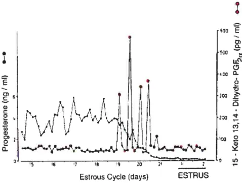
Release of PGF2Œ from the uterus in a pulsatile fashion on days 17—1 $ of the estrous cycle is essential to induce regression of the CL in ruminants (16-17). It has been clearly shown that PGF2Œ, the luteolytic hormone that is released cyclicaiiy from the uterus at the time of corpus luteum regression, is elevated as a series of pulses in uterine venous blood during luteolysis in cow (1 8). Also, it lias been demonstrated that levels of PGFM in the peripheral blood of cows show pulsatile increases with a pattem of several series of pulses of short duration for 2-3 days during and afler luteolysis (19), thus supporting the role ofPGF2u as a luteolytic hormone (figure 1).
t
E ‘20 LL 0 L b4
(T) o w ‘ ‘6 ? 1 20Estrous Cycle (days) STRUS
I E o, Q, 0)
e
Figure 1: Concentration of 15-keto-13, 14-dihydroprostaglandin F2Πmetabolite (PGFM) in the peripheral plasma of a cyclic cow during luteolysis. Pulses of PGFM representing uterine secretion of PGF2Πcoincide with the decline of progesterone during luteolysis.
Taken from: Kindahi et al. (1984) Levels ofprostaglandin F2u metabolites in blood and urine during early pregnancy. Anim. Reprod. Sci, 7: 133-48.
When PGF2Πis administered in a pulsatile fashion (20), the CL appears to be particularly sensitive to the luteolytic effects of PGF2a. Although continuous exposure of target ceils to a ligand would down-regulate responses (desensitization), the pulsatile exposure of cells to the ligand may prevent desensitization and, in consequence, enhance or maintain cellular responses (21).
It is generally accepted that PGF2a is secreted primarily from the luminal epithelium of the endometrium (22). In addition to uterus-derived PGf2Œ, the functional CL of the cow produces and secretes at least three kinds of PGs, such as PGF2Œ, PGE2, and 6-keto-PGF1a, the stable inactive metabolite ofprostacyclin (PGI2)(23, 24, 25, 26).
In the bovine endornetrium, epithehal and stromal ceils have specific morphological and functional properties. Epithelial ceils preferentially produce PGF2a, whereas stromal celis produce mainly PGE2 (27, 28, 29, 30). In bovine .endometrial celis in primary culture, PGF2a content is increased after stimulation with oxytocin in epithelial celis but flot in stromal celis (31, 32).
In contrast, exposure to interferon-tau (IFN-t), the pregnancy recognition signal produced by the embryo in ruminants, increases PGE2 production by both epithelial and stromal celis when used at high physiological concentrations (28).
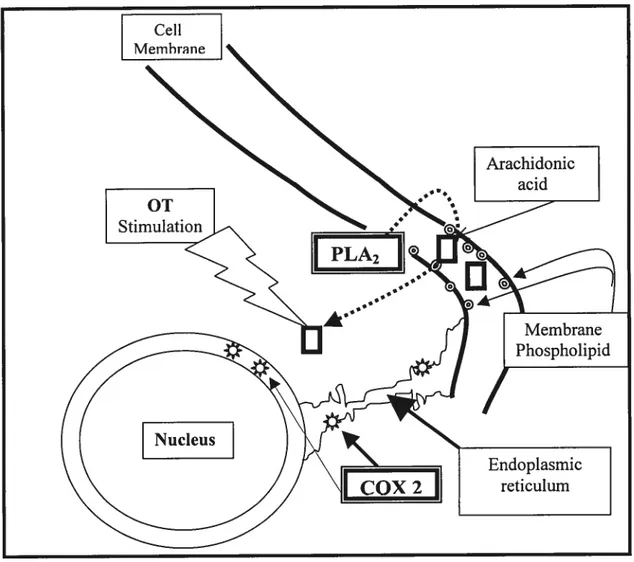
The first step involved in prostaglandin formation is the hydrolytic release of arachidonic acid rnediated by members of the phospholipase A2 family of enzymes (figure 2).
Figure 2: Location of the enzymes involved in the synthesis of prostaglandin F2Πin the ceils.
Following its release, arachidonate is converted to PGH2 by the action of prostaglandin GiN synthase (PGHS), which is situated on the luminal surface of the endoplasmic reticulum and the outer envelope of the nuclear membrane (33). Numerous studies have demonstrated the existence of two distinct genes encoding two isoforms ofPGHS, named PGHS-1 and PGHS-2. Both enzymes possess a PGG2-synthetic cyclooxygenase activity that has resulted in their being colloquially referred to as cyclooxygenase-1 and -2 (or COX-1 and -2), but they are also responsible for the rapid conversion of PGG2 to PGH2 via a peroxidase activity (34). Under basal
conditions, PGF2a, the principal prostaglandin produced by epithelial cells (29, 31), would be produced through constitutiveÏy expressed COX-1. An increase in COX-2 in response to stimulators of prostaglandin production (29) supports its identification as an inducible enzyme. This is further supported by the observation that expression of COX-2 protein in response to oxytocin or arachidonic acid treatment closely matched the production ofprostaglandins (35).
Sensitivity to oxytocin (OT) in a variety of target tissues is widely regarded to be controlled through changes in OT receptor (OTR) population density resulting from corresponding regulation of OTR gene transcription (16, 36). The ontogeny of endometrial expression of OTR in sheep and cattle is associated with development of uterine responsiveness to OT, resulting in OT-induced secretion of uterine PGF2Œ. (37, 38), which is critical for promoting corpus luteum regression in ewes (20, 39) and which leads to further follicular development, estrous behavior, ovulation, and the opportunity for mating and conception to occur. If mating and conception occur, then the initiation of endometrial OTR expression is blocked during early pregnancy (16, 40, 41); sensitivity to OT is abrogated, tuteolysis is prevented, and pregnancy is established. Endometrial sensitivity to OT is markedly reduced during early pregnancy in sheep (42, 43) and cattie (41, 44). In pregnant ruminants, this reduced sensitivity is controlled through inhibition of OTR expression (16, 40, 41). Although
there are reports, especially in the cow, that OT might flot be invoÏved in luteolysis (45), there is no conclusive evidence for other factors being involved in the regulation of pulsatile secretion of PGF2a. Thus, the acquisition of responsiveness to OT by the endometrial epithelium determines when endogenous secretion of PGF2Œ will occur during the estrous cycle, and this appears to require the coordinated action of P4 and E2. In ruminants, three hormones—P4, oxytocin, and estradiol (E2)—may play major roles in regulating uterine secretion of PGF2Œ (46). Progesterone administered early in the estrous cycle, before the normal rise in plasma P4, results in premature luteolysis (47), whereas delaying the action of P4 on the endornetrium by the use of an antagonist delays luteolysis. Thus, the length of time the endometrium is exposed to P4 determines the length ofthe luteal phase (48).
Oxytocin is an acute stimulus for PGF2u secretion. It is secreted from the pituitary and the CL acting via the OTR to stimulate the pulsatile release of PGF2Πfrom the luminal epithelium ofthe endometrium (15, 49).
P4 and E2 regulate uterine secretion of PGf2Œ, in part, by controlling both the timing and the magnitude of uterine secretory responsiveness to oxytocin (50). Lafrance and Goff showed that under the influence of P4, estradiol enhances the 0T-induced release of PGf2u and suggest a possible synergistic action of these hormones for the induction of luteolysis in heifers (51).
It is widely accepted that P4 downregulates OTR during the mid luteal phase (52-53) and that upregulation of the OTR is initiated by an increase in ER (54). However, studies in the cow have shown that there is an increase in OTR before an increase in ER is observed (55). This, together with the fact that prolonged treatment of ovariectomised ruminants with P4 alone is able to induce the response to 01 (51), suggests that the initial increase in OTR is not brought about by changes in E2 or ER. It is therefore possible that changes in the action of P4 results in the initial upregulation of OTR and that this is further increased when P4 decreases and ER increases.
Prostaj’tandin Sy,tthesis
In general, there are two large subgroups within the phospholipase family. The first is a group of small (approximately 14 kd), extensively disulfide cross-linked, secreted enzymes sharing a high degree of homology (56). The second subgroup of PLA2 enzymes is best characterized by the type W cytosolic PLA2 (cPLA2), an 85-kd enzyme without homology with other PLA2 enzymes (57). The nuclear envelope and endoplasmic reticulum are the primary sites for arachidonic acid (AA) metabolism initiated by cPLA2 in activated ceils. These are also the primary subcellular locations for the COX enzymes, 5-lipoxygenase that catalyzes conversion of arachidonic acid
to leukotrienes (58) and some of the terminal synthases like PGE synthase (PGES) and PGF synthase (PGFS). The downstream enzymes, PGES and PGFS, catalyze the conversion of PGH2 to PGE2 and PGf2a, respectively (59). Many studies indicate that PGFS and PGES are highly expressed in endometrium during mid- and late luteal phases of the bovine estrous cycle (59, 60, 61, 62, 63).
Foilowing its release, arachidonic acid is converted to PGH2 by the action of prostaglandin G/H synthase (PGHS), also known as cyclooxygenase (COX), whïch is situated on the luminal surface of the endoplasmic reticulum and the muer and outer membranes of the nuclear envelope (33).
COX proteins are rate-limiting enzymes for the conversion of arachidonic acid into PGH2, the common precursor of ail prostagiandins. There are two isoforms of cyclooxygenase: COX-1 and COX-2 (34). Numerous studies have demonstrated that COX-i and COX-2 are encoded by two distinct genes (64). Both enzymes possess a PGG2-synthetic cyclooxygenase activity and a peroxidase activity that converts PGG2 to PGH2. Despite cataiytic and structural simiiarities, COX-1 and -2 differ in most other respects, inciuding gene structure and regulation and mRNA stability (7, 34). After biosynthesis of PGH2, this endoperoxide is converted to one of several possible prostanoids by a terminal synthase. Prostaglandin F2Πis synthesized via three pathways from PGD2, PGE2, or PGH2 by PGD 1 1-ketoreductase, PGE 9-ketoreductase, or PGH 9-, 1 1-endoperoxide reductase, respectively (65).
The constitutive enzyme, COX- 1, is expressed in most nucieated ceils. On the other hand, the inducibie COX-2 is present only after induction by a variety of factors such as cytokines and tumor promoters (29) and plays a roie in various pathoiogicai and some physiological conditions. The same authors proposed that the two different enzymes couid be associated with distinct poois of arachidonic acid and different downstream enzymes (34, 66).
Arosh and his colleagues demonstrated that during the bovine estrous cycle, COX-1 is flot expressed either at the mRNA or at the protein level, but that COX-2 is expressed throughout the cycle with maximal expression between day 16 and day 18
in the bovine endometriai celIs (59). Asselin et al. reported an increase in expression of COX-2 mRNA and PGF2Πproduction after stimulation with oxytocin (29).
In a study conducted by Parent et al. on primary bovine epithelial ceils, expression of COX-1 and COX-2 proteins was measured by western blot analysis after treatment of epithelial celis with optimal concentrations of oxytocin or arachidonic acid, both of which are stimulators of prostaglandin production. Under non-stimulated conditions, expression of COX- 1 protein by epithelial cells was high, whereas expression of COX-2 was low. Stimulation of the same cells with oxytocin or arachidonic acid at concentrations known to stimulate prostaglandin production increased the amounts of COX-2 but did not affect COX-1 (35).
Oxytocin
Ail neurohypophysiaÏ hormones inciuding OT are nanopeptides with a disuifide bridge between Cys residues 1 and 6. This results in a peptide constituted of a 6-amino acid cyclic part and a COOH-terminal Œ-amidated three-residue tau. Based on the amino acid at position 8, these peptides are classified into vasopressin and OT; OT contains a neutral amino acid at this position and the vasopressin contains a basic amino acid (lysine, arginine) at this position. Isoleucin in position 3 is essential for stimulating OTRs and Arg. or Lys. in position 8 for acting on vasopressin receptors. The difference in the polarity of these amino acids residues is believed to enable the vasopressin and OT peptides to contact with the respective receptors (67).
The actual source of neurohypophysial hormones including OT is the nerve ceils in the supraoptic and paraventricular nuclei of the hypothalamus. From here the hormones are carried to the neurohypophysis by way of the axoplasm in the nerve fibers that pass from the hypothalamus to the neurohypophysis of the pituitary, where they are stored until released (68).
pituitary (69) has been supported by the report of Schams et al. in the cow showing that the administration of several PGF2a analogs caused a large elevation of oxytocin injugular venous blood that reached a peak within 15—20 min. (70).
Regulation of secretory ftinction of hormone secreting neurons is via feedback mechanisms of hormones from the anterior hypophysis, adrenal cortex, thyroid and gonads or by neural pathways in the hypothalamus (71). However, OT is also synthesized in peripheral tissues, e.g., uterus, placenta (72), corpus luteum (73, 74), testis (75) and heart (76).
It is well known that the bovine CL is a site of P4, prostaglandin and OT production (74). In several species, the ovary has been shown to contain OT and may be a site of local OT production (77). Functional OTRs have been detected in bovine granulosa ceils, suggesting that OT may be an autocrine factor during follicular growth (78). The scnsitivity of bovine endometrium to oxytocin varies during the estrous cycle. The stimulatory effects of oxytocin on PGF2Πsecretion by the bovine endometrium were observed at the follicular phase, estnts, and early luteal stages of the estrous cycle (days 1-4), whereas oxytocin had no effect during the rnid-to-Iate luteal stages (days 5-17) (79). These results confirmed the previous reports that the high responsiveness of the endometrium to oxytocin was maintained during the period of luteal regression until the early luteal stage of the next estrous cycle (40, 80, 81). However, there is increasing evidence that oxytocin is not essential for the initiation of PGF2Πoutput during luteolysis in the cow (82, 83, 84). In fact, concentrations of oxytocin in blood (85), and in intact and rnicrodialyzed CL ($6) are extremely low at the time of spontaneous luteolysis. Moreover, the blockade of uterine oxytocin receptors with a specific oxytocin antagonist from Days 15 to 22 of the cycle affected neither luteolysis nor the duration of the estrous cycle in heifers (83). Therefore, PGF2a secretion by the endometrium may be regulated not only by oxytocin but also by one or more other factors in cattie. Moreover, since the blockade of oxytocin receptors decreased the magnitude ofPGF2a release without preventing an increase of PGFM in blood (84), oxytocin may play a supportive and modulatory role as a regulator of the amplitude of pulsatile PGF2Πsecretion after the initiation of luteolysis in cattle (87).
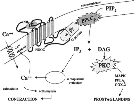
Wathes et al. showed that OT concentration in the CL of bovine ovary increased from stage 1 (early luteal phase days 1-4) to stage 2 (mid-luteal phase days 5-10) but declined during stage 3 (late luteal phase days 11-17) and were low in follicles, whole ovaries and pregnancy corpora lutea ($8). Binding of OT to its specific receptors in the uterus activates phospholipase C and release triphosphoinositol (1P3) and diacyloglycerol (DAG). The DAG stimulates protein kinase C (PKC) which might lead to several actions to increase contractility. The 1P3 stimulates Ca release from intracellular stores. This intracellular mobilization of Ca is followed by increases in PGF2Πsecretion (89, 90). The rnechanism of action of oxytocin in rnyometrium is illustrated in figure 3:
Figure 3: Mechanism of action 0foxytocin (OT). Interaction of OT with its specific
membrane receptor triggers G-protein (a/13’y) mediated activation of phospholipase C (PPLC) resulting in production of inositol trisphosphate (1P3) and diacylglycerol (DAG). The DAG stimulates protein kinase C (PKC) which might lead to several actions to increase contractility. The 1P3 stimulates Ca release from intracellular stores. The oxytocin receptor (OTR) might also be connected directly to a receptor
++ . . . ++
activated Ca chaimel. The increase in intracellular Ca stirnulates actrnlmyosrn coupling resulting in muscle fiber contraction. COX-2 = cyclooxygenase 2; PIP2 =
phosphatidylinositol-4, 5-bisphosphate; MAPK = mitogen-activated protein kinase;
PPLA =phospholipase A.
Taken from: Mitcheil B.f, Schmid 3 (2001) Oxytocin and its receptor in the process of parturition,J Soc GynecoÏ Investig; 8: 122-33.
ce”
Ca
DAG
Ca
sarcopla.smfc MAPKPPLA2rikuhim
COX-2 calmodulin
aetirnyosin
Lutinizing hormone (LH), PGf2Πand tumor necrosis factor-a (TNF-u) are arnong the other regulators of endometrial PGf2Πsecretion (87). A brief discussion of their effects follows:
There is conflicting evidence that LH is involved in uterine functions, since LH is traditionally known to drive progesterone synthesis of CL. Nevertheless, since LH induced endornetrial COX-2 expression and increased PGF2Πrelease by bovine -endometrial ceils, LH has been suggested to be involved in the regulation of PGf2Πrelease by bovine endometrium during the late luteal phase. In support of this, it was found that the concentration of the uterine LH/hCG receptor varied during the estrous cycle, with higher values at Days 15 to 17 and lower values at Days 2 to 4 (91, 92). Moreover, Canino et al. (93) demonstrated that LH stimulated PGF2Πsecretion in the cow. However, the effect of LH on PGF2Πsynthesis in bovine endometrium appears to be lirnited to a specific time frame, narnely the mid-to-late luteal phase ofthe cycle (93). Therefore, LH mayplay a reinforcing role rather than an initial role in luteolysis in cattle (92).
Wade and Lewis (94) demonstrated that exogenous PGF2a stimulates the utero ovarian release of PGF2Πin the ewe, suggesting that the utero-ovarian PGF2Πauto-amplification unit is a component of the luteolytic mechanism of PGf2u. furthermore, a PGF2Πanalogue increased PGF2 release from the bovine uterus on day 18 of the estrous cycle (84). It bas been shown that PGf2a activates PKC and increases intracellular calcium mobilization (21), which may in tum stimulate PGF2a production in the endometrium (95, 96, 97). Therefore, the endogenous PGF2Πmay be an essential component in the mechanism regulating its own production (87).
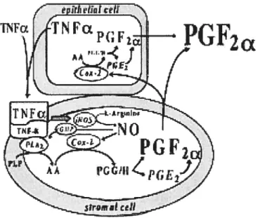
Some recent studies indicate the presence of functional TNF-a receptors in the bovine cyclic endometrium and suggest a possible role of TNF-Πin the regulation of endometrial PGF2u production in cattle (85, 97, 98, 99). TNF-Πstimulated PGF2Πproduction only in the stromal cells via the activation of PLA2 and nitric oxide synthase (98) (figure 4). Although both oxytocin and TNF-Πaffected PGF2a output at the follicular stage, TNF-a, in contrast to oxytocin, also affected PGF2Πoutput at the mid and late luteal stages.
2a
Figure 4: Hypothetical model for TNF-a control of PGf2Πsynthesis in bovine endometrial ceils during luteolysis. TNF-Πthat is produced by endometrial epithelial celis andlor uterine macrophages stimulates PGF2Πproduction only in the stromal celis via the activation of phospholipase A2 (PLA2) and nitric oxide (NO) synthase. TNF-u induced PGF2Πoutput from stromal ceils may be the first component of an auto-amplification cascade within the bovine endometrium and switches on the positive feedback loop between the uterine PGF2Πand the luteal oxytocin to complete luteolysis. Taken from: Okuda K (2002) Regulation of endometrial prostaglandin F2Πsynthesis during luteolysis and early pregnancy in cattie. Domestic Animal Endocrinology; 23: 255-64.
Moreover, just before luteolysis, there are dramatic increases in TNF-Πgene expression (100) as well as TNF-Πrelease (101) from the CL. Therefore, the overall findings lead us to hypothesize that endometrial andlor luteal TNF-Πmay be a trigger
for the output of PGf2Πfrom the uterus in the initiation of luteolysis. $ince PGF2a is produced preferentially by epithelial cells (31, 32, 98, 102), TNF-u-induced PGF2Πoutput from stromal celis may be the first component of an auto-amplification cascade within the bovine endometrium and switch on the positive feedback loop between the epithelial PGF2a and the luteal oxytocin to complete luteolysis
(
See Fig. 4). The endometrium apparently consists of many more stromal celis than epithelial celis, so the TNTu-stimulated PGF2Œ from stromal celis could be sufficient to play a role inthe initiation of luteolysis. Altematively, TNFa-induced PGF2Œ may stimulate PGf2Œ production in both stromal and epithelial celis as a paracrine and autocrine regulator, as has been suggested to occur in the ovine corpus luteum at luteolysis (103). Consistent with this auto-amplification cascade, there are reports that PGf2Œ treatment acutely increased PGF2Œ output from the bovine (104) and ovine (94) uterus. Therefore, along with these findings, Skarzynski et al suggested that TNFa induced PGf2Œ from the stromal celis can initiate luteolysis in cattie. Furthermore, TNFŒ-induced PGF2u from the strornal ceils may switch on the positive feedback Ïoop between epithelial PGF2a and luteal OT that completes luteolysis in cattie (70). It should be noted that TNF-u induces the output of both PGF2Œ (85, 98) and PGE2 (97) in cuÏtured bovine stromal cells. Moreover, it is well known that PGE2 isconverted into PGF2Œ (105, 106). Thus, the potential capacity of the uterus to
generate luteolytic PGf2Πin both epithelial and strornal cells from PGE2 may be the next mechanism involved in TNF-a-induced luteolysis in cattle (87).
Oxytocin Receptor (OTR)
Peripheral vasopressin and OT receptors have been classified on the basis ofboth the second messenger system coupled to the receptors and the affinity of a series of vasopressin and 01 analogues with enhanced seÏectivity for a certain receptor type. A
great number of molecular probes, including agonists and antagonists, and radio labelled, fluorescent or photosensitive ligands make the OTR family a good model with which to study structure-function relationships. Today, OTR along with the vasopressin receptors have been cloned in mammals, lower vertebrates and invertebrates, and molecular cloning of this family has confirmed that vasopressinlOTR subtypes are members of the G-protein-coupled receptor (GPCR) superfamily, consisting of 7 hydrophobie transmembrane Œ-helices joined by altemating intracellular and extracellular loops, an extracellular N-terminal domain, and a cytoplasmic C-terminal domain. They display a high degree of sequence identity, showing about 102 invariant amino acids among the 370-420 amino acids in the human receptors. The appearance of OTRs in the endometrium of ruminants controls the onset of a feed back loop and episodic PGF2Œ secretion. In ruminants, OT is produced in the CL and its release is stimulated by PGF2u. Thus a positive feedback loop is established that amplifies neural OT signaIs (107); neural OT stimulates PGF2u secretion, which stirnulates release of luteal OT, which in turn further stimulates release of PGF2Œ. Once concentrations of the OTRs have increased during late diestrus (3$), the ability of OT to stimulate synthesis of uterine PGF2a in ruminants may be mediated through a rapid increase in phospholipase C activity (108, 109). Activated PLC hydrolyses phosphoinositides (PI) presumably leading to the formation ofDAG and inositol (1, 4, 5)-triphosphate (1P3). Synthesis ofPGF2u in the endornetrium is stimulated by DAG which may act as a second messenger to activate PKC (110). Lafrance and Goff reported that stimulators of PKC activity (i.e. DAG analogues and phorbol ester 12-myristate 13 acetate) also stimulate PGF2Œ secretion from the bovine endometrium to the same extent as OT. They concluded that stimulation of PGF2Œ by OT is via the PKC effector pathway (111). Similar experiments in sheep have shown that oxytocin stimulated release of PGF2Œ and activity of PLC in explants of ovine endornetrial tissue in vitro. Second messengers associated with activation ofPLC enhanced release ofPGF2a from ovine endometrial tissue (110).
By Northem blot analysis and in situ hybridization, Zingg et al. determined that, at term, the rat uterine epithelium represents a major site of oxytocin gene expression. Estrogens act as a strong inducer of uterine OT gene expression in vivo, and this effect is potentiated 7-fold by concomitant progesterone P4 administration. Whereas OTR mRNA is strongly induced by estrogen, P4 does not potentiate but slightly attenuates the estrogen-induced rise. However, estrogen-induced 01 binding is completely reversed by concomitant P4 administration, suggesting an additional post transcriptional effect ofprogesterone (112).
Even if the receptors are downregulated in vivo, they show upregulation when explanted and cultured in vitro (113). This indicates that the OTR regulation is partly due to gene suppression in vivo. Despite the presence of steroid receptors in bovine endometrial ceils, the level of OTR mRNA could neither be affected by progesterone or estradiol nor by a progesterone withdrawal protocol. The only factor that affected the OTR mRNA level was interferon-r. As in vivo, this cytokine suppressed the OTR mRNA production (114).
As shown with knock-out mice, estradiol receptor u (ERu) is flot necessary for basal OTR synthesis but is absolutely necessary for the induction of OTR binding in the brain by estrogen (115). However, it is unclear whether OTR gene transcription is predominantly regulated by estrogen. The continuous presence of receptors in certain brain regions after gonadectomy suggests the existence of altemate mechanisms of regulation (116).
While there is no estrogen response element (ERE) on the bovine or ovine OTR gene promoter region (117), ER can act through SPi and possibly APi sites on gene promoters (11$). This is a possible mechanism by which estradiol can up-regulate OTR and is supported by recent findings that ERu likely stimulates OTR promoter through both protein-DNA and protein-protein interactions with SPi and AP-1 (119). Gonadal steroids have an important influence on the uterine OT receptor mRNA accumulation in vivo. Estrogens administered to ovariectomized rats increased 01 receptor binding sites and increased OT receptor mRNA accumulation several fold. Although progesterone leads to a marked decline of OT receptor binding sites, the mRNA levels of OT receptor were nearly unchanged (120). Estradiol treatment in
vivo induces an initial up-regulation of endometrial OTR expression in ewes (121, 122, 123) but if estradiol treatment is continued over several days, OTR concentration decreases (124, 125). Some researchers like Soloff et al., concluded these findings imply the involvement ofnongenomic effects ofprogesterone (126).
Taken together, these resuits indicate that estradiol can induce a short-term up regulation in OTR expression but that the effect cannot be maintained for more than 1—2 days. However, the exact mechanism of action of E2 lias been difficult to elucidate.
Steroids:
Five major classes of steroid hormones are derived from cholesterol: progestagens, glucocorticoids, mineralocorticoids, androgens, and estrogens. Hydroxylations by P450 monooxygenases that use NADPH and 02 play an important role in the synthesis of steroid hormones and bile salts from cholesterol. P450 enzymes, a large superfamily, also participate in the detoxification of drugs and other foreign substances. Pregnenolone (C21) is an essential intermediate in the synthesis of steroids. This steroid is formed by scission of the side chain of cholesterol. Progesterone (C21), synthesized from pregnenolone, is the precursor of cortisol and aldosterone. Hydroxylation of progesterone and cleavage of its side chain yields androstenedione, an androgen (C19). Estrogens (C18) are synthesized from androgens by the loss of an angular methyl group and the formation of an aromatic A ring (127).
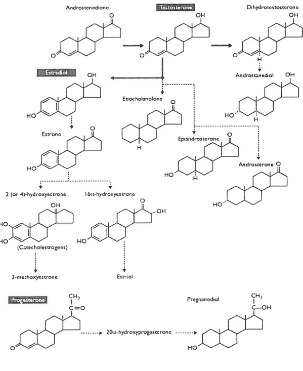
The following figure illustrates major metabolic pathways of the three principal steroids secreted by the gonads.
Figure 5: Major metabolic pathways of the three principal steroids (Progesterone, Estradiol and Testosterone) secreted by the gonads.
Taken from: Endocrinology: An Integrated Approach by Stephen Nussey, Saffron Whitehead © BIOS Scientific Publishers Ltd, (2001) St. George Hospitat Medical $chooÏ, London, UK. Andrtcriidcn Tiastéron. OH Dihdrvtcvrono H Andrs’erono O 2 r 4-Idryrrr HO H HO HO J CH c,=o O CH Prcnancdci! Ç---OH . 2O-h>’drQyprvescronc
In addition to OT, E2 and P4 play a significant rote in the initiation of luteolysis in ruminants. Uterine secretory responsiveness to OT increases at luteolysis, when endogenous, pulsatile secretion of PGf2Œ normaily begins. Therefore, the acquisition by the uterus of responsiveness to OT may determine when endogenous secretion of PGF2Œ occurs during the estrus cycle. In the presence of P4 uterine secretory responsiveness to OT develops slowly. Progesterone exerts two types of effects that contribute to the regulation of PGF2Œ secretion. first, prolonged exposure to progesterone appears to promote uterine accumulation of arachidonic acid, prostaglandin endoperoxide synthase, and other substances needed for synthesis of PGf2Œ. Second, P4 exerts a suppressive effect on secretion, which wanes afier prolonged exposure. Together, these effects of P4 ensure that PGF2Œ is secreted only at the proper time to induce luteolysis (45).
In an in vivo study conducted by Mmm et ai, they exposed 8 long-term ovariectomized cows to proionged treatment of P4, but not estradiol. This ied to the induction of responsiveness to OT, appearing within 2 days of progesterone treatment, reached a maximum by 6 days and was maintained until day 18. In ovariectomized ewes, while oestradiol treatment did induce temporary responsiveness to OT after 3 d treatment, progesterone treatment was required to induce sustained responsiveness that appeared by day 9 oftreatment and was maintained up to day 12. Measurement of endometrial receptors for OT revealed a significant decline in OTRs by day 6 of treatment when responsiveness to OT was maximal, showing that receptor concentrations were not a limiting factor. The most iikely mechanism by which progesterone treatment induces responsiveness to OT may be through the upregulation of post receptor signaling pathways andlor enzymes involved in prostaglandin synthesis (128). The resuits ofsome investigations show that there is a dose-dependent inhibition of OTR concentration by progesterone and a dose dependent stimulation of basal PGf2Πrelease by estradiol (15, 46, 129, 130).
Mann (131) investigated the effect of progesterone and estradiol on basal and OT stimulated PGF2Πproduction and on OTR concentrations in endometrium from long term ovariectomized cows using an expiant culture system. As a result he found that basai PGF2Πproduction was unaffected by progesterone treatment but was stimulated
by estradiol treatment in a dose-dependent manner. OTR concentration remained unchanged in control culture and were unaffected by treatment with estradiol while treatment with progesterone caused a dose-dependent inhibition. Responsiveness to OT in terms of increased PGF production developed spontaneously over the first 24 h of culture and was unaffected by treatment with progesterone and estradiol. The reason for spontaneous developrnent of responsiveness to OT remains unknown but may resuit from the removal of tissue from the influence of an as yet unidentified inhibitory factor (131).
The effect of prolonged progesterone treatment of bovine endometrial celis resulted in an increase in OT-stimulated PGf7a secretion but not in its basal secretion. It was demonstrated that estradiol had no effect on basal secretion ofPGF2Πbut did enhance the OT-stimulated secretion in celis exposed to progesterone for 17 and 21 days, but flot in ceils exposed to progesterone foronly 10 days (132).
Overali, the action of progesterone was more celi type specific than receptor specific. The progesterone doses that are required to affect the signaling function ofreceptors are much higher than the progesterone levels found in plasma or in nonsteroidogenic tissues such as the myometrium. In steroidogenic tissues, however, huge amounts of progesterone have been measured. In steroidogenic celis as well as in their environment, progesterone might nongenomically influence the signaling of receptors. The molecular mechanisms underlying this progesterone action are flot understood (133).
It is now clear that estradiol-17f3 and progesterone have multiple roles in controlling the luteolytic process, not only by regulating the enzymes necessary for the endometrial biosynthesis of PGF2a, but also by controling endometrial receptors for oxytocin in a number of species (15). Measurement of oxytocin in jugular plasma indicated that plasma levels of oxytocin increased markedly during hour-long bursts of intrarnyometrial pressure (llvIP), whereas plasma levels of vasopressin remained unchanged. The concentration of oxytocin during the first large burst of IMP reached -200 pg!ml of plasma, but peak concentration of oxytocin declined by —50% during each subsequent burst ofTMP (134, 135).
Other studies have also reported large pulses of oxytocin or its neurophysin during luteolysis in sheep (136), and cows (137). These resuits suggested that the large hour long episodic releases of oxytocin in ruminants, interacting with rising levels of endometrial oxytocin receptors, evoked the large episodic pulses of uterine PGF2Œ
that cause luteolysis in these species. Moreover, evidence has accumulated that ovarian steroids atso modulate the synthesis and secretion of oxytocin from the hypothalarnic/posterior pituitary system, which explains, at least in part, the pulsatile nature ofuterine PGF2a secretion (15).
Progesterone is considered to be essential to maintain the uterine quiescence. Grazzini et al. (138) postulated that progesterone specifically binds to the rat OTR with high affinity (Kd; 20 nM) and thereby inhibits receptor function. In case of the human OTR, a direct inhibitory interaction [inhibitory constant (Ki) 30 nM] with a progesterone metabolite, 53 pregnane-3, 20-dione, has been reported by the same
authors. Grazzini et al. (13$) clairned that progesterone could act as a negative modulator of the OTR and thus offered a plausible mechanism of how progesterone could contribute to uterine quiescence. However, these flndings could flot be reproduced in several other laboratories. Instead, the resuits of other investigations were totally different. Gimpl et al found that high concentrations of progesterone (>1 OjiM) attenuated or blocked the signaling of several GPCRs, including the OTR. The progesterone effects occurred within minutes, were reversible, and could not be blocked by a protein synthesis inhibitor (139). Therefore, they concluded that these effects of progesterone probably were flot mediated through genomic mechanisms, either activating or suppressing transcription of specific genes (e.g., the OTR or PGH2 synthase 2). In addition, this effect of progesterone is extrernely transient and progesterone bas no inhibitory effect on the response of cultured bovine uterine epithelial ceils to OT when administered for 72 h and then withdrawn for 6 h immediatelypriorto OT exposure (140).
Proj’esterone receptor (PR)
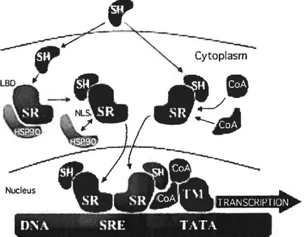
Binding of steroid hormones to the specific steroid hormone receptor tSR) ligand binding domain (LBD) induces a conformational modification of the receptor, followed by the separation of the receptor from cytoplasmic chaperone proteins such as heat shock protein 90 (Hsp90) and by the exposure of nuclear localization sequences. This allows nuclear transiocation and homo/heterodimerization of the ligand-bound receptors, and their binding to steroid response elements (i.e. nucleotide sequences specifically recognized by SRs) on the promoter regions of the target genes, thus regulating gene expression by interacting with the transcription machinery (141, figure 6).
Figure 6: Steroid receptors’ tSR) genomic actions. Binding of steroid hormones (SH) to the LBD of the SR induces a conformational modification of the receptor. This causes the separation of the receptor from cytoplasmic chaperone proteins such as Hsp9O, the interaction with celi-specific co-activators (CoA), and the exposure of nuclear localization sequences (NLS5). This allows nuclear transiocation and homo/heterodimerization of the ligand-bound receptors, and their binding to steroid
response elements (SREs, i.e. nucleotide sequences specifically recognized by SRs) on the promoter regions of the target genes, thus regulating gene expression by interfering with the transcription machinery (TM).
Taken from: Tommaso Simoncini and Andrea R Genazzani (2003) Non-genomic actions of sex steroid hormones. Europeait Journal ofEndocrinology; 148: 281—92.
The actions of progesterone are mediated by PR. Changes in the number of progesterone and oestradiol receptors in the endometrium are thought to play a role in the induction of luteolysis. The effect of oestradiol and progesterone on the regulation oftheir receptors in cultured bovine uterine epithelial and stromai ceils was examined by Xiao et al. They showed that the number ofPR was higher in the stromal ceils than in epithelial celis, whereas the number of estradiol receptors was higher in the epithelial celis than in stromal celis. Estradiol upregulates its own receptor and increases the number of progesterone receptors in both ceil types in vitro, whereas progesterone has littie effect, but inhibits the effects ofoestradiol on PRs. (142). P4 is the hormone of pregnancy and unequivocaiiy required in ail mammais for maternai support of conceptus (embryo/fetus and associated membranes) survival and development. However, the endometrial lumenai (LE) and giandular epithelia (GE) of a number of species exhibit a loss of PR expression prior to the stages of uterine receptivity and implantation. In sheep, PR expression becomes undetectabie in the endometrial LE aller Day 11 and then in the GE after Day 13. The actions of progesterone on endometrial epithelia during most of gestation appear to be mediated by the endometrial stroma that remains PR-positive throughout pregnancy. Stromai cells produce several growth factors, such as hepatocyte growth factor (HGF) and fibrobiast growth factors-7 and -10 (FGF-7, FGF-10), that have receptors expressed specificaliy in the endometrial epithelia (143). These factors may be progesterone responsive and mediate epitheiial-mesenchymai interactions that are crnciai for support of pregnancy. At estrns, estrogen increases PR expression in the endometrial epitheiia (144). High levels of endogenous Jaagsiekte sheep retroviruses (enJSRVs) are expressed in the PR-positive endometrial LE and GE in response to increasing
progesterone and are hypothesized to stimulate trophoblast proliferation and production of interferon (IFN)-tau. IFN-tau, the pregnancy recognition hormone produced by the trophoblast from Days 10 to 21, acts in a paracrine manner on the PR-negative endometrial LE and superficial GE to inhibit transcription of estrogen receptor alpha (ERa) and OTR genes. These actions of 1FN-tau maintain progesterone production from the corpus luteum by abrogating release of luteolytic pulses of PGF2Πfrom the endometrial epithelium. The antiluteolytic effects of WN tau are dependent on progesterone. Progesterone stimulation over 8-10 days suppresses expression of the PR gene in the LE and then GE. Loss of the PR in the LE is concomitant with decreases in mucin glycoprotein one (MUC-l), an inhibitor of blastocyst implantation. As the conceptus begins implantation on Day 15, the binucleate trophectodermal celis then differentiate and produce placental lactogen (PL), a member of the prolactin (PRL) and growth hormone (GH) family. PL stimulates GE proliferation and production of secretory proteins, such as UTMP and OPN. Interestingly, the effects of PL on the GE appear to require the absence of PR and prior exposure to IFN tau. During mid-pregnancy, the mononuclear trophectodermal cells produce GH that can also act on a progestinized uterus to stimulate GE hypertrophy and secretory function. The actions of this servomechanisrn are proposed to stimulate GE hyperpiasia from Days 20 to 50 and then GE hypertrophy and maximal differentiated function afier Day 50 when the majority of fetal growth and development occtirs during gestation (143).
Mechanism ofAction ofProesterone
Progesterone is an essential reproductive hormone that acts on the estrogen primed endometrium to induce conditions favorable for embryo implantation (145, 146), and the maintenance of pregnancy (147). The genomic effects of progesterone are mediated in target cells through interactions with specific intracellular progesterone
receptors (148). A welÏ-known progesterone binding protein is the multidntg resistance P-glycoprotein (133). In addition to their role in detoxification, P glycoproteins are involved in intracellular choiesterol transport. It is known that progesterone markedly interferes with the intracellular transport (and metabolism?) of cholesterol (sec model in figure 7). At concentrations in the micromolar range, it inhibits both the cholesterol esterification and the transport of cholesterol to and from the plasma membrane (149).
Paradoxically, at the same time, progesterone stimulates the activity of 3-hydroxy-3-methylgiutaryl (HMG)-CoA reductase, the key enzyme of de novo cholesterol biosynthesis. Hence, cholesterol precursors like lanosterol begin to enrich in the membranes ofthe celi (150). The OTR needs a cholesterol-rich microenvironment to become stabilized in its high-affinity state (151). Because the cholesterol precursors, particularly lanosterol, are completely inactive to support the OT receptor in its high affinity state (152), the responsiveness of the OT system may flot be fui ly operative during the continuous presence of high progesterone concentrations. According to this scenario, progesterone withdrawal would restore the cholesterol transport so that the highly enriched amounts of cholesterol prectirsors would now becorne rapidly converted to cholesterol (153). According to this postulated mechanism, progesterone could affect the signaling of ail those receptors that are functionaliy dependent on cholesterol. It is important to note that the nongenomic actions of progesterone including its influence on the cholesterol transport require progesterone concentrations in the micrornolar range. This suggests that the described effects may be limited to the steroidogenic tissues and to their environment. Most iikely, progesterone acts in these tissues via both genomic and nongenomic pathways (surnmarized in figure 7 on the next page) together with other steroids to control receptor activity (154).
Chût+ M2 4
OTR
LDL (CE)
Figure 7: Schematic moUd of nongenomic inhibitory effects of progesterone. Progesterone inhibits both the signal transduction ofGq coupled receptors (as shown here for the OT receptor) and the intracellular trafficking of cholesterol. PrincipaÏly, eukaryotic ceils can obtain the required cholesterol (Chol, gray ellipses) by two sources: endogenously by de novo synthesis of cholesterol and exogenously by uptake of cholesteryl ester (CE)-rich low-density lipoprotein (LDL) particles via receptor-mediated (R) endocytosis. De novo synthesized cholesterol first arrives at cholesterol-rich domains in the plasma membrane (caveolae and/or “lipid rafts”) that may function as cholesterol “sorting centers” within the plasma membrane, where most of the cellular cholesterol resides. Progesterone blocks several intracellular transport pathways of cholesterol (red bars) except for the LDL receptor-mediated uptake of cholesterol. Moreover, cholesterol esterification does not occur in the presence of progesterone, presumably due to the lack of cholesterol substrate for acyl CoA cholesterol acetyltransferase (ACAT). As a consequence, unesterified cholesterol accumulates in lysosomes (or late endosomes) and lysosome-like compartments (designated as “lamellar bodies”) (marked by red background). The key enzyme for the cholesterol de novo synthesis, 3-hydroxy-3-methylglutaryl-CoA reductase (HIvIG-CoA Red), is stimulated in the presence ofprogesterone (feU arrow),
but the cholesterol biosynthesis stops at the level of precursors (e.g., lanosterol). Enzymes involved in the conversion of cholesterol precursors reside in the endoplasmic reticulum tER), and progesterone most likely prevents sterol precursors localized in the plasma membrane from reaching the ER-resident enzymes, thereby preventing their conversion to cholesterol. Overali, progesterone induces a state of cholesterol auxotrophy. However, afler progesterone withdrawal, the accumulated precursors will be rapidly converted to cholesterol. Thus ceils will become overloaded with cholesterol for a certain period of time, afier which the cholesterol homeostasis will be reestablished. We hypothesize that these reversible progesterone induced changes of the cholesterol trafficking could have a strong influence on signal transduction processes, particularly in case of the OT receptor (OTRH and OTRL, high-affinity and low-affinity OT receptor, respectively; receptor in blue; OT in yellow).
Taken front: Gimpl G and fahrenholz f (2001) The Oxytocin Receptor System: Structure, Function, and Regulation. Physiol. Rev; 81: 629-83.
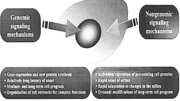
Non-genomic actions of steroid hormones resuit from the recmitrnent of signaling pathways that are often associated with celi membrane receptors such as GPCRs, ion channels or enzyrne-linked receptors (155). Through the recruitment of sucli pathways, steroids rapidly regulate multiple cellular functions, but can also modulate longer-terrn processes such as gene expression, protein or DNA synthesis and ceil proliferation(156).
To name only a few of non-genomic effects of P4 or its metabolites, we may mention induction of oocyte maturation (157) and interactions with the GABAA receptor (158). Other researchers have reported that P4 might be acting via a non-genomic mechanism to regulate uterine production of OT in the rat (112). They have shown that regulation of uterine OT binding involves at least two different mechanisms: E2-induced upregulation is accompanied by an increase in OTR mRNA accumulation, implying that the E2 effect is mediated via increased OTR gene transcription and/or OTR mRNA stabilization. In contrast, P4-induced OTR down-regulation occurs via a
novel non-genomic mechanism, involving a direct interaction of P4 with the OTR at the level of the celi membrane. These effects are specific as signalling and binding functions of the closely related Via vasopressin receptor remain unaffected by P4, and as other, related steroids are devoid of any effect on OTR binding or signalling functions. The observation of a specific interaction of a steroid with a G-protein linked receptor defines a new mechanism ofnon-genomic steroid action and uncovers a novel level of crosstaÏk between steroid and peptide hormone action (120). In a review article, Simoncini and Genazzani (2003) offered a tentative interpretation of genomic and non-genomic actions ofsteroid hormones (159). See figure 8:
*
Gen..zpm *d newprølnsyatbe*b
• tencyelonseÉ
MedIsmndksg4erm oeil prorim
•OrgsnLzatto* oTcdtntworIs
torc.mplêructlons
Figure 8: Genomic and non-genomic actions of steroid honnones: a tentative interpretation. Genomic actions of steroid hormones may serve to program celis, organs and systems for complex steroid hormone-regulated ftinctions, providing the single ceils and the celi networks with the tools to accomplish these tasks via gene expression and new protein synthesis. They are usually characterized by a relatively longer latency of action and could be meant to determine the medium- and long-term program of the cells. Non-genomic actions of steroids may instead serve to signal to the celis changes in the suirounding milieu, and to rapidly activate or repress the cellular functionalities needed to adapt to these changes, which are already present in the celis. The tirne of onset of these effects is usually extremely rapid, but the effects of these mechanisms may also serve to dynamically modulate the long-term ccli program.
Taken from: Simoncini T and Genazzani A.R (2003) Non-genomic actions of sex steroid hormones. Eztropean Journal ofEndocrinology; 148: 22 1—92.
White there is no evidence at present to show that P4 is acting at the genomic level to regulate the bovine OTR, there has been a report to suggest that it might be acting at the ccli membrane directly on the rat OTR protein (138). Grazzini et al showed that the effect of P4 on uterine sensitivity to 01 involves direct, non-genornic action of P4 on the uterine OTR. P4 inhibits 01 binding to OTR-containing membranes in vitro, binds with high affinity to recombinant rat OTR expressed in CHO ceils, and suppresses oxytocin-induced inositol phosphate production and calcium mobilization. These effects are highiy steroid- and receptor-specific, because binding and signaling functions of the closely related human OTR are not affected by P4 itself but by the P4 metabolite 5-dihydroprogesterone (138). They reported that addition of P4 in vitro to membranes derived from a parturient rat uterus inhibited binding of the OTR-specific ligand OTA. P4 also induced a dose-dependent reduction of specific OT binding to recombinant rat OTR expressed in CHO celis. By contrast, P4 had no effect on binding of the Via vasopressin -receptor-specific ligand LVA1O to recombinant V1a receptor (V1aR). Similar results were obtained with 3H-iabelied natural agonists. P4 coupled to bovine serum albumin also reduced oxytocin binding to intact OTR expressing CHO celis, thereby exciuding a cytoplasmic site of P4 action. b determine whether the P4 effect was ligand- or receptor-specific, Grazzini et al took advantage of the fact that arginine vasopressin (AVP) binds to both the OTR and the V1 aR with comparable affinity. [3H] AVP binding to the OTR was repressed by P4, whereas [3H] AVP biding to V1aR was unaffected, indicating that the specificity of the P4 effect is determined by the receptor and not the ligand. Their resuits indicate that the P4 interactions with the OTR are affected by guanine-nuclotide-induced changes of receptor conformation and that the observed effects of P4 are highly receptor-and steroid-specific and are unlikely to be due to non-specific membrane interactions or to interactions with another unknown binding protein (138).
A similar effect of P4 has been observed in the cow (140) and the sheep (160). However, this nongenomic action of P4 on OTR is somewhat controversiai because other groups have been unable to reproduce this effect of P4 or a variety of other naturally occurring progesterone metabolites on the human OTR and the bovine OTR (139, 161) and importantiy, there is no P4 response element on the promoter region of
the OTR gene in bovine (117) or otherspecies (162). Thus, the role ofa nongenomic action of P4 in the regulation of OT action in ruminants during the estrous cycle remains to be elucidated (163).
One of the other functions of progesterone is maintenance of uterine quiescence by decreasing uterine sensitivity to OT (164). And this, in tum, suppresses the ability of OT to induce endometrial secretion of PGF2a. This effect appears to be mediated through a direct influence in the interaction of 01 with its own receptor (140).
PrOÀ’esterone antaf’onists (FA)
Antihormones are potent antagonists of hormone action in vivo, but the mechanism underlying this antagonism is not fully understood. The antiprogesterone RU 486 (mifepristone) has partial agonistic and antagonistic actions. Jnterestingly, this compound displays partial progestational and glucocorticoid action and is therefore considered not to be pure antiprogestin (165). Hormone antagonists normally act by preventing the action of agonists like the effect of RU 486 that induces parturition in the rat.
Fang et at. has studied the changes in OT and OTR mRNA and peptide synthesis within the pregnant rat uterus during RU 486-induced parturition. Pregnant rats were given a single injection of RU 486 on day 15 of pregnancy (normal delivery occurs on day 22). The average time to delivery, after RU 486 injection, was 27 h. In controls, OT mRNA increased significantly, and this increase was blocked in the RU 486 treatment group. OTR mRNA levels increased within 6 h of RU- 486 and remained elevated until delivery. PGF2u was increased 1 6-fold. They indicated that the mechanism of action of RU 486 is to inhibit the P4 suppression of OTR synthesis, allowing increased expression of OTR, which may directly stimulate myometrial contractions or act indirectly through increased synthesis ofprostaglandins (166). Several steroid hormones transform (activate) their receptors from a cytosolic,
non-DNA binding 8$ sedimentation form to a nuclear, non-DNA binding 4$ form. A number of antiprogestins, inciuding RU486, induce an equaily dramatic, but distinct, structural alteration of the ligand binding domain. The distinction centers upon the final 30 to 40 amino acids at the carboxyl terminus. The conformational change can be induced by ligand prior to dissociation of the 8$ complex and is not induced by heat shock protein removai in the absence of hormone (167). Using protease digestion and antibody mapping, Vegeto et al demonstrated that progesterone and RU 486 induce different conformational changes in the PR (168). Binding to progesterone receptors, RU 486 may change their conformation. In an investigation by Weigei et ai, they have prepared a monoclonal antibody, C-262, to a synthetic peptide that contains the carboxy-terminal 14 amino acids from PR. This sequence is 100% conserved in ail species of PRs that have been cioned to date, suggesting that this antibody will recognize ail mammalian and avian PR. The C-262 antibody recognizes both native and denatured fonrLs of the receptor. However, it does not recognize PR when they are bound to the hormone agonists, progesterone or R5020. $urprisingly the antibody does recognize PR when they are bound to the steroid antagonist RU 486. This suggests that progestin agonists induce a conformational change in the receptor that occiudes the C-262 epitope in the carboxyi-terminus, whereas free receptors and receptors bound with RU 486 assume distinct conformation that ieaves the C-terminai tau accessible to the C-262 antibody (169).
The actions of PAs as well as progesterone are mediated by the PR. In the target celi, progesterone produces a dramatic change in conformation of the PR that is associated with transforming (or activating) PR from a non-DNA binding form to one that wiii bind to DNA. This transformation is accompanied by a loss of associated heat shock proteins and dimerization. The activated PR dimer binds to the progesterone responsive elements (PREs). The agonist-bound PR then activates transcription by associating with coactivators. The effect of the corepressors is blocked. With a progesterone antagonist the same process occurs initiaily. However, there is impaired interaction with coactivators. Corepressors are recruited in their place. This is the most likely expianation for the antagonist activity of steroidai antihormones (170). Androgen receptor (AR) antagonists are compounds that interfere in some way in the