HAL Id: hal-00380484
https://hal.archives-ouvertes.fr/hal-00380484
Submitted on 30 Apr 2009HAL is a multi-disciplinary open access archive for the deposit and dissemination of sci-entific research documents, whether they are pub-lished or not. The documents may come from teaching and research institutions in France or abroad, or from public or private research centers.
L’archive ouverte pluridisciplinaire HAL, est destinée au dépôt et à la diffusion de documents scientifiques de niveau recherche, publiés ou non, émanant des établissements d’enseignement et de recherche français ou étrangers, des laboratoires publics ou privés.
Tip60 acetyltransferase activity is controlled by
phosphorylation.
Claudie Lemercier, Gaëlle Legube, Cécile Caron, Mathilde Louwagie, Jérôme
Garin, Didier Trouche, Saadi Khochbin
To cite this version:
Claudie Lemercier, Gaëlle Legube, Cécile Caron, Mathilde Louwagie, Jérôme Garin, et al.. Tip60 acetyltransferase activity is controlled by phosphorylation.. Journal of Biological Chemistry, American Society for Biochemistry and Molecular Biology, 2003, 278 (7), pp.4713-8. �10.1074/jbc.M211811200�. �hal-00380484�
J. Biol. Chem., Vol. 278, Issue 7, 4713-4718, February 14, 2003
Tip60 Acetyltransferase Activity Is Controlled by
Phosphorylation
*Claudie Lemercier §, Gaëlle Legube¶ , Cécile Caron **, Mathilde Louwagie , Jérôme Garin , Didier Trouche¶, and Saadi Khochbin §§
From the Laboratoire de Biologie Moléculaire et Cellulaire de la Différenciation, INSERM U309, Institut Albert Bonniot, Faculté de Médecine, Domaine de la Merci, 38706 La Tronche Cedex, France, Laboratoire de Chimie des Proteines, ERIT-M0201, 38054 CEA-Grenoble, France, and ¶ Laboratoire de Biologie Moléculaire Eucaryote, UPR 9006 CNRS, 118, route de Narbonne, 31062 Toulouse, France
Received for publication, November 20, 2002, and in revised form, December 2, 2002
Abstract
Here we show that the phosphorylation of histone acetyltransferase Tip60, a target of human immunodeficiency virus, type 1-encodedtransactivator Tat, plays a crucial role in the control of itscatalytic activity. Baculovirus-based expression and purificationof Tip60 combined with mass spectrometry allowed the identification of serines 86 and 90 as two major sites of phosphorylation in vivo. The phosphorylation of Tip60 was found to modulate its histone
acetyltransferase activity. One of the identified phosphorylatedserines, Ser-90, was within a consensus cyclin B/Cdc2 site. Ser-90was specifically phosphorylated in vitro by the cyclin B/Cdc2complex. Accordingly, the phosphorylation of Tip60 was enhancedafter drug-induced arrest of cells in G2/M. This G2/M-dependent phosphorylation of Tip60 was abolished by
treating cells witha specific inhibitor of the cyclin-dependent kinase, roscovitin.All together, these results strongly suggest a G2/M-dependentcontrol of Tip60activity.
Introduction
Tip60 was first identified as a partner of human immunodeficiency virus, type 1-encoded transactivator protein Tat (1).Later, investigations clearly showed it to be a specific member of the MYST family of nuclear histone acetyltransferases (HATs)1 (2). Although the targeting of Tip60 by Tat was found to interferewith its HAT activity and to disturb the expression of at leastone cellular gene (3), the function of Tip60 remained elusiveuntil recently. Indeed, the identification of proteins associatedwith ectopically expressed Tip60 showed the presence of associated ATPase and DNA helicase activities. Functional tests showed that Tip60 and associated proteins may play an essential role in DNA repair and apoptosis (4). Moreover, considering other recentreports, it appears that Tip60 is involved in a wide variety ofcellular functions. For instance, Tip60 was found to interact with the androgen receptor and to
enhance its transactivation in a ligand-dependent manner (5). Moreover, androgen receptor was found to be a substrate for Tip60 (6). Other steroid receptors such as estrogen and progesterone receptors have also shown an enhanced activity in the presence of Tip60 and their ligands (5).Tip60 has also been involved in the NF B response system, becauseit was found to interact directly with BCL-3, a member of I Bfamily. BCL-3 is thought to serve as an adaptor-bridging Tip60 to the NF B p50/p52 and to participate in the gene activation function of this transcription factor (7). Moreover, it hasrecently been shown that interleukin-1 induces the activation of a specific group of NF B-responsive genes including KAl1 in relation to the selective recruitment of Tip60 to the promoterof this gene (8). Other studies (9) showed that Tip60 caninteract with interleukin-9 (IL-9) receptor, suggesting its possiblerole in IL-9 signaling. Tip60 was also found to interact directly with cAMP response element-binding protein (CREB) and interfere with its activity, implicating this HAT in the cAMP-dependentsignaling process (10). Moreover, Tip60 was also shown to interactwith one of the endothelin receptors, ETA (11). All of theseexamples point to an important involvement of Tip60 in variousreceptor-mediated signaling processes. In agreement with thishypothesis, it has recently been shown that Tip60 is an essentialcomponent linking the proteolytic cleavage of Amyloid- precursorprotein to transcriptional activation (8, 12).
The important function of Tip60 in cell signaling is probably the reason for its targeting by the HIV-1-encoded transactivator Tat. Indeed, it appears that Tat uses Tip60 to control cellularevents for the benefit of the virus (13). Here we evidenceda new property of Tip60. First, Tip60 has been shown to be phosphorylated in vivo, and second, two major sites of
Tip60 phosphorylation have been discovered. The sequence of one of these sites encompassingserine 90 perfectly matches a Cdc2 phosphorylation site. We showedthat cyclin B/Cdc2 complex can specifically phosphorylate Ser-90 in vitro and in vivo and that the
phosphorylation of Tip60 wasenhanced in G2/M phase of the cell cycle. This specific G2/M
phosphorylation of Tip60 was inhibited when cells were treated with a specificinhibitor of cyclin-dependent kinase, roscovitin. Our data clearlyshow that the phosphorylation of Tip60 controls its HAT activityand strongly suggest a role for Tip60 HAT activity in the controlof G2/M-relatedevents.
Materials and Methods
Production and Purification of Proteins in Baculovirus-- Wild type or mutant Tip60 cDNA
were cloned into pBacPAK9 transfer vector (Clontech) in-frame with a histidine tag at the C terminus or at the N terminus of the coding sequence. Viral particleswere generated using the BacPAK baculovirus expression system (Clontech) and Sf21 insect cells. 2-3 days after infection, Tip60 proteins were purified from Sf21 cells by nickel affinity column (NiTA-agarose, Qiagen), eluted with 250 mM imidazole, and finallydialyzed against 20 mM Tris, pH 7.5, 10% glycerol, and 1 mM dithiothreitol.The purified proteins were kept at 20 °C and used in phosphatasetreatment experiment, mass spectrometry analysis, and HAT assay (see below).
Plasmids-- The plasmid pcDNA-HA-Tip60 has been described previously (14). The
Tip60-(1-211) mutant was generated by PCR and clonedin-frame with the HA tag in the pcDNA-HA vector. The point mutations were generated by PCR, and the incorporation of all of the mutations was confirmed by DNA sequencing. In the Gly-380 mutant, glycine 380 was replaced by an alanine. In the Ser-86, Ser-90, and Ser-86/Ser-90 mutants, the serines were replaced by alanines. In the Leu-254/Leu-257 mutant, the two indicated leucines were replaced byalanines.
Phosphatase Treatment-- Five hundred nanograms of His-Tip60 proteins produced in
baculovirus were incubated with 10 units of calf intestine phosphatase (CIP) (New England BioLabs) in the presence or absence of phosphataseinhibitor (5 mM NaF) for 30 min at 37 °C (or 1 h on ice when thedephosphorylation was followed by a HAT assay). Tip60 was then removed by incubation of the reaction mixture with NiTA-agarose beads and eluted with Laemmli sample buffer and subsequently analyzed by SDS-PAGE and Western blotting. Phosphatase treatment of endogenous Tip60 was performed as follows. HeLa cell nuclei isolated from107 cells were incubated with 50,000 units of CIP for 30 min at 37°C. Nuclei were washed three times in the lysis buffer (15 mMNaCl, 60 mM KCl, 12% sucrose, 2 mM
EDTA, 0.5 mM EGTA, 0.65 mM spermidine, 1 mM dithiothreitol, 0.5 mM
phenylmethylsulfonyl fluoride, 0.5% Triton X-100) and directly lysed in protein loading buffer.Tip60 was detected using an anti-Tip60 antibody described by Legubeet. al. (14).
Histone Acetyltransferase Assays-- HAT assays were performed using 4 µg of free core
histones or 5 µg of oligonucleosomes, wild type, or mutant Tip60 proteins(100-200 ng) and 0.15 µCi of [14C]acetyl-CoA (65 mCi/mmol) (ICN) in HAT buffer (25 mM Tris, pH8.0, 10% glycerol, 100 mM NaCl, 1 mM dithiothreitol, 0.2 mM EDTA, 0.2 mM phenylmethylsulfonyl fluoride, 5 mM sodium butyrate) for30 min at 30 °C. 50% of the reactions was loaded on a 18% SDS-polyacrylamide gel followed by Coomassie Blue staining to ensure equivalent loadingof histones in each lane, and fluorography was performed afterincubation of the gel in the Amplify solution (Amersham Biosciences). In some assays, the HAT activity was analyzed with Tip60 immobilized on NiTA-agarose beads after alkaline phosphatase treatment.
MALDI-MS Analysis-- Mass spectra of the tryptic digests were acquired on a Biflex
(Bruker-Franzen Analytik, Bremen, Germany) MALDI-TOF mass spectrometer equipped with a gridless delayed extraction. The instrument wasoperated in linear mode. 0.5 µl of the digest solution (in 25mM NH4HCO3) was deposited directly onto the sample probe on a dry thin
layer of matrix made of -cyano-4-hydroxy-trans-cinnamic acid mixed with nitrocellulose (mixture (4:3, v/v) of a saturated solution of -cyano-4-hydroxy-trans-cinnamic acid in acetone and a solution consisting of 10 mg of nitrocellulose dissolved in1 ml of isopropyl alcohol/acetone (1:1, v/v)). The deposits werewashed with 5 µl of 0.1% trifluoroacetic acid before the analysis.A mass list of peptides was obtained for each proteindigest.
Peptide Dephosphorylation-- Phosphorylated tryptic peptides were identified on the peptide
mass fingerprint by a 80-Da (or multiples of 80 Da) mass shiftsafter the dephosphorylation step. The dephosphorylation reaction was done by treating the tryptic peptide mixture with bovine alkaline phosphatase in which the peptide digest (5 µl) was mixed with5 µl of calf intestinal alkaline phosphatase immobilized on agarose beads (P0762, Sigma) in 25 mM
NH4HCO3 buffer. After 30 min (37 °C), the peptide digest was directly deposited on the
MALDI sampleprobe foranalysis.
Phosphopeptide Microcropurification-- Phosphopeptides were purified from the crude tryptic
digest using immobilized metal ion affinity chromatography essentially as previously described (15). The tryptic digest (2 µl in 10mM NH4HCO3, 5% acetic acid, 30% acetonitrile)
was loaded on a ZipTipMC (ZT0MCS, Millipore,) that had been previously equilibrated
following the recommendations of the manufacturer. The ZipTipMCcolumn was then washed
with 0.1% acetic acid followed by 0.1%acetic acid, 30% acetonitrile, and 0.1% acetic acid and finallyeluted in 2% ammonium hydroxide (2 µl). Eluted peptides were thenmixed with 50 mM NH4HCO3 (v/v) and analyzed by MALDI-MS.
Nanoelectrospray-MS/MS Analysis-- The digest solution was dried in a vacuum centrifuge
and desalted with ZipTip C18 (Millipore, Bedford, MA) before the nanospray MS/MS analysis. A Q-TOF instrument (Micromass, Manchester, UnitedKingdom) was used with a Z-spray ion-source working in the nanoZ-spray mode. Approximately 3-5 µl of the desalted sample was introduced into a needle (medium sample needle, PROTANA Inc. Odense, Denmark)to run MS and MS/MS experiments. The capillary voltage was set to an average voltage of 1000 V, and the sample cone was set to 50 V. Glufibrinopeptide was used to calibrate the instrument in the MS/MS mode. MS/MS spectra were transformed using MaxEnt3 (MassLynx),and amino acid sequences were analyzed using PepSeq(BioLynx).
Kinase Assays-- Wild type or mutant Tip60 peptides (mutant Ser-90 and mutant
Ser-86/Ser-90) encompassing serines at positions 86 and 90 were synthesized, purified (Sigma), and reconstituted in distilledwater at 5 mM. For the phosphorylation assays, 15 nmol of wildtype or mutant peptides at a final concentration of 500 µM wereincubated in Cdc2 kinase buffer supplemented with 100 µM cold ATP, 100 µM sodium vanadate, 10 µCi of [ -32P]ATP (3000 Ci/mmol, ICN), and 5 units of Cdc2 kinase/cyclin B (New England BioLabs) for 30 min at 30 °C (final volume of 30µl). In control reactions, the kinase or the peptide was omitted.Two volumes of 10% trichloroacetic acid and 20 µg of carrier bovineserum albumin were added at the end of the reaction, and the samples were spotted on P81 cellulose phosphate paper. Filters were thenwashed in 0.5% phosphoric acid (4 times, 10 min), rinsed in acetone,and finally used to measure 32P incorporation in a scintillationcounter.
Cell Culture, Transfection, Drug Treatments, and Western Blot-- HeLa cells were grown as
described previously (14) and transfected using Exgen (Euromedex) or FuGENE 6 (Roche Molecular Biochemicals) as indicated by the suppliers. 24-48 h later, cells extracts were prepared by lysing cells directly in Laemmli sample buffer and sonicated. Stably Tip60 expressing HeLa cells (a giftof Dr. V. Ogrysko) were grown in Dulbecco's modified Eagle's mediumsupplemented with fetal calf serum (10%) andantibiotics.
Results
Phosphorylation of Tip60 in Vivo-- To investigate the biochemical property of Tip60, we have
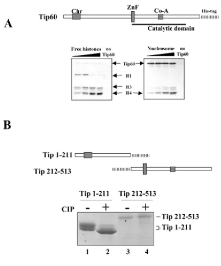
expressed a histidine-tagged human Tip60 in insect cells using abaculovirus-based expression system. The recombinant protein wasfound to be an active HAT on purified histones as well as on nucleosomes(Fig. 1A). Interestingly, whereas Tip60 efficiently acetylatedfree histone H4 and H3, in nucleosomes, it essentially acetylatedhistone H4. However, the acetylation of nucleomosal H4 was muchless efficient than that of free histones. Indeed, the gel containing nucleosomal histones had to be exposed for a longer period oftime to obtain a comparable signal for the acetylation of freeand nucleosomal H4. We have also noticed that the purified recombinant Tip60 migrated as a doublet on a 8% SDS-PAGE, and the treatment with the protein calf intestinal alkaline phosphatase prior toelectrophoresis caused a shift of Tip60 to a faster migratingband (data not shown). This observation showed that protein phosphorylation was responsible for the observed shift in the electrophoreticmobility of Tip60. To determine the region of Tip60 concernedby phosphorylation, two fragments of Tip60 were expressed in insect cells, purified, and analyzed on a 15% SDS-PAGE, one containing the N-terminal chromodomain of Tip60 encompassing the first 211amino acids and the other corresponding to the rest of the protein (Fig. 1B, schemes). Fig. 1B shows that the N-terminal fragment migrated as a doublet and that the CIP treatment caused a shiftof the majority of the protein into a single fast migrating band (compare lane 1 with 2). The 212-513 region of Tip60
migrated as a single band, and the CIP treatment did not affect its electrophoreticmobility (Fig 1B, lanes 3 and 4).
Two Major Sites of Tip60 Phosphorylation in Vivo-- Data presented above showed that the
major sites of Tip60 phosphorylation are located in the first 211 amino acids of theprotein. To determine precisely the sites of Tip60 phosphorylation,we chose a mass spectrometry-based approach. First, the mass spectraof the tryptic fragments of the 1-211 region of Tip60 were compared with that of the tryptic fragments of the CIP-treated protein. The MALDI-TOF analysis showed that peptide-(81-93) bears two phosphategroups. The Q-TOF sequencing of peptide-(81-93) from phosphorylated and CIP-treated proteins confirmed the above conclusion and showedthat serines 86 and 90 are phosphorylated. Finally, tryptic fragmentsof Tip60-(1-211) were loaded on a ZipTip-MC column capable ofretaining phospho-peptides, and the MALDI-TOF analysis of these peptides confirmed again the presence of two phosphate groups on peptide-(81-93). This analysis also showed the possible presence of phosphate groups on 190-211 and 204-211 tryptic fragments (datanot shown). To evaluate the respective participation of serines 86 and 90 in Tip60 phosphorylation, we mutated either serine 86or serine 90 or both to alanine and monitored their migration on 8% SDS-PAGE after an ectopic expression in HeLa cells. As controls, we also expressed either a Tip60 mutant in which glycine 380 wasreplaced by an alanine or Tip60 Leu-254/Leu-257 mutant in which the two indicated leucines potentially involved in a nuclear export signal were also changed to alanines. Fig. 2B shows that onlymutations affecting serines 86 and 90 affect the mobility of Tip60, which strongly suggests that these sites are majors sites of Tip60 phosphorylation in mammalian cells. This hypothesis was furtherconfirmed by showing that CIP-treated Tip60-(1-211) fragment expressedin HeLa cells migrated at the same position as a Tip60-(1-211)fragment containing the Ser-86/Ser-90 mutation (Fig. 2C). Allof these results showed that Tip60 can be phosphorylated both in insect and mammalian cells, and serines 86 and 90 represent two major sites of phosphorylation in vivo. Finally, we showed that endogenous Tip60 detected in HeLa cell nuclear extracts migratedas a doublet and that the CIP treatment of the nuclei before theextract preparation led to the migration of Tip60 as a singleband (Fig. 2D).
Phosphorylation of Tip60 Modulates Its HAT Activity-- To evaluate the role of
phosphorylation in the activity of Tip60, the HAT activity of purified phosphorylated Tip60 was compared with that of the CIP-treated protein. Fig. 3A shows that the pretreatment of Tip60 with increasing amounts of CIP considerably reduced theHAT activity of the protein (compare lanes 2 and 3 with 1). Inthe same experiment when CIP was added in the presence of itsinhibitor NaF (Fig. 3A, lanes 4-6) or heat-inactivated prior toits addition (Fig. 3B, lane
9), no detectable modification of Tip60 HAT activity was observed compared with the control. Theseexperiments show that the inhibition of Tip60 HAT activity isnot because of the presence of CIP itself in the reaction mediumbut because of its ability to dephosphorylate Tip60. Moreover,the activity of the CIP-treated Tip60 was comparable with thatof a mutated Tip60, Tip60Gly-380, containing a mutation severely affecting its HAT activity (Fig.3B, lane
was replaced by an alanine. This mutation had been previously shown to inactivate the DrosophilaTip60 homologue MOF (16) and was also one of the two mutationsintroduced by Ikura et al. (4) to inactivate Tip60. The above experiment clearly showed that dephosphorylated Tip60 has a poorcatalytic activity. To show the specific involvement of the serines86 and 90 in the control of the HAT activity of Tip60, baculovirus-basedexpression was used to express and purify a mutated form of Tip60(Tip60Ser-86/Ser-90) in which these two serines were replaced by alanines. A HATassay was set up to compare the HAT activity of the wild type, phosphorylated Tip60 with that of Tip60Ser-86/Ser-90. Fig. 3C shows that, as in mammalian cells, the double mutantTip60 migrated faster than the wild type protein (upper
panel). Moreover, this experiment also showed that Tip60Ser-86/Ser-90 was not as efficient as wild-type Tip60 at acetylating histones.
Kinases Involved in the Phosphorylation of Tip60-- The data presented thus far showed the
critical role of Tip60 phosphorylation in the control of its catalytic activity. Therefore, it appeared very important to identify the kinases involved tobetter understand the functional significance of this regulationof Tip60 activity by its phosphorylation. The analysis of the sequence encompassing Ser-86 and Ser-90 showed that Ser-90 corresponded to a potential p34 Cdc2 phosphorylation site XSPX(R/K) (17). To test the ability of cyclin B/Cdc2 to phosphorylate Ser-90,we have synthesized three peptides encompassing the amino acid82-96 region of Tip60. One peptide corresponded to the wild type sequence, and the two others contained serine replacement (Ser to Ala) of either Ser-90 or both Ser-86 and Ser-90 (Fig. 4A).The peptides were incubated with purified cyclin B/Cdc2 complexin the presence of 32 P-labeled ATP, and peptide phosphorylation was monitored. Fig.4B shows that only the wild type peptide was phosphorylated bythe purified cyclin B/Cdc2 complex. The phosphorylation of a peptidecontaining the "Ser-90 to Ala" mutation was comparable with thatof a peptide containing replacements of both serines, Ser-86 andSer-90, to alanines or that of the wild type peptide incubated with [32P]ATP in the absence of the enzyme. This experiment shows that cyclin B/Cdc2 specifically phosphorylates Ser-90 of Tip60. To show the participation of cyclin-dependent kinases (CDKs) in the phosphorylation of Tip60, HeLa cells were treated with nocodazole,inducing an arrest at G2/M phase of the cell cycle because ofits ability to
depolymerize microtubules. Nocodazole-treated cells were lysed, and Tip60 was detected using an anti-Tip60 antibody (14) when ~90% of cells were in the G2/M phase of the cell
cycle(data not shown). Fig. 5A shows that, as expected, in cyclingcells, Tip60 appeared as a doublet. Interestingly, after the treatment of cells with nocodazole, almost only the phosphorylated formof Tip60 was visible. However, in our hands for unknown reasons,the endogenous Tip60 could not be detected with a reasonable sensitivity in a reproducible manner. Therefore, to better investigate theinvolvement of the CDKs in the phosphorylation of Tip60, we used a characterized HeLa cell line stably expressing HA-tagged Tip60(4) to evaluate Tip60 phosphorylation in response to specificdrugs. In cycling HeLa cells, the HA-Tip60 appeared also as adoublet. After the treatment of cells with nocodazole, the HA-Tip60 behaved like the endogenous protein because the amount of thephosphorylated form of the protein increased (Fig. 5B, lane 3).This nocodazole-induced accumulation of phosphorylated HA-Tip60 was severely reduced when the nocodazole-treated cells were treated with
roscovitin, a specific inhibitor of CDKs (18), for 7 hbefore the cell harvest (Fig. 5B, lane 4). All together, thesedata show the participation of CDKs in the phosphorylation ofTip60 and strongly suggest a role for Tip60 in the control ofcell cycle-related events.
Discussion
The MYST family of histone acetyltransferases are evolutionary conserved enzymes from yeast to humans. The specific functionof several members of this family is now emerging in different species (19). For instance, in yeast, two HATs of the MYST family, SAS2 and SAS3, play a role in silencing. The HAT activity of SAS3has been shown in vitro (20), and moreover, it has been foundwithin the NuA3 complex, which specifically acetylates histone H3 in nucleosomes (21). The third member of MYST HATs in yeast, Esa1, appears to be responsible for cell cycle progression (22).This HAT is also present in a complex known as NuA4 (23) andis required for DNA double-stranded break repair (24). In Drosophila,MOF, a MYST HAT, has been clearly shown to be involved in the male X chromosome hyperactivation by specifically acetylatinghistone H4 lysine 16 (16). Interestingly, as Tip60, MOF has acanonic chromodomain, which possesses the ability to bind to RNAin vitro and to
the X chromosome-associated RNA in vivo (25). The role of other vertebrate MYST members, HBO1, MORF, and MOZ,has not yet been defined (19). Therefore, it appears that the HATs of the MYST family are involved in the control of a variety of critical cellular events. This conclusion implies that theactivity and the expression of these enzymes should be tightly regulated. In the literature, there is almost no hint regardingthis issue. Our data provide the first indication of the regulatedexpression and activity of a member of the MYST acetyltransferases,Tip60. Indeed, we have previously shown that the cellular concentrationof Tip60 is controlled by Mdm2-mediated ubiquitination and proteasome-dependentdegradation of the protein (14). Here we show that the phosphorylation of Tip60 is a controlled phenomenon and modulates the activity of the protein. Tip60 is not the only HAT whose activity is controlled by phosphorylation. Indeed, it has been shown that the transcription factor ATF-2 possesses an intrinsic HAT activity and that its phosphorylation considerably stimulates its HAT activity (26). Another example is CREB-binding protein, which is phosphorylatedat the G1/S boundary, and this phosphorylation was also shownto stimulate its
HAT activity (27). This phosphorylation-dependentcontrol of CREB-binding protein activity allows this HAT to beresponsive to distinct signaling pathways. Indeed, kinases asdifferent as p44 MAPK/ERK1 (28), cell cycle-dependent kinasessuch as cyclin E/Cdk2 (27), as well as MEKK1 (29) are capableof phosphorylating and, hence, activating the CREB-binding protein catalytic activity. In the case of Tip60, Ser-90 is located withina canonic cyclin B/Cdc2 site and the enzyme was found here to specifically phosphorylate this specific serine. Interestingly,Ser-90 is highly conserved among Tip60 homologues in differentspecies (data not shown), suggesting that this mode of regulation is also conserved during evolution. Furthermore, our data providea new basis to reconsider the many functions of Tip60. Indeed, Tip60 appears as an important intermediate in several unrelatedreceptor-mediated signaling processes (5, 6, 8, 9,11). Therefore, our data suggest that an additional controllevel, that of Tip60 phosphorylation, needs to be coordinatedwith signals induced by steroids (5, 6), IL-1 (8), IL-9 (9), or endothelin (11). Besides, the HAT activity of Tip60has been shown to be essential in the cellular response to DNAdamage (4). Therefore, the phosphorylation of Tip60 regulatingits HAT activity could well play an important role in this processand somehow link Tip60 function to the replication checkpoint.Finally, the HAT activity of Tip60 was found to mediate gene activation following the cleavage of amyloid- precursor (12). These data
should also be reconsidered in the light of a linkage betweenTip60 catalytic activity and its phosphorylation.
In summary, this work has allowed to establish a link between CDKs and the activity of Tip60. However, other kinases may alsophosphorylate Tip60, establishing a specific linkage between Tip60 activity and one or several of the signaling pathways discussed above.
Acknowledgements
We thank Dr. Vasily Ogrysko for the generous gift of the stable Tip60-expressing HeLa cell line, Sandrine Curtet and Marie-Paule Brocard for technical assistance, and Dr. Sophie Rousseaux forthe critical reading of thepaper.
Footnotes
*
This work was supported by a grant from "Sidaction - Ensemble Contre Le Sida" (to S. K.) and a grant from "La Ligue NationaleContre le Cancer" as a "équipe labelisée" (to D. T.).
§
Recipient of a postdoctoral fellowship from "Sidaction-Ensemble, Contre Le Sida from 1999 to2001.
Recipient of a postdoctoral fellowship from Ligue Nationale Contre leCancer.
**
Supported by the INSERM "Delegation"program.
§§
To whom correspondence should be addressed. Tel.: 334-7654-9583; Fax: 334-7654-9595.
Abbreviations
The abbreviations used are: HAT, histone acetyltransferase; IL, interleukin; CIP, calf intestine phosphatase; CREB, cAMP response element-binding protein; CDK, cyclin-dependent kinase; MALDI-TOF, matrix-assisted laser desorption ionization time-of-flight; MS, mass spectrometry; HA, hemagglutinin; MAPK, mitogen-activated protein kinase; ERK, extracellular signal-regulated kinase; MEKK, mitogen-activated protein kinase/extracellular signal-regulated kinase kinase kinase.
References
1. Kamine, J., Elangovan, B., Subramanian, T., Coleman, D., and Chinnadurai, G. (1996)
Virology 216, 357-366
2. Yamamoto, T., and Horikoshi, M. (1997) J. Biol. Chem. 272, 30595-30598
3. Creaven, M., Hans, F., Mutskov, V., Col, E., Caron, C., Dimitrov, S., and Khochbin, S. (1999) Biochemistry 38, 8826-8830
4. Ikura, T., Ogryzko, V. V., Grigoriev, M., Groisman, R., Wang, J., Horikoshi, M., Scully, R., Qin, J., and Nakatani, Y. (2000) Cell 102, 463-473
5. Brady, M. E., Ozanne, D. M., Gaughan, L., Waite, I., Cook, S., Neal, D. E., and Robson, C. N. (1999) J. Biol. Chem. 274, 17599-17604
6. Gaughan, L., Logan, I. R., Cook, S., Neal, D. E., and Robson, C. N. (2002) J. Biol. Chem. 277, 25904-25913
7. Dechend, R., Hirano, F., Lehmann, K., Heissmeyer, V., Ansieau, S., Wulczyn, F. G., Scheidereit, C., and Leutz, A. (1999) Oncogene 18, 3316-3323
8. Baek, S. H., Ohgi, K. A., Rose, D. W., Koo, E. H., Glass, C. K., and Rosenfeld, M. G. (2002) Cell 110, 55-67
9. Sliva, D., Zhu, Y. X., Tsai, S., Kamine, J., and Yang, Y. C. (1999) Biochem. Biophys. Res.
Commun. 263, 149-155
10. Gavaravarapu, S., and Kamine, J. (2000) Biochem. Biophys. Res. Commun. 269, 758-766 11. Lee, H. J., Chun, M., and Kandror, K. V. (2001) J. Biol. Chem. 276, 16597-16600 12. Cao, X., and Sudhof, T. C. (2001) Science 293, 115-120
13. Caron, C., Col, E., and Khochbin, S. (2003) Bioessays 25, 58-65
14. Legube, G., Linares, L. K., Lemercier, C., Scheffner, M., Khochbin, S., and Trouche, D. (2002) EMBO J. 21, 1704-1712
15. Posewitz, M. C., and Tempst, P. (1999) Anal. Chem. 71, 2883-2892 16. Akhtar, A., and Becker, P. B. (2000) Mol. Cell 5, 367-375
17. Kennelly, P. J., and Krebs, E. G. (1991) J. Biol. Chem. 266, 15555-15558
18. Meijer, L., Borgne, A., Mulner, O., Chong, J., Blow, J., Inagaki, N., Inagaki, M., Delcros, J., and Moulinoux, J. (1997) Eur. J. Biochem. 243, 527-536
19. Sterner, D. E., and Berger, S. L. (2000) Microbiol. Mol. Biol. Rev. 64, 435-459
20. Takechi, S., and Nakayama, T. (1999) Biochem. Biophys. Res. Commun. 266, 405-410 21. John, S., Howe, L., Tafrov, S. T., Grant, P. A., Sternglanz, R., and Workman, J. L. (2000)
Genes Dev. 14, 1196-1208
22. Clarke, A. S., Lowell, J. E., Jacobson, S. J., and Pillus, L. (1999) Mol. Cell. Biol. 19, 2515-2526
23. Allard, S., Utley, R. T., Savard, J., Clarke, A., Grant, P., Brandl, C. J., Pillus, L., Workman, J. L., and Cote, J. (1999) EMBO J. 18, 5108-5119
24. Bird, A. W., Yu, D. Y., Pray-Grant, M. G., Qiu, Q., Harmon, K. E., Megee, P. C., Grant, P. A., Smith, M. M., and Christman, M. F. (2002) Nature 419, 411-415
25. Akhtar, A., Zink, D., and Becker, P. B. (2000) Nature 407, 405-40
26. Kawasaki, H., Schiltz, L., Chiu, R., Itakura, K., Taira, K., Nakatani, Y., and Yokoyama, K. K. (2000) Nature 405, 195-200
27. Ait-Si-Ali, S., Ramirez, S., Barre, F. X., Dkhissi, F., Magnaghi-Jaulin, L., Girault, J. A., Robin, P., Knibiehler, M., Pritchard, L. L., Ducommun, B., Trouche, D., and Harel-Bellan, A. (1998) Nature 396, 184-186
28. Ait-Si-Ali, S., Carlisi, D., Ramirez, S., Upegui-Gonzalez, L. C., Duquet, A., Robin, P., Rudkin, B., Harel-Bellan, A., and Trouche, D. (1999) Biochem. Biophys. Res. Commun. 262, 157-162
29. See, R. H., Calvo, D., Shi, Y., Kawa, H., Luke, M. P., and Yuan, Z. (2001) J. Biol. Chem. 276, 16310-16317
Fig. 1. The enzymatically active Tip60 expressed in insect cells is phosphorylated. Baculovirus‐ based expression system was used to produce His‐tagged human Tip60 (scheme) in insect cells. A, the purified enzyme is capable of efficiently acetylating free histones (left panel) and nucleosomal histone H4 (right panel). B, the two indicated His‐tagged fragments of Tip60 (scheme) were produced in insect cells, treated or not with CIP (+ and , respectively) and analyzed on 15% SDS‐PAGE stained with Coomassie Blue.
Fig. 2. Serines 86 and 90 are two major sites of Tip60 phosphorylation. A, amino acid 1‐211 region of Tip60 expressed and purified from insect cells was digested with trypsin, and the CIP‐treated and untreated tryptic fragments were analyzed by mass spectrometry. The CIP treatment led to the complete displacement of a peak at 1483.30 kDa and the appearance of a new one at 1323.30. The loss of mass attributed to the CIP treatment (160 Da) shows the removal of two phosphate groups by CIP. The Q‐TOF sequencing of this peptide shows that it corresponds to the amino acid 81‐93 region of Tip60 containing only two phosphorylable residues, serine 86 and serine 90 (gray box). B, serines 86 (S86) and 90 (S90) are sites of phosphorylation in mammalian cells. Site‐directed mutagenesis was used to replace Ser‐86, Ser‐90, or both by alanines. As a control, other irrelevant sites of Tip60 have been also mutated to alanines. These sites are glycine 380 (G380) and leucines 254 (L254) and 257. These proteins were expressed in HeLa cells, and their mobility was monitored after a Western blot and immunodetection with an anti‐HA antibody. C, Tip60‐(1‐211) fragment containing the wild type sequence or mutated on Ser‐86 and Ser‐90 (Tip1‐211 S86/90) was also expressed in HeLa cells, and the mobility of the CIP‐treated wild type peptide was compared with that of the Ser‐86/Ser‐90 mutant. D, purified HeLa cell nuclei were isolated, and a fraction was incubated with CIP before extract preparation (+). The extracts were then analyzed by Western blotting using an anti ‐Tip60 antibody (14).
Fig. 3. Tip60 HAT activity is controlled by phosphorylation. A, purified Tip60 expressed in insect cells was treated with increasing amounts of CIP in the absence (lanes 2 and 3) or presence of CIP inhibitor NaF (lanes 4‐6). Lanes 1 and 4 show the activity of untreated Tip60. Treated and untreated Tip60 proteins were incubated with purified histone and [14C]acetyl‐CoA. The reaction was stopped, loaded on a 15% SDS‐PAGE, and analyzed by autoradiography. B, purified Tip60 was left untreated (lane 7) or treated with CIP or heat‐inactivated CIP (lanes 8 and 9, respectively). The activity of wild type Tip60 was also compared with a protein harboring a G to A mutation at position 380 (lanes
10 and 11, respectively). C, the Ser‐86/Ser‐90 Tip60 mutant shows a reduced HAT activity. Wild type
Tip60 or a mutated version of the protein harboring double S to A mutations at positions 86 and 90 was produced in insect cells and purified, and their HAT activity was tested as noted above. After the reaction, a fraction was analyzed on a silver‐stained 8% SDS‐PAGE to compare the amounts of enzyme used (upper panel). The middle panel (Autorad) shows the labeling of histone after the reaction by wild type or mutated Tip60. The corresponding Coomassie Blue‐stained gel before autoradiography shows histones used in each reaction (lower panel).
Fig. 4. Tip60 Serine 90 is phosphorylated by cyclin B/Cdc2 in vitro. A, peptides corresponding to the Tip60 amino acids 82‐96 region, either wild type (wt) or containing the indicated mutations, were synthesized and used in a kinase reaction. B, the histogram shows that only the wild type peptide is a substrate for Cdc2 kinase.
Fig. 5. The arrest of cells at the G2/M phase of the cell cycle is associated with an enhancement of Tip60 phosphorylation. A, HeLa cells were treated with nocodazole (100 ng/ml) for 16 h and lysed, and the migration of the endogenous Tip60 was monitored by Western blotting using an anti‐Tip60 antibody. B, HeLa cells stably expressing HA‐Tip60 were treated with nocodazole for 23 h, and 7 h before harvest, roscovitin (50 µM) was added where indicated. Cells were lysed and analyzed as above using an anti‐HA antibody. ‐, non‐treated cells.