Nature © Macmillan Publishers Ltd 1997
NATURE
|
VOL 390
|
20 NOVEMBER 1997
249
articles
The complete genome sequence
of the Gram-positive bacterium
Bacillus subtilis
F. Kunst
1, N. Ogasawara
2, I. Moszer
3, A. M. Albertini
4, G. Alloni
4, V. Azevedo
5, M. G. Bertero
3,4, P. Bessie`res
5, A. Bolotin
5, S. Borchert
6,
R. Borriss
7, L. Boursier
3, A. Brans
8, M. Braun
9, S. C. Brignell
10, S. Bron
11, S. Brouillet
3,12, C. V. Bruschi
13, B. Caldwell
14, V. Capuano
5,
N. M. Carter
10, S.-K. Choi
15, J.-J. Codani
16, I. F. Connerton
17, N. J. Cummings
17, R. A. Daniel
18, F. Denizot
19, K. M. Devine
20, A. Du¨sterho¨ft
9,
S. D. Ehrlich
5, P. T. Emmerson
21, K. D. Entian
6, J. Errington
18, C. Fabret
19, E. Ferrari
14, D. Foulger
18, C. Fritz
9, M. Fujita
22, Y. Fujita
23, S. Fuma
24,
A. Galizzi
4, N. Galleron
5, S.-Y. Ghim
15, P. Glaser
3, A. Goffeau
25, E. J. Golightly
26, G. Grandi
27, G. Guiseppi
19, B. J. Guy
10, K. Haga
28, J. Haiech
19,
C. R. Harwood
10, A. He´naut
29, H. Hilbert
9, S. Holsappel
11, S. Hosono
30, M.-F. Hullo
3, M. Itaya
31, L. Jones
32, B. Joris
8, D. Karamata
33,
Y. Kasahara
2, M. Klaerr-Blanchard
3, C. Klein
6, Y. Kobayashi
30, P. Koetter
6, G. Koningstein
34, S. Krogh
20, M. Kumano
24, K. Kurita
24, A. Lapidus
5,
S. Lardinois
8, J. Lauber
9, V. Lazarevic
33, S.-M. Lee
35, A. Levine
36, H. Liu
28, S. Masuda
30, C. Maue¨l
33, C. Me´digue
3,12, N. Medina
36,
R. P. Mellado
37, M. Mizuno
30, D. Moestl
9, S. Nakai
2, M. Noback
11, D. Noone
20, M. O’Reilly
20, K. Ogawa
24, A. Ogiwara
38, B. Oudega
34,
S.-H. Park
15, V. Parro
37, T. M. Pohl
39, D. Portetelle
40, S. Porwollik
7, A. M. Prescott
18, E. Presecan
3, P. Pujic
5, B. Purnelle
25, G. Rapoport
1,
M. Rey
26, S. Reynolds
33, M. Rieger
41, C. Rivolta
33, E. Rocha
3,12, B. Roche
36, M. Rose
6, Y. Sadaie
22, T. Sato
30, E. Scanlan
20, S. Schleich
3,
R. Schroeter
7, F. Scoffone
4, J. Sekiguchi
42, A. Sekowska
3, S. J. Seror
36, P. Serror
5, B.-S. Shin
15, B. Soldo
33, A. Sorokin
5, E. Tacconi
4,
T. Takagi
43, H. Takahashi
28, K. Takemaru
30, M. Takeuchi
30, A. Tamakoshi
24, T. Tanaka
44, P. Terpstra
11, A. Tognoni
27, V. Tosato
13, S. Uchiyama
42,
M. Vandenbol
40, F. Vannier
36, A. Vassarotti
45, A. Viari
12, R. Wambutt
46, E. Wedler
46, H. Wedler
46, T. Weitzenegger
39, P. Winters
14, A. Wipat
10,
H. Yamamoto
42, K. Yamane
24, K. Yasumoto
28, K. Yata
22, K. Yoshida
23, H.-F. Yoshikawa
28, E. Zumstein
5, H. Yoshikawa
2& A. Danchin
31Institut Pasteur, Unite´ de Biochimie Microbienne, 25 rue du Docteur Roux, 75724 Paris Cedex 15, France 2Nara Institute of Science and Technology, Graduate School of Biological Sciences, Ikoma, Nara 630-01, Japan 3Institut Pasteur, Unite´ de Re´gulation de l’Expression Ge´ne´tique, 28 rue du Docteur Roux, 75724 Paris Cedex 15, France 4Dipartimento di Genetica e Microbiologia, Universita di Pavia, Via Abbiategrasso 207, 27100 Pavia, Italy 5INRA, Ge´ne´tique Microbienne, Domaine de Vilvert, 78352 Jouy-en-Josas Cedex, France
6Institut fu¨r Mikrobiologie, J. W. Goethe-Universita¨t, Marie Curie Strasse 9, 60439 Frankfurt/Maine, Germany 7Institut fu¨r Genetik und Mikrobiologie, Humboldt Universita¨t, Chausseestrasse 17, D-10115 Berlin, Germany 8Centre d’Inge´nierie des Prote´ines, Universite´ de Lie`ge, Institut de Chimie B6, Sart Tilman, B-4000 Lie`ge, Belgium 9QIAGEN GmbH, Max-Volmer-Strasse 4, D-40724 Hilden, Germany
10Department of Microbiological, Immunological and Virological Sciences, The Medical School, University of Newcastle, Framlington Place, Newcastle upon Tyne NE2 4HH, UK 11Department of Genetics, University of Groningen, Kerklaan 30, 9751 NN Haren, The Netherlands
12Atelier de BioInformatique, Universite´ Paris VI, 12 rue Cuvier, 75005 Paris, France 13ICGEB, AREA Science Park, Padriciano 99, I-34012 Trieste, Italy
14Genencor International, 925 Page Mill Road, Palo Alto, California 94304-1013, USA
15Bacterial Molecular Genetics Research Unit, Applied Microbiology Research Division, KRIBB, PO Box 115, Yusong, Taejon 305-600, Korea 16INRIA, Domaine de Voluceau, PB 105, 78153 Le Chesnay Cedex, France
17Institute of Food Research, Department of Food Macromolecular Science, Reading Laboratory, Earley Gate, Whiteknights Road, Reading RG6 6BZ, UK 18Sir William Dunn School of Pathology, University of Oxford, South Parks Road, Oxford, OX1 3RE, UK
19Laboratoire de Chimie Bacte´rienne, CNRS BP 71, 31 Chemin Joseph Aiguier, 13402 Marseille Cedex 09, France 20Department of Genetics, Trinity College, Lincoln Place Gate, Dublin 2, Republic of Ireland
21Department of Biochemistry and Genetics, The Medical School, University of Newcastle, Framlington Place, Newcastle upon Tyne, NE2 4HH, UK 22Radioisotope Center, National Insitute of Genetics, Mishima, Shizuoka-ken 411, Japan
23Department of Biotechnology, Faculty of Engineering, Fukuyama University, Higashimura-cho, Fukuyama-shi, Hiroshima 729-02, Japan 24Institute of Biological Sciences, Tsukuba University, Tsuiuba-shi, Ibaraki 305, Japan
25Faculte´ des Sciences Agronomiques, Unite´ de Biochimie Physiologique, Universite´ Catholique de Louvain, Place Croix du Sud, 2-20 B-1348 Louvain-la-Neuve, Belgium 26Novo Nordisk Biotech, 1445 Drew Avenue, Davis, California 95616-4880, USA
27Eniricerche, Via Maritano 26, San Donato Milanese, 20097 Milan, Italy
28Institute of Molecular and Cellular Biology, The University of Tokyo, Bunkyo-ku, Tokyo 113, Japan
29Laboratoire Ge´nome et Informatique, Universite´ de Versailles, Baˆtiment Buffon, 45 Avenue des E´tats-Unis, 78035 Versailles Cedex, France 30Faculty of Agriculture, Tokyo University of Agriculture and Technology, Fuchu, Tokyo 183, Japan
31Mitsubishi Kasei Institute of Life Sciences, 11 Minamyiooa, Machida-shi, Tokyo 194, Japan
32Institut Pasteur, Service d’Informatique Scientifique, 28 rue du Docteur Roux, 75724 Paris Cedex 15, France 33Institut de Ge´ne´tique et Biologie Microbiennes, Universite´ de Lausanne, 19 rue Ce´sar Roux, 1005 Lausanne, Switzerland
34Department of Molecular Microbiology, MBW/BCA, Faculty of Biology, Vrije Universiteit Amsterdam, De Boelelaan 1087, 1081 HV Amsterdam, The Netherlands 35Chongju University College of Science and Engineering, Chongju City, Korea
36Institut de Ge´ne´tique et Microbiologie, Universite´ Paris Sud, URA CNRS 2225, Universite´ Paris XI–Baˆtiment 409, 91405 Orsay Cedex, France 37Centro Nacional de Biotecnologia (CSIC), Campus Universidad Autonoma, Cantoblanco, 28049 Madrid, Spain
38National Institute of Basic Biology, 38 Nishigounaka, Myoudaiji-chou, Okazaki 444, Japan
39Gesellschaft fu¨r Analyse-Technik und Consulting mbH, Fritz-Arnold Straße 23, D-78467 Konstanz, Germany 40Department of Microbiology, Faculty of Agronomy, 6 Avenue du Mare´chal Juin, B-5030 Gembloux, Belgium 41Biotech Research, BMF, Wilhelmsfeld, Klingelstrasse 35, D-69434 Hirschhorn, Germany
42Department of Applied Biology, Faculty of Textile Science and Technology, Shinshu University 3-15-1, Tokida, Ueda-shi, Nagano 386, Japan 43Human Genome Center, Institute of Medical Science, University of Tokyo, 4-6-1 Shirokanedai, Minato-ku, Tokyo 108, Japan
44Department of Marine Science, School of Marine Science and Technology, Tokai University, 3-20-1 Orido Shimizu, Shizuoka 424, Japan 45European Commission, DG XII-E-1, SDME 8/78, Rue de la Loi 200, B-1049 Brussels, Belgium
46AGOWA GmbH, Glienicker Weg 185, 12489 Berlin, Germany
. . . .
Bacillus subtilis is the best-characterized member of the Gram-positive bacteria. Its genome of 4,214,810 base pairs
comprises 4,100 protein-coding genes. Of these protein-coding genes, 53% are represented once, while a quarter of
the genome corresponds to several gene families that have been greatly expanded by gene duplication, the largest
family containing 77 putative ATP-binding transport proteins. In addition, a large proportion of the genetic capacity is
devoted to the utilization of a variety of carbon sources, including many plant-derived molecules. The identification of
five signal peptidase genes, as well as several genes for components of the secretion apparatus, is important given the
capacity of Bacillus strains to secrete large amounts of industrially important enzymes. Many of the genes are involved
in the synthesis of secondary metabolites, including antibiotics, that are more typically associated with Streptomyces
species. The genome contains at least ten prophages or remnants of prophages, indicating that bacteriophage
infection has played an important evolutionary role in horizontal gene transfer, in particular in the propagation of
bacterial pathogenesis.
Techniques for large-scale DNA sequencing have brought about a
revolution in our perception of genomes. Together with our
under-standing of intermediary metabolism, it is now realistic to envisage
a time when it should be possible to provide an extensive chemical
definition of many living organisms. During the past couple of
years,
the
genome
sequences
of
Haemophilus
influenzae,
Mycoplasma genitalium, Synechocystis PCC6803, Methanococcus
jannaschii, M. pneumoniae, Escherichia coli, Helicobacter pylori,
Archaeoglobus fulgidus and the yeast Saccharomyces cerevisiae have
been published in their entirety
1–8, and at least 40 prokaryotic
genomes are currently being sequenced. Regularly updated lists of
genome sequencing projects are available at http://www.mcs.anl.
gov/home/gaasterl/genomes.html (Argonne National Laboratory,
Illinois, USA) and http://www.tigr.org (TIGR, Rockville, Maryland,
USA).
The list of sequenced microorganisms does not currently include
a paradigm for Gram-positive bacteria, which are known to be
important for the environment, medicine and industry. Bacillus
subtilis has been chosen to fill this gap
9,10as its biochemistry,
physiology and genetics have been studied intensely for more
than 40 years. B. subtilis is an aerobic, endospore-forming,
rod-shaped bacterium commonly found in soil, water sources and in
association with plants. B. subtilis and its close relatives are an
important source of industrial enzymes (such as amylases and
proteases), and much of the commercial interest in these bacteria
arises from their capacity to secrete these enzymes at gram per litre
concentrations. It has therefore been used for the study of protein
secretion and for development as a host for the production of
heterologous proteins
11. B. subtilis (natto) is also used in the
production of Natto, a traditional Japanese dish of fermented
soya beans.
Under conditions of nutritional starvation, B. subtilis stops
growing and initiates responses to restore growth by increasing
metabolic diversity. These responses include the induction of
motility and chemotaxis, and the production of macromolecular
hydrolases (proteases and carbohydrases) and antibiotics. If these
responses fail to re-establish growth, the cells are induced to form
chemically, irradiation- and desiccation-resistant endospores.
Sporulation involves a perturbation of the normal cell cycle and
the differentiation of a binucleate cell into two cell types. The
division of the cell into a smaller forespore and a larger mother cell,
each with an entire copy of the chromosome, is the first
morpho-logical indication of sporulation. The former is engulfed by the
latter and differential expression of their respective genomes,
coupled to a complex network of interconnected regulatory
path-ways and developmental checkpoints, culminates in the
pro-grammed death and lysis of the mother cell and release of the
mature spore
12. In an alternative developmental process, B. subtilis is
also able to differentiate into a physiological state, the competent
state, that allows it to undergo genetic transformation
13.
General features of the DNA sequence
Analysis at the replicon level. The B. subtilis chromosome has
4,214,810 base pairs (bp), with the origin of replication coinciding
with the base numbering start point
14, and the terminus at about
2,017 kilobases (kb)
15. The average G þ C ratio is 43.5%, but it
varies considerably throughout the chromosome. This average is
also different if one considers the nucleotide content of coding
sequences, for which G and A (24% and 30%) are relatively more
abundant than their counterparts C and T (20% and 26%). A
significant inversion of the relative G
2 C=G þ C ratio is visible at
the origin of replication, indicating asymmetry of the nucleotide
composition between the replication leading strand and the lagging
strand
16. Several A þ T-rich islands are likely to reveal the signature
of bacteriophage lysogens or other inserted elements (Fig. 1, see
below).
We have analysed the abundance of oligonucleotides (‘words’) in
the genome in various ways: absolute number of words in the
genomic text, or comparison with the expected count derived from
several models of the chromosome (for example, Markov models, or
simulated sequences in which previously known features of the
genome were conserved
17). Comparing the experimental data with
various models allowed us to define under- and overrepresentation
of words in the experimental data set by reference to the model
chosen. In general, the dinucleotide bias follows closely what has
been described for other prokaryotes
18,19, in that the dinucleotides
most overrepresented are AA, TT and GC, whereas those less
represented are TA, AC and GT. Plots of the frequencies of AG,
GA, CT and TC in sliding windows along the chromosome show
dramatic decreases or increases around the origin and terminus of
replication (data not shown). Trinucleotide frequency, directly
related to the coding frame, will be discussed below. The
distribu-tion of words of four, five and six nucleotides shows significant
correlations between the usage of some words and replication
(several such oligonucleotides are very significantly overrepresented
in one of the strands and underrepresented in the other one).
Setting a statistical cut-off for the significance of duplications at
10
−
3
, we expected duplication by chance of words longer than 24
nucleotides to be rare
20. In fact, the genome of B. subtilis contains a
plethora of such duplications, some of them appearing more than
0.50
3,000,000
3,500,000
4,000,000
skin 7
0.30
0.35
0.40
0.45
0
500,000
1,000,000
1,500,000
2,000,000
2,500,000
Position (base pairs)
G+C (%)
SPß
2
3
PBSX
5
6
1
4
Figure 1 Distribution of A þ T-rich islands along the chromosome of B. subtilis, in
sliding windows of 10,000 nucleotides, with a step of 5,000 nucleotides. Location
of genes from class 3 according to codon usage analysis (see Fig. 4) is indicated
by dots at the bottom of the graph. Known prophages (PBSX, SPb and skin) are
indicated by their names, and prophage-like elements are numbered from 1 to 7.
Nature © Macmillan Publishers Ltd 1997
twice. Among the duplications, we identified, as expected, the
ribosomal RNA genes and their flanking regions, but also regions
known to correspond to genes comprising long sequence repeats
(such as pks and srf ). We also found several regions that were not
expected: a 182-bp repetition within the yyaL and yyaO genes; a 410-bp
repetition between the yxaK and yxaL genes; an internal duplication
of 174 bp inside ydcI; and significant duplications in the regions
involved in the transcriptional control of several genes (such as
118 bp repeated three times between yxbB and yxbC). Finally, we
found several repetitions at the borders of regions that might be
involved in bacteriophage integration.
The most prominent duplication was a 190-bp element that was
repeated 10 times in the chromosome. Multiple alignment of the ten
repeats showed that they could be classified into two subfamilies
with six and three copies each, plus a copy of what appears to be a
chimaera. Similar sequences have also been described in the closely
related species Bacillus licheniformis
21,22. A striking feature of these
repeats is that they are only found in half of the chromosome, at
either side of the origin of replication, with five repeats on each side.
Furthermore, with the exception of the most distal repeat at
position 737,062, they lie in the same orientation with respect to
the movement of the replication fork (Figs 2 and 3). Putative
secondary structures conserved by compensatory mutations, as
well as an insert in three of the copies, suggest that this element
could indicate a structural RNA molecule.
Analysis at the transcription and translation level. Over 4,000
putative protein coding sequences (CDSs) have been identified,
with an average size of 890 bp, covering 87% of the genome
sequence (Fig. 2). We found that 78% of the genes started with
ATG, 13% with TTG and 9% with GTG, which compares with 85%,
3% and 14%, respectively, in E. coli
8. Fifteen genes (eight in the
predicted CDSs in bacteriophage SPb) exhibiting unusual start
codons (namely ATT and CTG) were also identified through their
similarities to known genes in other organisms or because they had a
good GeneMark prediction (see Methods). This has not yet been
substantiated experimentally. However, in the case of the gene
coding for translation initiation factor 3, the similarity with its E.
coli counterpart strongly suggests that the initiation codon is ATT,
as is the case in E. coli.
We have not annotated CDSs that largely or entirely overlap
existing genes, although such genes (for example, comS inside
srfAA) certainly exist. It is also likely that some of the short CDSs
present in the B. subtilis genome have been overlooked. For these
reasons and possible sequencing errors, the estimated number of B.
subtilis CDSs will fluctuate around the present figure of 4,100.
In several cases, in-frame termination codons or frameshifts were
confirmed to be present on the chromosome (for example, an
internal termination codon in ywtF, or the known programmed
translational frameshift in prfB), indicating that the genes are either
non-functional (pseudogenes) or subject to regulatory processes. It
will therefore be of interest to determine whether these gene features
are conserved in related Bacillus species, especially as strain 168 is
derived from the Marburg strain that was subjected to X-ray
irradiation
23.
A few regions do not have any identifiable feature indicating that
they are transcribed: they could be ‘grey holes’ of the type described
in E. coli
24. Preliminary studies involving all regions of more than
400 bp without annotated CDSs indicated that, of
,300 such
regions, only 15% were likely to be really devoid of
protein-coding sequences. One of the longest such regions, located between
yfjO and yfjN, is 1,628 bp long. Grey holes seem generally to be
clustered near the terminus of replication. However, a grey-hole
cluster located at
,600 kb might be related to the temporary
chromosome partition observed during the first stages of
sporula-tion, when a segment of about one-third of the chromosome enters
the prespore, and remains the sole part of the chromosome in the
prespore for a significant transition period
25.
The codon usage of B. subtilis CDSs was analysed using factorial
correspondence analysis
17. We found that the CDSs of B. subtilis
could be separated into three well-defined classes (Fig. 4). Class 1
comprises the majority of the B. subtilis genes (3,375 CDSs),
including most of the genes involved in sporulation. Class 2 (188
CDSs) includes genes that are highly expressed under exponential
growth conditions, such as genes encoding the transcription and
translation machineries, core intermediary metabolism, stress
pro-teins, and one-third of genes of unknown function. Class 3 (537
CDSs) contains a very high proportion of genes of unidentified
function (84%), and the members of this class have codons enriched
in A þ T residues. These genes are usually clustered into groups
between 15 and 160 genes (for example, bacteriophage SPb) and
correspond to the A þ T-rich islands described above (Fig. 1).
When they are of known function, or when their products display
similarity to proteins of known function, they usually correspond to
functions found in, or associated with, bacteriophages or
trans-posons, as well as functions related to the cell envelope. This
includes the region ydc/ydd/yde (40 genes that are missing in
some B. subtilis strains
26), where gene products showing similarities
to bacteriophage and transposon proteins are intertwined. Many of
these genes are associated with virulence genes identified in
patho-genic Gram-positive bacteria, suggesting that such virulence factors
are transmitted horizontally among bacteria at a much higher
frequency than previously thought. If we include these A þ T-rich
regions as possible cryptic phages, together with known
bacterio-phages or bacteriophage-like elements (SPb, PBSX and the skin
element), we find that the genome of B. subtilis 168 contains at least
10 such elements (Figs 2 and 3). Annotation of the corresponding
regions often reveals the presence of genes that are similar to
bacteriophage lytic enzymes, perhaps accounting for the
observa-tion that B. subtilis cultures are extremely prone to lysis.
The ribosomal RNA genes have been previously identified and
articles
NATURE
|
VOL 390
|
20 NOVEMBER 1997
251
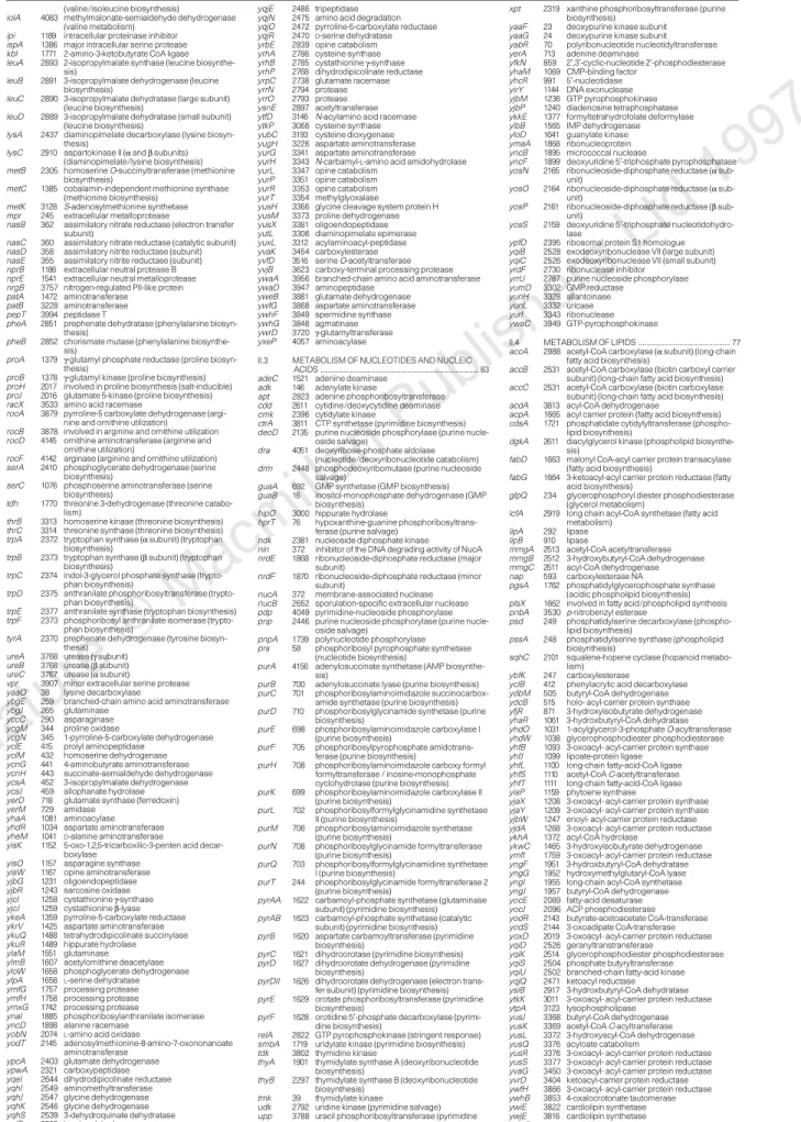
Table 1 Functional classification of the Bacillus subtilis protein-coding
genes
The genes of known function or encoding products similar to known proteins in B.
subtilis or in other organisms have been classified into functional categories
(2,379 genes). The total number of genes in each category is indicated after the
category title. Genes are listed in alphabetical order within each category, and
their positions (in kilobases) on the B. subtilis chromosome are indicated after the
gene names. A brief description is given for each gene. In some cases, interacting
proteins have been indicated between brackets (for example, histidine kinases
and response regulator, phosphatases and their substrates). More detailed and
constantly updated information is available in the SubtiList database (see
Methods). A preliminary assessment of the significance of sequence similarities
was obtained through an automated procedure involving a combination between
the BLAST2P probability and the percentage of amino-acid identity. Matches
considered significant were re-examined manually. It should be emphasized that
functions assigned to ‘y’ genes are based only on sequence similarity information
with the best counterparts in protein databanks. Genes whose products are only
similar to other unknown proteins, or not significantly similar to any other proteins
in databanks (categories V and VI), were omitted.
Figure 2 General view of the B. subtilis chromosome. Arrows indicate the
orientation of transcription. Genes are coloured according to their classification
into six broad functional categories (blue, category I; green, category II; red,
category III; orange, category IV; purple, category V; pink, category VI; see Table
1). Class 2 CDSs according to codon usage analysis are indicated by oblique
hatches, and class 3 CDSs are indicated by vertical hatches. Ribosomal RNA
genes are coloured in yellow. Transfer RNA genes are marked by triangles. Other
RNA genes are represented as white arrows. Known genes (non-‘y’ genes) are
printed in bold type. Putative transcription termination sites are represented as
loops. Known prophages and prophage-like elements are indicated by brown
hatches on the chromosome line. The 190-bp element repeated ten times is
represented by hatched boxes.
R
R
shown to be organized into ten rRNA operons, mainly clustered
around the origin of replication of the chromosome (Figs 2 and 3).
In addition to the 84 previously identified tRNA genes, by using the
Palingol
27and tRNAscan
28programs, we propose four putative new
tRNA loci (at 1,262 kb, 1,945 kb, 2,003 kb and 2,899 kb), specific for
lysine, proline and arginine (UUU, GGG, CCU and UCU
antic-odons, respectively). The 10S RNA involved in degradation of
proteins made from truncated mRNA has been identified (ssrA),
as well as the RNA component of RNase P (rnpB) and the 4.5S RNA
involved in the secretion apparatus (scr).
There is a strong transcription orientation bias with respect to the
movement of the replication fork: 75% of the predicted genes are
transcribed in the direction of replication. Plotting the density of
coding nucleotides in each strand along the chromosome readily
identifies the replication origin and terminus (Fig. 3). To identify
putative operons, we followed ref. 29 for describing
Rho-independent transcription termination sites. This yielded
,1,630
putative terminators (340 of which were bidirectional). We retained
only those that were located less than 100 bp downstream of a gene,
or that were considered by the program to be ‘very strong’ (in order
to account for possible erroneous CDSs). This yielded a total of
,1,250 terminators, with a mean operon size of three genes. A
similar approach to the identification of promoters is
problema-tical, especially because at least 14 sigma factors, recognizing
different promoter sequences, have been identified in B. subtilis.
Nevertheless, the consensus of the main vegetative sigma factor (j
A
)
appears to be identical to its counterpart in E. coli (j
70
):
59-TTGACA-n
17
-TATAAT-3
9. Relaxing the constraints of the similarity
to sigma-specific consensus sequences led to an extremely high
number of false-positive results, suggesting that the
consensus-oriented approach to the identification of promoters should be
replaced by another approach
17.
Classification of gene products
Genes were classified according to ref. 14, based on the
representa-tion of cells as Turing machines in which one distinguishes between
the machine and the program (Table 1). Using the BLAST2P
software running against a composite protein databank compound
of SWISS-PROT (release 34), TREMBL (release 3, update 1) and B.
subtilis proteins, we assigned at least one significant counterpart
with a known function to 58% of the B. subtilis proteins. Thus for up
to 42% of the gene products, the function cannot be predicted by
similarity to proteins of known function: 4% of the proteins are
similar only to other unknown proteins of B. subtilis; 12% are
similar to unknown proteins from some other organism; and 26%
of the proteins are not significantly similar to any other proteins in
databanks. This preliminary analysis should be interpreted with
caution, because only
,1,200 gene functions (30%) have been
experimentally identified in B. subtilis. We used the ‘y’ prefix in gene
names to emphasize that the function has not been ascertained
(2,853 ‘y’ genes, representing 70%).
Regulatory systems. Transcription regulatory proteins. Helix–
turn–helix proteins form a large family of regulatory proteins
found in both prokaryotes and eukaryotes. There are several classes,
including repressors, activators and sigma factors. Using BLAST
searches, we constructed consensus matrices for helix–turn–helix
proteins to analyse the B. subtilis protein library. We identified 18
sigma or sigma-like factors, of which nine (including a new one) are
of the SigA type. We also putatively identified 20 regulators (among
which 18 were products of ‘y’ genes) of the GntR family, 19
regulators (15 ‘y’ genes) of the LysR family, and 12 regulators (5
‘y’ genes) of the LacI family. Other transcription regulatory proteins
were of the AraC family (11 members, 10 ‘y’), the Lrp family (7
members, 3 ‘y’), the DeoR family (6 members, 3 ‘y’), or additional
families (such as the MarR, ArsR or TetR families). A puzzling
observation is that several regulatory proteins display significant
similarity to aminotransferases (seven such enzymes have been
identified as showing similarity to repressors).
Two-component signal-transduction pathways. Two-component
regulatory systems, consisting of a sensor protein kinase and a
response regulator, are widespread among prokaryotes. We have
identified 34 genes encoding response regulators in B. subtilis, most
of which have adjacent genes encoding histidine kinases. Response
regulators possess a well-conserved N-terminal phospho-acceptor
domain
30, whereas their C-terminal DNA-binding domains share
similarities with previously identified response regulators in E. coli,
Rhizobium meliloti, Klebsiella pneumoniae or Staphylococcus aureus.
Representatives of the four subfamilies recently identified in E. coli
31100%
80%
oriC
terC
0%
1
3 4 PBSX 5 6SP
β
skin
7
rrnO rrnA
rrnJ-W
rrnI-H-G
rrnE
rrnB
rrnD
2
Figure 3 Density of coding nucleotides along the
B. subtilis chromosome. Yellow stands for the
density of coding nucleotides in both strands of
the sequence; red indicates the density of coding
nucleotides in the clockwise strand (nucleotides
involved in genes transcribed in the clockwise
orientation). The movement of the replication
forks is represented by arrows. Ribosomal RNA
operons are indicated by brown boxes. Known
prophages and prophage-like elements are
represented as blue lines. The 190-bp element
repeated ten times is represented by green lines.
Nature © Macmillan Publishers Ltd 1997
(OmpR, FixJ, CitB and LytR) have been identified in B. subtilis. In a
fifth subfamily, CheY, the DNA-binding domain is absent. The
DNA-binding domain of a single B. subtilis response regulator,
YesN, shares similarity with regulatory proteins of the AraC family.
Quorum sensing. The B. subtilis genome contains 11 aspartate
phosphatase genes, whose products are involved in
dephosphoryla-tion of response regulators, that do not seem to have counterparts in
Gram-negative bacteria such as E. coli. Downstream from the
corresponding genes are some small genes, called phr, encoding
regulatory peptides that may serve as quorum sensors
32. Seven phr
genes have been identified so far, including three new genes (phrG,
phrI and phrK).
Protein secretion. It is known that B. subtilis and related Bacillus
species, in particular B. licheniformis and B. amyloliquefaciens, have
a high capacity to secrete proteins into the culture medium. Several
genes encoding proteins of the major secretion pathway have been
identified: secA, secD, secE, secF, secY, ffh and ftsY. Surprisingly, there
is no gene for the SecB chaperone. It is thought that other
chaperone(s) and targeting factor(s), such as Ffh and FtsY, may
take over the SecB function. Further, although there is only one such
gene in E. coli, five type I signal peptidase genes (sipS, sipT, sipU, sipV
and sipW) have been found
33. The lsp gene, encoding a type II signal
peptidase required for processing of lipo-modified precursors, was
also identified. PrsA, located at the outer side of the membrane, is
important for the refolding of several mature proteins after their
translocation through the membrane.
Other families of proteins. ABC transporters were the most
frequent class of proteins found in B. subtilis. They must be
extremely important in Gram-positive bacteria, because they have
an envelope comprising a single membrane. ABC transporters will
therefore allow such bacteria to escape the toxic action of many
compounds. We propose that 77 such transporters are encoded in
the genome. In general they involve the interaction of at least three
gene products, specified by genes organized into an operon. Other
families comprised 47 transport proteins similar to facilitators (and
perhaps sometimes part of the ABC transport systems), 18
amino-acid permeases (probably antiporters), and at least 16 sugar
trans-porters belonging to the PEP-dependent phosphotransferase
system.
General stress proteins are important for the survival of bacteria
under a variety of environmental conditions. We identified 43
temperature-shock and general stress proteins displaying strong
similarity to E. coli counterparts.
Missing genes. Histone-like proteins such as HU and H-NS have
been identified in E. coli. We found that B. subtilis encodes two
putative histone-like proteins that show similarity to E. coli HU,
namely HBsu and YonN, but found no homologue to H-NS. It is
known that the hbs gene encoding HBsu is essential, but we do not
expect the yonN gene to be essential because it is present in the SPb
prophage. IHF is similar to HU, and it is not known whether HBsu
plays a similar role to that of IHF in E. coli. Similarly, no protein
similar to FIS could be found.
Genes encoding products that interact with methylated DNA,
such as seqA in E. coli, involved in the regulation of replication
initiation timing, or mutH, the endonuclease recognizing the newly
synthesized strand during mismatch repair at hemi-methylated
articles
NATURE
|
VOL 390
|
20 NOVEMBER 1997
253
Figure 4 Factorial correspondence analysis of codon usage in the B. subtilis
CDSs. Red dots, genes from class 1; green triangles, genes from class 2; blue
crosses, genes from class 3. Class 2 contains genes coding for the translation
and transcription machineries, and genes of the core intermediary metabolism.
Class 3 genes correspond to codons strongly enriched in A or T in the wobble
position; they generally belong to prophage-like inserts in the genome.
112 (3%) 72 (2%) 210 38 47 57 64 77 2,126 (53%) 568 (14%) 273 (7%) 168 (4%) 100 (3%) 84 (2%) singlets doublets triplets quadruplets quintuplets sextuplets heptuplets octuplets 9 to 19 genes 38 genes 47 genes 57 genes 64 genes 77 genes
Figure 5 Gene paralogue distribution in the genome of B. subtilis. Each B. subtilis
protein has been compared with all other proteins in the genome, using a Smith
and Waterman algorithm. The baseline is established by making a similar
comparison using 100 independent random shuffles of the protein sequence
(Z-score
. 13).
GATC sites, are also missing. This is in line with the absence of
known methylation in B. subtilis, equivalent to Dam methylation in
E. coli. Similarly, E. coli sfiA, encoding an inhibitor of FtsZ action in
the SOS response, has no counterpart in B. subtilis. In contrast, B.
subtilis replication initiation-specific genes, such as dnaB and dnaD,
are missing in E. coli. The exact counterpart of the E. coli mukB gene,
involved in chromosome partitioning, does not exist in B. subtilis,
but genes spo0J and smc (Smc is weakly similar to MukB), which are
suggested to be involved in partitioning of the B. subtilis
chromo-some, are missing in E. coli.
Turnover of mRNA is controlled in E. coli by a ‘degradosome’
comprising RNase E. It has a counterpart in B. subtilis, but we failed
to find a clear homologue of RNase E in this organism. Whether this
is related to the role of ribosomal protein S1 as an RNA helicase
involved in mRNA turnover in E. coli requires further investigation.
In particular, a homologue of rpsA (S1 structural gene), ypfD, might
be involved in a structure homologous to the degradosome
34.
Structurally unrelated genes of similar function. Several genes
encode products that have similar functions in E. coli and B. subtilis,
but have no evident common structure. This is the case for the
helicase loader genes, E. coli dnaC and B. subtilis dnaI; the genes
coding for the replication termination protein, E. coli tus and B.
subtilis rtp; and the division topology specifier genes, E. coli minE
and B. subtilis divIVA. The situation may even be more complex in
multisubunit enzymes: B. subtilis synthesizes two DNA polymerase
III a chains, one having 3
9–59 proofreading exonuclease activity
(PolC) and the other without the exonuclease activity (DnaE); in E.
coli, only the latter exists. E. coli DNA polymerase II is structurally
related to DNA polymerase a of eukaryotes, whereas B. subtilis YshC
is related to DNA polymerase b.
Metabolism of small molecules
The type and range of metabolism used for the interconversion of
low-molecular-weight compounds provide important clues to an
organism’s natural environment(s) and its biological activity. Here
we briefly outline the main metabolic pathways of B. subtilis before
the reconstruction of these pathways in silico, the correlation of
genes with specific steps in the pathway, and ultimately the
predic-tion of patterns of gene expression.
Intermediary metabolism. It has long been known that B. subtilis
can use a variety of carbohydrates. As expected, it encodes an
Embden–Meyerhof–Parnas glycolytic pathway, coupled to a
func-tional tricarboxylic acid cycle. Further, B. subtilis is also able to grow
anaerobically in the presence of nitrate as an electron acceptor. This
metabolism is, at least in part, regulated by the FNR protein,
binding to sites upstream of at least eight genes (four sites
experi-mentally confirmed and four putative sites). A noteworthy feature
of B. subtilis metabolism is an apparent requirement of branched
short-chain carboxylic acids for lipid biosynthesis
35.
Branched-chain 2-keto acid decarboxylase activity exists and may be linked
to a variety of genes, suggesting that B. subtilis can synthesize and
utilize linear branched short-chain carboxylic acids and alcohols.
Amino-acid and nucleotide metabolism. Pyrimidine metabolism
of B. subtilis seems to be regulated in a way fundamentally different
from that of E. coli, as it has two carbamylphosphate synthetases
(one specific for arginine synthesis, the other for pyrimidine).
Additionally, the aspartate transcarbamylase of B. subtilis does not
act as an allosteric regulator as it does in E. coli. As in other
microorganisms, pyrimidine deoxyribonucleotides are synthesized
from ribonucleoside diphosphates, not triphosphates. The cytidine
diphosphate required for DNA synthesis is derived from either the
salvage pathway of mRNA turnover or from the synthesis of
phospholipids and components of the cell wall. This means that
polynucleotide phosphorylase is of fundamental importance in
nucleic acid metabolism, and may account for its important role
in competence
36. Two ribonucleoside reductases, both of class I,
NrdEF type, are encoded by the B. subtilis chromosome, in one case
from within the SPb genome. In this latter case, the gene
corre-sponding to the large subunit both contains an intron and codes for
an intein (V.L., unpublished data). The gene of the small subunit of
this enzyme also contains an intron, encoding an endonuclease, as
was found for the homologue in bacteriophage T4.
By similarity with genes from other organisms, there appears to
be, in addition to genes involved in amino-acid degradation (such
as the roc operon, which degrades arginine and related amino acids),
a large number of genes involved in the degradation of molecules
such as opines and related molecules, derived from plants. This is
also in line with the fact that B. subtilis degrades polygalacturonate,
and suggests that, in its biotope, it forms specific relations with
plants.
Secondary metabolism. In addition to many genes coding for
degradative enzymes, almost 4% of the B. subtilis genome codes
for large multifunctional enzymes (for example, the srf, pps and pks
loci), similar to those involved in the synthesis of antibiotics in other
genera of Gram-positive bacteria such as Streptomyces. Natural
isolates of B. subtilis produce compounds with antibiotic activity,
such as surfactin, fengycin and difficidin, that can be related to the
above-mentioned loci. This bacterium therefore provides a simple
and genetically amenable model in which to study the synthesis of
antibiotics and its regulation. These pathways are often organized in
very long operons (for example, the pks region spans 78.5 kb, about
2% of the genome). The corresponding sequences are mostly
located near the terminus of replication, together with prophages
and prophage-like sequences.
Paralogues and orthologues
It is important to relate intermediary metabolism to genome
structure, function and evolution. We therefore compared the B.
subtilis proteins with themselves, as well as with proteins from
known complete genomes, using a consistent statistical method that
allows the evaluation of unbiased probabilities of similarities
between proteins
37,38. For Z-scores higher than 13, the number of
proteins similar to each given protein does not vary, indicating that
this cut-off value identifies sets of proteins that are significantly
similar.
Families of paralogues. Many of the paralogues constitute large
families of functionally related proteins, involved in the transport of
compounds into and out of the cell, or involved in transcription
regulation. Another part of the genome consists of gene doublets
(568 genes), triplets (273 genes), quadruplets (168 genes) and
quintuplets (100 genes). Finally, about half of the genome is made
of genes coding for proteins with no apparent paralogues (Fig. 5).
No large family comprises only proteins without any similarity to
proteins of known function.
The process by which paralogues are generated is not well
understood, but we might find clues by studying some of the
duplications in the genome. Several approximate DNA repetitions,
associated with very high levels of protein identity, were found,
mainly within regions putatively or previously identified as
pro-phages. This is in line with previous observations about PBSX and
the skin element
39,40, and suggests that these prophage-like elements
share a common ancestor and have diverged relatively recently. In
addition, several protein duplications are in genes that are located
very close to each other, such as yukL and dhbF (the corresponding
proteins are 65% identical in an overlap of 580 amino acids), yugJ
and yugK (proteins 73% identical), yxjG and yxjH (proteins 70%
identical), and the entire opuB operon, which is duplicated 3 kb
away (opuC operon, yielding
,80% of amino-acid identity in the
corresponding proteins).
The study of paralogues showed that, as in other genomes, a few
classes of genes have been highly expanded. This argues against the
idea of the genome evolving through a series of duplications of
ancestral genomes, but rather for the idea of genes as living
organisms, subject to evolutionary constraints, some being
sub-Nature © Macmillan Publishers Ltd 1997
mitted to expansion and natural selection, and others to local
duplications of DNA regions.
Among paralogue doublets, some were unexpected, such as the
three aminoacyl tRNA synthetases doublets (hisS (2,817 kb) and
hisZ (3,588 kb); thrS (2,960 kb) and thrZ (3,855 kb); tyrS (3,036 kb)
and tyrZ (3,945 kb)) or the two mutS paralogues (mutS and yshD).
This latter situation is similar to that found in Synechocystis. In the
case of B. subtilis, the presence of two MutS proteins could indicate
that there are two different pathways for long-patch mismatch
repair, possibly a consequence of the active genetic transformation
mechanism of B. subtilis.
Families of orthologues. Because Mycoplasma spp. are thought to
be derived from Gram-positive bacteria similar to B. subtilis, we
compared the B. subtilis genome with that of M. genitalium. Among
the 450 genes encoded by M. genitalium, the products of 300 are
similar to proteins of B. subtilis. Among the 146 remaining gene
products, a further 3 are similar to proteins of other Bacillus species,
and 9 to proteins of other Gram-positive bacteria; 25 are similar to
proteins of Gram-negative bacteria; and 19 are similar to proteins of
other Mycoplasma spp. This leaves only 90 genes that would be
specific to M. genitalium and might be involved in the interaction of
this organism with its host.
The B. subtilis genome is similar in size to that of E. coli. Because
these bacteria probably diverged more than one billion years ago, it
is of evolutionary value to investigate their relative similarity. About
1,000 B. subtilis genes have clear orthologous counterparts in E. coli
(one-quarter of the genome). These genes did not belong either to
the prophage-like regions or to regions coding for secondary
metabolism (
,15% of the B. subtilis genome). This indicates that
a large fraction of these genomes shared similar functions. At first
sight, however, it seems that little of the operon structure has been
conserved. We nevertheless found that
,100 putative operons or
parts of operons were conserved between E. coli and B. subtilis.
Among these,
,12 exhibited a reshuffled gene order (typically, the
arabinose operon is araABD in B. subtilis and araBAD in E. coli). In
addition to the core of the translation and transcription machinery,
we identified other classes of operons that were well conserved
between the two organisms, including major integrated functions
such as ATP synthesis (atp operon) and electron transfer (cta and
qox operons). As well as being well preserved, the murein
bio-synthetic region was partly duplicated, allowing creation of part of
the genes required for the sporulation division machinery
41. The
amino-acid biosynthesis genes differ more in their organization: the
E. coli genes for arginine biosynthesis are spread throughout
the chromosome, whereas the arginine biosynthesis genes of
B. subtilis form an operon. The same is true for purine biosynthetic
genes. Genes responsible for the biosynthesis of coenzymes and
prosthetic groups in B. subtilis are often clustered in operons that
differ from those found in E. coli. Finally, several operons conserved
in E. coli and B. subtilis correspond to unknown functions, and
should therefore be priority targets for the functional analysis of
these model genomes.
Comparison with Synechocystis PCC6803 revealed about 800
orthologues. However, in this case the putative operon structure
is extremely poorly conserved, apart from four of the ribosomal
protein operons, the groES–groEL operon, yfnHG (respectively in
Synechocystis rfbFG), rpsB-tsf, ylxS-nusA-infB, asd-dapGA-ymfA,
spmAB, efp-accB, grpE-dnaK, yurXW. The nine-gene atp operon of
B. subtilis is split into two parts in Synechocystis: atpBE and
atpIHGFDAC.
Conclusion
The biochemistry, physiology and molecular biology of B. subtilis
have been extensively studied over the past 40 years. In particular, B.
subtilis has been used to study postexponential phase phenomena
such as sporulation and competence for DNA uptake. The genome
sequences of E. coli and B. subtilis provide a means of studying the
evolutionary divergence, one billion years ago, of eubacteria into the
Gram-positive and Gram-negative groups. The availability of
powerful genetic tools will allow the B. subtilis genome sequence
data to be exploited fully within the framework of a systematic
functional analysis program, undertaken by a consortium of 19
European and 7 Japanese laboratories coordinated by S. D. Ehrlich
(INRA, Jouy-en-Josas, France) and by N. Ogasawara and H.
Yoshikawa (Nara Institute of Science and Technology, Nara,
Japan).
M
. . . .
Methods
Genome cloning and sequencing.
An international consortium was
established to sequence the genome of B. subtilis strain 168 (refs 9, 10, 42).
At its peak, 25 European, seven Japanese and one Korean laboratory
partici-pated in the program, together with two biotechnology companies. Five
contiguous DNA regions totalling 0.94 Mb, and two additional regions of
0.28 and 0.14 Mb, were sequenced by the Japanese partners, while the European
partners sequenced a total of 2.68 Mb. A few sequences from strain 168
published previously were not resequenced when long overlaps did not indicate
differences.
A major technical difficulty was the inability to construct in E. coli gene
banks representative of the entire B. subtilis chromosome using vectors that
have proved efficient for other sources of bacterial DNA (such as bacteriophage
or cosmid vectors). This was due to the generally very high level of expression of
B. subtilis genes in E. coli, leading to toxic effects. This limitation was overcome
by: cloning into a variety of vectors
9,43,44; using an E. coli strain maintaining
low-copy number plasmids
44; using an integrative plasmid/marker rescue
genome-walking strategy
44; and in vitro amplification using polymerase chain reaction
(PCR) techniques
45,46.
Although cloning vectors were used in the early stages as templates for
sequencing reactions, they were largely superseded in the later stages by
long-range and inverse PCR techniques. To reduce sequencing errors resulting from
PCR amplification artefacts, at least eight amplification reactions were
performed independently and subsequently pooled. The various sequencing
groups were free to choose their own strategy, except that all DNA sequences
had to be determined entirely on both strands.
Sequence annotation and verification.
The sequences were annotated by the
groups, and sent to a central depository at the Institut Pasteur
14. The Japanese
sequences were also sent there through the Japanese depository at the Nara
Institute of Science and Technology. The same procedures were used to identify
CDSs and to detect frameshifts. They were embedded within a cooperative
computer environment dedicated to automatic sequence annotation and
analysis
39. In a first step, we identified in all six possible frames the open
reading frames (ORFs) that were at least 100 codons in length. In a second step,
three independent methods were used: the first method used the GeneMark
coding-sequence prediction method
47together with the search for CDSs
preceded by typical translation initiation signals (5
9-AAGGAGGTG-39),
located 4–13 bases upstream of the putative start codons (ATG, TTG or
GTG); the second method used the results of a BLAST2X analysis performed on
the entire B. subtilis genome against the non-redundant protein databank at the
NCBI; and the third method was based on the distribution of non-overlapping
trinucleotides or hexanucleotides in the three frames of an ORF
48.
In general, frameshifts and missense mutations generating termination
codons or eliminating start codons are relatively easy to detect. We shall devise a
procedure for detecting another type of error, GC instead of CG or vice versa,
which are much more difficult to identify. It should be noted that putative
frameshift errors should not be corrected automatically. The sequences of the
flanking regions of a 500-bp fragment centred around a putative error were sent
to an independent verification group, which performed PCR amplifications
using chromosomal DNA as template, and sequenced the corresponding DNA
products.
Organization and accessibility of data.
The B. subtilis sequence data have
been combined with data from other sources (biochemical, physiological and
genetic) in a specialized database, SubtiList
49, available as a Macintosh or
Windows stand-alone application (4th Dimension runtime) by anonymous
FTP at ftp://ftp.pasteur.fr/pub/GenomeDB/SubtiList. SubtiList is also accessible
through a World-Wide Web server at http://www.pasteur.fr/Bio/SubtiList.html,
articles
where it has been implemented on a UNIX system using the Sybase relational
database management system. A completely rewritten version of SubtiList is in
preparation to facilitate browsing of the information of the whole
chromo-some. Flat files of the whole DNA and protein sequences in EMBL and FASTA
format will be made available at the above ftp address. Another B. subtilis
genome database is also under development at the Human Genome Center of
Tokyo University (http://www.genome.ad.jp), and SubtiList will also be
avail-able there.
Received 16 July; 29 September 1997.
1. Fleischmann, R. D. et al. Whole-genome random sequencing and assembly of Haemophilus influenzae Rd. Science 269, 496–512 (1995).
2. Fraser, C. M. et al. The minimal gene complement of Mycoplasma genitalium. Science 270, 397–403 (1995).
3. Kaneko, T. et al. Sequence analysis of the genome of the unicellular Cyanobacterium Synechocystis sp. strain PCC6803. II. Sequence determination of the entire genome and assignment of potential protein-coding regions. DNA Res. 3, 109–136 (1996).
4. Bult, C. J. et al. Complete genome sequence of the methanogenic archaeon, Methanococcus jannaschii.
Science 273, 1058–1073 (1996).
5. Himmelreich, R. et al. Complete sequence analysis of the genome of the bacterium Mycoplasma
pneumoniae. Nucleic Acids Res. 24, 4420–4449 (1996).
6. Goffeau, A. et al. The yeast genome directory. Nature 387, 5–105 (1997).
7. Tomb, J.-F. et al. The complete genome sequence of the gastric pathogen Helicobacter pylori. Nature
388, 539–547 (1997).
8. Blattner, F. R. et al. The complete genome sequence of Escherichia coli K-12. Science 277, 1453–1462 (1997).
9. Kunst, F., Vassarotti, A. & Danchin, A. Organization of the European Bacillus subtilis genome sequencing project. Microbiology 389, 84–87 (1995).
10. Ogasawara, N. & Yoshikawa, H. The systematic sequencing of the Bacillus subtilis genome in Japan.
Microbiology 142, 2993–2994 (1996).
11. Harwood, C. R. Bacillus subtilis and its relatives: molecular biological and industrial workhorses.
Trends Biotechnol. 10, 247–256 (1992).
12. Stragier, P. & Losick, R. Molecular genetics of sporulation in Bacillus subtilis. Annu. Rev. Genet. 30, 297–341 (1996).
13. Solomon, J. M. & Grossman, A. D. Who’s competent and when: regulation of natural genetic competence in bacteria. Trends Genet. 12, 150–155 (1996).
14. Moszer, I., Kunst, F. & Danchin, A. The European Bacillus subtilis genome sequencing project: current status and accessibility of the data from a new World Wide Web site. Microbiology 142, 2987–2991 (1996).
15. Franks, A. H., Griffiths, A. A. & Wake, R. G. Identification and characterization of new DNA replication terminators in Bacillus subtilis. Mol. Microbiol. 17, 13–23 (1995).
16. Lobry, J. R. Asymmetric substitution patterns in the two DNA strands of bacteria. Mol. Biol. Evol. 13, 660–665 (1996).
17. He´naut, A. & Danchin, A. in Escherichia coli and Salmonella: Cellular and Molecular Biology (eds Neidhardt, F. et al.) 2047–2066 (ASM, Washington DC, 1996).
18. Nussinov, R. The universal dinucleotide asymmetry rules in DNA and amino acid codon choice.
Nucleic Acids Res. 17, 237–244 (1981).
19. Karlin, S., Burge, C. & Campbell, A. M. Statistical analyses of counts and distributions of restriction sites in DNA sequences. Nucleic Acids Res. 20, 1363–1370 (1992).
20. Burge, C., Campbell, A. M. & Karlin, S. Over- and under-representation of short oligonucleotides in DNA sequences. Proc. Natl Acad. Sci. USA 89, 1358–1362 (1992).
21. Kasahara, Y., Nakai, S. & Ogasawara, H. Sequence analysis of the 36-kb region between gntZ and trnY genes of Bacillus subtilis genome. DNA Res. 4, 155–159 (1997).
22. Presecan, E. et al. The Bacillus subtilis genome from gerBC (3118) to licR (3348). Microbiology 143, 3313–3328 (1997).
23. Burkholder, P. R. & Giles, N. H. Induced biochemical mutations in Bacillus subtilis. Am. J. Bot. 33, 345–348 (1947).
24. Daniels, D. L., Plunkett, G. III, Burland, V. & Blattner, F. R. Analysis of the Escherichia coli genome: DNA sequence of the region from 84.5 to 86.5 minutes. Science 257, 771–778 (1992).
25. Wu, L. J. & Errington, J. Bacillus subtilis SpoIIIE protein required for DNA segregation during asymmetric cell division. Science 264, 572–575 (1994).
26. Itaya, M. Stability and asymmetric replication of the Bacillus subtilis 168 chromosome structure. J.
Bacteriol. 175, 741–749 (1993).
27. Billoud, B., Kontic, M. & Viari, A. Palingol: a declarative programming language to describe nucleic acids’ secondary structures and to scan sequence database. Nucleic Acids Res. 24, 1395–1403 (1996). 28. Fichant, G. A. & Burks, C. Identifying potential tRNA genes in genomic DNA sequences. J. Mol. Biol.
220, 659–671 (1991).
29. d’Aubenton Carafa, Y., Brody, E. & Thermes, C. Prediction of rho-independent Escherichia coli transcription terminators. A statistical analysis of their RNA stem-loop structures. J. Mol. Biol. 216, 835–858 (1990).
30. Stock, J. B., Surette, M. G., Levitt, M. & Park, P. in Two-Component Signal Transduction (eds Hoch, J. A. & Silhavy, T. J.) 25–51 (ASM, Washington DC, 1995).
31. Mizuno, T. Compilation of all genes encoding two-component phosphotransfer signal transducers in the genome of Escherichia coli. DNA Res. 4, 161–168 (1997).
32. Perego, M., Glaser, P. & Hoch, J. A. Aspartyl-phosphate phosphatases deactivate the response regulator components of the sporulation signal transduction system in Bacillus subtilis. Mol.
Microbiol. 19, 1151–1157 (1996).
33. Tjalsma, H. et al. Bacillus subtilis contains four closely related type I signal peptidases with overlapping substrate specificities: constitutive and temporally controlled expression of different sip genes. J. Biol.
Chem. 272, 25983–25992 (1997).
34. Danchin, A. Comparison between the Escherichia coli and Bacillus subtilis genomes suggests that a major function of polynucleotide phosphorylase is to synthesize CDP. DNA Res. 4, 9–18 (1997). 35. Suutari, M. & Laakso, S. Unsaturated and branched chain-fatty acids in temperature adaptation of
Bacillus subtilis and Bacillus megaterium. Biochim. Biophys. Acta 1126, 119–124 (1992).
36. Luttinger, A., Hahn, J. & Dubnau, D. Polynucleotide phosphorylase is necessary for competence development in Bacillus subtilis. Mol. Microbiol. 19, 343–356 (1996).
37. Lande`s, C., He´naut, A. & Risler, J.-L. A comparison of several similarity indices used in the classification of protein sequences: a multivariate analysis. Nucleic Acids Res. 20, 3631–3637 (1992). 38. Gle´met, E. & Codani, J.-J. LASSAP, a LArge Scale Sequence compArison Package. Comput. Appl.
Biosci. 13, 137–143 (1997).
39. Me´digue, C., Moszer, I., Viari, A. & Danchin, A. Analysis of a Bacillus subtilis genome fragment using a co-operative computer system prototype. Gene 165, GC37–GC51 (1995).
40. Krogh, S., O’Reilly, M., Nolan, N. & Devine, K. M. The phage-like element PBSX and part of the skin element, which are resident at different locations on the Bacillus subtilis chromosome, are highly homologous. Microbiology 142, 2031–2040 (1996).
41. Daniel, R. A., Drake, S., Buchanan, C. E., Scholle, R. & Errington, J. The Bacillus subtilis spoVD gene encodes a mother-cell-specific penicillin-binding protein required for spore morphogenesis. J. Mol.
Biol. 235, 209–220 (1994).
42. Anagnostopoulos, C. & Spizizen, J. Requirements for transformation in Bacillus subtilis. J. Bacteriol.
81, 741–746 (1961).
43. Azevedo, V. et al. An ordered collection of Bacillus subtilis DNA segments cloned in yeast artificial chromosomes. Proc. Natl Acad. Sci. USA 90, 6047–6051 (1993).
44. Glaser, P. et al. Bacillus subtilis genome project: cloning and sequencing of the 97 kb region from 3258 to 3338. Mol. Microbiol. 10, 371–384 (1993).
45. Ogasawara, N., Nakai, S. & Yoshikawa, H. Systematic sequencing of the 180 kilobase region of the
Bacillus subtilis chromosome containing the replication origin. DNA Res. 1, 1–14 (1994).
46. Sorokin, A. et al. A new approach using multiplex long accurate PCR and yeast artificial chromosomes for bacterial chromosome mapping and sequencing. Genome Res. 6, 448–453 (1996).
47. Borodovsky, M. & McIninch, J. GENMARK: parallel gene recognition for both DNA strands. Comput.
Chem. 17, 123–133 (1993).
48. Fichant, G. A. & Quentin, Y. A frameshift error detection algorithm for DNA sequencing projects.
Nucleic Acids Res. 23, 2900–2908 (1995).
49. Moszer, I., Glaser, P. & Danchin, A. SubtiList: a relational database for the Bacillus subtilis genome.
Microbiology 141, 261–268 (1995).
Acknowledgements. We thank C. Anagnostopoulos, R. Dedonder and J. Hoch for their pioneering
efforts, and A. Bairoch for advice in annotating B. subtilis protein data. The main funding of the European network was provided by the European Commission under the Biotechnology program. The Japanese project was included in the Human Genome Program, and supported by a research grant from the Ministry of Education, Science and Culture, and the Proposal-Based Advanced Industrial Technology R&D Program from New Energy and Industrial Technology Development Organization. The Swiss and Korean projects were funded by the Swiss National Fund and the Korean government, respectively. An industrial platform was set up to facilitate contacts between participants of the European consortium and some European biotechnology companies: DuPont de Nemours (France, USA), Frimond (Belgium), Genencor (Finland, USA), Gist Brocades (The Netherlands), Glaxo-Wellcome (UK, Italy), Hoechst Marion Roussel (France, Germany), F. Hoffmann-La Roche AG (Switzerland), Novo Nordisk (Denmark), SmithKline Beecham (UK).
Correspondence and requests for materials should be addressed to F.K. (e-mail: [email protected]), N.O. ([email protected]), H.Y. (hyoshika:bs.aist-nara.ac.jp) or A.D. ([email protected]). The sequence has been deposited in EMBL/GenBank/DDBJ with accession numbers from Z99104 to Z99124.