DEPARTMENT OF ECOLOGY AND PLANT BIOLOGY
THESIS
Presented byBOUSSAHEL Soulef
For the fulfillment of the requirements for the degree of
Doctorate of Sciences in Biology
Option: Plant Biology
TOPIC
Study of the phytochemical composition and
biological activities of Rhamnus alaternus and
Retama sphaerocarpa
Presented publically in: 13/04/2016Jury:
President: CHAREF N. MCA. U. Setif 1
Supervisor: SERAICHE DAHAMNA S. Prof. U. Setif 1
Examiners: AKKAL S. Prof. U. Constantine 1
SARI M. Prof. U. M’Sila
LALAOUI K. MCA. U. Constantine 1
ةيبعشلا ةيطارقميدلا ةيرئازجلا ةيروهمجلا
ثحبلا و يلاعلا ميلعتلا ةرازو
يملعلا
فيطس سابع تاحرف ةعماج
1
ةايحلا و ةعيبطلا مولع ةيلك
Ferhat Abbas University, Setif 1Faculty of Nature and Life Sciences
N°……….……/SNV/2016
DEDICATION
I dedicate this dissertation to:
My dearest parents
My brother, sisters and their families
All my family
All my professors and teachers
My friends and colleagues
Acknowledgements
ACKNOWLEDGEMENTS
I take this opportunity to express my gratitude to the ministry of high education of Algeria for opening the doctorate post and give me the opportunity to continue my research and reach this scientific level, and to give us the help to ameliorate the researches in our laboratory at Setif University.
I would like to express my special appreciation and thanks to my advisor Professor Dahamna Seraiche Saliha, thank you for your encouragement, advices and help.
I would like also to thank: Prof. Charef Noureddine; Prof. Akkal Salah; Prof. Sari Madani and Prof. Lalaoui Korrichi for serving as my committee members and to give from their precious time and expertise to discuss my thesis.
My deep gratitude goes also to the Erasmus Mundus programme of the European Union (PROJECT EU-MARE NOSTRUM, Erasmus Mundus Action 2, Strand 1, lot 1) for the project and the financial support to my research in Italy.
I express my worm thanks to Prof. Antonella Saïja, thank you my dear Professor to accept to host me in your laboratories for such long period of research, to trust in my capacities, to give me the hand of help; a chance to study better, to be brilliant as a student and to provide me with scientific guidance.
I would like to thank: Prof. Harzallah Daoud; Prof. Ferlazzo Guido, Prof. Luigi Mondello, Prof. Paola Dugo; ,Prof. Domenico Trombetta, Prof. Giuseppe Ruberto; Prof. Vita Di Stefano to accept my presence and my research work in their laboratories and to be so kind and helpful to me. I am also grateful to: Dr. Cristani Mariateresa; Dr. Cimino Francesco; Dr. Francesco Cacciola, Dr. Laura Siracusa, Dr. Bonaccorsi Irene; Dr. Speciale Antonio; Dr. Deborah Fratantonio; Dr. Daniela Ferrari; Dr. Sirajudheen Anwar and Dr. Soundara Rajan for their assistance, advices, comments, and thank you to provide your insight and expertise in this research thesis.
I would especially like to thank deeply all my colleagues at the:
Laboratory of Phytotherapy Applied to Chronic Diseases, Department of Animal physiology, University Ferhat Abbas, Setif 1;
Department of Drug Sciences and Health Products, Messina University (Italy);
Department of Environmental Sciences, Security of the Territory of the Food and Health, Messina University (Italy);
Department of Human Pathology, Laboratory of Immunology and Biotherapy, University of Messina (Italy);
Institute of C.N.R. Biomolecular Chemistry, Catania, Italy;
Department of Biological Science and Chemical and Pharmaceutical Technology University of Palermo, Italy.
All of you have been there to support me when I recruited patients, when I needed help in experiments, in work, you shared with me the research, and you participated in my work. Thank you for every precious moment you have given to me.
Acknowledgements
I would like to thank the people of Italy as they were so kind with me during my stay for research in this beautiful country.
A special thanks to my family. Words cannot express how grateful I am to you my parents, for all of the sacrifices that you made on my behalf. Your prayer for me was what sustained me thus far, your encouragement, hopes and your beliefs gave me the strength to go ahead.
I would also like to thank my brother and sisters. Thank you for supporting me in everything and all the time.
To my friends that are too many for writing all their names in this few lines, I would like to express my thanks for all of you being such a good friends always cheering me up.
I also place on record, my sense of gratitude to everyone, who directly or indirectly, supported me during my thesis research.
Abstract
ABSTRACT
Rhamnus alaternus L (Rhamnaceae) commonly known as M‟lilez and Retama sphaerocarpa (Retama) known as R‟tem are two medicinal plants growing in the Mediterranean Basin including Algeria. The aim of this study was to evaluate the phytochemical composition by HPLC-MS and GC-MS and to study the biological activities of extracts (methanolic, aqueous and traditional) prepared from different parts of these two plants. For R.alaternus results the flavonoid investigation showed that the composition of the plant is dominated with the presence of flavonols derivatives (quercetin, kaempferol and rhamnetin derivatives). The extracts showed a good antioxidant/free radical scavenger activity, hence in FRAP and ORAC tests the results of the bark methanolic extract were (1.71±0.07 Fe2+/g; 6.55±0.03 mmol TE/g, respectively), Furthermore, the bark methanolic extract was the best in cytotoxicity test because it reduced significantly the proliferation of U937 cancer cells (6.39 µg/ml), and oppositely produced less PBMC cell death (220.35 µg/ml), PE Annexin V test showed that R.alaternus methanolic extract induce apoptosis to U937 cancer cells in a dose dependent manner. Regarding R sphaerocarpa results the isoflavones (daidzein, genistein, Glycitein derivatives) dominated the composition of the extracts and the alkaloids study showed that R. sphaerocarpa stems methanolic extract contains quinolizidine alkaloids; the main components were cytisine and retamine. In another hand a good antioxidant activity was found, hence in FRAP and ORAC the results of the fruits methanolic extract were (0.65±0.05 Fe2+/g; 7.31± 0.07mmol TE/g, respectively). All the extracts of R. sphaerocarpa showed a good activity in advanced glycation end-products (AGEs) formation assay; the methanolic extract of the stems was the most active (IC50= 9.36%), this extract was tested for acute toxicity and was found to be slightly toxic (LD50 =2488.86 mg/kg). It was used also for antidiabetic activity in rats and showed at the end of the experiment at the dose 50 mg/kg a significant decrease in feed and water intake, blood glucose level (75±3.60 mg/dl), plasma triglycerides and cholesterol levels of diabetic rats (48.66±4.72 mg/dl,
3.00 ±
89.00 mg/dl, respectively). Similarly the hematological parameters studied were improved and the effect was not dose dependent. As a conclusion R. alaternus and R. sphaerocarpa especially bark and stems (respectively) can be regarded as good sources of bioactive compounds. The results confirm that the decoction, usually employed in traditional medicine of these plants is of a therapeutic importance related directly to their content in phytochemical active compounds.
Key words: Rhamnus alaternus; Retama sphaerocarpa; flavonoids; alkaloids; antioxidants, cytotoxicity; antidiabetic.
Résumé
RESUME
Rhamnus alaternus L (Rhamnaceae) communément connue sous le nom M‟lilez et Retama sphaerocarpa (Retama) connue sous le nom R‟tem sont deux plantes médicinales croissant dans le bassin
méditerranéen incluant l‟Algérie. Le but de cette etude est l‟evaluation de la composition phytochemique on utilisant les methodes: HPLC-MS and GC-MS, ansi que l‟étude des activités biologiques des extraits (methanolic, aqueux et traditionnelle) préparés à partir de différentes organes de ces deux plantes. Pour les resultats de R.alaternus l‟analyse des flavonoïdes a montré que la plante est dominée par la presence des dérivés des flavonols (quercétine, kaempférol et rhamnétine). Les extraits ont montré une bonne activité antioxidante, ansi dans les assais de FRAP et ORAC les resultat de l‟extrait methanolique de l‟ecorce était (1.71±0.07 Fe2+/g; 6.55±0.03 mmol TE/g, respectivement). D‟autre part; l‟extrait methanolique de l‟écorce a présenté les meilleur résultats dans le test de cytotoxicité réduisant de façon significative la prolifération des cellules cancéreuses U937 (6.39 µg/ml), et de manière opposée il a produit moins de mort chez les cellules mononucléaires de sang périphérique (220.35 µg/ml). PE Annexin V test montre que cet extrait provoque une apoptose des cellules cancéreuses (U937) dépendante de la dose. Concernant les resultats de R.sphaerocarpa les dérivés des isoflavones (daidzéine, génistéine, glycitéine) dominent la composition des extraits. L'étude des alcaloïdes a montré que l‟extrait methanolique des rameaux contient les alcaloïdes quinolizidinique et les principaux composants sont cytisine et retamine. Les extrait de
R.sphaerocarpa ont demontré une bonne activité antioxidante, ansi les resultats de l‟extrait des fruits dans
les assais FRAP et ORAC était (0.65±0.05 Fe2+/g; 7.31±0.07 mmol TE/g, respectivement). De même tous les extraits ont montrés une bonne activité dans le test de la formation des produits de glycation avancée, cependant l'extrait methanolique des rameaux était le plus actif (IC50 9.36%). Ce même extrait a était utilisé pour le test de la toxicité aigue et il a était trouvé comme légèrement toxique (LD50 =2488.86 mg/kg). Cet extrait a était aussi utilisé pour l‟évaluation de l‟activité antidiabetique sur des rats, et il a montré une bonne activité se manifestant a la fin de l‟experience à la dose 50mg/kg par une diminution significative dans la consommation d‟aliments et d'eau, ainsi qu‟une réduction de la glycémie (75±3.60 mg/dl) et des triglycérides et du cholestérol plasmatiques des rats diabétique (48.66±4.72 mg/dl,
3.00 ±
89.00 mg/dl, respectivement). De façon similaire les paramètres hématologiques étudiés ont été améliorées, et l‟effet n‟est pas dose dependant. En conclusion, R. sphaerocarpa et R. alaternus particulièrement les parties rameaux et écorce peuvent être considérés comme de bonnes sources de composés bioactifs. Les résultats confirment que la décoction, habituellement utilisé dans la médecine traditionnelle de ces plantes est d'une importance thérapeutique directement liés à leur composition phytochemique.
Mots clés: Rhamnus alaternus; Retama sphaerocarpa; flavonoids; alcaloïdes; antioxydants, cytotoxicité; antidiabétique.
صخٍِ
صخلم
ُٓخخبٌٕا شبخعح Rhamnus alaternus L ( (Rhamnaceae ـب تفوشعٌّا : ضٍٍُِ و Retama sphaerocarpa (Retama) ـب تفوشعٌّا : ُحس ػسىخٌّا طُبلأا شحبٌا تمطّٕب ىّٕح ٍخٌا تُبطٌا ثاحابٌٕا ِٓ هٌر ٍف اّب شئاضجٌا . زه ِٓ فذهٌا ا ثحبٌا ىه ٍئاُُّىٌا بُوشخٌا تساسد تطساىب ًٍُحخٌا قشغ ياّعخساب HPLC-MS و GC-MS ُُُمح و تُجىٌىُبٌا تُغاشٌٕا ًٌ ثاصٍخخسِ ( ٌٍىٔاثٌُّا ، ٌذٍُمخٌا و ٍئاٌّا ) ةشعحٌّا ِٓ تفٍخخِ ءاععأ ِٓ ُٓخخبٌٕا . تخبٔ جئاخٔ صخَ اِ ٍف R. alaternus ذمف يىٔىفلافٌا ثابوشِ ثامخشِ ْأ ٍئاُُّىٌا ًٍُحخٌا شهظأ ( quercetin, kaempferol و rhamnetin ) ًٍع تُّٕهٌّا ٍه تخبٌٍٕ ٍئاُُّىٌا بُوشخٌا . اّو ًو ثشهظأ ثاصٍخخسٌّا ،ةذسولأٌ ةداعٌّا تُغاشٌٕا ٍف ةذُج جئاخٔ صٍخخسٌّا ْأ ثُح ٍخبشجح ٍف ءاحٌٍ ٌٍىٔاثٌُّا ORAC و FRAP تٌُاخٌا جئاخٌٕا َذل ( 1.71 ± 0.07 يىٍُِّ Fe2+ / ، غ 0.03 ± 6.55 يىٍُِّ TE / ٌٍاىخٌا ًٍع غ ) ، ٌٍىٔاثٌُّا صٍخخسٌّا ْاو ،يشخأ تبشجح ٍف ءاحٌٍ ىه طفخ هٔلأ تَىٍخٌا تُّسٌا سابخخا ٍف ًعفلأا تُٔاغشسٌا اَلاخٌا ساشخٔا ِٓ شُبو ًىشب 937 U ( 6.39 غوشىُِ / ًٍِ ) َذٌا اَلاخٌ ثىِ تبسٔ ًلأ ببس سواعِ ًىشبو ، ( تَداعٌا ) ةاىٌٕا ةذُحو ( 220.35 غوشىُِ / ًٍِ ) ازه ، تبشجح جُٕب ذلو PE Annexin V ْأ ثىٌّا ٍف ببسح صٍخخسٌّا تُٔاغشسٌا اَلاخٌٍ جِشبٌّا U937 . تخبٔ جئاخٔ اِأ R.sphaerocarpa جُّٕه ذمف ْىفلافىسَلإا ثامخشِ ( daidzein, genistein و glycitein ) ًٍع ،ْاصؼلأا و ساّثٌٍ ٍئاُُّىٌا بُوشخٌا ٌٍىٔاثٌُّا صٍخخسٌّا ْأ جُٕب ذمف ثاذَىٍمٌا تساسد اِأ ْاصؼلأ تخبٔ R. sphaerocarpa تَُٕذَضٌُىُٕىٌا ثاذَىٍمٌاب ٍٕؼ تُّٕهٌّا و ٍه اهِٕ ( cytisine و retamine .) ثشهظأ اّو ثاصٍخخسٌّا ةذسولأٌ ةداعٌّا تُغاشٌٕا ٍف ةذُج جئاخٔ ٍخبشجح ٍف شهظأساّثٌٍ ٌٍىٔاثٌُّا صٍخخسٌّا ْأ ثُح ، ORAC و FRAP تٌُاخٌا جئاخٌٕا ( 0.65 ± 0.05 يىٍُِّ Fe2+ / ،غ 7.31 ± 0.07 يىٍُِّ TE / ٌٍاىخٌا ًٍع غ ) ، ًو يشخأ تهج ِٓ ثاصٍخخسِ R. sphaerocarpa ٍف ذُج غاشٔ اهٌ ْاو سابخخا (AGEs) ، ٌٍىٔاثٌُّا صٍخخسٌّا ْاو و ىه اغاشٔ شثولأا ( % 9.36 (IC50= . ازه ًّعخسا صٍخخسٌّا تساسذٌ ةداحٌا تُّسٌا ، و هَذٌ ْأ جئاخٌٕا جُٕب ذل تُّس تٍٍُل (LD50=2488.86) تساسد ٍف هٌزو صٍخخسٌّا ازه ًّعخساو ٌشىسٌٍ ةداعٌّا تُغاشٌٕا ، تعشجٌا ذٕع تبشجخٌا تَاهٔ ٍف شهظأ ثُح 50 عِ / ػو ٍف اشُبو اظافخٔا ثأاىُحٌا تُهاشش لأٌ و ًو عِ ءاٌّا نلاهخسا ضافخٔا هٌزو َذٌا ٍف شىسٌا يىخسِ ٍف ( غم/لد 3.60±75 ) ؛ و هٌزو ْىهذٌا جعفخٔا ذَشسٍجٌا تُثلاث ْارشجٌٍ اِصلابٌا ٍف يوشخسٌىىٌا ثاَىخسِ و ٌشىسٌا ءاذب تباصٌّا ( 48.66 ± 4.72 ػِ / ،يد 89.00 ± 3.00 ػِ / ٌٍاىخٌا ًٍع ،يد ) ، اّو عحىٌ ٓسحح ٍف وسذٌّا تَىِذٌا ثاششؤٌّا ثاَىخسِ شُثأخٌا ٓىَ ٌُ و ،تس تعشجٌا ًٍع افلىخِ . تصلاخو Rhamnus alaternus و ءاحٌٍا تصاخ Retama sphaerocarpa تصاخ و ْاصؼلأا ٍه سداصِ ةذُج اَىُح تطشٌٕا ثابوشٌٍّ . ذوؤحو جئاخٌٕا ْأ ُٓخخبٌٍٕ ٌذٍُمخٌا ٍبطٌا َاذخخسلاا ( ثابٌٕا ٍٍؼ ) ، تُّهأ ور ىه كٍعخح ةششابِ اّهٌ ٍئاُُّوىخُفٌا بُوشخٌاب . حيتافملا تاملكلا : Rhamnus alaternus ، Retama sphaerocarpa ، ،ةذسولاا ثاداعِ ،ثاذَىٍمٌا ،ثاذَوىٔىفلافٌا تَىٍخٌا تُّسٌا ، شىسٌا داعِ ٌ .Abbreviations
ABBREVIATIONS
AAD amino-Actinomycin D
AAPH 2,2‟-azobis (2-methylpropionamidine) dihydrochloride ABTS 2,2'-azino-bis(3-ethylbenzothiazoline-6-sulphonic acid) AGEs advanced glycation end products
AUC area under the curve
CE catechin
DM diabetes mellitus
DMSO dimethyl sulfoxide
DNA deoxyribonucleic acid
DPPH 1,1-diphenyl-2-picrylhydrazyl radical
DAD diode array detector
ESI electrospray ionisation
ET electron transfer
FBS fetal bovine serum
FCS fetal calf serum
FID flame ionization detector
FL fluorescein
FRAP ferric reducing/antioxidant power
GaE gallic acid
GC gas chromatography
HAT hydrogen atom transfer
Hb hemoblobin
HPLC high-performance liquid chromatography IC50 half maximal inhibitory concentration
LD50 median lethal dose
MS mass spectrometery
NMR nuclear Magnetic Resonance
OGTT oral glucose tolerance test
ORAC oxygen radical absorbance capacity
Abbreviations
PDA photo diode array
PE Phycoerythrin
PBMCs human peripheral blood mononuclear cells
PBS phosphate buffered saline
PCV hematocrit
QE quercetin equivalents
RBC red blood cells
ROS reactive oxygen species
RPMI roswell Park Memorial Institute medium RSD relative standard deviation
RSM Retama sphaerocarpa stems methanolic extract
STZ streptozotocin
TE trolox equivalent
TEA trimethylamine
TEAC trolox equivalent antioxidant capacity
TLC thin layer chromatography
TPTZ 2,4,6-tripyridyl-s-triazine
tR retention time
U937 human monocytic leukemia cells UV-vis ultraviolet visible
VWR company involved in the distribution of laboratory products
Figures
FIGURES
Figure 1. Chemical structure of the flavonoids (1-3) isolated from R. alaternus leaves. _______ 5
Figure 2. Sketch of a shoot of Retama sphaerocarpa Boissier (L.). _______________________ 7
Figure 3. Structures of Cytisine and Retamine. _______________________________________ 8
Figure 4. Plants pictures in their environment. _____________________________________ 12
Figure 5. Extraction procedure __________________________________________________ 14
Figure 6. Calibration curve of catechin used for the determination of condensed tannins _____ 27
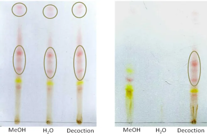
Figure 7. TLC fingerprint of A: R. alaternus bark extracts and B: R. alaternus leaves extracts 28
Figure 8. The chemical structures of the compounds found in R. alaternus leaves extracts. ___ 29
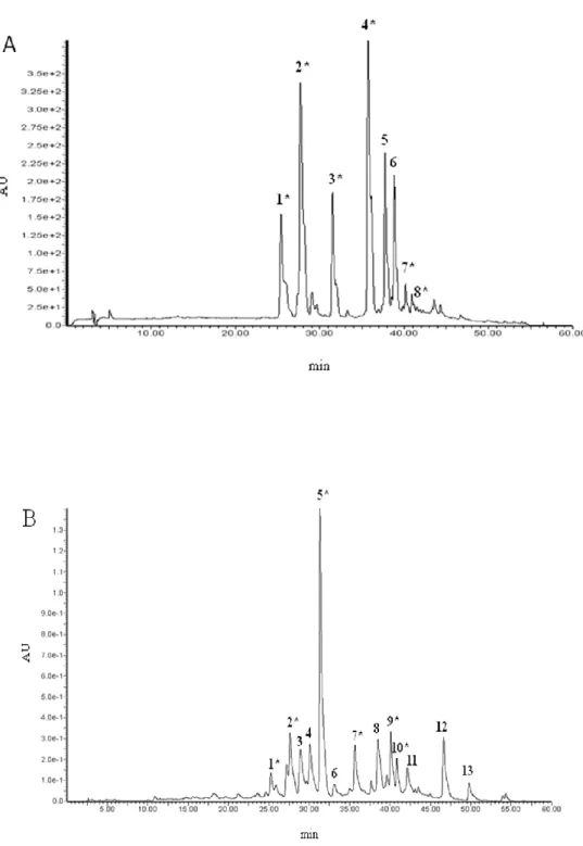
Figure 9. HPLC/MS representative chromatograms of flavonoids from Rhamnus alaternus bark
(extracted at 280nm). A: methanolic extract, B: aqueous extract and C: traditional extract. ____________________________________________________________ 30
Figure 10. HPLC-PDA representative chromatograms of flavonoids from Rhamnus alaternus
leaves (visualized at at 3500nm). A: methanolic extract, B: aqueous extract. ______ 31
Figure 11. Gallic acid calibration curve used in folin-Ciocalteau experiment _______________ 34
Figure 12. Trolox calibration curve used in 1,1-diphenyl-2-picrylhydrazyl (DPPH•) test _____ 34
Figure 13. Trolox calibration curve used in ABTS·+ radical assay _______________________ 34
Figure 14. FeSO4·7H2O calibration curves used in ferric reducing/antioxidant power assay. __ 35
Figure 15. Morphology of U937 cancer cells seen under microscope. ____________________ 37
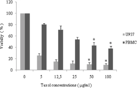
Figure 16. Effect of taxol at different concentrations on U937 and PBMCs cell viability.. ____ 38
Figure 17. Effect of R. alaternus bark extracts at different concentrations on U937 and PBMCs
cell viability. ________________________________________________________ 39
Figure 18. Effect of R. alaternus leaves extracts at different concentrations on U937 and PBMCs
cell viability. ________________________________________________________ 40
Figure 19. Morphology of U937 cancer cells and PBMCs normal cells after 24 hours of
treatment with R. alaternus bark methanolic extract. ________________________ 42
Figures
Figure 21. Apoptosis induction to U937 cells by R. alaternus bark methanolic extract. _______ 46
Figure 22. HPLC-PDA representative chromatograms of flavonoids from R. sphaerocarpa fruits
(extracted at 280nm). A: methanolic extract, B: aqueous extract and C: traditional extract. ____________________________________________________________ 50
Figure 23. Isoflavones found in the aqueous extract of Retama sphaerocarpa stems. ________ 52
Figure 24. HPLC profile of Retama sphaerocarpa aqueous extract (visualised at 260nm). ____ 53
Figure 25. GC/FID profiles of RSM. ______________________________________________ 56
Figure 26. Gallic acid calibration curve used in folin-Ciocalteau experiment. ______________ 57
Figure 27. Trolox calibration curves used in 1,1-diphenyl-2-picrylhydrazyl (DPPH•) test_____ 57
Figure 28. Trolox calibration curves used in ABTS·+ radical assay. ______________________ 57
Figure 29. FeSO4·7H2O calibration curves used in ferric reducing/antioxidant power assay. __ 58
Figure 30. Effect of the standard quercetin on in vitro formation of fluorescent AGEs. _______ 61
Figure 31. Effect of Retama sphaerocarpa fruits and stems extracts on in vitro formation of
fluorescent AGEs. ___________________________________________________ 62
Figure 32. Adjusted probit toxicity (log-dose) curve of RSM in rats. ____________________ 64
Figure 33. Effect of RSM on blood glucose levels in normoglycemic rats. ________________ 66
Figure 34. Effect of RSM on glucose tolerance in rats. ________________________________ 67
Tables
TABLES
Table 1. Extraction yields of Rhamnus alaternus bark and leaves. ______________________ 25
Table 2. Total flavonoids, total flavonols and total tanins contents in R. alaternus bark and
leaves ______________________________________________________________ 26
Table 3. The chemical nomination of R1 and R2 appearing in figure 9. Peak numbers refer to
figure 8. _____________________________________________________________ 29
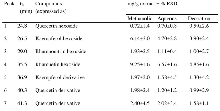
Table 4. HPLC/MS identification and quantification of flavonoids contained in methanolic,
aqueous and traditional extracts obtained from Rhamnus alaternus bark. __________ 29
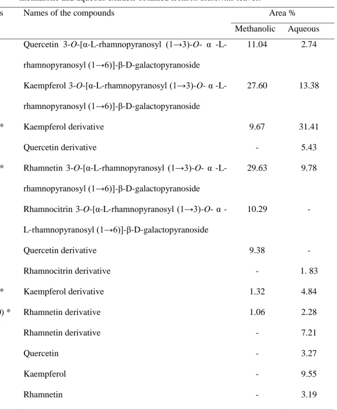
Table 5. HPLC/Uv-vis-DAD/MS identification and quantification of flavonoids contained in
the methanolic and aqueous extracts obtained from R. alaternus leaves. __________ 32
Table 6. Total phenolics content and antioxidant activity of Rhamnus alaternus bark extracts
measured by means of different in vitro tests. _______________________________ 35
Table 7. Total phenolics content and antioxidant activity of Rhamnus alaternus leaves extracts
measured by means of different in vitro tests. _______________________________ 35
Table 8. IC50 values (concentration eliciting 50% inhibition) for R. alaternus extracts and taxol
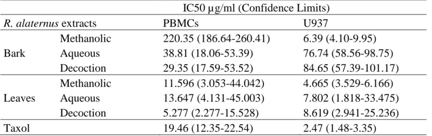
applicated to PBMCs and U937 cells. Cells were treated with various concentrations of the extract and taxol, and the cell number was counted after 24h of exposure. ______ 43
Table 9. Extraction yields of Retama sphaerocarpa fruits and stems extracts. _____________ 48
Table 10. Total flavonoids, flavonols and total tannins contents in R. sphaerocarpa fruits and
stems. ______________________________________________________________ 49
Table 11. HPLC-PDA/ESI-MS identification and quantification of flavonoids contained in
methanolic, aqueous and traditional extracts obtained from R. sphaerocarpa fruits. _ 51
Table 12. HPLC/MS quantification of flavonoids contained in the aqueous extract obtained from R. sphaerocarpa stems. _________________________________________________ 53 Table 13. Alkaloids found in RSM. Numbers correspond to figure 26.____________________ 56
Table 14. Total phenolic content and antioxidant activity of R. sphaerocarpa fruits extracts
measured by means of different in vitro tests ________________________________ 59
Table 15. Total phenolic content and antioxidant activity of R. sphaerocarpa stems extracts
measured by means of different in vitro tests. _______________________________ 59
Table 16. IC50 values (concentration reducing 50% of AGE formation) of R. sphaerocarpa fruits
Tables
Table 17. Results of probits determination __________________________________________ 64
Table 18. Effect of RSM on the water and feed intake of normal and diabetic rats. Values are the
mean±SD of 6 rats in each group; (*: P<0.05 and **: P<0.01) as compared with
diabetic control group. _________________________________________________ 71
Table 19. Effect of RSM on some hematological parameters of normal rats _______________ 72
Table 20. Effect of RSM on some hematological parameters of diabetic rats _______________ 72
Contents
Contents
INTRODUCTION ... 1
CHAPTER 01: LITERATURE REVIEW ... 3
1. Rhamnus alaternus L. ... 3
2. Retama sphaerocarpa (L.) Boisse. ... 5
3. Antioxidants ... 8
4. Diabetes mellitus (DM) ... 10
CHAPTER 02: MATERIELS AND METHODS ... 12
1. Plants ... 12
2. Animals ... 12
3. Chemicals ... 12
4. Extraction procedure ... 13
5. Folin-Ciocalteu colorimetric method ... 14
6 Total flavonoids content ... 14
7. Total flavonols content ... 15
8. Determination of condensed tannins ... 15
9. Thin layer chromatography (TLC) analysis of the extracts ... 15
10. HPLC-DAD/ESI-MS analysis of flavonoids ... 16
11. GC/MS/FID analyses of alkaloids in R. sphareocarpa stems methanolic extract ... 17
12. Antioxidant activity ... 18
13. Cell culture ... 20
14. Acute oral toxicity study of R. alaternus bark methanolic extract ... 21
15. Advanced glycation end products (AGEs) formation assay ... 21
Contents
17. Evaluation of RSM effect on normal healthy rats ... 22
18. Evaluation of RSM effect in oral glucose tolerance test ... 22
19. Evaluation of RSM effect in streptozotocin (STZ)-induced diabetic rats ... 23
20. Statistical analysis ... 23
CHAPTER 03: RESULTS AND DISCUSION ... 25
1 Rhamnus alaternus L. ... 25
1.1. Extraction yields ... 25
1.2. Total flavonoids, total flavonols and total tannins contents ... 25
1.3. Anthraquinones identification by TLC analysis ... 27
1.4. Identification and quantification of flavonoids ... 28
1.5. Antioxidant/radical scavenging activity ... 33
1.6. Cytotoxicity of R. alaternus bark extracts ... 36
1.7. Apoptosis analysis by flow cytometry ... 44
1.8. Acute toxicity test ... 44
2 Retama sphaerocarpa (L.) Boisse ... 48
2.1. Extraction yields ... 48
2.2. Total flavonoids, total flavonols and total tannins contents ... 48
2.3. Identification and quantification of flavonoids ... 49
2.4. GC/MS/FID analysis of alkaloids in R. sphaerocarpa stems methanolic extract . 54 2.5. Antioxidant/radical scavenging activity ... 56
2.6. Advanced glycation end products (AGE) formation assay ... 60
2.7. Acute oral toxicity of R. sphaerocarpa stems methanolic extract (RSM) ... 63
2.8. Effect of RSM on normoglycemic rats ... 65
2.9. Effect of RSM on oral glucose tolerance in normal rats ... 67 2.10. Effect of RSM on blood glucose level in normal and STZ-induced diabetic rats . 68
Contents
2.11. Effect of RSM on feed and water intake of normal and diabetic rats ... 70
2.12. Effect of RSM on haematological parameters of normal and diabetic rats ... 71
2.13. Effect of RSM on plasma lipids in normal and STZ-induced diabetic rats ... 73
CONCLUSION ... 76
Introduction
Introduction
1
INTRODUCTION
The biomolecular diversity of natural products still represents a valuable starting point for the development of new drug formulations, and in this vision the sustainable management of traditional medicinal plant resources remains an important aspect, being as in the past and recent history of drug discovery inexorably bound to the plant kingdom (Kingston, 2011, Newman and Cragg, 2007). Indeed, the experience of traditional medicine is precious in identifying possible target plant species or particular parts of them.
Rhamnus alaternus L. (Rhamnaceae) grows in the Mediterranean region, and is commonly found
in Algeria, where it is known locally as “M‟lilez”. The aerial parts of this plant are used in some North African countries for the treatment of liver complications such as jaundice (Boukef, 2001). Many antioxidant compounds were identified in Rhamnus alaternus like anthraquinones such as emodin, chrysophanol and flavonoids such as Kaempferol 3-O-bisorhamninoside (3) (Bhouri et
al., 2011b; Ben Ammar et al., 2009). Experimental studies have demonstrated that extracts and
purified compounds from aerial parts of R. alaternus possess different biological activities, including antibacterial, antimutagenic, antigenotoxic, antioxidant/free radical scavenging, and cytotoxic activities, and are able to modulate cellular gene expression (Ben Ammar et al., 2007a-b; 2008a-2007a-b; 2009).
Retama sphaerocarpa (L.) Boiss widely found within the Mediterranean area both in wet and dry
climates (Quezel and Santa, 1963). In Algeria and according to survey got from people, the stems of Retama were used traditionally to cure rabies in both humans and animals, to prepare a type of chewing tobacco commonly called "Chemma". Several papers in literature account for the composition of different extracts from R. sphaerocarpa, the majority of them dealing with the analysis of its alkaloids. Phytochemical studies on Retama sphaerocarpa report the presence of a particular class of alkaloids: the quinolizidine alkaloids, which are known to possess a wide range of pharmacological and toxicological properties (El-Shazly et al., 1996). Retama sphaerocarpa was shown to possess antimicrobial and cytotoxic activities (López-Lázaro et al., 2000; Louaar et
al., 2007). It is notified that other species from Retama genus but not Retama sphaerocarpa were
studied for their diuretic, hypoglycemic (Maghrani et al., 2005a-b), antioxidant, antiviral (Edziri
et al., 2010), antihypertensive (Eddouks et al., 2007), and anti-inflammatory (Bremner et al.,
Introduction
The purpose of this study can be listed as the following:
- Preparation of different extracts by maceration and decoction from R.alaternus and R.
sphaerocarpa.
- Charectirisation of flavonoids profile of the extracts by means of high-performance liquid chromatography-diode array detector/electrospray ionisation mass spectrometry (HPLC-DAD/ESI-MS) analysis.
- Alkaloids determination in R. sphaerocarpa stems methanolic extract by means of gas chromatographic (GC) separation coupled to a mass spectrometer (MS) and a flame ionization detector (FID).
- Evaluation of the antioxidant capacity of R.alaternus and R. sphaerocarpa extracts using a battery of five different simple redox-based assays differing in the mechanisms involved and the chemical environment used. The Folin-Ciocalteu assay; the bleaching of the stable 1,1-diphenyl-2-picrylhydrazyl radical (DPPH•); the trolox equivalent antioxidant capacity (TEAC) assay; the ferric reducing/antioxidant power (FRAP) assay; and, the oxygen radical absorbance capacity (ORAC) assay.
- The study of antiproliferative effect of R. alaternus extracts on cultures of human monocytic leukemia U937 cells, and of human normal peripheral blood mononuclear cells.
- Evaluation of apoptosis/necrosis effect of R. alaternus using phycoerythrin-labeled annexin V (PE Annexin V) and 7-Amino-Actinomycin D (7-AAD) flow cytometry assay. - Determination of the antiglycation activity of R. sphaerocarpa extracts by advanced
glycation end products (AGEs) formation assay.
- Estimation of the acute toxicity of R. sphaerocarpa in Albino Wistar rats through the calculation of the corresponding LD50 value.
- Investigation of the effect of R. sphaerocarpa after long-term treatment of normal and streptozotocin induced diabetes rats, the biochemical and hematological parameters were analyzed for all animals.
Literature review
Chapter 01
Literature review
Chapter 01
LITERATURE REVIEW
1. Rhamnus alaternus L.
1.1. Arabic vernacular names
- Aoud el kheir; méliles; mlîlâ, Quaced ((Quezel and Santa, 1963; Beloued, 2005).
1.2. Berber names
- Ajroudj; Khalis n'imidekh; Amliles ((Quezel and Santa, 1963; Beloued, 2005).
1.3. Taxonomy
Kingdom: Plants Subkingdom: Tracheophytes Branch: Spermatophytes Sub-Branch: Angiosperms Class: Dicotyledons Order: Rhamnales Family: Rhamnaceae Genus: RhamnusSpecie: Rhamnus alaternus L. (Quezel and Santa, 1963).
1.4. Botany and ecology
Rhamnus alaternus is a native Mediterranean, dioecius, evergreen shrub or tree, 2-6 m in
height. It is a cosmopolitan plant but mostly found in temperate and tropical regions; forest, scrub and Algerian tell (Tsahar et al., 2002). The stems have reddish bark and pubescent branches, the leaves are rounded, compact and alternating. Flowering occurs between February and April, and fleshy fruit appear between April and August (Quezel and Santa, 1962-1963). Unripe fruits are green, which turn red, then black when ripe (Spichiger et al., 2004). Its fleshy fruits are an important source of nutriment they are regularly consumed by birds and small mammals, it is a predominantly entomophilous species, and the seeds are dispersed by ants (Aronne and Wilcock 1994; Gómez et al., 2003; Arroyo et al., 2009). R. alaternus is one of the species commonly used
Literature review
4
in reforestation programs in the Mediterranean, due to its ability to survive in xeric environments (Gulias et al., 2004).
1.5. Traditional applications
In North Africa especially in Algeria, Tunisia and Morocco, R. alaternus have diverse uses, but the doses must be with precaution. The infusion of the bark or leaves of R. alaternus are mostly used to cure jaundice and liver troubles, for this 10g of root or trunk bark are mixed with half a liter of water, boiled for 10 minutes, filtered, and then two cups are drunk per day (Beloued, 2005). In the other hand, the fruits of this plant are used as purgative and laxative (bellakhdar, 1997), for this: two grams of crushed fruits are mixed with a quarter of water, the mixture is boiled, and then to be drunk in a fasting morning (Beloued, 2005). The wood of the plant was used to have a yellow coloration of fabric and wool (bellakhdar, 1997).
1.6. Phytohemical composition
The genus Rhamnus contains abundant phenolic substances, especially anthraquinones but also tannins and flavonoids (Tsahar et al., 2002). R. alaternus leave, and bark of the root and trunk are rich in anthraquinones (Paris and Moyse, 1976-1981). Four anthraquinone aglicones were identified in the above ground parts of R. alaternus (emodin, chrysophanol, alaterin and physcion); emodin is the most abundant (Tsahar et al., 2002). Three triglycoside flavonoids were isolated from the leaves of R. alaternus (kaempferol O-b-isorhamninoside (1), rhamnocitrin 3-O-b-isorhamninoside (2) and rhamnetin-3-O-bisorhamninoside (3) (Figure 1) along with apigenin, kaempferol and quercetin. (Ben Ammar et al., 2009).
Literature review
Compound name R1 R2
1- Kaempferol 3-O-b-isorhamninoside OH H 2- Rhamnocitrin 3-O-b-isorhamninoside OCH3 H
3- Rhamnetin-3-O-bisorhamninoside OCH3 OH
Figure 1. Chemical structure of the flavonoids (1-3) isolated from R. alaternus leaves (Ben
Ammar et al., 2009).
1.7. Biological activities
Previous studies have shown potent antioxidant, free radical scavenging, antimutagenic and antigenotoxic activities of crude extracts from R. alaternus (Chevolleau et al, 1992; Ben Ammar
et al., 2005; 2008a-b). It was also reported other biological activities of R. alaternus extracts:
antibacterial and antiproliferative (Kosalec et al. 2013; Ben Ammar et al., 2007a; 2008b; 2011). In human cells, extracts of R. alaternus leaves modulate the expression levels of genes implicated in both DNA repair and oxidative defense systems (Ben Ammar et al., 2007a-b). It was proved that kaempferol 3-O-b-isorhamninoside and rhamnocitrin 3-O-b-isorhamninoside isolated from
R. alaternus Induce apoptosis in human lymphoblastoid cells (Bhouri et al., 2011a; 2012).
2. Retama sphaerocarpa (L.) Boiss.
2.1. Arabic vernacular name
- R‟tem (Quezel and Santa, 1963).
2.2. Berber names
Literature review 6
2.3. Taxonomy
Kingdom: Plants Subkingdom: Tracheophytes Branch: Spermatophytes Sub-Branch: Angiosperms Class: Dicotyledons Order: FabalesFamily: Fabaceae (leguminosae) Genus: Retama
Specie: Retama sphaerocarpa (L.) Boisse (Quezel and Santa, 1963).
2.4. Botany and ecology
From Retama genus three species are growing in Algeria, Retama monosperma, Retama
raetam, and Retama sphaerocarpa (Belmokhtar and Kaid-Harche, 2012; Boulila et al., 2009). R. sphaerocarpa is a perennial shrub up to 5m height with a simple modular structure consisting of
several branches (figure 2), its open structure allows adequate amount of light to pass through it (Fungairiño et al., 2005; Padilla and Pugnaire, 2009). Its small, ephemeral leaves are shed within a couple of weeks, the flowers are small (5-6mm) of yellow color and appear from April to May, while the fruits are of a hard cover and appear in winter (January–February) (Domingo et al., 1997; 1998; 1999; Pugnaire et al., 1996). Shrubby legumes of Retama genus are endemic to the Mediterranean Basin and they are distributed in the various Mediterranean climates (from humid to arid) and ecosystems including coastal, dunes, maquis, and also deserts, since Retama shrubs are tolerant to extreme drought conditions (Martin-Cordero et al., 2000; Quezel and Santa, 1962-1963). Retama species are of ecological interest for dune stabilization, soil fixation, and revegetation of semi arid ecosystems (Caravaca et al., 2003). An important trait of shrub legumes that contributes to their adaptation to low fertile (nitrogen-deficient) soils, and to improve fertility, is their capacity to establish N2-fixing symbiosis with rhizobia, which are the soil
bacteria that can fix N2 in the nodules formed with legumes (Boulila et al., 2009). R.
sphaerocarpa has a remarkable capacity to withstand drought owing to its crown architecture and
its deep root system, which can penetrate to depths of 25m (Haase et al. 1996), providing access to deep water sources (Caravaca et al., 2005).
Literature review
Figure 2. Sketch of a shoot of Retama sphaerocarpa (Haase et al., 2000).
2.5. Traditional application
Retama species including R. sphaerocarpa have same traditional applications. They are mostly known to cure rabies and to reduce blood glucose level in the medicinal folk traditions of the east of Algeria (Louaar et al., 2005. Djeddi et al., 2013), in Morocco Retama is largely recommended by traditional herbal healers for diabetes control (Maghrani et al., 2005b).
2.6. Phytochemical composition
Phytochemical studies have shown that R. sphaerocarpa contains quinolizidine alkaloids (Figure 3). El-Shazly and his coworkers (1996) detected the following alkaloids: cytisine retamine, sparteine, Ammodendrine, 5,6 dehydrolupanine, lupanine, Anagyrine, the phytochemical structures of the most abundant alkaloids in R. sphaerocarpa are shown in figure 4. Also phenolics were isolated from the aerial parts of the plant and they are: Retamatrioside, rhamnazin, 6-methoxypseudobaptegenin, genistein 8-C-glucoside, genistein O-b-glucoside, 7-hydroxy-6‟methoxy-3‟,4‟-methylenedioxyioflavone 7-O-ß-glucoside, genistein, daidzein, rhamnazin-3-O-ß-glucopyranozyl-(1-5)-α-arabinofuranoside (López-Lázaro et al., 1998; Martín-Cordero et al., 1999; 2000; Louaar et al., 2005; 2007). Fatty acids were detected in R.
sphaerocarpa stems and grain, whereas sterols were detected in stems and monoglycerides in
Literature review
8
Figure 3. Structures of Cytisine and Retamine (Orhan et al., 2007).
2.7. Biological activities
Diverse biological activities were attributed to R. sphaerocarpa; hence flavonoids such as rhamnazin extracted from the aerial parts of this plant showed a cytotoxic activity and produced a dose-dependent inhibition of cell growth (López-Lázaro et al., 2000). The leaves of Retama
sphaerocarpa were reported to have antimicrobial properties against Staphylococcus aureus
(Louaar et al., 2007). Moreover, several studies investigated Retama Genus for various pharmacological effects, including hypoglycemic, diuretic (Maghrani et al., 2005b), antioxidant, antiviral (Edziri et al., 2010), antihypertensive (Eddouks et al., 2007) and anti-inflammatory (Bremner et al., 2009) activities.
3. Antioxidants
3.1. The concept of free radicals and antioxidants
A free radical is a chemical species that has an odd number of electrons, highly unstable and active toward chemical reactions (Jensen, 2003; Wu et al., 2013) and seek to stabilize themselves by „„stealing‟‟ electrons from other chemicals or compounds (including proteins, carbohydrates, lipids and DNA) (Talaulikar and Manyonda, 2011). The free radical species are called reactive oxygen species (ROS). They are generated naturally in the human body and additional free radicals can be developed when we‟re exposed to environmental toxins, pollutants and others, radicals derived from oxygen represent the most important class of such species generated in living systems (Valko et al., 2004). ROS includes radicals such as anion superoxide
Literature review
(O2•
-), hydroxyl radical (•OH-), nitric oxide (NO-), and other species like hydrogen peroxide (H2O2) (Sen et al., 2010).
An antioxidant is a compound that inhibits or delays the oxidation of substrates even if the compound is present in a significantly lower concentration than the oxidized substrate (Matkowski, 2008). Antioxidants neutralize free radicals by donating one of their own electrons, ending the electron-„„stealing‟‟ reaction (Talaulikar and Manyonda, 2011). The antioxidants can have various mechanism of action for instance: the scavenging of reactive oxygen species (ROS); prevention of ROS formation and chain breaking effects (Gopi et al., 2014). The antioxidant compounds can be recycled in the cell or are irreversibly damaged, but their oxidation products are less harmful or can be further converted to harmless substances (Blassan et al., 2014).
3.2. Natural antioxidants
The human organisms possess antioxidant defense systems (endogenous) that deal with reactive oxygen species; it is divided into two major groups: enzymatic antioxidants and non-enzymatic antioxidants (Yapo et al., 2013). Despite its remarkable efficiency, this endogenous antioxidant system does not suffice. humans depend on various types of antioxidants that are present in the diet to maintain free radical concentrations at low levels like: vitamins C and E that are generic names for ascorbic acid and tocopherols (Barros et al., 2011), other vitamins are also important like vitamin E and vitamin K (Vervoort et al., 1997; Carocho and Ferreira, 2013). Phenolic compounds are also an important antioxidants present in plants they are secondary plant metabolites possessing an aromatic ring bearing one or more hydroxyl groups (Balasundram et
al., 2006). They are considered as the most widely occurring groups of phytochemicals, and are
of considerable physiological and morphological importance in plants (Popa et al., 2008; Naczk and Shahidi, 2006), many of the phenolic compounds are considered as strong antioxidants like flavonoids that the antioxidant properties is conferred to their phenolic hydroxyl groups attached to ring structures. Some of the most important flavonoids are catechin, quercetin and kaempferol (Rice Evans et al., 1996; Prochazkova et al., 2011).
3.3. Detection of antioxidants in plants
The presence of antioxidants in plants can be detected by different methods like spectrophotometric experiments, or by more specified complex analysis like High performance liquid chromatography (HPLC) and gas chromatography (GC). However, it is impossible to get all the antioxidant compounds in a plant by one or two types of experiments because there are
Literature review
10
other conditions to take in consideration, like the preparation of plante samples, the type of solvent, the extraction method and others. Recent technological advances and the development of new methods to improve the production, detection, isolation and/or characterization have revolutionized the screening of bioactive compounds such as antioxidants (Gil-Chávez et al., 2013). Indeed, in recent publications the chromatographic determination of antioxidants predominates, frequently accompanied by bioactivity-guided fractionation, spectral structure elucidation of isolated chemicals by MS or NMR. The use of hyphenated techniques such as LC-MS and LC-NMR is considered to be the best means for structure determination when novel compounds are present in vitro that have not been known from intact plants (Tian et al., 2005; Sanchez-Sampedro et al., 2007; Matkowski, 2008).
4. Diabetes mellitus (DM)
The word „diabetes‟ is derived from the Greek word “Diab” (meaning to pass through, referring to the cycle of heavy thirst and frequent urination); „mellitus‟ is the Latin word for “sweetened with honey” (presence of sugar in the urine) (Tiwari et al., 2013). This disease can be defined as a syndrome characterized by a state of chronic hyperglycemia, DM and its complications like the cardiovascular disorders are the fourth most important causes of mortality and the principal cause of irreversible blindness (Perez et al., 1998). The incidence of this disease affects 1–2% of the population worldwide (Ahmed, 2005). DM can be divided into two types that are: type 1 diabetes also known as Insulin-dependent DM and Type 2 diabetes mellitus, formerly called non-insulin-dependent DM (American diabetes association, 2008; Li and Hölscherb, 2007).
4.1. Oxidative stress and DM
Different studies assessed the role of the oxidative stress in the prognosis of diabetes and diabetic complications (Baynes and Thorpe 1999; Giacco and Brownlee 2010). Generally, these studies attribute the high oxidative stress level in diabetes to the imbalance between the production of the reactive oxygen species and the decline in the endogenous antioxidants in different tissues. The magnitude of such imbalance contributes to the severity of the oxidant-mediated damage present in the diabetic context (Halliwell, 1994). Brownlee suggested the model that reactive oxygen species initiate the development of diabetic complications in his unifying hypothesis of diabetes. He hypothesized that the overproduction of the reactive oxygen species (ROS) like superoxide anion O2•-, in the mitochondrial electron-transport chain of glucose treated
Literature review
cells may alter important biochemical reactions responsible for diabetic complications development (Brownlee, 2005).
4.2. Plants as a natural medicine used for therapy of DM
Before the discovery of antidiabetic drugs, diabetic patients used medicinal plants and traditional medicine (Sewell and Rafieiann, 2014). Plants may operate through different mechanisms that may affect for example the blood sugar, Insulin, or also the pancreatic β cells (Bailey and Day, 1989).
The ethnobotanical information reports about 800 plants that may possess antidiabetic potential (Grover et al., 2002; Jung et al., 2006). Medicinal foods are prescribed widely even when their biologically active compounds are unknown, because of their effectiveness and availability (Dewanjee et al., 2009; Patel et al., 2012b). The World Health Organization (WHO) has recommended the evaluation of traditional plant treatments for diabetes as they are effective, with less or no side effects and are considered excellent candidates for oral therapy (Shokeen et al., 2008).
Finding the promising candidates for the treatment of diabetes mellitus could be achieved through the evaluation of the anti-diabetic properties of different medicinal plant extracts, their fractions and isolated components, followed by preliminary phytochemical screening, and tests of their probable toxicities (Nasri and Shirzad, 2013; Bnouham et al., 2006). Although various compounds with different mechanism of actions have been shown to reduce blood glucose, however, antioxidant activity of these plants has a crucial role in their antidiabetic actions (Nasri and Rafieian, 2014; Bahmani et al., 2014). The phytoconstituents that may act positively against diabetes are belonging to different classes of metabolites in plants such as quinolizidine alkaloids; flavonoids; terpenoids and others. These constituents may be considered as a promising source of hypoglycemic agents with minimal side effects (Sarkhail et al., 2010; Mohammadi et al., 2010; Patel et al., 2012). To recognize the most active candidates, pharmacological evaluation will be determined through measuring different biochemical parameters such as serum glucose, insulin, hemoglobin, lipid profile, serum urea and creatinine, plasma aspartate transaminase and alanine transaminase. The antidiabetic properties will also be further confirmed through microscopical examinations of the pancreatic sections (Baradaran et al., 2013).
Materials and methods
Chapter 02
Materials and methods
Chapter 02
Materials and methods
1. Plants
The bark and leaves of Rhamnus alaternus were collected in 2010 from Teniet En Nasr (Bordj Bou Arreridj, Algeria), the climate of this region is semi arid with a cold winter and a hot summer and knows as a forest place characterized by the abundance of trees and herbaceous plants (DPSB-BBA, 2014). whereas Retama sphearocarpa fruits and stems were collected in 2011 from Ras El Aioun (Batna, Algeria) and this region is characterized by a semi-arid climate, the temperature change between day and night; summer and winter, hence in summer it can reach up to 38°C as maximum especially in August, whereas in winter it may go down into 0°C especially in January; the quantity of rain that falls is between 200-350 Mm per year considered very few for vegetation growing; (Messaoudene, 2009). The identification of the two plants was based on the work of Quezel and Santa (1963), and validated by botanists in the Department of Ecology and Plant Biology at Setif 1 University (Algeria). The plants (figure 4) were dried and stored at room temperature until use.
Figure 4. Plants pictures in their environment. A: Rhamnus alaternus in Teniet En Nasr; B: Retama sphearocarpa in Ras El Aioun. (2014).
Materials and methods
13
2. Animals
Male and female Wistar rats weighing between 150 and 180g were obtained from Pasteur Institute (Algiers, Algeria) They were housed under standard environmental conditions (temperature 28±1°C photoperiod: 12 h light and 12 h dark cycle). The animals were allowed free access to water and ad libitum standard laboratory diet.
3. Chemicals
Unless otherwise stated, all reagents and solvents were of analytical grade and used without further purification. High-performance liquid chromatography (HPLC) grade methanol, fluorescein (FL) sodium nitrite, Folin-Ciocalteu phenol reagent and hydrochloride acid were purchased from Carlo Erba (Milan, Italy). High-performance liquid chromatography grade water and acetonitrile, aluminum chloride anhydrous and potassium peroxodisulfate were from VWR. Pure reference standards daidzein and genistein were purchased from Extrasynthese (Lyon, France). All other reagents, if not specified, were purchased from Sigma-Aldrich (Milan, Italy).
4. Extraction procedure
The extraction procedure is summarized in figure 5. The methanolic and aqueous extracts from R. alaternus bark and leaves and R. sphearocarpa fruits and stems were prepared by maceration. Briefly, drug samples were powdered by a mill and 50g of the powdered drugs were mixed with 500ml of methanol or water and then left for 24 hours under occasional stirring. Furthermore, a traditional aqueous extract was prepared from both plants by decoction: Boiling 10g of the powdered drug in 500ml of water for 10min (Beloued, 2005), then to obtain the dried extracts, the mixtures were filtered, and the filtrates were evaporated to dryness under vacuum by using a rotavapor (Büchi). The residues were kept into brown vial until use (Gnanaprakash et al., 2010; Thanabhorn et al., 2006). For the chemical and biochemical assays, the appropriate volumes of methanol or water were used to redissolve the methanolic, aqueous and traditional extracts respectively. All solutions were freshly prepared before experiments and used immediately.
Materials and methods
Figure 5. Extraction procedure
5. Folin-Ciocalteu colorimetric method
The antioxidant capacity (expressed as total phenols content) of the extracts was determined by means of the Folin-Ciocalteu reagent (Spagna et al., 2002; Cimino et al., 2007). 50µl of the solutions containing different concentrations of the extracts to be tested were added to 450µl of deionized water; 500µl of Folin-Ciocalteu reagent and 500µl of 10% aqueous sodium carbonate solution; samples were then maintained at room temperature for 1h. Absorbance was measured at 786nm (UV-Vis Spectrophotometer, Shimadzu Japan) against the blank containing 50µl of the same solvent used to dissolve the extracts. Total phenolic content was expressed as µg of gallic acid equivalents per mg of extract, using calibration curve prepared with gallic acid standard solutions. Each determination was performed in duplicate and repeated at least two times.
6. Total flavonoids content
The flavonoid content was measured using the colorimetric assay with little modification in the procedure reported by Lenucci and coworker (2006). Aliquots (50µl) of the methanol/water extracts were diluted with distilled water to a final volume of 0.5ml, and 30µl of 5% NaNO2 were
Materials and methods
15
200µl of 1M NaOH and 210µl of distilled water. Absorbance was recorded at 510nm using a UV- visible spectrophotometer (Shimadszu, Japan) and flavonoid content was expressed as µg of catechin equivalents (CatE) per mg of extract. Each determination was carried out in duplicate and repeated at least two times.
7. Total flavonols content
The content of flavonols was determined by the method described by Tomaino and coworker (2010). Aliquots (125µl) of the solutions containing the different extracts to be tested were mixed with 125µl of AlCl3 (2mg/ml), and 750µl of sodium acetate (50mg/ml). The
absorbance at 440nm was detected by a spectrophotometer after 2.5h. Total flavonol content was expressed as µg of quercetin equivalents (QE) per mg of dried extract. All determinations were carried out in duplicate and repeated at least two times.
8. Determination of condensed tannins
In presence of concentrated H2SO4, condensed tannins were transformed by the reaction
with vanillin to anthocyanidols. The experiment is previously described by Tounsi and coworkers (2009). 50µl of each extract appropriately diluted was mixed with 3ml of 4% methanol vanillin solution and 1.5ml of H2SO4. After 15 min, the absorbance was measured at 500nm. Condensed
tannin contents of the extracts (three replicates per treatment) were expressed as ng catechin equivalents (CE) per milligram of extract through the calibration curve of catechin.
9. Thin layer chromatography (TLC) analysis of the extracts
Thin layer chromatography (TLC) analyses were performed as previously described by Sakulpanich and coworker (2009). The TLC fingerprints were performed on a precoated aluminum plate of silica gel 60 F254 (10x20cm) using ethyl acetate: methanol: water (100:17:13)
as the mobile phase. The developing distance was 8.0cm. After removing the plate from the chamber, the plate was dried with an air dryer, and sprayed with 10% alcoholic potassium hydroxide solution. Anthraquinones show pink spots.
Materials and methods
10. Flavonoid analysis by high-performance liquid chromatography-diode
array detector/electrospray ionisation mass spectrometry
(HPLC-DAD/ESI-MS)
In this section two methods were used; the first was for R. alaternus bark and R.
sphaerocarpa fruits extracts and the second for R. sphaerocarpa stems aqueous extract and R. alaternus leaves methanolic and aqueous extracts.
The solutions containing the different extracts (R. alaternus bark and R. sphaerocarpa fruits extracts) to be tested (10mg/ml) were filtered through a 0.45µm membrane filter (Whatman, Clifton, NJ, USA). HPLC-DAD/ESI-MS analyses of flavonoids were performed on a Prominence LC system (Shimadzu, Milan, Italy) consisting of a CBM-20A controller, two LC-20AD pumps, a CTO-20AC, a DGU-20A3 degasser, a SIL-20AC autosampler, a SPD-M20A
photo diode array (PDA) detector, and a quadrupolar mass analyzer (LCMS-2020), equipped with an electrospray ionization (ESI) interface, operated in the negative mode. Data acquisition was performed by Shimadzu LabSolution software ver. 5.10.153.
Separation was carried out on an Ascentis Express C18 column, 15 cm x 4.6 mm i.d. packed with 2.7 m partially porous particles (Supelco, Bellefonte, PA, USA). The injection volume was 5µl, and the mobile phase consisted of water/acetic acid (0.075%) at pH=3 (solvent A) and acetonitrile/acetic acid (0.075%) (solvent B), respectively in the following linear gradient mode: 0min, 0% B; 60min, 40% B; 70min, 100% B; 71min, 0% B. The mobile phase flow rate was 1.0ml/min, and it was splitted to 0.2ml/min prior to MS detection. PDA wavelength range was 190-400nm and the chromatograms were extracted at 280nm
The quantitative determination of each compound was carried out by means of the external standard method using kaempferol; quercetin, Apigenin; Luteolin; Genistein; Daidzein as reference compounds in a concentration range of 1-150ppm, The results were obtained from the average of three determinations and are expressed as mg/g dried extract ± percent relative standard deviation (%RSD).
For the analytical determinations of R. sphaerocarpa stems aqueous extract and R. alaternus leaves methanol and aqueous extracts; variable aliquots of the powders were accurately weighted and solved in HPLC grade water immediately prior analyses to obtain at least three samples for each extract (10mg/ml). Quantification was carried out using daidzein derivatives (correlation coefficient R2= 0.9998), whilst the other signals were quantified using genistein (R2= 0.9999).
Materials and methods
17
Waters instrument equipped with a 1525 Binary HPLC pump, a Micromass ZQ with a ESI Z-spray source, and a 996 Photo Diode Array Detector (DAD). The extracts were analysed using solvent system A (2.5% formic acid in water) and B (2.5% formic acid in acetonitrile) with the following gradient: 0min: 5% B; 10min: 15% B; 30min: 25 % B; 35min: 30% B; 50min: 90% B; then kept for 7min at 100% B. The solvent flow rate was 1ml/min, the temperature was kept at 25°C with a column oven (Hitachi L-2300, VWR International, Milan, Italy), and the injector volume selected was 20μl.
DAD analyses were carried out in the range between 800 and 190nm, setting the detector at 245 and 260nm for isoflavones and 350nm for flavonols. Collected data were processed through a MassLynx v. 4.00 software (Waters S.p.A. Milano, Italy).
11. Gas chromatographic (GC) separation coupled to a mass spectrometer
(MS) and a flame ionization detector (FID) analyses of alkaloids in R.
sphareocarpa stems methanolic extract (RSM)
For the GC/MS analyses, a small aliquot (50mg) of the methanolic extract was put in an 8ml amber vial and 1ml HCl was added. The solution was vigorously shaken (250rpm) for 10 minutes at room temperature. Then the pH value was brought to 8 with TEA (triethylamine), 1ml CH2Cl2 was added and the two layers left to separate for 5 minutes. The organic layer was then
removed and dried over anhydrous Na2SO4. For the GC/MS analyses, 4µl of the sample was
taken and injected. Gas chromatogaphic (GC) analyses were run on a Hewlett-Packard gas-chromatograph mod. 5890, equipped with a flame ionization detector (FID). GC-FID analyses were carried out in the following analytical conditions: ZB-5 capillary column (30m 0.25mm i.d. 0.25μm film thickness); helium as carrier gas; injector and detector temperature were set at 250 and 300°C, respectively. Oven temperature was programmed as follows: 0min. 100°C, then kept at 100°C for 1min; from 100 to 150°C at 5°C/min, then kept at 150°C for a further 2min; from 150 to 300°C at 3°C/min, then kept at 300°C for a further 10 minutes.
Gas chromatography-mass spectrometry (GC-MS) was carried out on the same gas chromatograph connected to a Hewlett-Packard mass spectrometer model 5971A, ionization voltage 70eV, electron multiplier 1700V; ion source temperature 180°C. Gas chromatographic conditions were the same as above.
The identity of components was based on their GC retention time, and computer matching of spectral MS data with those from Wiley 275 library, and the comparison of the fragmentation patterns with those reported in literature (El-Shazly et al., 1996).
Materials and methods
12. Antioxidant activity
12.1 1,1-diphenyl-2-picrylhydrazyl (DPPH) test
The free radical-scavenging capacity of the extracts was determined by the DPPH assay (Rapisarda et al., 1999; Morabito et al., 2010), a method based on the reduction of the stable radical 1,1-diphenyl-2-picrylhydrazyl. The reagent mixture consisted in 1.5ml of 100mM DPPH• in methanol, to which 37.5µl of various concentrations of the extracts to be tested were added; an equal volume of the solvent employed to dissolve the extracts was added to control tubes. After 20min of incubation at room temperature, the absorbance was recorded at 517nm in a UV-Vis spectrophotometer (Shimadzu, Japan). Results were expressed as mmol trolox equivalents/g of dried extract, using calibration curve prepared with trolox as standard solutions. Each determination was performed in duplicate and repeated at least two times.
12.2 ABTS·+ radical assay
The ability of the extracts under study, to scavenge the ABTS·+ radical was evaluated as described by Morabito and coworkers (2010). Briefly, this method determines the capacity of antioxidants to quench the ABTS·+ radical. The ABTS·+ radical cation was produced by the oxidation of 1.7mM ABTS·+ (2,2‟-azinobis-(3-ethylbenzothiazine- 6-sulfonic acid) with potassium persulphate (4.3mM final concentration) in water. The mixture was allowed to stand in the dark at room temperature for 12-16h before use, and then the ABTS·+ solution was diluted with phosphate buffered saline (PBS) at pH 7.4 to give an absorbance of 0.7±0.02 at 734nm. 50µl of a solution containing different concentrations of the extracts to be tested or of the vehicle alone (methanol or water) were added to 2ml of the ABTS·+ solution, and the absorbance was recorded at 734nm in a UV-Vis spectrophotometer (Shimadzu, Japan) after allowing the reaction to stand for 6min in the dark at room temperature. Each determination was performed in duplicate and repeated at least two times. Results were expressed as mmol troloxequivalents/g of dried extract, using calibration curve prepared with trolox standard solutions.
12.3 Ferric reducing/antioxidant power (FRAP) assay
The ferric reducing ability of the extracts under study was evaluated according to the method described by Cimino and coworkers (2013), with some modifications. The method is based on the reduction of a ferric–tripyridyltriazine complex to its colored ferrous form in the
Materials and methods
19
2.5ml of 10mM 2,4,6-tripyridyl-s-triazine (TPTZ) solution (prepared in 40mM HCl), with 25ml of 0.3M acetate buffer (pH 3.6), and 2.5ml of 20mM FeCl3∙6H2O solution, and then warmed
(preheated) at 37°C before use. 50µl of a methanolic or water solution containing different concentrations of the extracts to be tested, or of the vehicle alone (methanol or water), were added to 1ml of FRAP reagent, and the absorbance was measured at 593nm in a
spectrophotometer (Shimadzu, Japan) after incubation at 20°C for 4min. A standard curve was prepared using various concentrations of FeSO4·7H2O. Each determination was performed in
duplicate and repeated at least two times, and results were expressed as mmol Fe2+/g of dried extract.
12.4 Oxygen radical absorbance capacity assay using fluorescein (ORAC-FL)
assay
The ORAC-FL assay was performed as described by Dávalos and coworkers (2004). This assay is based upon the effect of peroxyl radicals generated from the thermal decomposition of 2,2‟-azobis (2-methylpropionamidine) dihydrochloride (AAPH) on the signal intensity from the fluorescent probe fluorescein (FL) in the presence of an oxygen-radical absorbing substance. The reaction was carried out in 75mM phosphate buffer (pH 7.4), and the final reaction mixture was 2ml. An aliquot of the solution containing different concentrations of the extracts to be tested (200μl), and 1.2ml of FL solution (70nM, final concentration) were placed in cuvette. The mixture was pre-incubated for 15min at 37°C in the spectrofluorimeter. Freshly prepared AAPH solution (600μl; 12mM, final concentration) was rapidly added. Fluorescence was recorded every minute for 80min at 37°C; the fluorescence conditions were as follows: excitation at 485nm, and emission at 520nm. A blank containing FL and AAPH with only the solvent employed to dissolve the extracts, different calibration solutions using Trolox (1-7.5μM, final concentration) as reference antioxidant, were also carried out in each assay. All the reaction mixtures were prepared in triplicate and at least three independent assays were performed for each sample. The area under the curve (AUC) was calculated for each sample by integrating the relative fluorescence curve. The net AUC was calculated by subtracting the AUC of the blank. The final ORAC values were determined by linear regression equation of trolox concentrations, and expressed as mmol Trolox equivalents/g dried extract.