POSTER PRESENTATION
Open Access
Auditory object feature maps with a hierarchical
network of independent components?
Jean Rouat
*, Simon Brodeur, Éric Plourde
From The Twenty Third Annual Computational Neuroscience Meeting: CNS*2014
Québec City, Canada. 26-31 July 2014
Auditory Object representation in the brain is still a controversial question [1,2]. Kumaret al. [3] discuss the hierarchical organization for auditory object perception and observe that the Planum Temporale (PT) area of the cortex encodes invariant representations of the spec-tral envelops of sounds. Many other studies find maps of representations elsewhere in the brain (Cochlear
Nucleus, Inferior Nucleus, etc.). Sparse representations with minimum overlap could be considered, according to Barlow [4]. Griffiths and Warren [5] propose that auditory object representations might be segmented or segregated in the Planum Temporal (PT) by increasing the independence between the neural activities. We therefore explore the potential of a hierarchical neural
* Correspondence: [email protected]
NECOTIS, Département génie électrique, génie informatique, Université de Sherbrooke, Québec, Canada, J1K 2R1
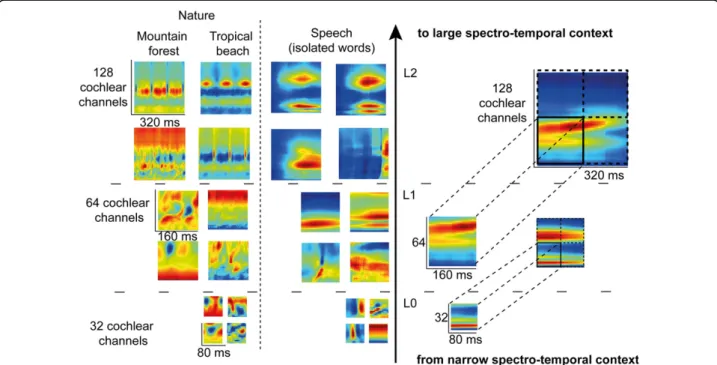
Figure 1 Learned representations illustrated for speech and natural sounds. FastICA training is first done on patches of 80ms x 32 cochlear channels of the envelopes coming from a 128 channels cochleagram (Level L0). Then, patches of 160ms x 64 cochlear channels are created at level L1 with a concatenation through time and space of the learned L0 features. FastICA is then performed on these larger patches to generate the new L1 representations. The same procedure is repeated for level L2.
Rouatet al. BMC Neuroscience 2014, 15(Suppl 1):P66 http://www.biomedcentral.com/1471-2202/15/S1/P66
© 2014 Rouat et al; licensee BioMed Central Ltd. This is an Open Access article distributed under the terms of the Creative Commons Attribution License (http://creativecommons.org/licenses/by/4.0), which permits unrestricted use, distribution, and reproduction in any medium, provided the original work is properly cited. The Creative Commons Public Domain Dedication waiver (http:// creativecommons.org/publicdomain/zero/1.0/) applies to the data made available in this article, unless otherwise stated.
assembly with the use of a computer simulation -whose layers increase the feature independence during training, to represent auditory object parts. It is observed that learned features are organized into non-overlapping maps (Figure 1) and that redundancy of the representation is in fact reduced. Learning was done on three categories of sounds having distinct acoustical sta-tistics: speech, music and natural sounds. We observed that the learned feature maps are very different from one sound category to another and might be, to some extend, comparable to receptive fields measured in the brain. We discuss of their potential similarity with receptive fields measured in the Inferior Colliculus of the Guinea Pig and how they might be part of a repre-sentation of auditory objects in the brain.
Published: 21 July 2014 References
1. Bizley JK, Cohen YE: The what, where and how of auditory-object perception. Nat Rev. Neuro 2013, 14(10):693-707.
2. Griffiths TD, Warren JD: What is an auditory object? Nat Rev Neurosci 2004, 5(887):892.
3. Kumar S, Stephan KE, Warren JD, Friston KJ, Griffiths TD: Hierarchical processing of auditory objects in humans. PLoS Comput Biol, Public Library of Science, 2007, 3(6):e100-e100.
4. Barlow H: Redundancy reduction revisited. Network : Comput. Neural Syst 2001, 12:241-253.
5. Griffiths TD, Warren JD: The planum temporale as a computational hub. Trends in neurosciences 2002, 25(7):348-353.
doi:10.1186/1471-2202-15-S1-P66
Cite this article as: Rouat et al.: Auditory object feature maps with a hierarchical network of independent components? BMC Neuroscience 2014 15(Suppl 1):P66.
Submit your next manuscript to BioMed Central and take full advantage of:
• Convenient online submission
• Thorough peer review
• No space constraints or color figure charges
• Immediate publication on acceptance
• Inclusion in PubMed, CAS, Scopus and Google Scholar
• Research which is freely available for redistribution
Submit your manuscript at www.biomedcentral.com/submit
Rouatet al. BMC Neuroscience 2014, 15(Suppl 1):P66 http://www.biomedcentral.com/1471-2202/15/S1/P66