affiliée à l’Université de Montréal
The Biopigment Eumelanin in the Sustainability Challenge: Interfaces with
Metal Electrodes, UV-Absorption Enhancement of Plastics and its
Biodegradability
EDUARDO DI MAURO
Département de génie physique
Thèse présentée en vue de l’obtention du diplôme de Philosophiae Doctor Génie physique
Mai 2019
affiliée à l’Université de Montréal
Cette thèse intitulée :
The Biopigment Eumelanin in the Sustainability Challenge: Interfaces with
Metal Electrodes, UV-Absorption Enhancement of Plastics and its
Biodegradability
présentée par Eduardo DI MAURO
en vue de l’obtention du diplôme de Philosophiae Doctor a été dûment acceptée par le jury d’examen constitué de :
Alain ROCHEFORT, président
Clara SANTATO, membre et directrice de recherche Fabio CICOIRA, membre et codirecteur de recherche Noémie-Manuelle DORVAL COURCHESNE, membre Rafael A. AURAS, membre externe
ACKNOWLEDGEMENTS
First, I would like to thank my supervisor for the opportunity she has given to me: I had the chance to have a very interdisciplinary experience, from working in the clean room to dealing with compost from municipal waste. I also thank her for being manager of the laboratory and responsible of safety as well as for leading teams of interns during four summers. Finally, I am thankful for her trust in my perseverance in finding a collaboration to carry-out the biodegradability test. I would also like to thank my co-supervisor for the opportunity he has given to me to be teaching assistant in Électrochimie et Applications and for the fruitful discussions and support he has always provided, in a very discrete and professional manner. Furthermore, I am grateful to the jury members prof. Rochefort, prof. Dorval Courchesne and prof. Auras, for their time and interest in my work.
I am thankful to Dr. Denis Rho for opening the doors of his laboratory of the Canadian National Research Council to me and letting me use his pieces of equipment for the biodegradability tests: an email sent in April 2017 to know more about the biodegradability test in composting conditions lead to an extraordinary collaboration, during which I could learn from him plenty of new concepts about biodegradability and microbiology and have a great hands-on experience. I would like also to thank Dr. Rho’s team/colleagues at the CNRC (Ms. M.-J. Lorrain, Ms. S. Dodard, Dr. P. Fobert) that made that collaboration possible. I am also thankful to Mr. R. Walling, Advanced Materials Center (Ottawa, IL) and Dr. A. Klamczynski, United States Department of Agriculture, Agricultural Research Service (Albany, CA), for fruitful discussions to start the biodegradability tests as well as to Mr. B. Lamarche and Ms. M. Brassard (Englobe Corp.) for providing the compost.
I am thankful to prof. C. Pellerin and to Dr. B. Baloukas for their time and dedication to acquire IR/UV-visible spectra and for all the related explanations. I am grateful to Ms. P. Moraille, Dr. N. MacDonald and Dr. J. Lefebvre for help with AFM microscopy, SEM microscopy and ToF-SIMS. I thank prof. Ajji for the joint project on eumelanin-including polymers.
I am also grateful to Dr. A. Pezzella for support and insights regarding eumelanin’s chemistry, as well as Dr. Irimia-Vladu for fruitful discussions.
A special thanks goes also to all the interns and master students who collaborated with my project: Olivier, Ndembi, Myriam, Sergio, Émilie, Yasmina, Caleb, Nils, Patrice, Guillaume, Anthony, Matteo, Julien and Jordan: without them the projects could have never advanced.
I would like to thank all my colleagues who helped me out during those years: Shiming, Xiang, Prajwal, Fred, Francis, Xu, Tian, Manuel, Nicolò, Ben and Abdelaziz. Un merci spécial à Michael, qui s’est révélé un ami sincère, un collègue toujours prêt à m’aider et formidable co-équipier de football.
Je remercie Yves Drolet et Christophe Clément pour le support technique.
Un grand merci à tous les amis de Montréal qui m’ont soutenu dans ces années : Julien, John, Juan Manuel, Franco, Fabrizio et Alessia, entre autres. Muchas gracias a Chris por su apoyo y ayuda, y por todas las charlas después de misa.
Je voudrais aussi remercier Solène pour son soutien, son amour et son sourire, même pendant les moments les plus durs : je n’aurais jamais réussi sans toi.
Grazie agli amici della Nunziatella, che dopo avermi sopportato per tre anni in camerata hanno dovuto sorbirsi aggiornamenti transoceanici con messaggi vocali di milioni di minuti: Giuseppe, Ettore, Cono e Luca.
Ringrazio di cuore la mia famiglia che mi ha sempre supportato ed aiutato da tutti i punti di vista in questi anni. Un ringraziamento va anche a Bruno ed Edda, per avermi sempre trattato come parte della loro famiglia.
Finally, I also thank Dr. M. Wakeman and Dr. P. Dafniotis of the European Technical Center of DuPontTM in Geneva for their advice of pursuing a PhD in 2013.
RÉSUMÉ
L’Organisation des Nations Unies (ONU) définit le développement durable comme la capacité d’une génération de satisfaire ses propres besoins « sans compromettre la possibilité des générations suivantes de satisfaire les leurs ». Le domaine de l’électronique est marqué par la croissance effrénée des déchets d’équipements électriques et électroniques (DEEE) et par l’épuisement des ressources nécessaires à la fabrication des EEE. L’utilisation de matériaux organiques (constitués de carbone) naturels (biosourcés), biodégradables et traités à l’aide de solvants non toxiques, est alors une solution à considérer pour réduire l’empreinte écologique de l’électronique.
L’eumélanine, sous-catégorie noire/marron de la mélanine (pigment omniprésent dans la faune et la flore), présente une absorption optique étendue sur les spectres ultraviolet (UV) et visible, une réponse électrique dépendante du niveau d’hydratation, des propriétés de chélation des métaux et de piégeage des radicaux ainsi qu’une structure moléculaire qui comporte des groupements fonctionnels redox.
L’eumélanine est donc un matériau prometteur dans l’électronique organique verte. L’électronique organique utilise des matériaux conducteurs ou semiconducteurs à base de carbone, qui présentent une alternance de liaisons simples et doubles carbone-carbone (systèmes conjugués). Ces matériaux, outre leur flexibilité mécanique, peuvent être traités en solution. Les dispositifs à base de matériaux organiques se distinguent, par conséquent, par leur faible énergie intrinsèque (l’énergie consommée pendant leur fabrication), comparés à la majorité des dispositifs à base de matériaux inorganiques pour lesquels le processus de fabrication implique de hautes températures et des très baisses pressions (vide élevé).
Les efforts pour rendre le développement plus durable concernent aussi les matériaux organiques isolants (plastiques) pour les emballages et leurs additifs nécessaires pour améliorer certaines propriétés telles que la stabilité thermique et l’absorption des rayons UV.
Le cœur de cette thèse est consacré à l’étude de plusieurs propriétés fonctionnelles de l’eumélanine dans le cadre d’une utilisation potentielle dans les technologies liées à l’électronique organique verte ainsi que dans le domaine des additifs plus respectueux de l’environnent pour les plastiques. Le Chapitre 1 présente la mélanine, avec une attention particulière portée à l’eumélanine et ses propriétés. Le Chapitre 2 concerne l’état de l’art des potentielles applications de l’eumélanine qui
ont été prouvées dans la littérature. Les objectifs de la thèse sont énumérés dans le Chapitre 3 : l’étude des interfaces eumélanine/métal sous tension, l’évaluation de l’eumélanine comme additif pour un matériau plastique utilisé dans les emballages ainsi que l’étude de la biodégradation du biopigment. Le Chapitre 4 explique brièvement les techniques de caractérisation utilisées.
Dans les Chapitres 5 et 8 de cette thèse, les interfaces entre une électrode métallique (Au, Pd, Cu, Ni et Fe) et une couche d’eumélanine (synthétique et naturelle) hydratée ont été étudiées en configuration planaire (métal/eumélanine/métal), sous tension électrique, à différents degrés d’humidité relative, par microscopie à force atomique, microscopie électronique à balayage et spectrométrie de masse des ions secondaires en temps de vol. Ces interfaces ont une importance primordiale dans l’électronique verte : pour bien caractériser la réponse électrique intrinsèque des couches d’eumélanine, la stabilité de ces interfaces est indispensable (absence de phénomènes électrochimiques). L’étude des interfaces entre les couches de mélanine hydratées à un degré d’humidité relative de 90%, avec une teneur en chlorure similaire à celle de l’eumélanine naturelle, et les électrodes métalliques a révélé une dissolution possible de l’électrode ainsi que la formation de structures conductrices qui établissent un contact entre les électrodes dans le cas de l’Au, du Pd et du Cu. Une teneur en chlorure plus faible et un degré d’humidité relative plus bas causent l’absence de dissolution pour le Pd et Cu. Dans le cas de l’or, le changement de résistivité se produisant lorsqu’une structure établit un contact entre les électrodes peut être modulé par l’humidité relative. Le Ni présent, quant à lui, une forte dissolution localisée, mais aucune structure conductrice établissant un contact entre les électrodes n’est visible. Les électrodes de Fe présentent des instabilités sur de larges portions des électrodes, sans qu’il y ait formation de structures établissant un contact entre les électrodes. En résumé, les électrodes de Pd et Ni peuvent être utilisées pour la caractérisation de la réponse électrique de l’eumélanine à un degré d’humidité relative inférieure à 90%.
Dans le Chapitre 6, deux types d’eumélanine synthétiques et une naturelle ont été étudiées dans l’objectif d’améliorer l’absorption UV d’un copolymère commercial (éthylène-acétate de vinyle, EVA) utilisé dans les emballages. Avant d’être mélangée avec le polymère, l’eumélanine a subi un traitement chimique (Melanin Free Acid) pour augmenter sa dispersion dans le polymère et réduire son absorption dans le visible. De plus, les effets de l’exposition prolongée aux rayons UV ont été évalués par un test de vieillissement UV. Comme les polymères commerciaux incluent des additifs antioxydants, les effets synergétiques ou antagonistes de l’eumélanine avec l’antioxydant présent
dans l’EVA commercial (le butylhydroxytoluène) ont été étudiés à l’aide de l’analyse thermogravimétrique. L’ajout d’eumélanines synthétiques et d’eumélanine naturelle ont permis d’augmenter l’absorption UV du polymère commercial EVA. Toutefois, en raison de la réduction de la capacité à piéger les radicaux, causée par le traitement Melanin Free Acid, et de la génération de dérivés réactives de l'oxygène sous UVA, l’eumélanine a aussi fonctionné comme un photo pro-oxydant (elle a favorisé la photo-dégradation). L’optimisation du traitement chimique
Melanin Free Acid est à considérer pour conserver la capacité de piéger les radicaux, afin que
l’eumélanine puisse augmenter l’absorption UV et être un stabilisant (empêcher la photo-dégradation).
Dans l’électronique, les matériaux et les dispositifs ont toujours été conçus de façon à privilégier leurs performances, avec peu d’attention accordée à leur devenir après utilisation. En revanche, dans l’électronique organique verte, la biodégradation comme possible scenario après utilisation est d’importance primordiale. Dans le Chapitre 7, nous avons réalisé une étude de biodégradabilité de l’eumélanine extraite de l’encre de seiche mélangée à du compost provenant de déchets municipaux, en conditions mésophiliques (25°C) et thermophiliques (compostage, 58°C). L’étude a aussi concerné deux matériaux synthétiques bien connus en électronique organique : la phtalocyanine de cuivre (Cu-PC) et le sulfure de polyphénylène (PPS). L’eumélanine a atteint un degré de biodégradation de 37% en conditions de compostage en 100 jours, alors que le Cu-Pc et PPS n’ont montré aucune biodégradation. Ce résultat a confirmé que recourir à des matériaux biosourcés est une option à considérer pour l’écoconception de dispositifs électroniques organiques biodégradables. Cependant, le seuil (90% de biodégradation en 6 mois) imposé par la norme ASTM D6400 (conçue pour les plastiques pour emballer et servir la nourriture) n’a pas été passé. Établir une norme internationale pour mesurer la biodégradabilité des matériaux de l’électronique organique serait primordial. Enfin, le degré presque négligeable de biodégradation de l’eumélanine à 25 °C (conditions similaires à un écosystème naturel) a démontré le besoin de confier les déchets organiques à des installations de compostage industrielles.
Pour conclure, dans cette thèse, trois différentes facettes du biopigment eumélanine ont été étudiées : ses interfaces avec électrodes métalliques sous tension, son utilisation comme additif pour les plastiques et sa biodégradabilité. Notre travail suggère que clarifier les liens structure moléculaire-propriétés, ainsi que contrôler la composition chimique des matériaux biosourcés, est primordial pour leur intégration dans des technologies plus vertes.
ABSTRACT
The United Nations define sustainability as the ability to meet one generation’s needs “without compromising the ability of future generations to meet their own needs”. The field of electronics features a dramatic increase of waste electrical and electronic equipment (WEEE) and the depletion of key elements necessary for EEE fabrication. The use of biodegradable organic (carbon-based) materials extracted from natural sources (bio-sourced) and processed with non-toxic solvents represents a valuable option to alleviate the environmental footprint of the electronic sector. Eumelanin, a dark-brown subcategory of melanins (a ubiquitous biopigment in flora and fauna), features broad ultraviolet-visible absorption, hydration-dependent electrical response as well as metal chelation, radical scavenging and redox activity. Eumelanin is a promising candidate in the field of green (sustainable) organic electronics. Organic (plastic) electronics is based on carbon-based conducting and semiconducting polymers and small molecules that feature conjugation (alternance of single and double carbon-carbon bonds) in their molecular structure. In addition of being mechanically flexible, devices based on organic electronic materials can be solution-processable and thus stand for their lower embodied energy (i.e. “energy spent in the production phase and stored in the inner constituents”) with respect to most inorganic ones, which are processed at high-temperature and under high-vacuum conditions.
Sustainability is an issue also in the field of (non-conducting) plastics for packaging, where it concerns not only the packaging polymers but also the additives needed to enhance certain properties, such as thermal stability or ultraviolet (UV) radiation absorption.
The core of this PhD thesis is devoted to the study of a number of functional properties of eumelanin in view of its use in sustainable organic electronic technologies as well as a greener additive for plastic packaging.
Chapter 1 gives an overview on melanins, with a focus on the subcategory eumelanin and its properties. Chapter 2 provides a review of the state of the art of the potential applications of eumelanin demonstrated in the literature. Chapter 3 details the targets of the research: the investigation of eumelanin-metal interfaces under bias, the study of eumelanin as an additive for plastics and the assessment of eumelanin’s biodegradability. Chapter 4 briefly explains the characterization techniques used.
Chapters 5 and 8 of this thesis deal with interfaces between metal electrodes (Au, Pd, Cu, Ni and Fe) and hydrated films of different types of eumelanin (synthetic and natural). These interfaces are of paramount importance for applications in green electronics: the proper characterization of the intrinsic electrical response of eumelanin films requires indeed interface stability (absence of electrochemical processes). The metal/melanin interfaces were investigated in planar configuration (metal/eumelanin/metal), at different RH (relative humidity) levels, by atomic force microscopy (AFM), scanning electron microscopy (SEM) and time-of-flight secondary ion mass spectrometry (ToF-SIMS), after transient current measurements. We found that electrode dissolution and formation bridging structures take place at the interface with eumelanin films, with a chloride content similar to natural eumelanin, and hydrated at 90% RH, in the case of Au, Pd and Cu electrodes. Reducing the RH level and chloride content rules out the occurrence of dissolution for Pd and Cu. For Au, varying the RH level allows to modulate the change in resistivity that takes place when bridging structures connect one electrode to the other. Ni presented only very localized dissolution, with no formation of bridging structures. Fe presented instabilities over large sections of the electrodes, with no formation of bridging structures. All in all, Pd and Ni electrodes can be used for the characterization of eumelanin’s electrical response, for RH levels lower than 90%. In Chapter 6, synthetic as well as natural eumelanins were investigated as UV absorption enhancers of a commercial grade packaging polymer (ethylene-vinyl acetate copolymer, EVA). Prior to melt compounding with the polymer, eumelanin underwent the Melanin Free Acid (MFA) treatment to improve its dispersion in the polymer matrix and reduce its visible absorption. The effect of long-term UV exposure was evaluated by means of a UV-aging test. As commercial grade polymers contain oxidants, possible synergistic or antagonistic effects between eumelanin and the anti-oxidant present in the commercial grade EVA (butylated hydroxytoluene) were studied using thermogravimetric analysis (TGA). Both synthetic and natural eumelanin proved to work as UV-absorption enhancers for the commercial grade EVA. However, due the reduced radical scavenging ability related the MFA treatment concomitant to reactive oxygen species (ROS) production under UVA irradiation, eumelanin also worked as a photo-prooxidant (i.e. favoring photodegradation). The optimization of the MFA treatment has to be envisaged, to keep the radical scavenging properties of eumelanin, so that it can work as UV-absorption enhancer and photostabilizer (i.e. hindering photodegradation).
In the field of electronics, materials and devices have been designed, so far, focusing mainly on the performance, with limited attention to the scenarios after service (end of life). As opposed to that, green organic electronics puts great emphasis on one of the possible end-of-life scenarios of materials and devices, i.e. biodegradation. In Chapter 7 we conducted a biodegradability study of eumelanin extracted from cuttlefish ink (Sepia Melanin) blended with compost from municipal waste, both under mesophilic (25 °C) and thermophilic (composting, 58 °C) conditions. The investigation under composting conditions was extended to two well-investigated synthetic organic electronic materials, copper (II) phthalocyanine (Cu-Pc) and poly(p-phenylensulphide) (PPS). Eumelanin reached a level of biodegradation of 37% in composting conditions in 100 days, strikingly higher than Cu-Pc and PPS, that showed no biodegradation. This result confirmed that recurring to bio-sourced materials can be a valuable option to eco-design biodegradable organic electronic devices. However, the threshold (90% in 6 months) set by the standard ASTM D6400 (that addresses plastics for packaging and food serving) was not passed. It would be paramount to have an international protocol to assess the biodegradability of organic electronic materials. The low level of biodegradation at 25 °C (conditions similar to a natural ecosystem) highlighted the need of sending organic waste to industrial composting facilities.
In conclusion, in this thesis, three different aspects of the biopigment eumelanin were investigated: metal/eumelanin interfaces under bias, its use as an additive for plastics and its biodegradability. Our work confirmed that elucidating the molecular structure-property relationship of bio-sourced materials and controlling their chemical composition (after extraction from natural sources) is paramount for their integration in greener technologies.
TABLE OF CONTENTS
ACKNOWLEDGEMENTS ... III RÉSUMÉ ... V ABSTRACT ... VIII TABLE OF CONTENTS ... XI LIST OF TABLES ... XVII LIST OF FIGURES ... XVIII LIST OF SYMBOLS AND ABBREVIATIONS... XXIII LIST OF APPENDICES ... XXV
INTRODUCTION ... 1
1.1 Classification ... 1
1.2 Eumelanin ... 2
1.2.1 Eumelanin in the Biosphere ... 2
1.2.2 Skin Complexations ... 3
1.2.3 Synthetic Pathways and Redox Forms ... 4
1.2.4 Supramolecular Structure ... 6
1.3 Other Types of Melanin ... 7
1.3.1 Pheomelanin ... 7
1.3.2 Neuromelanin ... 8
1.3.3 Melanins in Insects, Microorganisms and Plants ... 9
1.4 Physical and Chemical Properties of Eumelanin ... 10
1.4.1 UV-visible Absorption and Photoprotection ... 10
1.4.3 Radical Scavenging ... 13
1.4.4 Electrical Properties ... 15
1.4.5 Other Proprieties of Eumelanin ... 16
1.5 Context of the Investigation: Green electronics, Compostability and Additives for Packaging ... 16
1.5.1 Organic Electronics, Bioelectronics and Green Electronics ... 16
1.5.2 Electrochemical Metallization Memory Cells: towards Green Electronics ... 17
1.5.3 Biodegradation as an Option for Waste Disposal ... 18
1.5.4 Additives for Packaging ... 19
LITERATURE REVIEW 2.1 Organic Electronics Applications of Eumelanin ... 20
2.2 Interfaces between Eumelanin and Metal Electrodes ... 21
2.3 Melanin’s Biodegradability: Not to Be Taken for Granted ... 22
2.4 Biodegradability in Green Electronics ... 23
2.5 Eumelanin as an Additive for Plastic Polymers ... 24
2.6 Biomedical Applications of Eumelanin ... 27
2.7 Further Diverse Applications of Eumelanin ... 28
2.8 Diverse Synthetic Melanins ... 29
OBJECTIVES ... 31
METHODOLOGY ... 34
4.1 Atomic Force Microscopy ... 34
4.2 Scanning Electron Microscopy and Energy-Dispersive X-ray Spectrometry ... 35
4.3 Time-of-Flight Secondary Ion Mass Spectrometry ... 38
4.4 UV-visible-NIR Spectroscopy ... 39 20
4.5 Infrared Spectroscopy ... 42
4.6 Neutron Activation Analysis ... 44
4.7 Thermogravimetric Analysis ... 44
4.8 Biodegradation ... 46
4.8.1 Aerobic Biodegradation ... 46
4.8.2 Quantifying the Biodegradation of a Material ... 46
4.8.3 CHNS Elemental Analysis and Inorganic Carbon Analysis ... 47
4.8.4 Phytotoxicity Test ... 48
ARTICLE 1: RESISTIVE SWITCHING CONTROLLED BY THE HYDRATION LEVEL IN THIN FILMS OF THE BIOPIGMENT EUMELANIN ... 50
5.1 Authors ... 50
5.2 Introduction ... 50
5.3 Experimental ... 53
5.3.1 Sample Preparation ... 53
5.3.2 Electrical Measurements ... 53
5.3.3 Other Characterization Techniques ... 54
5.4 Results and discussion ... 55
5.4.1 Dendrite Formation Time ... 55
5.4.2 Effects of the Hydration Time ... 57
5.4.3 Effects of Chloride Content and Eumelanin Type (Natural Eumelanin and DMSO-melanin) ... 58
5.4.4 The Combined Effect of Cl- and Voltage ... 60
5.4.5 The Combined Effect of Cl- and Hydration Times Longer than 1 hour ... 61
5.4.6 Different ON/OFF Ratios for Different Hydration Levels... 61
5.4.8 Retention Time ... 66
5.4.9 Conclusions ... 66
5.4.10 Acknowledgements ... 67
ARTICLE 2: EUMELANIN FOR NATURE-INSPIRED UV-ABSORPTION ENHANCEMENT OF PLASTICS ... 68
6.1 Authors ... 68
6.2 Abstract ... 68
6.3 Introduction ... 69
6.4 Materials and Methods ... 72
6.4.1 Materials and Processing ... 72
6.4.2 Characterizations ... 73
6.5 Results and Discussion ... 74
6.6 Conclusions ... 81
6.7 Funding Sources ... 81
6.8 Acknowledgment ... 81
ARTICLE 3: BIODEGRADATION OF BIO-SOURCED AND SYNTHETIC ORGANIC ELECTRONIC MATERIALS: TOWARDS GREEN ORGANIC ELECTRONICS 82 7.1 Authors ... 82
7.2 Abstract ... 82
7.3 Introduction ... 83
7.4 Materials and Methods ... 86
7.4.1 Chemicals ... 86
7.4.2 Compost Characteristics ... 86
7.4.3 Biodegradability Test under Mesophilic Conditions ... 86
7.4.5 Statistics and Mineralization Computations ... 87
7.4.6 Phytotoxicity Test ... 88
7.5 Results ... 89
7.5.1 Eumelanin: Biodegradability Test under Mesophilic Conditions (25°C) ... 89
7.5.2 Eumelanin: Biodegradability Test under Thermophilic Conditions (58°C) ... 91
7.5.3 PPS and Cu-Pc: Biodegradability Test under Thermophilic Conditions (58°C) ... 93
7.5.4 Phytotoxicity Test ... 95
7.6 Discussion ... 96
7.7 Conclusion ... 100
7.8 Acknowledgments ... 101
7.9 Funding Sources ... 101
ARTICLE 4: ON THE INTERFACES BETWEEN ORGANIC BIO-SOURCED MATERIALS AND METALS FOR SUSTAINABLE ELECTRONICS: THE EUMELANIN CASE 102 8.1 Authors ... 102
8.2 Abstract ... 102
8.3 Introduction ... 103
8.4 Experimental Methods ... 105
8.5 Results and Discussion ... 107
8.5.1 Eumelanin and Palladium Electrodes ... 107
8.5.2 Eumelanin and Copper Electrodes ... 109
8.5.3 Eumelanin and Iron Electrodes ... 111
8.5.4 Eumelanin and Nickel Electrodes ... 112
8.5.5 The Role of Chloride-containing Water Drops ... 113
8.6 Conclusions ... 113 102
8.7 Acknowledgments ... 114
GENERAL DISCUSSION ... 115
9.1 Interfaces between Eumelanin and Metal Electrodes under Bias ... 115
9.2 Eumelanin as a Natural UV-absorption Enhancer of the Ethylene-Vinyl Acetate Copolymer (EVA) ... 119
9.3 Biodegradability of Eumelanin and Synthetic Materials for Organic Electronics ... 121
CONCLUSIONS AND PERSPECTIVES ... 126
REFERENCES ... 131
LIST OF TABLES
Table 2.1. Literature regarding eumelanin added to polymers as a UV-absorber and/or anti-oxidant. ... 24 Table 7.1 Biodegradation summary. Respiration rates under mesophilic (25°C) and thermophilic
(58°C) conditions for various combinations of compost and test material buried into 100 g and 250 g of compost, respectively. ... 95 Table 7.2 Phytotoxicity tests. Seedling emergence and plant biomass in wet sandy soil, used as a
LIST OF FIGURES
Figure 1.1: (A) 4-hydroxyphenylalanine, tyrosine; (B) 3-(3,4-dihydroxyphenyl)-alanine, L-dopa; (C) 5-6 dihydroxyindole, DHI; (D) 5,6-dihydroxyindole-2-carboxylic acid, DHICA; (E) cynsteinyl-dopa; (F) benzothiazine; (G) benzothiazole; (H) 3,4-dihydroxyphenethylamine, dopamine; (I) homogentisic acid, (2,5-Dihydroxyphenyl)acetic acid); (L)
1,8-dihydroxynaphthalene, DHN. ... 2
Figure 1.2 Localization and function of melanins in vertebrates. Reprinted with permission from Ref. [7]. ... 3
Figure 1.3 Biosynthetic pathway of eumelanin. Reprinted with permission from Ref. [14]. ... 5
Figure 1.4 Redox forms of eumelanin’s building blocks, R=H for DHI and R=COOH for DHICA. Reprinted with permission (granted by co-authorship) from Ref. [25]. ... 6
Figure 1.5 Supramolecular buildup of eumelanin. Reprinted with permission (granted by Creative Commons Attribution License of Open Access) from Ref. [32]. ... 7
Figure 1.6 The dual role of NM. Reprinted with permission from Ref. [44]. ... 8
Figure 1.7 Absorbance spectra of synthetic dopa-melanin in water, pH 7 (dotted line) and in dimethyl formamide (dashed line), 0.005% (w/w). Reprinted with permission from Ref. [27]. ... 11
Figure 1.8 Differences in structure and properties between DHI-melanin and DHICA-melanin. Reprinted with permission from Ref. [77]. ... 12
Figure 1.9 Comproportionation equilibrium. Reprinted with permission (granted by noncommercial educational use) from Ref. [113]. ... 15
Figure 2.1 DMSO-melanin’s building blocks. Reprinted with permission from Ref. [311]. ... 30
Figure 3.1 (A) Cu (II) phthalocyanine; (B) monomer of poly(p-phenylene sulphide). ... 33
Figure 4.1 Basic AFM set-up. Reprinted with permission from Ref. [320]. ... 34
Figure 4.2 Basic components of a SEM. Reprinted with permission of JEOL USA Inc. from Ref. [323]. ... 36
Figure 4.3 Schematic representation of reflection and transmission from a scattering material. Reprinted with permission from Ref. [326]. ... 40 Figure 4.4 (A) Typical layout of an integrating sphere; FOV stands for field of view of the detector;
(B) set-up of an integrating sphere system for total transmittance measurements; (C) commercial integrating sphere with the center-mount set-up. Reprinted with permission from Ref. [326]. ... 41 Figure 4.5 An ATR accessory. The sample is placed on the internal reflection plate assuring
intimate contact. Adapted with permission from Ref. [327]. ... 43 Figure 5.1 AFM images: (a) 25 μm × 25 μm, interelectrode area of a thin film of Sigma eumelanin,
hydrated for 1 hour at 90% RH and biased at 1 V for 3 hours: nanoclusters form; (b) 25 μm × 25 μm, interelectrode area of a thin film of Sigma eumelanin, hydrated for 1 hour at 90% RH and biased at 1 V for 15 hours: nanoclusters migrate towards the negative electrode; (c) 18 μm × 18 μm, interelectrode area of a thin film of Sigma eumelanin, hydrated for 1 hour at 90% RH and biased at 1 V for ≈ 28 hours: dendrites grow from the negative electrode towards the positive electrode. ... 56 Figure 5.2 SEM image of the dendrite bridging one electrode to the other in a thin film of Sigma
eumelanin with high Cl- content (1h½ hydration at 90% RH). The resistive switch took place after 3 minutes and the biasing voltage (1 V) was applied for 42 minutes. The sample was metallized (2 nm Au on the surface) prior to imaging. Image taken at 20 kV. ... 58 Figure 5.3 SEM image of a dendrite growing in a thin film of Sigma melanin (8% wt. Cl-), hydrated
for 1 hour at 90% RH and biased at 0.7 V for 3 hours. No dendrite bridged the electrodes during the 3 hours of the measurement. Image taken at 10 kV. ... 61 Figure 5.4 (a) and (b) SEM images of dendrites bridging the two electrodes in two thin films of
Sigma eumelanin (8% wt. Cl-), hydrated for 1 hour at ≈ 80% RH and biased for 1 hour at 1 V. A hybrid resistive switch takes place after ≈4 minutes (a) and ≈12 minutes (b) of biasing. Images taken at 5 kV(a) and 10 kV (b); (c) Current-time plot for sample (a); (d) Zoom of the current-time diagram after the occurrence of the hybrid resistive switch: the dotted line represents the double exponential law that fits the trend I(t)=I0+I1e^[–(t-trs)/τ1] + I2
e^[–(t-trs)/τ2], where I0 is 6.2554e-7 A, I1 is 5.8985e-6A, τ1 is 0.553 min, I2 is 2.0187e-6 A, τ2 is 5.892
min and trs is the time to the hybrid resistive switch (4.07 min). ... 62 Figure 5.5 (a) Diagram of the phenomena taking place at the interface between thin films of
different types of eumelanin and gold electrodes, for 1 hour-hydration at 90% RH, at different biasing voltages (in parentheses the probability). (b) Scheme representing how the water level influences the type of resistive switch for Sigma eumelanin with 8% wt. Cl-. Between approximately 12% wt. and 19% wt. a standard resistive switch occurs (ON/OFF ratio ~104); below 12% wt., a hybrid resistive switch takes place (ON/OFF ratio ~102) whereas, above 19% wt., the resistive switch is hindered by the thin film destabilization. ... 64 Figure 6.1 (A) Featureless UV-visible absorbance of eumelanin (adapted from [409]); Molecular
structure of the building blocks of eumelanin, (B) DHI (5,6-dihydroxyindole) and (C) DHICA (5,6-dihydroxyindole-2-carboxylic acid); (D) Monomer of polyethylene-vinyl acetate copolymer, EVA; (E) Phenol, 2,6-bis(1,1-dimethylethyl)-4-methyl- or butylated hydroxytoluene (BHT), a commercial anti-oxidant; (F) 2-hydroxy-4-(octyloxy) benzophenone or BLS®531, a commercial UV-absorber... 71 Figure 6.2 Equivalent absorption coefficient of the control film and films including different types
of eumelanin 0.2% wt. (A) in the UV range and (B) in the UV and visible ranges. ... 75 Figure 6.3 Equivalent absorption coefficient of the control film and films including different
DHICA-melanin amounts (A) in the UV range and (B) in the UV and visible ranges. ... 76 Figure 6.4 Equivalent absorption coefficient (A) in the UV range and (B) in the UV and visible
ranges (B) of the control film and films including DHICA-melanin 0.8% wt. at different times of UV-aging (0, 48 and 144 days). ... 78 Figure 6.5 IR spectra of the control film and the films including different concentrations of DHICA-melanin at: 48 days of UV-aging in the range (A) 1800 cm-1 – 1650 cm-1 and (B) 1230 cm-1 – 850 cm-1; 144 days of UV-aging, in the range (C) 1800 cm-1 – 1650 cm-1 and (D) 1230 cm-1 – 850 cm-1. The control film at day 0 of UV-aging is reported for the sake of comparison. .... 79 Figure 7.1 Organic electronic materials investigated. a, Molecular structure of DHI and b DHICA,
the building blocks of the bio-sourced eumelanin. c, Cu (II) phthalocyanine. d, monomer of poly(p-phenylene sulfide). ... 85
Figure 7.2 Biodegradation under mesophilic conditions. a, Cumulative O2 consumed at 25°C: blank
compost, Sepia Melanin and cellulose. b, Net O2 consumed by Sepia Melanin and cellulose,
with respect to the O2 consumption of the blank compost. c, Mineralization of Sepia Melanin
and cellulose. ... 90 Figure 7.3 Biodegradation of Sepia Melanin in composting conditions. a, Cumulative CO2 evolved
at 58°C from blank compost, polyethylene (PE), Sepia Melanin and cellulose. For the first two, only half of the points are shown for the sake of clarity, as they overlapped (see Supplementary Figure 3). b, Net CO2 evolved from PE, Sepia Melanin and cellulose. c,
Mineralization of PE, Sepia Melanin and cellulose. ... 92 Figure 7.4 Biodegradation in composting conditions of synthetic materials. a, Cumulative CO2
evolved from blank compost, PE, PPS and Cu-Pc. b, Net cumulative CO2 evolved from PE,
PPS and Cu-Pc, with respect to blank compost. ... 94 Figure 8.1 Building blocks of eumelanin: (a) DHI, (b) DHICA. ... 103 Figure 8.2 (a) Side view and (b) top view of the experimental configuration used to conduct
electrical measurements in this work, (c) side view of the water drop that can stretch over the entire interelectrode distance during the measurements at high relative humidity. Images not in scale. ... 105 Figure 8.3 Current vs time plot obtained with (a) a synthetic eumelanin film, 8% wt. Cl-, hydrated
for 1 hour at 90% RH, spin-coated on a substrate patterned with Pd electrodes, bias time of 30 min (current increase after ~ 46 s, compliance of 1 mA); (b) Sepia Melanin film, 7% wt. Cl-, hydrated for 1 hour at 90% RH, spin coated on a substrate patterned with Pd electrodes, bias time of 3 hours (current increase after ~ 16 min); 1 V electrical bias. The interelectrode areas, delimited by the dotted blue lines, are shown in the two insets; SEM images taken in secondary electron mode, at 10 kV (a) and 5 kV (b). ... 107 Figure 8.4 Current vs time plot obtained with synthetic eumelanin films, 8% wt. Cl-, hydrated for
1 hour at 90% RH, spin coated on a substrate patterned with Cu electrodes, and corresponding SEM image of the interelectrode area (delimited by the dotted lines): (a-b) treated Cu electrodes, bias of 17 hours (current increase after ~ 3 min); (c-d) non treated Cu electrodes, bias of 18 hours (current increase after ~ 30 s); 1 V electrical bias; images taken in secondary electron mode, at 10 kV (b) and 5 kV (d) . ... 110
Figure 8.5 Current vs time plots and corresponding SEM images of the interelectrode area obtained with Sepia Melanin films, 7% wt. Cl-, hydrated for 1 hour at 90% RH and spin coated on (a-b) a substrate patterned with untreated Fe electrodes, bias time of 19 hours; (c-d) on a substrate patterned with untreated Ni electrodes, bias time of 18 hours; points 3-6 of (b) and 1-5 of (d) indicate aggregates of Sepia Melanin; 1 V electrical bias; images taken in secondary electron mode, at 5 kV. ... 112 Figure 9.1 Plants growing after 19 days of incubation in (A) plain sandy soil and (B) sandy soil
mixed with compost and Cu-Pc (compost blended with Cu-Pc had undergone the 98-day biodegradability in composting conditions test prior to the phytotoxicity test). ... 125
LIST OF SYMBOLS AND ABBREVIATIONS
AFM Atomic force microscopyATR Attenuated total reflection
BHT Phenol, 2,6-bis(1,1-dimethylethyl)-4-methyl- or butylated hydroxytoluene BLS®531 2-hydroxy-4-(octyloxy) benzophenone
Cu-Pc Copper (II) phthalocyanine DHI 5,6-dihydroxyindole
DHICA 5,6-dihydroxyindole-2-carboxylic acid DMSO Dimethyl sulfoxide
ECMs Electrochemical metallization memory cells EDX Energy-dispersive X-ray spectroscopy EVA Ethylene-vinyl acetate copolymer FT-IR Fourier transform infrared spectroscopy L-dopa L-3-(3,4-dihydroxyphenyl)-alanine NAA Neutron activation analysis
NIR Near infrared
NM Neuromelanin PC Bisphenol A polycarbonate PCL Poly-ε-caprolactone PE Polyethylene PHB Polyhydroxy Butyrate PI Polyimide
PLA Poly(lactic acid)
PON Peroxynitrate PP Polypropylene
PPS Poly(p-phenylene sulphide) PS Polystyrene
PTT Photothermal therapeutic agent PVA Poly(vinyl alcohol)
ReRAM Resistive random access memories RH Relative humidity
ROS Reactive oxygen species SEM Scanning electron microscopy TGA Thermogravimetric analysis
ToF-SIMS Time of flight-secondary ion mass spectrometry UV Ultraviolet
LIST OF APPENDICES
Appendix A – SUPPORTING INFORMATION OF ARTICLE 1... 183 Appendix B – ARTICLE 5: NATURAL MELANIN PIGMENTS AND THEIR INTERFACES
WITH METAL IONS AND OXIDES: EMERGING CONCEPTS AND TECHNOLOGIES ... 203 Appendix C – SUPPORTING INFORMATION OF ARTICLE 2 ... 215 Appendix D – SUPPORTING INFORMATION OF ARTICLE 3... 230 Appendix E – SUPPORTING INFORMATION OF ARTICLE 4 ... 242 Appendix F – PARTICIPATION TO CONFERENCES ... 254 Appendix H – AWARD ... 254
INTRODUCTION
In our daily life, we might have already noticed black spots on moldy bread, black incrustations on shower curtains, dark parts of banana peels or the different complexations of human skin: in all such occasions, we came across melanin, a vast category of natural pigments [1], [2]. Even melanin’s absence cannot pass unnoticed: albino animals have fascinated humans for centuries [3]. Melanins are biopigments present in humans, animals, plants and microorganisms, such as fungi and bacteria [2].
1.1 Classification
The first scientist to use the term melanin was the Swedish chemist Berzelius, in 1840, referring to black animal pigments [4]. The first classification categorized melanins based on the living beings where they could be found [5]: (i) eumelanin (from ancient Greek εὖ=well and μέλας=black) and pheomelanins (from ancient Greek φαιός=dusky, grey) present in humans and animals; (ii) allomelanins, present in plants and microorganisms.
The second classification, however, puts more emphasis on the chemistry of the pigments [2]. Melanins are “pigments of diverse structure and origin derived by the oxidation and polymerization of tyrosine in animals or phenolic compounds in lower organisms” (Figure 1.1, A) [2]. Several subgroups can be identified:
1. Eumelanins are “a black-brown subclass of insoluble melanin pigments derived at least in part from the oxidative polymerization of L-3-(3,4-dihydroxyphenyl)-alanine (L-dopa) from 5-6 dihydroxyindole (DHI) intermediates” [2] (Figure 1.1, B-D).
2. The term pheomelanins denotes a yellow to reddish-brown subgroup of melanin pigments containing sulfur. They are alkali-soluble, obtained from the oxidation of cysteinyl-dopa precursors (Figure 1.1, E) [2]. Their building blocks are benzothiazine and benzothiazole (Figure 1.1, F-G) [6].
3. The dark pigment neuromelanin (NM) forms from the oxidation of dopamine and other catecholamine1 precursors within neurons [2] (Figure 1.1, H).
4. Pyomelanins are “dark pigments produced by microorganisms mainly, but not exclusively, from 2,5-dihydroxyphenyl-acetate (homogentisate)” [2] (Figure 1.1, I).
Eumelanin, the type of melanin most studied by materials scientists, is the focus of this thesis.
Figure 1.1: (A) 4-hydroxyphenylalanine, tyrosine; (B) 3-(3,4-dihydroxyphenyl)-alanine, L-dopa; (C) 5-6 dihydroxyindole, DHI; (D) 5,6-dihydroxyindole-2-carboxylic acid, DHICA; (E) cynsteinyl-dopa; (F) benzothiazine; (G) benzothiazole; (H) 3,4-dihydroxyphenethylamine, dopamine; (I) homogentisic acid, (2,5-Dihydroxyphenyl)acetic acid); (L)
1,8-dihydroxynaphthalene, DHN.
1.2 Eumelanin
1.2.1 Eumelanin in the Biosphere
In vertebrates, eumelanins can be found in the epidermis, in the eyes, in the hairs, as well as in several internal parts of the body (spleen, liver, kidney, inner ear, lung, connective tissues, heart, peritoneum, muscles) [7] (Figure 1.2). NM is present in the brain of humans and large primates [8] (Figure 1.2).
Figure 1.2 Localization and function of melanins in vertebrates. Reprinted with permission from Ref. [7].
Eumelanin can be found in invertebrates, too: cephalopods eject black ink to escape from predators. The black color is due to the presence of eumelanin. One of the most studied natural eumelanins is indeed Sepia Melanin, extracted from the ink sac of the cephalopod Sepia officinalis (cuttlefish) [9].
1.2.2 Skin Complexations
As melanins are the only pigments to be endogenously synthesized in humans (apart from hemoglobin), eumelanin synthesis has become synonym of pigmentation [10]. Melanogenesis in human epidermis is controlled by a very complex regulatory system [11]. Eumelanin in humans’ epidermis blocks harmful ultraviolet (UV) radiation that can cause damage to the DNA as well as skin cancer and mitigates the effects of the radicals formed because of UV light [12]. However, the syntheses of the vitamin D3 and some endorphins are mediated by UV light, so that a minimum
latitude (i.e. on the exposure to UV radiation), the human body developed an amount of eumelanin in the skin that is a trade-off between the need to block harmful UV radiation from one side, and, from the other, the need to absorb some UV radiation in the skin to promote the aforementioned photo-mediated syntheses. This trade-off explains why humans living at different latitudes developed differently pigmented skins: from eumelanin-rich pigmentations near the Equator (high levels of UV radiation exposure) to eumelanin-devoid skins outside of tropical latitudes (low levels of UV radiation exposure) [1]. As early as the IV century BC, Aristotle and his followers in their “climatic theory” had already mentioned an association between dark skin pigmentation with intense sunshine and heat [1].
1.2.3 Synthetic Pathways and Redox Forms
In humans, eumelanin is synthesized in specialized organelles of the cytoplasm (melanosomes), belonging to cells known as melanocytes [13]. Eumelanin’s precursor is tyrosine (Figure 1.1, A), an amino acid. During eumelanin biosynthesis, tyrosine is oxidized to dopaquinone by the enzyme tyrosinase. From the intramolecular cyclization of dopaquinone, cyclodopa is formed which, following redox exchange with dopaquinone itself, produces dopachrome and L-dopa (Figure 1.1, B). This latter can be further enzymatically oxidized to dopaquinone (Figure 1.3).
Figure 1.3 Biosynthetic pathway of eumelanin. Reprinted with permission from Ref. [14].
In presence of the enzyme dopachrome tautomerase (also known as tyrosine-related protein 2, Tyrp2), dopachrome isomerizes to DHICA (5,6-dihydroxyindole-2-carboxylic acid, Figure 1.1, D). In absence of enzymatic assistance, a spontaneous decarboxylation takes place to give DHI (5-6 dihydroxyindole) [15], [16] (Figure 1.1, C). The building blocks of eumelanin are thus DHI and DHICA [2].
Isolation of natural eumelanins has proven to be a challenging task [17]; nonetheless, it is possible to mimic their formation by synthetic procedures in vitro [18]. The oxidative polymerization of synthetic melanins does not stop until monomers are still available: already-polymerized eumelanin is always able to integrate in its structure new monomers, thus it acts as a “living bio-polymer” [19].
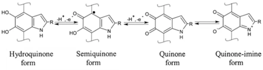
As early as in 1939, Figge reported about the redox properties of eumelanin as a natural reversible oxidation-reduction system and an intracellular redox indicator dye (i.e., its change in color indicates whether redox reactions related to certain enzymes’ activity took place) [20]. Eumelanin was demonstrated to be an efficient electron transfer agent in various reduction-oxidation systems [21]. DHI and DHICA are indeed catecholic subunits, that can be reversibly oxidized into the corresponding orto(o)-quinone forms in two processes of one-electron one-proton removal process [22], [23]. Various redox forms of the building blocks coexist in eumelanin [24] (Figure 1.4).
Figure 1.4 Redox forms of eumelanin’s building blocks, R=H for DHI and R=COOH for DHICA. Reprinted with permission (granted by co-authorship) from Ref. [25].
1.2.4 Supramolecular Structure
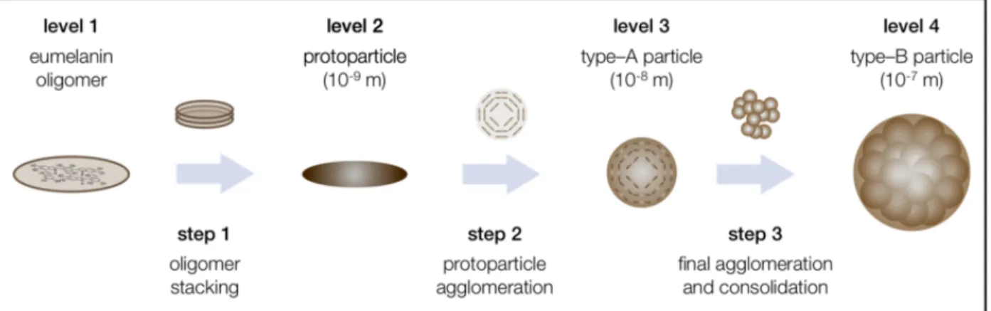
The supramolecular buildup of eumelanin largely determines its properties. Its monomers (DHI and DHICA) form oligomers of up to a few tens of monomers [26]–[31]. The oligomers stack one on the other via π-π stacking; the characteristic d-spacing is of 3.4 Å [26]–[31]. The oligomeric sheets form protoparticles. Such protoparticles then arrange in an onion-like structure and densify into spherical particles and finally undergo aggregation in larger spherical particles [32]. Consistently, Sepia Melanin is constituted of 150 nm-sized aggregates [33] that assemble into granules.
Figure 1.5 Supramolecular buildup of eumelanin. Reprinted with permission (granted by Creative Commons Attribution License of Open Access) from Ref. [32].
Molecular dynamics simulations and first-principles density-functional theory (DFT) calculations were recently used by Antidormi et al. to simulate the polymerization of DHI and DHICA in methanol. Such a work confirmed that the oligomer size of 10 units is the most likely to occur for eumelanin [34]. To gain more insights into eumelanin polymerization, a bottom-up approach was adopted by De Marchi et al. They studied the self-assembled monolayer networks of DHI and DHICA on Au and Ag surfaces using scanning tunneling microscopy (STM) [35], [36].
It was also demonstrated that the supramolecular buildup can be stopped at each level by enzymatic oxidation in vitro [37]. Cations and small molecules are able to influence the supramolecular aggregation of eumelanin [38]. Belitsky et al. recently investigated the ability of small molecules to influence aggregation of dopa-melanin during its polymerization and aggregation (measured considering the acceleration or delay in the appearance of macroscopic particles) [38].
1.3 Other Types of Melanin
1.3.1 Pheomelanin
In presence of cysteine, tyrosine oxidation leads to the formation of pheomelanin. A weakly acidic pH in melanosomes renders pheomelanogenesis favored over eumelanogenesis. Not only red hairs, but also black and brown hairs contain pheomelanin [39]. The ratio of eumelanin/pheomelanin in hairs can be measured by electron spin resonance [40].
Pheomelanin has poor photoprotective properties compared to eumelanin [41]. It acts as photosensitizer, generating reactive oxygen species (ROS) upon UV irradiation [41], [42].
1.3.2 Neuromelanin
Neuromelanin is present only in the brain of humans and large primates. This complicates the ethics of procurement, so that Sepia Melanin is used as a NM simulant [8], [43]. In humans, NM piles up within the dopaminergic neurons of the substantia nigra and in the neurons of the locus coeruleus of the brain [44]. NM is largely depleted in patients with Parkinson’s disease through a selective loss of NM-containing neurons, suggesting that NM may be involved in the neurodegenerative processes of Parkinson’s disease [45]. In Parkinsonian brains, after neuronal death, NM is released in the extracellular environment. As it is insoluble, it can remain for a long time acting as a pro-inflammatory stimulus leading to further neuronal damage [44]. NM may have both a beneficial and a detrimental role (Figure 1.6). An example of the beneficial role is the chelation of iron and other potentially toxic metals with formation of non-redox active species as well as the binding of environmental toxins, such as pesticides. The detrimental role can occur, for example, when high iron levels are present. In such a scenario, NM accumulates iron in low affinity binding sites where iron remains redox active [44], [46].
Figure 1.6 The dual role of NM. Reprinted with permission from Ref. [44].
1.3.3 Melanins in Insects, Microorganisms and Plants
Melanins are present in insects, too, where they play several roles: wound healing, defense reactions, cuticular coloration, and camouflage. Melanins in insects originate from dopamine, hence the term dopamine-melanin [47].
The building block of most (but not all [48], [49]) fungal melanins is DHN, 1,8-dihydroxynaphthalene (often referred to as DHN-melanin)2 (Figure 1.1, L) [6]. Melanins in fungi are located in the cell walls or secreted into the environment, while eumelanins in animals are synthesized and maintained almost entirely as melanosomes as well as granules within melanocytes [50]. In fungi, melanins provide mechanical strength and chemical resistance to the cell, protection against the detrimental effects of UV radiation, high concentrations of salts and heavy metals [51]. They also allow fungi to tolerate extreme conditions, from the Earth’s poles to deserts [52]. The exposure of certain fungi to gamma rays lead to increased production of melanin [53]. Melanized fungi have been found in Chernobyl’s reactors, responding to ionizing radiation with enhanced growth [54]. Such a response lead to the intriguing hypothesis that fungal melanin is able to transduce radiation into metabolic energy in a sort of radiotropism [54]. In pathogenic fungi, melanin contributes to virulence, promoting tissue invasion and inactivation of the plant defense system [55]. Lately, interest in the electrical conductivity of fungal melanin has grown, too [56]. Fungi have been studied for the production of melanin from food or agricultural waste, such as wheat extracts [57]–[60]. However, in such cases, the pigment extraction from cell walls proved to be cumbersome. Recently, Ribera et al. demonstrated a scalable method to obtain eumelanin from a fungus that secretes the pigment in the surrounding liquid environment, thus facilitating its extraction [61].
Bacteria associated with darkly pigmented sponges [62] and other bacteria [63] have been studied as a source of melanin, too.
2 Fungi: “Eukaryotic microorganisms of the kingdom Fungi, that possess cell walls and lack chlorophyll. Some species
are pathogens of humans, animals and plants. Certain fungi are used commercially (e.g. in the production of enzymes and fermented foods).” [529]
Melanins extracted from plants are also based on oligomers: their building blocks can be either conventional eumelanin-like indoles [64] or phenolic units (such as p-cumaric acid for black oat hull [65]). Natural melanins in plants contribute to the defense mechanisms against pathogens and to the strengthening of the cell walls [65]. Melanin can be extracted from waste such as sunflower husk [66], flesh fruit [67] and fruit kernel skin [68].
1.4 Physical and Chemical Properties of Eumelanin
Eumelanin presents a vast set of properties such as UV-visible absorption, radical scavenging, hydration dependent electrical conduction [69], and metal ion chelation [70].
In a biological environment, an anti-oxidant can be defined as ‘‘any substance that, when present at low concentrations compared to that of an oxidizable substrate, would significantly delay or prevent oxidation of that substrate”: radical scavengers, metal ion chelators and reducing agents fall within this definition [71]–[73]. Consequently, eumelanin is often defined as an anti-oxidant [69].
Eumelanin is available from natural sources and environmentally-friendly extraction routes (e.g. from natural fibers, such as fleece from alpaca [74]): eumelanin is therefore considered a carbon-based bio-sourced material.
1.4.1 UV-visible Absorption and Photoprotection
Eumelanin presents a featureless UV-visible absorption spectrum (Figure 1.7), in virtue of which it plays the role of photoprotector [75]. If there had been any gaps in the spectrum, the living organisms deploying eumelanin for protection against UV-radiation would have been left vulnerable to these specific wavelengths of the sun’s radiation [76].
Figure 1.7 Absorbance spectra of synthetic dopa-melanin in water, pH 7 (dotted line) and in dimethyl formamide (dashed line), 0.005% (w/w). Reprinted with permission from Ref. [27].
The main photoprotection path entails that eumelanin converts the energy of the photons into heat (rotational and vibrational energy), in the so called “photothermal effect” (which is the main contribution of the “photoacoustic effect”, so that eumelanin is often defined to have “photoacoustic properties”) [77], [78]. Such a property is not affected by temperature until 80 °C [79].
The featureless absorption spectrum of eumelanins has been explained with the chemical disorder model. According to this model, eumelanin is constituted of several chemically distinct species, i.e. oligomers that differ by number of monomers, type of monomers and polymerization sites. Its broadband absorption spectrum would thus result from the averaging over the spectra of these species (superposition principle) [75]. The inconsistencies of such a model were summarized in a recent study by Chen et al. [76]. Inter alia, the chemical disorder model did not explain the reason for the monotonic increase of the spectrum toward lower wavelengths [76]. Furthermore, interactions among eumelanin oligomers can significantly affect the spectrum so that the superposition principle does not hold [80]. The work by Chen et al. does not stress the focus on the chemical heterogeneity but rather on the geometric disorder: the stacked eumelanin oligomers
differ by size and are randomly oriented, so that peculiar excitonic interactions take place among them [76].
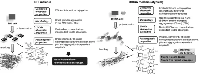
The ratio DHI/DHICA monomers in eumelanin dramatically influences light absorption (as well as other properties such as paramagnetic and redox behavior, particle morphology, surface properties and metal chelation) [81]. These differences can be understood studying synthetic eumelanins made up of only one of the two monomers, DHI-melanin and DHICA-melanin. The oligomers of DHI-melanin are planar, with a high level of stacking and inter-unit π-conjugation. DHICA features carboxyl groups that cause steric hindrance, so that DHICA forms non-planar oligomers, with limited inter-unit π-conjugation [18]. The negative charge of the deprotonated carboxyl groups can further limit efficient stacking, so that rod-shaped assemblies form [77] (Figure 1.8).
Figure 1.8 Differences in structure and properties between DHI-melanin and DHICA-melanin. Reprinted with permission from Ref. [77].
In general, stacking increases the visible absorption [80], so that a higher content in DHICA decreases visible absorption [81], [82]. The light absorbing properties and redox behavior are intimately connected: it has been suggested that the nonradiative decay processes of DHI can lead to DHI in a different oxidation state [83].
1.4.2 Binding of Metals and Organic Compounds
In the fields of dermatology, materials chemistry and physics many scientists have advanced arguments that melanin does not play a merely pigmentary role [84]. Eumelanin can bind both organic compounds [85], [86] and metal ions [70], [87]. The binding capacity of melanin towards metal ions is large but finite [88]: it depends on the cation, on the pH and on the binding site [89]. When eumelanin binds reactive metals, it mitigates their potential role in inducing oxidative stress [70]. A detailed literature review concerning the binding of eumelanin towards metal ions is provided in Article 5 [89].
1.4.3 Radical Scavenging
Eumelanin is a radical scavenger3 [69] and its redox state determines its radical scavenging ability. Eumelanin must be in a reduced state to donate electrons to scavenge oxidative radicals or must be oxidized to receive electrons from reductive radicals [90].
Eumelanin’s photoprotective role is strongly related to the radical scavenging properties. In particular, it has been suggested that the reactive oxygen species (ROS) generated as an inflammatory response to UV radiation may be scavenged by eumelanin [91].
However, eumelanin itself, under UV irradiation, can form ROS [92]–[94], thus explaining the dichotomous role (photoprotective and photodamaging) played by its light-absorbing properties [95], [96]. The factors that determine whether the photoprotective or photodamaging effect of the UV-visible absorption properties prevails are the radiation wavelength [78], [84], the UV exposure time [97], [98] and the pigment’s aggregation [99], [100].
It has been suggested that the main photoprotective mechanism, the photothermal conversion, may take place at long wavelengths, i.e. UVB, i.e. 280–320 nm, UVA, i.e. 320–400 nm and visible [78]. Conversely, at short wavelengths (UVC, i.e. 220–280 nm) eumelanin may become cytotoxic by generating radicals [84].
3 A radical is a molecular entity with an unpaired electron; in this thesis, the term “free radical”, belonging to an old
In the human eye, eumelanin has a supportive role in the visual process: it reduces intraocular light scatter [101]. However, a prolonged exposure to UVA could lead to its photodegradation and photoaging [92], with the loss of its radical scavenging abilities [102], [103]. In that case, eumelanin of the retinal pigment epithelium (RPE) can even become an efficient pro-oxidant [97]. It was proved that small particles of natural eumelanin are involved in UVA-induced photochemical processes that can lead to the DNA damage in skin cells [104]. Conversely, large particles efficiently dissipate UVA energy through the nonradiative decay processes (the aforementioned photothermal conversion). It is thus clear that aggregation limits ROS photoproduction related to UV irradiation, suggesting that the aggregation of the biopigment and the integrity of melanosomes in tissues is another factor determining whether the photoprotective or the photodamaging behavior prevails in nature [99], [100].
The radical scavenging property, too, is influenced by the DHI/DHICA ratio [81]. In DHICA-melanin, relatively homogeneous radical species, spatially confined within restricted segments of the polymers, are present [81]. On the contrary, DHI-melanin is characterized by a large variety of radical species that could be generated within the delocalized π-electron systems [81]. DHICA-melanin was also found to be a more efficient hydroxyl radical-scavenger: its aggregates would indeed be more accessible to radicals compared to compact π-π stacked DHI-melanin [81]. Recently, the ability to scavenge radicals by natural and synthetic eumelanins, as well as by their precursors DHI and DHICA, has been quantified [105]. Such ability is higher in the monomers with respect to synthetic DHICA-melanin and DHI-melanin, as polymerization involves the oxidation of some building blocks from the catechol to the quinone form, reducing the amount of OH groups involved in radical scavenging [105]. In agreement with the literature, DHICA-melanin proved to have superior radical scavenging than DHI-melanin, as explained above [105].
Furthermore, it was proved by Agapito and Cabral that the enthalpy of formation of the OH· radical from the hydroxyl groups of catechol subunits is lower in the DHI monomer than in DHICA. The lower “energetic cost” for OH· formation in DHI than in DHICA could potentially explain why DHI-melanin features superior ROS generation rates than DHICA-melanin [42].
The differences in ROS generation between DHI and DHICA could also tentatively explain why nature “opts” for an enzymatically-controlled pathway to DHICA instead of leveraging on the spontaneous, biologically “low cost” process of DHI formation (Figure 1.3) [81].
1.4.4 Electrical Properties
Eumelanin is one of the first biomaterials whose electrical properties have been investigated [106]. Eumelanin’s electrical conductivity ranges from 10-3S cm-1in the fully hydrated state to 10-8S cm
-1in the dehydrated state [107], [108].
It was defined a “mixed semiconductor” in 1970 by Powell and Rosenberg, with a proton to electron ratio of ≈1:2: the experiment was carried out on synthetic melanin (dopa-melanin) powders, placed in a sandwich cell made of platinum foil and such a ratio did not change in the hydration range 10 – 35% wt. [106]. Bistable switching behavior depending on eumelanin’s hydration was reported by McGinness et al. in 1974 [109], leading to the definition of eumelanin as an amorphous semiconductor. The investigation of the parameters related to the amorphous semiconductor model followed: optical band gap [110], band structure and charge carrier density [111], [112].
In 2012, a new model was suggested: a hydration-dependent electronic–ionic conduction [113]. The comproportionation equilibrium, already known since the 70s [22] (Figure 1.9), was used to explain the conductivity increase with the amount of water. The presence of two indole moieties in different oxidation states (catechol and quinone) together with adsorbed water would bring about the (thermodynamically favorable) generation of semiquinone moieties and protons, responsible for the doping of electrons and ions into the system (Figure 1.9) [113].
Figure 1.9 Comproportionation equilibrium. Reprinted with permission (granted by noncommercial educational use) from Ref. [113].
Further experiments involving D2O (heavy water, an oxide of the hydrogen isotope deuterium), in
presence of which the forward reaction of Figure 1.9 is more favored than the backward one, were provided to corroborate such model [114].
However, the same authors who postulated the hybrid electronic–ionic conduction theory very recently demonstrated that the main component of eumelanin’s hydration dependent conductivity is actually protonic [115].
Other important properties are photoconductivity [110], [113], [116]–[118] and permanent paramagnetism [119]. The photoconductivity of eumelanin has been explained within the frame of the comproportionation equilibrium, too [120]. DHI-melanin was recently demonstrated to be a photocatalytic material, capable of reducing oxygen to hydrogen peroxide upon irradiation [121].
1.4.5 Other Proprieties of Eumelanin
Eumelanin is hydrophilic [122]: Sepia Melanin can absorb an amount of water equal to its mass in a water vapor saturated environment in 24 hours [123]. Studies on the water sorption of synthetic melanin (dopa-melanin) thin films, exposed at different vapor pressures of H2O and D2O, revealed
that water is taken up thoroughly through the films [124].
Eumelanin is biocompatible [125] and bioresorbable [126]. Tyrosine-melanin implants in vivo located nearby a peripheral nerve tissue caused a foreign body response similar to silicone and were almost fully eroded and bioresorbed after 8 weeks [126].
1.5 Context of the Investigation: Green electronics, Compostability
and Additives for Packaging
1.5.1 Organic Electronics, Bioelectronics and Green Electronics
Electronics have become indispensable in our daily routine. A great part of the electronic equipment that surrounds us belongs to what is known as conventional electronics, based on inorganic materials such as silicon and gallium arsenide [127].
However, with the life of the electric and electronic equipment becoming shorter and shorter, two major issues arise for both the scientific community and municipalities: the increasing amount of Waste of Electrical and Electronic Equipment (WEEE) and the depletion of natural resources [127], [128]. The United Nations define sustainability as the ability of satisfying one generation’s needs “without compromising the ability of future generations to meet their own needs” [129]. It is thus evident that the electronics sector lacks sustainability, at least so far [127]. Consequently, great
attention has been given to green (sustainable) electronics in the last years, having as core values (i) the use of abundant and low-cost precursors, (ii) the use of biodegradable materials, leveraging on processing routes that (iii) lack toxic solvents as well as toxic waste and (iv) are low cost. Sustainable electronics envisages devices with low embodied energy, i.e. “energy spent in the production phase and stored in the inner constituents” [128].
Organic electronics is based on organic (carbon-based) materials as the core semiconductor element. Such materials, polymers or small molecules, are characterized by conjugation, i.e. alternance of single and double carbon-carbon bonds. Organic electronics features solution processability, printability and tunable optical and electronic properties with chemical synthesis [130]. The feature of being solution-processable (printable) adds to organic electronic devices the further advantage of lower embodied energy with respect to their inorganic counterparts, that are processed by high-vacuum and high-temperature techniques [128].
Organic bioelectronics is a branch of organic electronics, which has the specific aims of “implementing biocompatible materials in different electronic architectures at the interface with living tissues” [131]. The main challenge of organic bioelectronics is to transduce signals between biology, featuring predominantly ionic signals, and semiconductors electronics, featuring electronic signals [132].
1.5.2 Electrochemical Metallization Memory Cells: towards Green Electronics
Electrochemical metallization memory cells (ECMs), also known as conductive bridge random access memories [133], belong to Resistive Random Access Memories (ReRAMs) [134], a subgroup of non-volatile memories, NVM [135]. ECM working mechanism entails the formation, under bias, of conductive filaments. As the active electrode dissolves, cations migrate through a solid electrolyte (ion conductor) and accumulate on the counter electrode (CE), where the conductive bridge starts growing [135]. Once the filament grows over the entire inter-electrode distance, it connects one electrode to the other, causing the system’s resistivity to plummet [135]. The advantages of ReRAMs are good reliability [133], low power consumption [134], [136]–[138], fast response time [133], [136], multilevel data storage [133], [136], [139], and high density [134]. Albeit initially constituted entirely of inorganic materials, ECMs have witnessed the use of organic synthetic materials for the ion conductive layer since 2011 [140]. The use of natural (bio-sourced)organic materials ensued in the last few years [141]. Organic and polymeric switching memories feature flexibility [142]–[148], optical transparency [144], printability [142], [149], electrical and physical properties tunable by means of molecular design and chemical synthesis [150], [151], which provide a potential for high density memory applications [135], [152]. Conductive bridge memories have been obtained using conductive polymers well-established in the field of organic electronics, polymers loaded with salts, small molecules and dyes [153]–[160].
Interest regarding biomaterials and bio-sourced materials for memories (Bio-ReRAMs) has increased, too [141]. Examples of natural materials used as ion conductors in ECMs are: dead leaves [161], lignin loaded with Au nanoparticles [162], eggshells [163], pectin extracted from orange peels [164], a DNA-based biomaterial [165], [166], electro-spun cellulose fibers [167], nano-cellulose [168], gelatin [169], [170], Aloe vera gel [171], [172], chicken [173] and duck [174] egg albumen, silk fibroin [175]–[179], glucose [180] and carrageenan, a polysaccharide from seaweeds [181]. Natural materials investigated as dielectrics for ECMs are chicken egg albumen [182], gelatin [183] and human hair keratin [184]. Further examples of biomaterials for Bio-ReRAMs can be found in the review by Raeis-Hosseini and Lee [141].
1.5.3 Biodegradation as an Option for Waste Disposal
At the end of life of materials, their biodegradability plays a key role in the challenge to achieve sustainability [127], [185]. Biodegradation is “the metabolic breakdown of materials into simpler components by living organisms” [186]. When a material is defined biodegradable, the conditions and the time range have to be specified, too [187].
In municipal waste management, composting (aerobic biodegradation, i.e. in presence of O2) has
been identified as a valuable disposal pathway [188]. During composting, the temperature is of ~ 60 °C is reached because of the microbial activity [189]. The ultimate end products of composting are CO2, minerals and water [187]. Composting has several advantages, such as reducing the
volume of waste for landfilling and incineration, avoiding methane production and release in the atmosphere (differently from anaerobic biodegradation) and providing a product that can be used for crop growth. Standard test methods have been developed for plastic materials used for food packaging and serving in order to evaluate their compostability, that is, biodegradability in composting conditions (e.g. CAN/BNQ 0017-088/2010 in Canada, ASTM D5338 and ASTM D6400 in the USA, EN 13432 and EN 14995 in the European Union as well as ISO 14855-1:2005).

![Figure 1.3 Biosynthetic pathway of eumelanin. Reprinted with permission from Ref. [14]](https://thumb-eu.123doks.com/thumbv2/123doknet/2328851.31158/30.918.238.689.103.808/figure-biosynthetic-pathway-eumelanin-reprinted-permission-ref.webp)
![Figure 1.6 The dual role of NM. Reprinted with permission from Ref. [44].](https://thumb-eu.123doks.com/thumbv2/123doknet/2328851.31158/33.918.146.799.652.975/figure-dual-role-nm-reprinted-permission-ref.webp)

![Figure 2.1 DMSO-melanin’s building blocks. Reprinted with permission from Ref. [311].](https://thumb-eu.123doks.com/thumbv2/123doknet/2328851.31158/55.918.302.613.174.315/figure-dmso-melanin-building-blocks-reprinted-permission-ref.webp)
![Figure 4.1 Basic AFM set-up. Reprinted with permission from Ref. [320].](https://thumb-eu.123doks.com/thumbv2/123doknet/2328851.31158/59.918.259.669.422.689/figure-basic-afm-set-reprinted-permission-ref.webp)

