Combined Radioimmunotherapy and Radiotherapy of Human Colon Carcinoma
Grafted in Nude Mice1
Franz Buchegger,2 Anamaria Rojas, Angelika Bischof Delaloye, Charles-André Vogel, René-OlivierMirimanoff,
Philippe Coucke, Lin-Quan Sun, Serge Raimondi, Julie Denekamp, AndréPèlegrin, Bernard Delaloye, and
Jean-Pierre
Mach
Division nf Nuclear Medicine [F. B., A. B. D.. C-A. V., L-Q. S., B. D.] and Department of Radio-Oncology ¡F.B., R-O. M., P. C.], Centre Hospitalier Universitaire Vaudois, CH 1011 Laminine, Switzerland; "InstituÃ- de Radioph\siÃ-¡ueAppliquée" IS. R.l, CH 1015 Lausanne, Switzerland; Cancer Research Campaign Gray Laboratory, Mount Vernon Hospital Nortliwood HA62 YR, United Kingdom [A. R., J. D.J: and Institute of Biochemistry, University of Lausanne. CH 1066 Epalinges, Switzerland ¡A.P., J-P. M./
ABSTRACT
The effect of combined radioimmunotherapy (RIT) and fractionated external beam radiotherapy (RT) was assessed in two human colon cancer xenografts, Col 12 and LS174T in nude mice. These tumors were selected for being resistant to RIT alone, as is usually the case in the clinical situation. Tumor-bearing mice were treated with a combination of five X-ray fractions over 5 days followed by RIT with two doses of 1.5 mCi 131I-labeled anticarcinoembryonic antigen monoclonal antibody F(ab')2. In Col 12 and LS174T, RIT alone achieved a regrowth delay similar to that of fractionated RT with total doses of 28 and 26 Gy, respectively. In both tumor types, an additive therapeutic effect, measured as increased regrowth delay or local control, was observed when combining RT of different dose levels with RIT. Normal tissue responses were assessed by monitoring acute peak skin reactions and blood cell count. Bone marrow depression for the combination treatment was similar to that of RIT alone; relative to skin, at equitoxic levels, no mice bearing Col 12 tumors were locally controlled with a 32 Gy RT dose alone, while this RT combined with RIT gave a local control of 100%. These studies show a therapeutic benefit when external beam RT is combined with RIT.
INTRODUCTION
Improving local tumor control has a significant impact on patient survival, especially in tumors of low metastatic potential (1). Follow ing or preceding surgery, adjuvant external beam RT3 improves local control in a significant number of patients suffering from breast, colorectal, and other carcinomas and produces relatively mild side effects. Radiosensitive organs in the radiation field, however, may limit the radiation dose in certain situations and therefore compromise the efficacy of RT. In addition, distant micrometastases that already exist at the time of adjuvant RT will not be affected by it and may be a determinant for the patient's prognosis.
A further increase in overall control rate could be achieved with combined therapies which have different normal tissue toxicities and if one modality is also effective against micrometastatic disease. Combination therapies of RT, RIT, and cytostatic drugs have been used with some success in patients with liver cancer (2). Additive therapeutic effects of RT and RIT have, however, not been demonstrated experimentally.
The therapeutic index of RJT administered systemically depends on the amount of antibody that can be localized in the tumors versus its bone marrow toxicity. In patients, mean radiation doses that can be delivered to solid tumors by i.v. injected radiolabeled antibodies are generally
Received 7/5/94; accepted 10/31/94.
The costs of publication of this article were defrayed in part by the payment of page charges. This article must therefore be hereby marked advertisement in accordance with
18 U.S.C. Section 1734 solely to indicate this fact.
' This work was supported by the Swiss Foundation for Scientific Research Grant 31—31238-91, the foundation "Swiss Research Against Cancer" (Akt 312). and the Swiss Robert Wenner Cancer Research Award 1991.
2 To whom requests for reprints should be addressed.
3 The abbreviations used are: RT, radiotherapy; R1T, radioimmunotherapy; RD, tumor regrowth delay; TDT. tumor (volume) doubling time; CEA, carcinoembryonic antigen; % ID/g, percentage of injected dose/g (tissue); mAb, monoclonal antibody.
estimated to be between 10 and 20 Gy when no bone marrow support is used (3-5). Such radiation doses are insufficient to produce significant regression of large nonhematopoietic tumors. However, some colorectal cancers show relatively high uptakes of radiolabeled antibodies of more than 0.05% ID/g (6). In such tumors, radiation doses might be as high as 60 Gy and could explain some anecdotal tumor remissions (7). Similarly, because of better vascularization and accessibility, micrometastatic dis ease can show higher uptake than large tumor masses (8). Such measure ments are, however, still sparse and need to be confirmed both experi mentally and clinically.
Here we have studied the effect of RIT combined with RT on two human colon cancer xenografts, Col 12 and LS174T, which, due to their relatively low antibody uptake, are resistant to RIT alone and represent quite well the average clinical situation. The experiments address the question of the complementarity of the two radiation treatments. Although the mechanism of cell kill by RT and RIT is in both cases through ionizing radiation, it has not been shown whether their combined use would yield an additive therapeutic effect. The interaction of fractionated external beam RT with RIT was therefore addressed and a therapeutic comparison made relative to two fast-proliferating normal tissues, i.e., skin and bone marrow.
MATERIALS AND METHODS
Tumor Grafting. All experiments in nude mice were performed according
to Swiss legislation and approved in 1993 by the official committee of surveillance of animal experiments. Female nude mice (Swiss nu/nu, produced at the Institute) aged 7-9 weeks received transplants on the middle of their backs at 2 cm from the tail with a volume of 30 mm1 of minced Col 12 or LSI 74T tumor. After 6-10 days, during which diminution of the graft size was observed, growth started and at 14 days about 90% of the tumors grew exponentially. These mice were randomized into the different treatment arms and therapy was initiated 2-3 weeks after transplantation of Col 12 tumors, at 18 days for LS174T.
Irradiation of Tumor Transplants. X-rays were generated by a Philips
RT 250 operating at 200 kV and 20 mA. The beam was filtered with 0.5-mm copper (half-value layer = 1-mm copper). Up to 6 mice per irradiation were restrained in 3-mm lead jigs designed with a cutout 20 X 15 mm to expose their lower dorsum (9). The jigs were placed in a perspex box with an additional lead shield with 60- X 17-mm openings; in each field 2 mice were exposed tail-on-tail. This set-up gives minimum scatter to the animals placed at 52.5 cm from the source. The X-ray beam hits the tumors tangentially to the dorsum. Dose rate was 0.64 Gy/min and dose variation was ±5.5% for an 8-mm tumor. To maximize dose homogeneity, tumors were exposed from opposite sides at alternate days with a regimen of five fractions over 5 days. Care was taken to have the whole area of transplantation (including the site of needle penetration) in the irradiation field. A control dosimetry was performed
in vivo: 12 microdosimeters (10, 11) were implanted in 5 tumors and these
were irradiated with 2 fractions of 8 Gy from alternate sides. For calibration, 6 dosimeters were irradiated in air with 16 Gy. For the tumors exposed in vivo the measured dose was 16.0 ±1.8 Gy.
Monoclonal Antibodies. F(ab')2 fragments of 3 monoclonal anti-CEA
antibodies (mAb 35, CE25-B7, and B93; Ref. 12), all of the IgGl subclass and of irrelevant control mouse IgGl secreted by the mouse myeloma P3x63, were 83
used. The three mAbs are directed against three independent epitopes of CEA: gold 2, 4, and 1 (13), respectively. The antibodies are highly specific for CEA and do not bind to radiolabeled NCA 55 or NCA 95 (14) or other granulocyte glycoproteins (15). These results were confirmed on tissue sections and by fluorescence-activated cell sorting analysis in an international exchange of antibodies (16).
F(ab')2 fragments were obtained from intact mAbs and control IgGl as described previously (12).
Radiolabeling of Fragments of mAbs and Control IgG. F(ab')2 of
pooled anti-CEA mAbs and control IgGl were labeled using the lodo-Gen method. Final specific activities were between 9 and 11 mCi 131I/mg protein. Radiolabeled protein was separated from free iodine by filtration on Sephadex G25 columns (Pharmacia). Immunoreactivity was determined as described (17): in a direct binding assay on CEA insolubilized on CNBr-Sepharose (Pharmacia) binding of radiolabeled mAb fragments was 80.3 ± 4.5% (1.3 ±0.7% binding on control protein-Sepharose), while that of radiolabeled control F(ab')2 was 1.5 ±0.8%. Analytic chromatography of ml-labeled proteins on Sephadex G200 gave a single peak, no detectable aggregates, and less than 2% free iodine.
Radioimmunotherapy and Combined Therapies. Radioimmunotherapy
consisted of 2 doses of 1.5 mCi each of 131I-labeled anti-CEA mAb F(ab')2 (~150 jn.g protein) injected via the tail vein. The first dose was administered on the last day of RT, followed by a second injection 4 days later. In the group treated with antibody alone, RIT was initiated the same day as RT in the other groups. The whole-body half-life of radiolabeled mAb and control F(ab')2, measured by placing the mice in a dose calibrator, was 11.3 ± l h and 11.5 ±0.7 h, respectively, for the first 2.5—3days. Then excretion gradually decreased, leading to a final half-life of about 24 h.
Antibody Localization in Tumors after RT. Tumor-bearing nude mice
were irradiated with 16 or 25 Gy in 5 fractions over 5 days. They were given injections 8 h after the last irradiation with 2 fid 131I-labeled mAb and I25I-labeled control F(ab')2. For some mice, unlabeled mAb F(ab'), was added to a total of 150 /xg, corresponding to the amount used for RIT. All mice were dissected 12 h after injection when maximum tumor uptake has been observed previously (18). Biodistribution was measured as follows: mice were killed by CO2 inhalation and 0.5 ml blood was taken from the vena cava. Tumor, normal tissues, and carcass were dissected, weighed, and counted in a dual-channel gamma counter. Tissue radioactivity per g was expressed in % ID.
Tumor Assays. The effect of treatment was assessed using both a local
control and RD assay. Tumor size was measured initially twice per week and later once a week: the three orthogonal diameters were measured with a caliper and tumor volume V was calculated by this formula:
v =
X d}Time to grow three times (Col 12) and six times (LS174T) above initial treatment size was calculated and RD was obtained by subtraction of the growth delay in untreated mice. (A few of the LS174T tumors started to ulcerate before they reached this end point volume. A third diameter, 60% of diameter d2, was used in these mice for the volumetric calculations.)
In the growth delay analysis, only regrowing tumors were included. Growth delay was plotted against dose (from external irradiation) as the mean and SE for the mice in each dose group. The curves (±95% confidence intervals) were obtained by fitting a generalized logistic equation to the growth delay for each mouse against the dose, using nonlinear least-squares regression (19).
Complete or partial regressions were assessed first twice, then once per week. The absence of palpable tumor mass 180 days after therapy was taken as a safe indication of local control, since no relapses were observed at later time points. The percentage of controlled tumors was plotted for each group and the data were fitted by logistic regression.
Only two mice in the whole series developed inguinal métastases(Col 12 tumor-bearing mice). Their macroscopic aspect and high amounts of CEA measured in the extracts (91 and 153 fig CEA/g) confirmed their human colorectal origin. Both primary tumors in the two mice regrew also and all mice contributed to the local control end point.
Tumor (volume) doubling Time (TDT) was calculated for each mouse from the start of tumor regrowth until they reached either 10 times this volume (Coll2) or the endpoint of treatment (LS174T).
Normal Tissue Side Effect Evaluation. Peak local skin toxicity of RT was
evaluated by inspection three times per week for 5 weeks: I, faint redness; II, partially necrotic irradiation field; and III, completely necrotic irradiation field. Occasionally, progressive ulcerative skin disease was observed after RT. Thus, 2 mice died with progressive skin disease 38 and 48 days after 32 and 35 Gy RT, respectively, and were excluded from further evaluation.
Toxicity following RIT was judged by weight measurements three times weekly for 3 weeks, then once weekly for 2 months. Formation of petechiae was controlled also on three inspections per week. Peripheral WBCs, hemo globin, and platelets were counted on day 13 after the first radiolabeled mAb injection (corresponding to the time when petechiae indicated the nadir of platelet depression). WBCs were counted in 15 jul blood (obtained from the tail vein) diluted 1/10 in Turck solution, hemoglobin, and platelets in a Coulter System 9000 counter (Serono, Baker Diagnostics). No late toxicity was ob served in mice treated with radiolabeled mAb alone but one mouse died 22 days after injection with extensive petechia and was not évaluablefor tumor growth control.
Statistical Analysis. Tumor regrowth delay after combined therapies was
compared with that after RT and RIT alone or after RT combined with 131I-labeled control F(ab')2 using the Student-Neuman-Keuls procedure: an ANOVA was first performed that showed for the three experiments with tumor relapses an F P <0.0001. In a second step, pairs of groups were sorted, reaching a significance level of 0.05. For analysis of the number of long-term complete remissions in combined treatments compared to their absence in groups treated with RT or RIT alone, the ^ test was used.
RESULTS
Tumor Size Evolution and End Point Analysis. In all three
experiments with Col 12 tumors, RT of either 16, 25, or 32 Gy (given in 5 fractions) combined with RIT (Table 1 and Fig. 1, b-d, respectively) showed an additive therapeutic effect measured as increased regrowth delay and/or local control (Fig. 2).
Transient regression and tumor regrowth in all mice were observed when Col 12 tumor transplants were treated with irradiation alone using doses of 16, 25, 32, and 35 Gy (Table 1; Fig. la). After irradiation of 50 Gy, 2 of 6 tumors regrew with a delay of 86 and 151 days while complete long-term remissions were observed in four mice.
RIT alone gave a mean RD of 34 days in 12 mice from three experiments. In two control groups that received 16 or 25 Gy RT
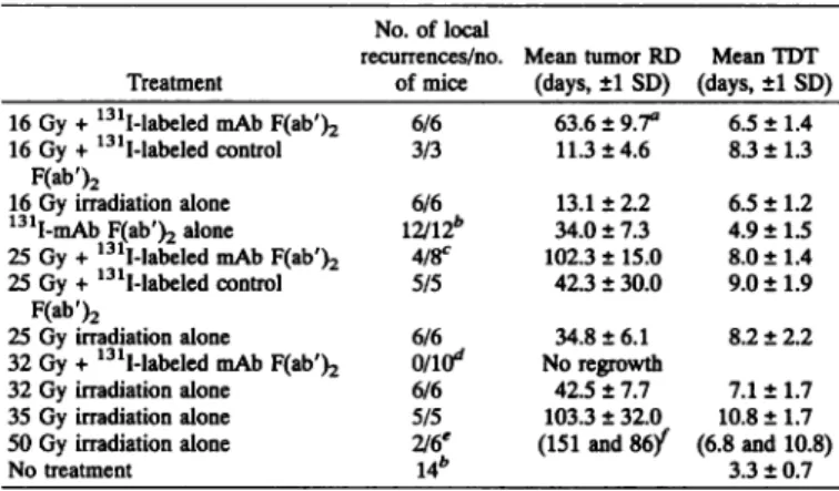
Table 1 Mean (±I SD) of tumor RD and TDT of colon cancer Col 12 after RT, RIT, or combined treatments
No. of local
recurrences/no. Mean tumor RD Mean TDT Treatment of mice (days, ±1SD) (days, ±1SD) 16 Gy + 131I-labeled mAb F(ab')216 Gy + 131I-labeled controlF(ab')216 Gy irradiation alone131I-mAb F(ab')2 alone25 Gy + 131I-labeled mAb F(ab')225 Gy + 131I-labeled controlF(ab')225 Gy irradiation alone32 Gy + 131I-labeled mAb F(ab')232 Gy irradiation alone35 Gy irradiation alone50 Gy irradiation aloneNo treatment6/63/36/612/12*4/8c5/56/6onff16/65/52/6'14*63.6 ± 9.7"11.3 + 4.613.1 ± 2.234.0 ± 7.3102.3 ± 15.042.3 ± 30.034.8 ±6.1No regrowth42.5 ± 7.7103.3 + 32.0(151 and 86/6.5 +8.3 +6.5 +4.9 +8.0 ±9.0 +8.2 +7.1 ±10.8 +(6.8 and3.3 +1.41.31.21.51.41.92.21.71.710.8)0.7 a Tumor regrowth delay is defined as the increased interval from treatment initiation
to reach a tumor size of three times the initial volume in treated mice as compared to untreated controls. (A mean delay of 4.7 days observed in untreated mice was subtracted.) Mean RDs were calculated only for mice that had tumor relapses.
Pooled results from three experiments.
' Four of 8 mice had no tumor relapse 180 days after therapy and this remained the case up to 360 days.
Ten of 10 mice were without a tumor relapse 180 days after therapy and this remained the case up to 270 days.**Four of 6 mice were without tumor relapse 180 days after therapy.
-^Numbers in parentheses, individual data of two mice with recurrences. 84
Fig. 1. Tumor size evolution (mean ±1 SE for 5-6 animals/group, except when stated differently) in mice transplanted with colon cancer Col 12. Ar rows, day of first treatment (day 14, a-c; day 21, d). a, treatment with external beam RT alone of 16 (•), 25 (A), 35 (•),and 50 Gy (O) and untreated mice (D). Four of 6 mice had no tumor relapse after 50 Gy RT. b, combination of 16 Gy RT followed by RIT (•)in comparison to single modality treatment of 16 Gy RT (•)or RIT (A, n = 9) and a control group of 3 mice receiving 16 Gy RT followed by two injections of nll-labeled control F(ab')2 (O). c, combination of 25 Gy RT followed by RÎT(•, n = 8) in comparison to 25 Gy RT alone ( •) or R1T alone (A, n = 9) and 5 mice treated with 25 Gy RT followed by two injections of '31I-labeled control F(ab')2 (O). In the group of mice treated with RT + RIT, tumors in 4 of 8 mice showed complete long-term remissions, d, combination of 32 Gy RT followed by RIT (•,n = 10) ¡ncomparison to 32 Gy RT alone (•)or RIT alone (A, n = 3).
followed by two injections of 131I-labeled control F(ab')2, the mean RDs were only slightly increased as compared to RT alone (Table 1; Fig. 2a). As another control, three mice treated with 32 Gy RT received two additional injections of 150 jj,g each of unlabeled mAb F(ab')2. Tumor remissions and relapses in these mice were superimposable on those observed in the three mice treated with 32 Gy RT alone and the six mice are presented as one group (Fig. Id).
RDs observed with Col 12 tumors were analyzed with the Student-Neuman-Keul test. The combined therapies of 16 and 25 Gy RT with RIT gave significantly longer RDs (P < 0.05) than all of the corre sponding treatments of RT alone (at identical dose level) or RIT alone, or also RT combined with 131I-labeled control F(ab')2. Additionally, the number of complete long-term remissions in 4 of 8 and 10 of 10 mice after the combined treatment of 25 and 32 Gy RT and RIT, respectively, were significantly different from the groups that had only transient tumor remissions after RT alone (at an identical dose level) or RIT alone, when analyzed with the x2
test-After RT, tumor growth (expressed as TDT) was progressively slower, depending on the radiation dose (Table 1): tumor growth plotted versus no treatment and RT of different doses gave a correlation coefficient of r = 0.8. TDT after RIT alone was less affected than after isoeffective RT alone. Additionally, RIT added to RT did not change significantly the TDT observed after the corresponding RT alone.
Tumor LS174T, after a latent period during which the size of the transplants regressed, grew very rapidly (doubling time, 2.1 days) and the mean tumor size at treatment initiation 18 days after transplanta tion was larger (90-150 mm3 in the different groups) than for the Col 12 transplants. The tumor size progressed further during treat ment, reaching on average about four times the initial size before growth arrest or tumor regression was measured. After 25 Gy RT or RIT alone, only tumor growth arrest without size diminution was found (Fig. 3). The LS174T tumor end point was therefore defined as growth to six times the initial size. This volume was reached on average after 4.5 days in untreated mice.
The combined treatment of LS174T tumors with 25 Gy RT and RIT produced a significantly longer tumor regrowth delay than the indi vidual treatments of RT or RIT alone, or RT followed by two injections of 131I-labeled control F(ab')2 (Table 2 and Fig. 4a, Stu-dent-Neuman-Keuls test). After 50 Gy RT alone, in 3 of 5 mice tumors either completely disappeared (n = 2) or remained very small (n = 1) 180 days after therapy (Fig. 4b); in the 2 remaining mice a RD of 72 and 93 days was observed.
Toxicity of the Treatments. Three types of toxicity were observed as side effects of RT or RIT: local skin toxicity was found after RT and hematological toxicity and some early weight loss after RIT. No local skin toxicity was observed after 16 or 25 Gy RT alone or when combined with RIT or 131I-labeled control F(ab')2. Following treat ment with higher doses of RT, skin lesions occurred in a dose-dependent manner (Table 3). They appeared about 14 days after treatment initiation and healed after an additional 2-3 weeks. Of 18 mice, 2 treated with 32 or 35 Gy RT alone developed progressive ulcerative skin disease, leading to death after less than 50 days, and could not be evaluated. There is no indication that irradiation from RIT when combined with RT produced additional skin toxicity as compared to RT alone at corresponding doses.
Bone marrow toxicity occurred in mice treated with radiolabeled mAb or radiolabeled control F(ab')2. Petechiae were observed be tween 11 and 14 days after the first mAb injection in 10 (53%) of 19 mice treated with RIT alone; 1 of them died 22 days after the initiation of therapy and was excluded from further analysis. Petechiae were observed in only 4 (10%) of 41 mice after combined treatment consisting of different radiation doses of RT followed by RIT or injection of 131I-labeled control F(ab')2.
The blood of mice treated with radiolabeled proteins from experi ments 2, 3, and 4 was analyzed 13 days after the beginning of treatment (when petechiae were strongest). Platelets decreased to a mean of 190-320 x 103/mm3 in mice treated by injection of radio-labeled mAb or control F(ab')2 (Table 4), compared to 1300 X IO3/ mm3 in untreated mice. Individual mice with discrete to extensive petechiae had platelet values of 120-200 X 103/mm3. WBCs dropped
85
to a mean of a 270-780 cells per mm3 with a large scatter (Table 4) while untreated tumor-bearing nude mice had a mean of 11,000/mm3. Hemoglobin dropped to about 55% of the normal level. In conclusion, in the group treated with RIT alone the drop in peripheral WBCs was slightly less but the percentage of mice with petechiae was higher as compared to mice with combined treatments.
A small weight loss (<10%) occurred in many mice 4-6 days after the second injection of radiolabeled mAb or control IgG fragments, as observed previously (20). Weight gain was recovered quite rapidly, generally within 1 week (results not shown).
Antibody Uptake after RT. In a double-labeling experiment, tu mor uptake of 125I-labeled mAb F(ab')2 was measured 12 h after injection and compared with coinjected 131I-labeled control F(ab')2. For Co 112, tumor uptake of about 11% ID/g was observed as reported previously (18). This was the case after injection of either low amounts of antibody fragments (5 ju,g) or high amounts (150 p,g) that correspond to the amounts used for RIT (Table 5). Since three mAbs directed against different CEA epitopes were used, only 50 jug of the individual mAb fragments were used against each individual epitope. A tendency (not significant) for slightly enhanced tumor uptakes of
16% and 14% ID/g was observed after 16 or 25 Gy RT, respectively, but only when low amounts of antibodies were injected.
1000 125 ID N « 100 "e x
to
o
75 § 50» *
25 co I 4/8.' O , i r'-V' , ; : ! I 10 20 30 40 50 60 70 80 1000 750-N 500 O 3 250-—i— 20 40 60 80 100days after tumor transplantation
120
Fig. 3. Tumor size evolution (mean ±1 SE for 5-6 animals/group, except when stated differently) in mice transplanted with colon cancer LS174T. Arrows, day of first treatment 18 days after transplantation, a, treatment with external beam RT alone with total radiation doses of 25 (•),35 ( 0 ), and 50 Gy (O) and a control group of untreated mice (D). Three of 5 mice had no tumor relapse after 50 Gy RT). b, combination treatment of 25 Gy RT followed by RIT (•)in comparison to 25 Gy RT alone (•)or RIT alone (A) and 3 mice treated with 25 Gy RT followed by 131I-labeled control F(ab')2 (O) and untreated mice (D).
20 40
Total X-ray dose
60 80
(Gy)
Fig. 2. Dose-response curves for growth delay to three times initial size (a) and local control (b) for Col 12 tumors plotted against X-ray dose (RT) with or without RIT: RT alone (O), RT + RIT (A), RT + I31l-labeled control F(ab')2 (0). Note that regrowth delay data include only mice that actually had tumor relapses; thus, for 25 Gy RT combined with RIT, only 4 of 8 mice relapsed while the other 4 mice had complete long-term remissions followed for 360 days. Dashed lines (a) and error bars (b), 95% confidence limits.
For LS174T, mAb F(ab')2 uptake into tumor was 10.6% ID/g when low amounts were injected. Under the conditions used here for RIT (injection of 150-/MgmAb fragments), antibody uptake into tumor was lower, 5.5% ID/g, whether pretreatment of 25 Gy RT was used or not (Table 5). Overall, we interpret the biodistribution results obtained with both types of tumors in the sense that no indication for diminished antibody localization is observed after irradiation with 16 or 25 Gy RT.
DISCUSSION
Two types of human colorectal cancer xenografts in nude mice were treated by combinations of external beam RT with RIT. An additive therapeutic effect of the combined treatments compared to size-matched controls was shown: a cytostatic effect of RIT alone similar to that of a 26 or 28 Gy RT could be added to 16, 25, or 32 Gy RT to give an increased combined effect similar to RT alone of between 35 and 50 Gy.
Colon cancer Col 12 xenografts grew relatively slowly and were still relatively small at therapy initiation. However, the long period of 2-3 weeks before therapy provides plenty of time for vascularization, since it has been shown that significant numbers of blood vessels develop as early as 4 days after tumor inoculation in nude mice (21). 86
Additionally, small tumor transplants give a higher antibody uptake per g than larger ones (20, 22), possibly due to the fact that tumors of <100 mm3 show a good blood perfusion, irrespective of whether the tumor bed is well or badly vascularized (21). While the results obtained with the small Col 12 xenografts probably cannot be extrap olated directly to large tumors, they are of interest if we consider RIT combined with RT in an adjuvant postoperative setting in patients with minimal disease.
Table 2 Mean (±1SD) of tumor RD and TDT of colon cancer LSI 74T after RT, RIT. or combined treatments
Table 3 Skin damage in mice treated with RT alone or RT combined with RIT
Treatment
No. of local
recurrences/no. Mean tumor RD Mean TDT of mice (days, ±1SD) (days, ±1SD) 25 Gy + 131I-labeled mAb F(ab')225 Gy + 131I-labeled control F(ab')225 Gy irradiation alone131I-mAb F(ab')2 alone35 Gy irradiation alone50 Gy irradiation aloneNo treatment5/6"3/36/66/65/52/5"10111.2+18.0* 10.8 + 2.156.7 ±5.0 9.5 ± 3.137.8 ±9.9 13.3 ± 5.342.2 ±5.4 10.7 ± 2.476.8 ±17.8 13.4 ± 7.0(76 and 98)r (9.5 and13.8)2.1 ±0.6 " No. of mice with tumor recurrences/no, of mice treated; remaining mice are those without tumor relapse 180 days after therapy.
* Tumor RD is defined as the increased interval from treatment initiation to reach a tumor size of three times the initial volume in treated mice as compared to untreated controls. (A mean delay of 4.5 days observed in untreated mice was subtracted.) Mean RDs were calculated only for mice that had tumor relapses.
' Numbers in parentheses, data of two individual mice with recurrences.
125 r
100
è 75 co o-2
50
S. 250
À-20 40 60Total X-ray dose (Gy)
80
Fig. 4. Dose-response curves for growth delay to six times initial size (a) and local control (b) for LS174T tumors plotted against X-ray dose (RT) with or without RIT: RT alone (O), RT + RIT (A), RT + "'I-labeled control F(ab')2 ( 0 ). Dashed lines (a), 95% confidence limits.
Total X-ray dose(Gy)1625
32 32 35 35 50Skin damageGrade0 0 l-II 111 II III IIIRT alone0/6" (0%) 0/12 (0%) 4/7 (57%) 1/7* (14%) 10/11 (91%) 1/11* (9%) 11/11 (100%)RT and RIT0/6 (0%) 0/14 (0%) 7/10 (70%) 1/10(10%)
" No. of mice with skin damage/no, of mice treated. Numbers in parentheses, percent age of mice with skin damage.
One mouse treated with 32 Gy and one mouse treated with 35 Gy died with progressive skin disease after less than 50 days after treatment initiation. They were not evaluated further.
In two experiments with Col 12 xenografts, a combination of 25 or 32 Gy RT with RIT gave complete long-term tumor remissions in a significant number of mice. This partially answers the question as to whether RIT is able to contribute to the eradication of hypoxic, badly nourished, and potentially radioresistant tumor cells. Indeed, it has been speculated that tumor cells with low antigen expression and limited accessibility for antibodies might concomitantly be relatively hypoxic and radioresistant and that this might render elimination of the "last tumor cell" (cure) very unlikely with RIT alone (23). As shown previously, colon cancer Col 12 develops CEA-rich pseudolu-mina that cannot be reached by intact antibodies (24). Additionally, both in vivo in tumor xenografts and in vitro in multicellular sphe roids, tumor Col 12 forms rims of 8-15 viable cell layers surrounding areas of mostly necrotic cells (24, 25). Multiple tight junctions be tween tumor cells have been found in Col 12 spheroids (25). Delivery of macromolecules such as monoclonal antibodies might therefore be difficult (26, 27). As has been shown repeatedly, low tissue oxygen and nutrient supply usually go hand in hand with a low pH and bioenergetic status of tumor cells, factors that can markedly influence the therapeutic response (28). In all of these respects, tumor Co 112 provides an excellent model which compares well with the average clinical situation. The observed complete long-term remissions in 4 of 8 and 10 of 10 mice after 25 or 32 Gy RT, respectively, combined with RIT, are therefore very encouraging.
In previous experiments we have treated mice bearing a different colon cancer xenograft, T380. In T380, tumor uptake reached 30% ID/g under therapy with 131I-labeled anti-CEA mAb F(ab')2, giving a tumor irradiation dose of about 90 Gy and complete long-term remis sions (12, 20). Tumor T380 has a higher blood flow and vascular volume and a better vascular permeability than three other colon carcinomas, including Col 12 and LS174T (29). In vivo, tumor T380 is rather radiosensitive, i.e., complete responses were observed at 35 Gy RT given in 5 fractions.4
With Col 12, the maximum tumor uptake of about 11% ID/g observed here is comparable with previous results (18). In comparison to tumor T380, where we calculated for RIT a tumor dose of 90 Gy, it can be assumed that the tumor dose delivered to Col 12 was much lower, but our data are insufficient to calculate a precise dose that would allow us to compare it with RT. In this respect, a large variation of radiation dose efficiency from RIT compared to fractionated RT has been reported previously (30-33), and Col 12 is probably well within this range.
With LS174T xenografts, a tumor uptake of 11% ID/g after injec tion of low amounts of antibodies decreased to 5.5% ID/g after injection of the larger amounts of mAb fragments that were needed for therapy. This relative reduction of tumor uptake after injection of high amounts of antibodies could be explained by a low amount of antigen (and its saturation) in this tumor, as has been determined previously
4 Unpublished data. 87
Table 4 Blood analysis of mice 13 days after the firsl injection of radiolabeled mAb or radiolabeled control F(ab')2 in comparison to untreated tumor-bearing nude mice
Hemoglobin Treatment Platelets X Kr/mrrr WBC/mm (g/liter) RT + RIT
(24)"RIT alone (15)RT
+ "'l-labeled control F(ab')2 (8)Untreated mice (6)3182311911307± 125*±118±67±20434877727711000±211±678± 198±29008.5 ±9.0 ±8.2 ±15.8 ±1.41.51.41.2
a Numbers in parentheses, no. of mice. h Mean i SD.
(29). Not unexpectedly, tumor response was rather modest after treatment with the maximum tolerated doses of RIT alone. The response was nevertheless markedly better after RT combined with RIT than after RT alone or RT combined with I3ll-labeled control F(ab')2. This might indicate that the radiolabeled mAb fragments taken up by tumor LS174T were more concentrated on viable, ex panding tumor cells and that longer tumor retention occurred for mAb F(ab')2 than for control F(ab')2.
In our study, we have measured acute skin reaction as a model of early reacting tissues comparable to the intestinal mucosa or oral squamous tissue that can be dose limiting for RT. Our results indicate that RIT does not increase toxicity in the combined therapy in com parison to RT alone. For RT, late-reacting tissues such as brain, lung, and liver are more frequently dose limiting (34, 35). Such toxicity has not been studied here. However, after RT slower growth of tumor Col 12 was observed compared to those of untreated mice. After RIT, tumor growth was less slowed down than after RT. Additionally, tumor doubling time after the combined treatments was similar to that after corresponding RT alone. Since a slower TDT after RT is generally interpreted as tumor bed effect related to vascular and connective tissue damage, our results might indicate a less pro nounced tumor bed effect after RIT than after RT. Similar results have been reported previously (32). This observation might therefore be interpreted as an indication for less normal tissue damage after RIT compared to RT concerning late-reacting tissues.
Since both RT and RIT exert their cytotoxic effect by ionizing radiation, a combined additive therapeutic effect of both therapies may be expected. However, infraadditive, supraadditive, or simply additive effects could possibly be obtained from this combination, depending on the time schedule. Relating to the number of days in the RD analysis, a supraadditive effect in all experiments was obtained with the combined therapy as compared to the simple addition of RDs after individual treatments. Such an interpretation appears however rather optimistic, if we consider that tumor growth delay after RT is not a linear function of dose. We therefore prefer to compare the
combined treatment data with data of RT alone. Since we have always given RT in five fractions irrespective of the dose, the effect on each dose level is only indicative and cannot be compared directly with lower levels, i.e., a single 10 Gy RT dose (used here to deliver 50 Gy in 5 fractions) is much more efficient (and more toxic to normal tissues) than five 2 Gy fractions (that are clinically used). For these reasons, a definitive conclusion regarding the type of additivity in these experiments cannot be drawn.
One major difference to RT is that radiolabeled antibodies give a heterogeneous distribution in most tumors and their irradiation is there fore uneven (23, 36). Furthermore, the radiation dose rate in RIT using medium energy ß-emittingradionuclides such as 131Ior 67Cu will be relatively low (with the possibility of target cell repair) so that a given radiation dose might be less efficient than a similar dose given by external beam RT (low-dose rate effect) (37). This disadvantage might be less than expected since, with respect to radiation dose, normal tissue toxicity might decrease more than tumor response at the low-dose rates delivered by RIT (38-41). The timing in combination treatments of RT and RIT might be very important because of this radiobiological difference be tween the two treatments. These questions have not been addressed in this first study of combining RT with RIT.
We can interpret our results in the sense that the side effects could be spread to different organs: RIT-induced bone marrow toxicity and RT of 32 Gy or less, moderate or no skin toxicity. RT alone of 50 Gy produced a similar tumor cytostatic effect as the combined treatments but gave severe skin lesions in all mice. In terms of isoeffective tumor doses for patients, it has been speculated that with a combination of RIT and RT it might be possible to reduce external beam RT by 10-20% (42) while maintaining the tumor radiation dose constant. Otherwise, the combination of RT with RIT might allow an increase of the tumor radiation dose while maintaining toxic side effects within acceptable limits. This complementation requires that RT is precisely directed to the tumor and induces minimal bone marrow toxicity.
A general advantage of combining systemic RIT with RT as com pared to local RT alone might be that potentially therapeutic radiation doses could be delivered to micrometastases throughout the body by RIT. This question was not addressed here because these xenografts rarely produce métastases. Interestingly, while only two inguinal métastasesoccurred, both of them developed in mice treated with a high dose of RT alone.
In these experiments, we have shown that a defined type of sequen tial application of RT and RIT can give an additive therapeutic effect. It remains to be tested whether a different timing of RT and RIT might allow a further increase of the therapeutic effect through mechanisms such as increased antibody uptake in the tumor after single-dose RT
Table 5 Radiolabeled antibody and control F(ab')2 localization in tumor and blood 12 h after injection" Tumor
X-ray dose No. of mice mAb F(ab')2 control F(ab')2
Tumor LS174T: low amounts of mAb F(ab')2 (5 (ig)
0 4 10.6 ±3.6
Tumor LS174T: high amounts of mAb F(ab')2 (150 ng)
0 3 5.5 ±1.4 25 Gy 4 5.3 ±1.0 3.7 ±1.0 3.9 ±0.4 Blood mAb F(ab')2 5.5 ±2.6 5.9 ±0.2 6.7 ±0.8 control F(ab')2 Tumor Col 12:016 Gy25 GyTumor Col 12:016 Gy25 Gylow
amounts of mAb F(ab')2 (5 ¿ig)433high
amounts of mAb F(ab')->(150¿tg)43310.9
± 2.2*15.9 ±4.214.1 ±3.110.8 ±1.211.8 ±2.010.2 ±2.42.24.24.93.44.34.3±0.5±0.9±1.5±0.4±0.6±0.86.3 ±10.6±9.6 ±6.9 ±10.3±10.4 ±1.51.21.11.50.30.46.89.48.97.59.881± 1.6±1.2± 1.1±1.4±0.3+ 04 4.4 ±1.7 6.0 4 0.4 6.8 ±1.0 " Mice bearing tumors of 0.1—0.7g were given injections of ' "I-labeled anti-CEA mAb F(ab')2 in low (5 /ig) or high (150 jig) amounts, along with ! I-labeled control F(ab')2. Tumors and blood were analyzed for the level of radioactivity expressed as % ID/g.
(43), or by exploiting phenomena described as the inverted low-dose rate effect or tumor sensitization by prolonged irradiation.
ACKNOWLEDGMENTS
We thank Dr. J-F. Valley for advice and help in radiation dosimetry and Dr. F. Healy for reviewing the manuscript.
REFERENCES
1. Suit, H. D. Local control and patient survival. Int. J. Radiât.Oncol. Biol. Phys., 23: 653-660, 1992.
2. Order, S. E., Klein, J. L., Leichner, P. K., and Ettinger, D. S. Hepatoma: model for radiolabeled antibody in cancer treatment. Nati. Cancer Inst. Monogr., 3: 37-41,
1987.
3. Mach, J-P., Pèlegrin, A., and Buchegger, F. Imaging and therapy with monoclonal antibodies in non-hematopoietic tumors. Curr. Opin. Immunol.. 3: 685-693, 1991. 4. Scheinberg, D. A. Current applications of monoclonal antibodies for the therapy of
hematopoietic cancers. Curr. Opin. Immunol., 3: 679-684, 1991.
5. Goldenberg, D. M., and Schlom, J. The coming of age of cancer radioimmunocon-jugates. Immunol. Today, 14: 5—7,1993.
6. Folli, S., Wagnières, G., Pèlegrin, A., et al. Localization and detection of fluores-ceinated chimeric antibodies against carcinoembryonic antigen in primary colorectal carcinomas: first approach to clinical immunophotodiagnosis. Proc. Nati. Acad. Sci. USA, 89; 7973-7977, 1992.
7. Breitz, H. B., Weiden, P. L., Vanderheyden, J-L., el al. Clinical experience with rhenium-186-labeled monoclonal antibodies for radioimmunotherapy: results of phase I trials. J. NucÃ-.Med., 33: 1099-1112, 1992.
8. Chatal, J-F., Saccavini, J-C, Gestin, J-F., Thedrez, P., Cutlet, C., Kremer, M., Guerreau, D., Nolibé,D., Fumoleau, P., and Guillard, Y. Biodistribution of indium-111-labeled OC125 monoclonal antibody intraperitoneally injected into patients operated on for ovarian carcinomas. Cancer Res., 49: 3087-3094, 1989. 9. Sheldon, P. W., and Hill, S. A. Hypoxie cell radiosensitizers and local control by
X-ray of a transplanted tumour in mice. Br. J. Cancer, 35: 795-808, 1977. 10. Wessels, B. W., and Griffith, M. H. Miniature thermoluminescent dosimeter absorbed
dose measurements in tumor phantom models. J. NucÃ-.Med., 27: 1308-1314, 1986. 11. Yorke, E. D., Williams, L. E., Demidecki, A. J., Heidorn, D. B., Roberson, P. L., and Wessels, B. W. Multicellular dosimetry for beta-emitting radionuclides: autoradiog-raphy thermoluminescent dosimetry and three-dimensional dose calculations. Med. Phys. (NY), 20: 543-550, 1993.
12. Buchegger, F., Pfister, C., Fournier, K., Frevel, F., Schreyer, M., Carrel, S., and Mach, J.-P. Ablation of human colon carcinoma in nude mice by 13'I-labeled monoclonal anti-carcinoembryonic antigen antibody F(ab')2 fragments. J. Clin. Invest., 83: 1449-1456, 1989.
13. Hammarstrom, S., Shively, J. E., Paxton, R. J., et al. Antigenic sites in carcinoem bryonic antigen. Cancer Res, 49: 4852-4858, 1989.
14. Buchegger, F., Schreyer, M., Carrel, S., and Mach, J-P. Monoclonal antibodies identify a CEA crossreacting antigen of 95 kD (NCA-95) distinct in antigenicity and tissue distribution from the previously described NCA of 55 kD. Int. J. Cancer, 33: 643-649, 1984.
15. Audette, M., Buchegger, F., Schreyer, M., and Mach, J-P. Monoclonal antibody against carcinoembryonic antigen (CEA) identifies two new forms of crossreacting antigens of molecular weight 90,000 and 160,000 in normal granulocytes. Mol. Immunol., 24: 1177-1186, 1987.
16. Nap, M., Hammarstrom, M. L., Bormer, O., et al. Specificity and affinity of monoclonal antibodies against carcinoembryonic antigen. Cancer Res., 52: 2329-2339, 1992. 17. Buchegger, F., Vacca, A., Carrel, S., Schreyer, M., and Mach, J-P. Radioimmuno
therapy of human colon carcinoma by '31l-labelled monoclonal anti-CEA antibodies in a nude mouse model. Int. J. Cancer, 41: 127-134, 1988.
18. Vogel, C. A., Bischof Delaloye, A., Mach, J-P., Pèlegrin,A., Hardman, N., Delaloye, B., and Buchegger, F. Direct comparison of a radioiodinated intact chimeric anti-CEA Mab with its F(ab')2 fragment in nude mice bearing different colon cancer xenografts. Br. J. Cancer, 68: 684-690, 1993.
19. Johns, H., and Joiner, M. C. A simple method for fitting curves to dose-effect data for functional damage. Int. J. Radial. Biol., 60: 533-541, 1991.
20. Buchegger, F., Pèlegrin,A., Delaloye, B., Bischof Delaloye, A., and Mach, J-P. 131I labeled F(ab')2 fragments are more efficient and less toxic than intact anti-CEA antibodies in radioimmunotherapy of large human colon carcinoma grafted in nude mice. J. NucÃ-.Med., 31: 1035-1044, 1990.
21. Okunieff, P., Dois, S., Lee, J., Singer, S., Vaupel, P., Neuringer, L. J., and Beshah, K. Angiogenesis determines blood flow, growth rate, and ATPase kinetics of tumors growing in an irradiated bed: 31P and 2H nuclear magnetic resonsnce studies. Cancer Res., 51: 3289-3295, 1991.
22. Hagan, P. L., Halpern, S. E., Dillman, R. O., et al. Tumor size: effect on monoclonal antibody uptake in tumor models. J. NucÃ-.Med., 27: 422-427, 1986.
23. Buchsbaum, D. J., and Wessels, B. W. Radiolabeled antibody tumor dosimetry. Med. Phys. (NY), 20: 499-501, 1993.
24. Buchegger, F., Haskell, C. M., Schreyer, M., Scazziga. B. R., Randin, S., Carrel, S., and Mach, J-P. Radiolabeled fragments of monoclonal antibodies against carcinoem bryonic antigen for localization of human colon carcinoma grafted into nude mice. J. Exp. Med., 158: 413-427, 1983.
25. Sutherland, R., Buchegger, F., Schreyer. M., Vacca, A., and Mach, J-P. Penetration and binding of radiolabeled anti-carcinoembryonic antigen monoclonal antibodies and their antigen binding fragments in human colon multicellular tumor spheroids. Cancer Res., 47: 1627-1633, 1987.
26. Jain, R. K. Physiological barriers to delivery of monoclonal antibodies and other macromolecules in tumors. Cancer Res., 50: 814s-819s, 1990.
27. Baxter, L. T., Zhu, H., Mackensen, D. G., and Jain, R. K. Physiologically based pharmacokinetic model for specific and nonspecific monoclonal antibodies and fragments in normal tissues and human tumor xenografts in nude mice. Cancer Res., 54: 1517-1528, 1994.
28. Vaupel, P., Kallinowski, F., and Okunieff, P. Blood flow, oxygen and nutrient supply, and metabolic microenvironment of human tumors: a review. Cancer Res., 49: 6449-6465, 1989.
29. Folli, S., Pèlegrin,A., Chalandon, Y., Yo, X., Buchegger, F., Lejeune, F., and Mach, J-P. Tumor necrosis factor can enhance radio-antibody uptake in human colon carcinoma xenografts by selective increase of vascular permeability. Int. J. Cancer, 53: 829-836, 1993.
30. Wessels, B. W., Vessella, R. L., Palme, 1.1., D. F., Berkopec, J. M., Smith, G. K., and Bradley, E. W. Radiobiological comparison of external beam irradiation and radio immunotherapy in renal cell carcinoma xenografts. Int. J. Radiât.Oncol. Biol. Phys.. 17: 1257-1263, 1989.
31. Buras, R. R., Wong, J. Y. C., Kühn.J. A., Beatty, B. G., Williams, L. E., Wanek, P. M., and Beatty, J. D. Comparison of radioimmunotherapy and external beam radiotherapy in colon cancer xenografts. Int. J. Radial. Oncol. Biol. Phys., 25: 473-479, 1992.
32. Williams, J. A., Edwards. J. A., and Dillehay, L. E. Quantitative comparison of radiolabeled antibody therapy and external beam radiotherapy in the treatment of human glioma xenografts. Int. J. Radial. Oncol. Biol. Phys., 24: 111-117, 1992. 33. Knox, S. J., Goris, M. L., and Wessels, B. W. Overview of animal studies comparing
radioimmunotherapy with dose equivalent external beam irradiation. Radiother. Oncol., 23: 111-117, 1992.
34. Emami, B., Lyman, J., Brown, A., Coia, L, Goitein, M., Munzenrider, J. E.. Shank, B., Solin, L. J., and Wesson, M. Tolerance of normal tissue to therapeutic irradiation. Int. J. Radial. Oncol. Biol. Phys., 21: 109-122, 1991.
35. Burman, C., Kutcher, G. J., Emami, B., and Goitein, M. Fitting of normal tissue tolerance data to an analytic function. Int. J. Radiât.Oncol. Biol. Phys., 21: 123-135, 1991.
36. Humm. J. L. Dosimetrie aspects of radiolabeled antibodies for tumor therapy. J. NucÃ-. Med., 27: 1490-1497, 1986.
37. Fowler, J. F. Radiobiological aspects of low dose rates in radioimmunotherapy. Int. J. Radial. Oncol. Biol. Phys., 18: 1261-1269, 1990.
38. Moulder, J. E., Fish, B. L., and Wilson, J. F. Tumor and normal tissue tolerance for fractionated low-dose-rate radiotherapy. Int. J. Radiât. Oncol. Biol. Phys., 19: 341-348, 1990.
39. Turesson, I. Radiobiological aspects of continuous low dose-rate irradiation and fractionated high dose-rate irradiation. Radiother. Oncol., 19: 1-16, 1990. 40. Williams, J. R., Zhang, Y-G.. and Dillehay, L. E. Sensitization processes in human
tumor cells during protacted irradiation: possible exploitation in the clinic. Int. J. Radial. Oncol. Biol. Phys., 24: 699-704, 1992.
41. Lambin, P., Marples, B., Fértil,B., Malaise, E. P., and Joiner, M-C. Hypersensitivity of a human tumour cell line to very low radiation doses. Int. J. Radiât.Biol., 63: 639-650, 1993.
42. Langmuir, V. K., Fowler, J. F., Knox, S. J., Wessels, B. W., Sutherland, R. M., and Wong, J. Y. C. Radiobiology of radiolabeled antibody therapy as applied to tumor dosimetry. Med. Phys. (NY), 20: 601-610, 1993.
43. Kalofonos, H., Rowlinson, G., and Epenetos, A. A. Enhancement of monoclonal antibody uptake in human colon tumor xenografts following irradiation. Cancer Res., 50: 159-163, 1990.