HAL Id: tel-01500096
https://tel.archives-ouvertes.fr/tel-01500096
Submitted on 2 Apr 2017
HAL is a multi-disciplinary open access
archive for the deposit and dissemination of sci-entific research documents, whether they are pub-lished or not. The documents may come from teaching and research institutions in France or abroad, or from public or private research centers.
L’archive ouverte pluridisciplinaire HAL, est destinée au dépôt et à la diffusion de documents scientifiques de niveau recherche, publiés ou non, émanant des établissements d’enseignement et de recherche français ou étrangers, des laboratoires publics ou privés.
imaging probe for radio-guided solid tumor surgery
Sara Spadola
To cite this version:
Sara Spadola. Development and evaluation of an intraoperative beta imaging probe for radio-guided solid tumor surgery. Medical Physics [physics.med-ph]. Université Paris Saclay (COmUE), 2016. English. �NNT : 2016SACLS257�. �tel-01500096�
NNT : 2016SACLS257
T
HESE DE
D
OCTORAT
DE
L
’U
NIVERSITE
P
ARIS
-S
ACLAY
PREPAREE A
L
’U
NIVERSITE
P
ARIS
-S
UD
ÉCOLE DOCTORALE N°576 (PHENIICS)
particules hadrons énergie et noyau : instrumentation, image, cosmos et simulation
Spécialité de doctorat : Imagerie médicale et radioactivité
Par
Mme SPADOLA Sara
Development and evaluation of an intraoperative beta imaging probe
for radio-guided solid tumor surgery
Thèse présentée et soutenue à Orsay, le 27 septembre 2016 :
Composition du Jury :
M. Riccardo Faccini, Professeur, Sapienza Università di Roma, Rapporteur M. Thomas Patzak, Professeur, Université Paris-Diderot, Rapporteur
M. Bruno Espagnon, Professeur, IPN Orsay, Président du jury
M. Stéphane Palfi, Professeur, Hôpitaux Universitaires Henri-Mondor, Examinateur M. Yves Charon, Professeur, Université Paris-Diderot, Directeur de thèse
Acknowledgments
I would like to thank all the people who contributed in some way to the work described in this thesis.
First and foremost I express my special appreciation and thanks to my thesis director Laurent Ménard, who have supervised me during these last three years. I would like to thank him, Marc-Antoine Verdier and Yves Charon for encouraging my research and for allowing me to grow on both the scientific and personal plane. Their advices on research and on my career have been priceless.
The results described in this thesis work were accomplished with the help and support of fellow labmates and collaborators. I am grateful to Laurent Pinot et Cédric Esnault for hav-ing helped me numerous times with the experimental set-up. I’m also indebted to Albertin Dubois, Françoise Bouvet-Lefebvre and Alexandre Liège for their support and availability to solve all the informatics issues.
I like to acknowledge all the members of IMNC for the nice time spent together and their support during those years. I would like to specially thank Christiane Robin for her kind helpfulness in the moment of needs, like when I broke my ankle, as well as Olivier Seksek, Nathalie Arlaud, Fred Pain and Hirac Gurden for their friendliness and encouragement to pursuit my objectives, and the PhD students of the laboratory for the funny moments spent together.
Finally, I would like to acknowledge friends and family who supported me during this time. First of all I would like to thank mamma Delizia, papà Massimo and le mie sorelline Nastasia and Eleonora for their love and support. A great thank to Amaury Hornbeck, who stayed to my side during the last and harder months of my thesis. Thanks to Héloise Beutier, Yan Chelminski, Renata Coura, Camilla De Rossi, Amandine Gnaedinger, Emilie Gon-tran, Valériane Guiraud, Carlo Mancini, Imma Martinez, Cécile Peucelle and Antonella Tramontana; they were there for me whenever I needed, helping me to get through this period with the smile and lots of great memories. A special thank goes as well to Javier Byford and his family for their friendship and their help with the correction of the English of this thesis.
Introduction 9
1 Intra-operative imaging for oncological surgery 13
1.1 Cancer treatment . . . 14
1.1.1 Evolution of cancer management . . . 15
1.1.2 Oncological surgery . . . 19
1.1.2.1 Clinical impact of surgery for the treatment of solid tumors 20 1.1.2.2 Interventional imaging techniques . . . 23
1.2 Radio-guided cancer surgery. . . 28
1.2.1 Radioactive tumor labelling . . . 29
1.2.1.1 Constraints on the radiotracer choice . . . 29
1.2.1.2 Particular radiotracers. . . 30
1.2.1.3 Metabolic radiotracers . . . 31
1.2.1.4 Monoclonal antibodies radiotracers . . . 32
1.2.2 Intraoperative detection devices . . . 33
1.2.2.1 Detection performance parameters . . . 33
1.2.2.2 Cerenkov light imaging . . . 35
1.2.2.3 Gamma detection . . . 36
1.2.2.4 Beta detection . . . 43
1.2.3 Clinical applications . . . 49
1.2.3.1 Cerenkov light imaging . . . 49
1.2.3.2 Gamma counting and imaging probes . . . 50
1.2.3.3 Beta counting and imaging probes . . . 55
1.3 New β intraoperative imaging devices based on SiPMs . . . 57
2 Beta imaging probes for intraoperative tumor resection 59 2.1 Challenges and constraints of intraoperative positron detection . . . 59
2.2 Radiation interactions . . . 60
2.2.1 β interaction mechanisms . . . 61
2.2.1.1 Collision loss . . . 61
2.2.1.2 Range and absorption of β particle. . . 63
2.2.1.3 Backscattering of low energy β . . . 64
2.2.1.4 Annihilation . . . 64
2.2.2 Interactions of gamma rays . . . 64
2.2.2.1 Photoelectric effect . . . 64
2.2.2.2 Compton effect . . . 65
2.2.2.3 Gamma rays attenuation . . . 66
2.3 Indirect radiation detection: scintillators and photodetectors . . . 67
2.3.1 Scintillators and scintillation phenomenon . . . 67
2.3.1.1 Organic scintillators . . . 68
2.3.1.3 Light collection and scintillator mounting . . . 71
2.3.2 Photodetection systems . . . 72
2.4 Influence of the γ noise background on the positron detection . . . 75
2.4.1 Nature and origin . . . 76
2.4.2 Strategies for noise minimization . . . 77
2.5 The miniaturized β imaging probes . . . 80
2.5.1 Design of the probe . . . 80
2.5.2 The scintillator choice . . . 81
2.5.2.1 Material nature and geometry . . . 81
2.5.2.2 Optical coating and light shielding . . . 83
2.5.3 The photodetection device. . . 84
2.5.3.1 Operation principles of SiPM . . . 85
2.5.3.2 SiPM intrinsic characteristics and drawbacks . . . 88
2.5.3.3 Advantages and drawbacks of SiPMs for the development of intraoperative beta probe . . . 94
2.5.4 Readout electronics . . . 95
2.5.4.1 Electronic characteristics . . . 98
2.5.4.2 Acquisition software . . . 101
2.5.5 Image reconstruction algorithms . . . 101
3 Development and optimization of the positron imaging probes 107 3.1 Characterization of the photodetection system . . . 107
3.1.1 Experimental set-up . . . 107
3.1.2 SiPM gain . . . 108
3.1.3 Response uniformity . . . 110
3.1.4 Temperature influence . . . 111
3.2 Optimization and performances of the single scintillator probe. . . 112
3.2.1 Measurements of the imaging performances . . . 112
3.2.2 Simulation study . . . 114
3.2.3 Comparison of scintillator materials and thicknesses . . . 115
3.2.3.1 Energy response and sensitivity . . . 115
3.2.3.2 Spatial performances . . . 117
3.2.4 Influence of the light spreading window . . . 119
3.2.5 Influence of optical coating and light shielding . . . 119
3.2.6 Sensitivity to the background noise and tumor detectability . . . 121
3.2.7 Focus on the optimal configuration . . . 123
3.2.7.1 Overvoltage optimization . . . 123
3.2.7.2 Spatial performances . . . 125
3.2.7.3 Correction of temperature dependance. . . 126
3.2.8 Comparison of reconstruction methods . . . 128
3.2.8.1 Standard barycenter method . . . 128
3.2.8.2 Iterative position weighted barycenter method . . . 128
3.2.8.3 Analytical model fitting method . . . 129
3.2.8.4 Neural network method . . . 130
3.3.1 Scintillators discrimination . . . 134
3.3.1.1 Automatic clustering . . . 134
3.3.1.2 Overvoltage and electronic gain optimization . . . 136
3.3.1.3 Events discrimination method with cut . . . 136
3.3.1.4 Influence of light guide thickness . . . 137
3.3.1.5 Focus on the optimal configuration. . . 137
3.4 Towards the clinical prototype . . . 140
3.4.1 Development of a new miniaturized readout electronics . . . 140
3.4.2 Mechanical housing design. . . 141
4 Pre-clinical evaluation of the probe performances 143 4.1 Experimental protocol . . . 143
4.1.1 Estimation of positron sensitivity . . . 143
4.1.2 Spatial resolution performances . . . 144
4.1.3 Phantoms geometry and uptakes . . . 145
4.1.4 Evaluation of tumor detectability . . . 146
4.2 Spatial and sensitivity performances . . . 149
4.2.1 Sensitivity and minimal detectable activity . . . 149
4.2.2 Spatial response . . . 151
4.3 Tumor detectability evaluation . . . 152
4.3.1 Influence of background noise . . . 152
4.3.2 Influence of radiotracer uptake in tumor . . . 154
4.3.3 Detectability of small tumors . . . 156
4.4 Discussion and conclusions. . . 157
Conclusions and prospectives 161 A Résumé 163 A.1 Imagerie per-opératoire pour guider l’exérèse des tumeurs solides . . . 163
A.2 Les imageurs β per-opératoires . . . 163
A.2.1 Principe de détection des sondes β . . . 164
A.2.2 La tête de détection . . . 164
A.2.3 Le photodetecteur et l’électronique d’acquisition . . . 165
A.3 Développement et optimisation des sondes positon . . . 166
A.3.1 Caractérisation du système de photodétection . . . 167
A.3.2 Optimisation et performances de l’imageur avec un scintillateur unique 167 A.3.2.1 Mesures des performances d’imagerie . . . 167
A.3.2.2 Etude par simulations Monte-Carlo . . . 168
A.3.2.3 Choix de la configuration optimale . . . 168
A.3.2.4 Focus sur la configuration optimale . . . 170
A.3.3 Optimisation et performances de l’imageur avec deux scintillateurs . . 172
A.3.4 Présentation du prototype clinique . . . 174
A.4 Evaluation préclinique des performances de la sonde . . . 175
Cancer is a large family of diseases characterized by abnormal cell growth with the po-tential to invade or spread to other parts of the body. This group of disease represents one of the leading cause of mortality worldwide. Many treatments strategies have been developed during the years to face the different cancerous pathologies. The major ones are surgery, radiotherapy and chemotherapy. Depending on the nature and development of the disease, the treatment may be curative. Surgery is a key step in the treatment of solid tumors, that grow as solid masses of tissues and have distinct structure from surrounding tissues. The accurate removal of a tumors is one of the major factor influencing the patient prognosis. The challenge is to precisely define the margins of the tumor mass in order to remove while preserving as much as possible the healthy surrounding tissues.
Surgery is usually planned for each patient images basis obtained with a Computed Tomo-graphy (CT) or Magnetic Resonance Imaging (MRI). Those techniques provide morphology and anatomic information allowing to characterized the tumor size, texture and vasculariza-tion. However, those images are acquired before the operation and are difficult to associate to the patient anatomy during the surgery. More generally, performances and ergonomics of pre-operative imaging technologies are not suited to accurately define the extend of a tumor lesion or di detect small tumors. In that context, intraoperative detection devices are useful tools to guide the surgeon in the accurate resection of the tumor lesion. Many devices have been developed based on different imaging techniques such as interventional ultrasounds, optical imaging, interventional MRI or radioguided imaging probes. Their aim is to provide real time evaluation of the tumor resection margins. The use of specific tumor-seeking radiotracers coupled with miniaturized sensors is more and more studied. Compared to anatomical imaging tools, in situ localization of tissues labeled with functional tracers provides higher sensitivity and specificity. Therefore, they are more suitable for the localiz-ation of small tumoral uptakes. Radioguided surgery protocols, using γ probes have proven usefullness utility for tumor resection in different pathologies. The development of new spe-cific radiotracers labeled with positron emitters and initially dedicated to Positron Emission Tomography, has created a renewed interest for radioguidance methods. As a result, several gamma counting probes specially dedicated to the detection of the of high energy gamma rays produced by the annihilation of positrons in tissue have been developed lately. How-ever, the high penetration of γ rays strongly decreases the sensitivity and spatial selectivity of such probes and requires the use of mechanical or electronic collimation in order to re-move the contamination coming from areas of non-specific accumulation of the radiotracer. Meanwhile, the intraoperative detection of positron is still rarely used today despite the intrinsic advantages that presents compared to the detection of γ rays. The short range of
β particles of a few millimeters in tissues allows to remove the contamination of non specific
uptake areas, improving the spatial resolution and the signal-to-noise ratio of the detection process. Due to this short range, no collimation system is required, allowing the develop-ment of compact detection system with very high sensitivity. The detection of β particles is therefore particularly promising technique to provide the surgeon useful information about
the resection margins and the control of the surgical cavity after resection.
In this context, the imaging and modelization in neurobiology and cancerology laborat-ory (IMNC UMR 8165) based in Orsay, France, has been developing a new generation of miniaturized intraoperative β probes based on Silicon Photomultipliers photosensors (SiPM). The SiPM presents the advantageous feature of being extremely compact and present per-formances comparable to that of the photomutipliers based on the vacuum technology. The project SONIM (Nouvelles SONdes miniaturisées pour la détection de particules chargées en Imagerie Moléculaire) has been initiated in a previous thesis [Hudin 2013], during which characterized the performances of SiPM as photodetectors for intraoperative beta detection of radiolabeled tumor tissue and aims develop two new prototypes of miniaturized positron imaging probes and to evaluate their ability to perform in real time tumor localization and post-operative control of the surgical cavity. The work characterized the performances of SiPM as photodetectors for intraoperative beta detection of radiolabeled tumor tissue. This manuscript is divided in four main chapters.
The first chapter presents the general clinical context of cancer therapy and especially the role of medical imaging in cancer treatment. Surgical procedures and the major tools developed to improve efficiency in the resection of solid tumors are described, by presenting different clinical pathologies for which the surgery plays a key role. Furthermore, the differ-ent intraoperative imaging technologies available nowadays for guided surgery are presdiffer-ents with a particular focus on radioguided imaging techniques. A state of the art of γ and β detection modalities is described, showing the interest for their use in an operating room, for different clinical applications.
The second chapter will make a general presentation of the fundamentals of interaction between radiation and matter. The basic components of a detection system based on the in-direct detection of the radiation through the coupling of a scintillator to a photodetector will be detailed. The nature and origin of the γ background noise in the positron detection and the different strategies that can be used to minimize it will be detailed. The design of two miniaturized imaging probe prototypes and detection components choices are also detailed, including the photodetection device based on a SiPM array. Then, the readout electronics are presented (components, characteristics, parameter optimization etc.). Finally, the ima-ging reconstruction algorithms which were tested during this work are described.
The third chapter introduces the detection system characterization and optimization for the two configurations of the imaging probe. Different designs of the positron imaging probes, including scintillator material and thickness, light spreading window and optical re-flector, were investigated with Monte Carlo simulations and measurements. Their impact on the probes performances were optimized in terms of positron sensitivity, gamma ray noise reduction, spatial resolution, distortion and uniformity of response. The effect of different reconstruction algorithm on spatial performances is also studied. Finally, the dedicated mini-aturized electronic readouts and mechanical housing designed to obtain a fully operational intraoperative probe in the operating room will be presented.
preclinical study using radioactive phantoms. This study allowed to evaluate the β sensitiv-ity and the abilsensitiv-ity of detecting small radiotracer uptake in present of a high background noise. In conclusion, we resume and comment performances and limits the two intraoperative imaging probe and give some perspectives that could guide the future improvements and implementation of the probe in a clinical environment.
Intra-operative imaging for
oncological surgery
Cancer is the second leading cause of death in the world after cardiovascular disease. In 2012 in Europe there were 3.7 million new cancer cases, 1.9 million cancer deaths and 9.7 million people living with cancer [Ferlay et al. 2013]. The word cancer comes from the Greek word karkinos, used by the physician Hippocrates (460-370 B.C.) to describe carcinoma tu-mors. Nevertheless, he was not the first to observe this disease; the world’s oldest recorded case of cancer hails from ancient Egypt in 1500 BC [Sudhakar 2010]. Nowadays the word cancer, medically known as malignant neoplasm, describes a class of diseases characterized by unregulated cell growth. Different factors can cause this disease, from genetic predisposi-tion to environmental factors that can directly damage genes or combine with existing genetic faults within cells. In humans over 200 different cancers are known. Tumors are classified by the type of cell that is initially affected. Normal cells in the body follow a preordained path of growth, division and death. The programmed cell death is called apoptosis. Unlike regular cells, tumor cells do not experience programmatic death and instead continue to grow and divide. This leads to a mass of abnormal cells that grows out of control. When a cell is mutated, the mutation can be transmitted to the daughter cells, which in turn continue to mutate and duplicate in an uncontrolled way. The uncontrolled proliferation of cells can lead to benign or malignant disease. If not treated in time, both may harm the patient and could lead to death.
Benign tumors do not invade neighboring tissues or spread throughout the body, although
some types can still produce a negative health effects as well as degenerate into cancerous tumors. Therefore, the patients may be cured with only local treatment with a high rate of survival.
Malignant tumors, on the other hand, can invade the organs near its primary site and may
also spread to more distant parts of the body through the lymphatic system or blood stream. When cancer cells break away from the primary (original) tumor and travel to other places in the body, another tumor may form. The new occurrences of disease thus generated are referred to as metastases. The secondary tumor is of the same type as the primary tumor. Metastatic tumors are very common in the late stages of cancer and are more difficult to treat.
Cancer is classified in five broad groups by the type of cell it originates from. Carcinomas originate from epithelial cells and begin in tissue linings in the inner or outer surfaces of the body. Sarcomas arise from mesenchymal cells so they are made of cells located in bone, cartilage, fat, connective tissue, muscle and other supportive tissue. Common malignancies, such as breast, colon and lung cancer, are almost always carcinoma. Lymphomas are tumors that develop from lymphatic cells while leukemia begins in the bone marrow and often
accumulates in the blood stream. Adenomas are cancers arising in the thyroid, the pituitary gland, the adrenal gland and other glandular tissue. With the exception of leukemia, most tumors grow as solid masses of tissue with a distinct structure, and we refer to them as solid tumor. Many diverse treatment strategies have been developed, throughout the years, to face the varied cancerous pathologies.
Figure 1.1 – Cell growth in normal cells (top) and cancer cells (bottom).
1.1
Cancer treatment
The goal of cancer treatment is to "cure" the cancer, or prolong survival in patients with the advanced disease, while preserving the highest possible quality of life in both the short and long term. Today, two main therapeutic strategies are available to treat solid tumors malignancies: locoregional and systemic treatments. A combination of these two type of therapy is frequently used.
Locoregional treatments are restricted to a localized region of the body and aim to
con-trol primitive tumors or macro-metastasis. This is commonly obtained by surgery and/or radiotherapy. The surgery consist in the ablation of the tumor (primary or secondary) in association or not to the biopsy of the lymph node that could have be invaded by the can-cerous cells. Radiotherapy is performed by the local irradiation of the organ and/or of the anatomical area containing the tumor. An irradiation of the lymphatic node can be as well associated.
Systematic treatments are complementary to locoregional ones. They consists in the
ad-ministration to the patient of a systematic drug treatment that diffuses in the whole body, throughout the blood stream, with the intent to kill cancerous cells and so to reduce primary tumors (in case they were not previously treated), and to eliminate metastasis and tumor focal area too small or of too complex access to be treated with a locoregional method. Sys-tematic treatments aim to a general control of cancer by reducing the risk of recurrence or
development of metastasis. Chemotherapy, hormonal therapy and immunotherapy are the treatments allowing this kind of control.
Presently, the therapeutic strategy is designed personally for each patient thanks to the collaboration of multiple specialized doctors (surgeon, oncologist, radiotherapist, pathologist, radiologist). Typically, the choice of the optimal therapy is determined by the compromise between the benefit and the risk related to the patient heath status, the nature of the cancer (histological type, interested organ, tumor size, infiltration to the nearby organs and lymph node, eventual metastasis presence) and the toxicity and morbidity risk of the treatment.
The actual approach to cancer treatment results from more than a century of technologies and the evolution of biomedical knowledge. In the following sections, the key innovations and breakthroughs in the field of cancer management will be synthetically presented, starting from the surgical approach, since that was the first and only effective treatment during the first decades of the 19th century, till the introduction and technological progresses in radiotherapy to finish with the more recent systemic drugs therapies. The development and worldwide access of structural and then molecular imaging, that allows accurate staging, localization and treatment monitoring of malignancies, had been an important driver for the development of all these treatments methods.
1.1.1 Evolution of cancer management
Management of cancer has drastically evolved in the last few decades. From the turn of the century up to the end of the second World War, thanks to the invention of anesthesia in 1846, surgery had been the primary method of treatment against cancer.
Surgery evolution
At the beginning of the 20th century, based on the belief that cancer spread outward by infiltration from the original site of generation, Halsted and Handley formulated their hypothesis, in which en block dissection was considered mandatory to improve loco regional control and mortality rates for carcinoma of the breast [Halsted 1907]. Accordingly, exten-ded procedures were devised for other malignancies, like colorectal cancer. Data collected between 1900 and 1950 report an increase of survival rates of cancer patients treated with surgery alone from near 0 to approximately 30 % in the US. Although radiotherapy was already available during this period, it caused such toxicity at its early stage that it had no impact on the overall survival of patient and scientific articles in the 1920s and 1930s questioned the utility of this new technique [DeVita 1983]. While Halsted and Handley were developing their radical operations, Stephen Paget formulates his ’seed and soil’ theory, ac-cording to which metastasis processes do not occur by chance but, rather, that metastasis development can take place only if there is an affinity between the originally tumor cells and new organ microenvironment. This concept has been confirmed by numerous publications thanks to the techniques of modern cellular and molecular biology almost a hundred years later [Ribatti et al. 2006] and represented a breakthrough for the understanding of tumor disease. One of the important notions in the last decades has been the growing awareness
that locoregional recurrence is not the main cause of a deadly prognosis: metastatic disease spreading to vital organs is the major cause of patient’s morbidity and death. Indeed, when radical surgery is necessary to obtain locoregional control, the patient is not likely to benefit from it in terms of survival if the metastatic disease disseminates to the vital organs. This understanding of metastasis became a key element in recognizing the limitation of cancer surgery. As a consequence, it began the tendency to perform more restricted radical surgery in order to put more emphasis on systematic treatment, in locally advanced tumors, and to give high importance to accurate lymph node staging.
Originally, due to the unavailability of accurate imaging devices, the treatment was planned when the patient had already reached the surgeon in operating theatre for an "exploration surgery". Starting from the 1970s, not only the existing imaging techniques, mainly based on X-ray radiation, saw a large improvement but new radiographic tools like ultrasound, computerized tomography (CT), and magnetic resonance (MRI) became also widely available [Scatliff & Morris 2014]. Those techniques provide anatomic and morpholo-gic information allowing non-invasive in vivo characterization of the tumor size, texture and vascularization. Overlapping of morphologic and anatomic images allows a more complete overview significantly improving the accuracy of the diagnosis and preoperative staging. As general rule, operability is now judged before-hand and individual planning treatments are carefully planned and discussed in advance. Surgical procedures have also evolved thanks to the development of new excision tools, like electrocautery or ultrasonic cavitation device (combination of an ultrasound probe with an suction device), minimally-invasive techniques for imaging of internal organs such as endoscopy, laparoscopy and natural orifice transluminal endoscopic surgery [Wang et al. 2015,Arezzo et al. 2015] or computer-assisted technologies including simulation-based surgical training, planning, navigation, augmented reality, tele-operation and robot-assisted surgery. Navigation, which is often used during brain tumor surgery, allows to accurately define the real spatial position of the surgical tools in the op-erative wound in agreement with the planning developed from preopop-erative images of the tumor and surrounding healthy tissue. The improvement of topical anesthesia has allowed to perform brain tumor procedures while the patient is awake and sedated. In this way, the neurosurgeon can map the areas around the tumor by electrostimulation, consisting in the electrical stimulation of nerve tissues and the recording of the neural electrical activity, while questioning the patient to precisely locate the functional areas of the brain and avoid them during resection of tumoural tissues [De Witt Hamer et al. 2012]. Finally, among the latest innovation introduced in the operation room, the robot-assisted surgery allows telemanipu-lated or computer controlled minimally-invasive surgical procedures. Several robot systems have been developed for specific applications, such as the da Vinci (bariatric surgery, gastrec-tomy for cancer, gynecological oncology etc. ), NeuroMate (neurological surgery) or Thor-aCAB surgical system (cardiovascular procedures) [Ficarra et al. 2007,Goldberg et al. 2015,
Ishikawa & Watanabe 2015]. The use and further development of these new techniques aims to reduce the overall postoperative morbidity (by enhancing the accuracy, safety and ease of use of surgical procedures), hospital stay and treatment costs while improving survival rate and one’s quality of life.
Other therapeutic approaches
The main challenge in radiotherapy treatments, from the first attempt of treating cancer with x-ray radiation in 1896, is to deliver controlled dose distribution to the tumor mass while sparing the surrounding healthy tissues. Advances in imaging techniques, computer technologies, radiation physics and radiobiology have led to rapid development in this field. An example is represented by intensity-modulated radiotherapy (IMRT) that was first in-troduced in 1960s. IMRT provides highly conformal dose distribution by the modulation of the radiation beam intensity using static or dynamic collimators [Nakamura et al. 2014]. In 1990s, with the introduction of inverse treatment-planning procedures, the quality of ra-diotherapy treatment has seen further improvement. A personalized treatment plan can be developed specifically for each patient to achieve an uniform dose distribution on the tumor target while minimizing the dose to the surrounding healthy tissues (Fig. 1.2). Another ra-diotherapy technique, that has been increasingly used worldwide over the last 20 years, is the stereotactic body radiotherapy (SBRT) [Martin et al. 2014]. SBRT refers to the precise irra-diation of an image-define extra-cranial lesion, by delivering a high total rairra-diation dose using a small number of fractions. In order to deliver safely large radiation fractions, the treatment system has to be able to produce uniform dose on target volume and steep dose gradient between the gross tumor target and the organs at risk. Inter- and intra-fraction movement of the target as to be take into account by image guidance to ensure that the dose deliver is perfectly matching the patient anatomy. SBRT treatment can be delivered successfully using a conventional gantry-based linear accelerator with appropriate image guidance based on a cone-beam computed tomography system and motion management techniques. However, a new special platform has been developed to allow greater non-coplanar beam arrangements with greater accuracy, such as, for example, the CyberKnife robotic stereotactic radiosur-gery system (Accuray) or the Vero SBRT system (BrainLab) [Oppenlander & Porter 2014]. Furthermore, hadrontherapy, which uses irradiating beams of charged particles (protons and other ions, such as carbon). Inversely to X-rays that gradually and mainly exponentially loss energy in tissues, the hadrons deposit almost all of their energy in a sharp peak at the end of their path, in the so-called Bragg Peak. This allows to obtain high conformal treatments, providing efficient sparing of the healthy tissues surrounding the targeted tu-mor. As a result, handron therapy presents the clear advantages over external radiotherapy in improving both local control of very aggressive tumors and providing a lower tissues tox-icity [Lodge et al. 2007]. Finally, a last innovative radiotherapeutic techniques which relies on a multidisciplinary approach, deserves to be quoted: the targeted radionuclide therapy. High-affinity radiopharmaceuticals, such as free radionuclides or radiolabeled peptides or an-tibodies, are transported in blood stream after injection and target molecules on the surface of the tumor cells. The radioactive decay of the radionuclide (β and α emitters) will then cause significant damage to the cancer cells with minimal damage to healthy tissues. This technique represents a further step towards personalized treatment and increase of the overall therapeutic efficiency. However, tangible therapeutic outcomes have been yet demonstrated only for therapy of hematological malignancies [Gudkov et al. 2016].
In the same way, new adjuvant treatments have also undergone impressive development in the past years. Gene therapy is probably the most promising field. We can distinguish
different therapy approaches such as immunotherapy and gene transfer. Immunotherapy in-cludes all treatments aiming to stimulate or support the immune system to attack the tumor cells [Wurz et al. 2015]. The potential of cancer immunotherapy is known since the 1890s when William Coley showed that the injection of killed bacterial cultures had curative ef-fects in cancer patients. However, first attempt to develop effective therapeutic vaccines and antibodies against solid tumor have been often unsuccessful. An increased knowledge of the mechanism of cancer reaction to immune system and the identification of tumor-associated antigens had led to the development and therapeutic successes of monoclonal antibody in 2013 and gave a renewed interest for anti-cancer immunotherapy. The gene transfer is a new treatment modality that delivers a foreign gene into patient cancerous cells, as a drug, to cause cell death or slow the growth of cancer [Herweijer & Wolff 2003]. Ideally, gene therapy should allow to treat primary and disseminated tumors with a minimal effect on normal cells, which would represent its main and most innovative advantage in comparison to the standard chemotherapy. So far, gene therapy failed or reported limited success in cancer therapy. Nevertheless, experts keep optimistic advice for future advances of this therapeutic mode [Das et al. 2015].
Figure 1.2 – Coronal and axial images of dose distribution for a 3D conformal radiotherapy plan (3D-CRT) plan (left) and IMRT (right) for esophageal cancer [Ling et al. 2014]. 3D-CRT utilized X-ray beams traversing the body, creating both entrance and exit doses; as a result the organs at risk are also exposed to radiation. IMRT, using multiple beam angles with modulated intensity, escalates dose at the target while sparing surrounding normal tissues.
Imaging contribution
The concept of medical imaging was born nearly 120 year ago with the discovery of X-ray by Rontgen. From this moment on, it has undergone remarkable developments. In a
cen-tury, we passed from the radiological planar representation of X-ray attenuation of tissues to the detection of submillimetric structures or to the quantification of molecular concentra-tions. CT scanner and MRI, became largely available respectively during 1970s and 1990s, are medical imaging technologies used to obtain anatomical and structural data on tissue. We can also have access to information regarding metabolism, blood flow, regional chemical composition and absorption of the interest tissues thanks to the functional imaging tech-niques. Among those, we can distinguish some specific functional sequence of MRI, so called functional MRI (fMRI) and multiple nuclear imaging techniques such as gamma camera scintigraphy, Photon Emission Tomography (PET) and Single Photon Emission Computed Tomography (SPECT). Alongside screening programs aiming for the early diagnosis of can-cer, all those imaging techniques plays also a fundamental and complementary role in the treatment of cancer. First of all, they allow to perform the tumor diagnosis, providing information on the location, size, shape and the stage of the malignancy. The medical ima-ging, together with other specific analysis results (e.g. biopsies, blood analysis etc.), are then consulted and evaluated from a multidisciplinary approach to choose the best therapeutical approach. The availability of detailed information on the biochemical nature of the tumor, its precise localization and delineation allows to plan an individualized and custom treat-ment that presents the optimal balance between the curative purpose and the undesired side effect such as unaesthetic scar, mutilations, or functional deficit. If surgery is included in the treatment planning, several imaging technologies can be used to guide it. For example, pre-operative MRI images can be used to localize brain lesions, define their extension and obtain functional information to define the eloquent surrounding area. To enhance the accuracy of the surgical procedure, some intraoperative imaging technologies such as interventional ultrasounds [Volders & Haloua 2016], optical imaging [Keereweer et al. 2013], interventional MRI [Hall & Truwit 2008] or radioguided imaging probes [Povoski et al. 2009], are available to provide a real time visual feed-back on the tumor resection margins. These technics and their clinical applications will be described in more details in the following sections. After surgery, imaging is used to control the quality of the excision and assess the following therapeutical steps. The radiation treatment planning uses imaging techniques to define the optimal dose distribution before treatment, patient position control during the treatment, and more recently, to guide tumor irradiation [Vargo & Beriwal 2014,Hsieh et al. 2016]. In chemotherapy, imaging is used for therapeutic monitoring and to predict prognosis. To date, PET imaging is the reference technique for therapeutic follow-up because it allows an accurate and early evaluation of metabolism and volume evolution of treated tumors [Ishiba et al. 2015,Zaniboni et al. 2015].
Despite the various adjuvant therapeutical approaches and the numerous technological advances, in part described above, surgery remains the primary treatment for solid tumors. In this chapter, we are going to focus on the surgical treatment of solid tumors and on the tools that have been developed to guide the surgeon in the operation room.
1.1.2 Oncological surgery
The excision surgery represents, in most of cases, the first therapeutic step recommended for solid tumor treatment. Depending on the pathology and the staging, two approaches
can be adopted. When solid tumors are discovered in an early stage (small size tumors and well-defined boundaries, no metastasis or involvement of lymph nodes), conservative surgery may be performed. This approach aims to complete removal of the tumoral tissues while preserving as much as possible the surrounding healthy tissues to achieve low morbidity and recurrence rate together with good esthetic results or preservation of organs functional-ity. On the other hand, when surgery is recommended for voluminous lesions conservative treatment is not allowed and radical treatment is performed: thyroidectomy, mastectomy or gastrectomy represents a few examples. In this scenario, the possibility of a complete ex-cision of malignancies, to reduce recurrence probability, has to be balanced with the risk of higher morbidity and poorer quality of life. In this context, intraoperative detection devices could be useful tools for guiding the surgeon in the accurate resection of the tumor lesions. They allow real-time visualization of tumors locations and boundaries during surgery and inspection of the surgical bed after resection and/or imaging of specimen from the resected tissues in order to control the tumor resection margins. If tumoral residuals are found, the surgeon can continue the operation until complete removal is achieved or sensitive structures are touched. In this way, recurrence and re-operation rate can be sensibly reduced.
1.1.2.1 Clinical impact of surgery for the treatment of solid tumors
Surgery is used both to obtain a therapeutic locoregional control of the cancer and ac-curate staging of the malignancy.
The surgeon has to perform an excision that removes all the gross tumor area plus a safety
margin around it to ensure that eventual infiltrated or migrated cells are as well removed.
A non-resected tumor mass, called residual tumor, may occasionally regrows resulting in a tumor recur. The resection margins are distinguished in positive, if residual tumor cells are present, or negative, if no trace of disease is detected. Depending on the tumor location and extension, sentinel lymph node or the entire nodal station may be excised as well to control the spread of the disease. The histopathological analysis of the extracted tissues and/or the lymph nodes is used for the cancer staging. The resection quality is classified according to the residual disease status (called ’R’ factor). The denomination R0 refers to no gross or mi-croscopic residual disease, R1 to mimi-croscopic residual malignancies (mimi-croscopically positive surgical margins but no gross residual disease) and R2, to grossly evident disease residual. Therefore, the histologic study and classification of the surgical specimens allows to predict the risk of locoregional and metastatic recurrence. These are key points that will influence the choice of the strategy to apply for the adjuvant treatment, if needed. In summary, the surgical treatment can improve survival and quality of life and provides the information to plan complementary therapies. These treatments can be curative or only palliative when patients have advanced cancer or widespread disease.
In the following paragraphs, the attention will be focused on a few selected malignancies for which surgery represents a milestone of curative treatment.
Breast - Breast cancer is the most frequent malignant tumor in women. Around 25% to 35% of these cancers are not palpable at the diagnosis. This stage distribution at diagnosis is mainly due to the wide spread use of mammography for screening and diagnosis together
with the awareness of women. Breast cancer at low stages is associated to a good prognosis: the 5-year survival rate for women diagnosed with localized breast cancer is 98.6%. However, the survival reduces to 83.8% for regional stage and to 23.3% for distant stages. In addition to the stage, the patient survival depends on tumor grade, hormone receptor status and hu-man epidemal growth factor receptor 2 (HER2) status. Surgical treatment for breast cancer involves mastectomy or, for most women diagnosed at early stage, breast-conserving surgery (BCS). This surgery consists of removing the whole tumor plus several millimeters of nor-mal tissues around it. These margins are grossly measured during surgery but until now, no standardized definition has been provided [Angarita et al. 2014]. The margin status is micro-scopically evaluated at the edges of the resected specimen. The majority of woman with early stage breast cancer who undergoes BCS receives complementary adjuvant treatments (radio-therapy and/or chemo(radio-therapy and/or hormonal (radio-therapy) to improve disease-free and overall survival. The goal of breast conserving treatment is to completely remove the cancer while obtaining low morbidity and good or excellent cosmetic results. However, BCS is also asso-ciated with a higher risk of locoregional recurrence than for mastectomy [Fisher et al. 2002]. Among others biological components, the strongest predictor of local recurrence is the status of surgical margin [Leong et al. 2004,Park et al. 2000]. Local recurrence from positive mar-gin is indeed 2 to 3 times greater than with a negative marmar-gin [Mullen et al. 2012]. After breast-conserving surgery, positive margins are found in 15% to 47% of breast cancer pa-tients and 20% to 81% of ductal carcinoma in situs papa-tients [Angarita et al. 2014]. Generally, management of positive margins includes re-excision or mastectomy, depending on the ex-tent of the positive margins and the remaining healthy breast tissues. However, re-excision may result in poor cosmetic results, improved morbidity, increased medical cost and patient stress. The factors that most significantly predict the risk of local recurrence are younger age, higher tumor grade and presence of microcalcifications on mammography and accuracy of lesion burden delineation. Preoperative imaging, such as mammography and ultrasound, and in some cases MRI, allows to define disease boundaries and helps to plan the surgical treat-ment. However, some lesions are difficult to visualize or occult with mammography. MRI can potentially overcome this limitation, but has a high rate of false-positive requiring sup-plementary biopsy to confirm the surgical plan [Lehman et al. 2009,Houssami et al. 2008]. Ultrasound presents a low capability to depict microcalcifications, especially when they are located inside echogenic and fibroglandular breast tissues, and in situ carcinoma.
In women with early-stage breast cancer, the regional lymph node status is one major pro-gnostic factor. Axillary lymph node dissection has been a standard component in the routine of breast cancer management to improve disease control. However, axillary clearance may cause lymphedema, nerve injury and shoulder dysfunction. Sentinel lymph node biopsy is now accepted as a standard of care to perform axillary staging in early breast cancer patients [Senkus et al. 2013]. The role of sentinel lymph node in clinical management of breast cancer and other pathologies will be described in more details in Section1.2.3.2.
Glioma - Gliomas are a group of central nervous system neoplasms that develop in the brain or the spine. Gliomas make up approximately 30% of all brain and central nervous system tumors and 80% of all malignant brain tumors. Gliomas are classified by cell type, grade and localization. A main distinction is made between low-grade gliomas and high-grade gliomas. Low-grade gliomas are rare and compared to high-grade gliomas are heterogeneous,
slowly growing, less aggressive and by consequence associated with a better prognosis. Con-versely, high-grade gliomas are highly vascular and infiltrative tumors and are therefore associated with a low survival, usually inferior to ∼15 months. Depending on the glioma classification, a combination of surgical, radiation therapeutic and chemotherapeutic ap-proaches is used to guarantee an optimal survival and outcome. The goal of surgery is to relieve symptoms and eliminate or control the tumor. The accuracy and extension of the surgical resection are believed to be a key factor to achieve a promising prognosis in neurolo-gical oncology [Carpentier 2008,Gil-Robles & Duffau 2010]. Low grade gliomas are usually associated to a better prognosis, however the 5-year survival percentage ranges from 42% to 92% [Sanai & Berger 2008]. Whatever the grade, the largest extent of resection has to be obtained without causing damages to the surrounding functional brain parenchyma (such as speech or balance). The main challenge that the surgeon has to face to perform total or near total resection is the intraoperative lack of information about the margins status and the eventual proximity of eloquent tissue limiting possible resectable area. Tradition-ally, excision of brain tumors relies on the neurosurgeon’s ability to identify slight variation in cortical topography. However, even experienced surgeon may be enable to detect those irregularities. For this reason, preoperative images obtained with ultrasounds, CT, MRI [Rössler et al. 2005] or PET [Kaschten et al. 1998] can be used to guide the surgeon action. Nevertheless, the brain parenchyma becomes distorted during surgery because of loss of cerebrospinal fluid, oedema and tumor resection. This phenomenon is called brain shift. As a result, preoperative images becomes less reliable during operation, limiting as well the ef-ficiency of neuronavigation [Barbosa et al. 2015]. Electrical stimulation of the brain is used in combination with awake craniotomy to guide the tumor resection. This technique allows to map the cortical area involved in motor, sensory, language, and cognitive function located within the tumor or along its margins. The intraoperative stimulation mapping procedure is nowadays an essential procedure in glioma resection and different approaches are available [Hamer et al. 2012,Szelényi et al. 2010].
Pancreas - Diagnosis of pancreatic cancer is associated to a poor prognosis. Large number of studies had highlight the aggressive nature of these tumors and their high local recurrence rate in combination with early metastasis spread. Complete surgical resection, associated with adjuvant treatments such as chemotherapy, is currently the only curative treatments for pancreatic cancer. However, only a minority of patients is suitable for resection of pan-creatic cancer [Hackert et al. 2009]. This depends on the localization of the tumor. If it is located in pancreatic areas adherent to or involving adjacent vascular structures, vascular reconstruction can be performed but with limitations [Varadhachary et al. 2006]. Pancreatic cancer surgery is performed only when high probability to completely remove all cancerous cells can be achieved (radical resection), since non-radical resection does not improve survival but rather seems to be related to higher morbidity and mortality [Barugola et al. 2009]. The 5-year survival rate for patients treated with surgery ranges from 61% to 16%, for patients with stage I and stage IV pancreatic neuroendocrine tumors (National Cancer Data Base). Preoperative imaging modalities such as CT and MRI are generally used for assessment of pancreatic tumor resectability (R0 margin classification). However, the efficacy of these techniques is limited due to difficult differentiation between necrosis, fibrosis and edematous tissue from malignant tumor cells, especially after neoadjuvant therapy [Buchs et al. 2010,
Cassinotto et al. 2013]. Combined use of endoscopic ultrasound and laparoscopic ultrasono-graphy improves the accuracy of resectability evaluation [Mortensen et al. 2011]. Despite this evaluation, microscopic resides (R1 resection) are reported in 75% of cases, resulting in local recurrence and poorer overall survival [Verbeke et al. 2006, Esposito et al. 2008]. The association of preoperative functional and anatomical images could enhance surgical plan-ning. However, translation of those images to the operating room is difficult due to the change in patient position, tissues swelling after surgeon manipulation and lack of sensitivity for subcentimetric lesion. Visualization and delineation of pancreatic cancer during surgery would greatly improve accuracy of resection [Handgraaf et al. 2014].
Thyroid - Thyroid cancer count 3.8% of all new cancer cases in US and its incidence, particularly in women, is rapidly raising [SEER 2014]. The most common type of thyroid cancer is the papillary thyroid tumors, which are the most common endocrine malignancy, slow-growing and highly curable, the follicular and the medullary tumors. Therapeutic man-agement consists in surgery, whose extension depends on the staging, followed by radioiodine therapy for ablation of thyroid remnants and micrometastasis or residual of tumor disease. Most of the patients receive a total (84%) or partial (15%) thyroidectomy [NCDB 2011]. Total thyroidectomy is associated with a increased frequency of surgical complications such as recurrent laryngeal nerve injury and permanent hypocalcemia [Kebebew & Clark 2000]. However, this radical surgery applied to papillary thyroid cancer bigger than 1 cm allows to reduce recurrence risk and improve long-term survival [Bilimoria et al. 2007]. Even by com-bining total thyroidectomy with radioiodine remnant ablative therapy as initial therapeutic strategy, locoregional neck recurrence occurs in nearly 5% to 20% of this patients. The ad-ditional application of radioiodine ablative therapy alone does not show adequate control of disease recurrence [Mazzaferri & Kloos 2001]. Therefore, total resection of thyroid tissues is fundamental to improve patient’s survival. Surgery outcome relies once again on accurate preoperative imaging, which enables complete visualization and delineation of the primary tumor. Ultrasound is the most important imaging modality in the evaluation of thyroid cancer while CT and MRI are of interest for cases of invasive and bulky tumors. Func-tional imaging with PET or PET-CT may also be useful for recurrent cancer not visible on anatomic imaging [Yeh et al. 2015].
1.1.2.2 Interventional imaging techniques
When a therapeutic surgical excision of the tumor can be performed, no matter the im-plied pathology, its success depends directly from the accurate localization and complete ablation of the malignant tissues. Depending on the intervention site, the surgeon can guide the operative procedures relying either on anatomical reference or, when this is in not pos-sible, uses extemporaneous exams of tissue samples, like the frozen-section analysis. The histopathological analysis of the samples requires a significant processing and evaluation time that significantly extend the time of the surgery. Even though this method is com-monly performed for resection margin assessment, it has been shown that for some patho-logies it has low sensitivity in evaluating final resection margin status [Nelson et al. 2013] [Moncrieff et al. 2015]. The limitation of this extemporaneous exams is connected to the limited and unrepresentative tissue sampling. Various other protocols have been developed
to guide the surgery and improve it in terms of accuracy, safety and duration. First, the surgeon can use diagnostic images, such as CT or MRI images, that have been previously acquired to plan the surgery. These images help to determine the localization of the tumor lesion. However, for some pathologies, the surgery itself induces modification of the anatomy making the previously acquired images of difficult interpretation to localize the malignancies (i.e. brain shift, see1.1.2.1). More generally, performances and ergonomics of pre-operative imaging technologies are not suited to accurately define the extend of a tumor lesion. Most detection challenges appear with small size lesions, when the lesion has not clearly delimited margins or is localized in an anatomical region difficult to access.
Different intraoperative imaging technologies have been developed to help the surgeon exploring the resection bed and evaluate the possible presence of tumor residual during surgery. These information can be used to perform more extent resection and potentially improve prognosis. These intraoperative tools are used in operating room from more than 20 years for different pathologies interventions (breast cancer, colorectal cancer, thyroid, brain tumors, etc.) We can identify two categories of intraoperative imaging technologies that are currently in clinical use. The first includes standard anatomic imaging systems adapted for intraoperative use, such as ultrasounds, interventional MRI or X ray imaging. The second one relies on the combination of radioactive or optical tumor seeking agents with miniatur-ized detection systems.
In the following paragraphs will be presented the physical principles, characteristic and limits of three of the main non-isotopic intraoperative imaging modalities: MRI, ultrasounds and optical imaging. Since radioguided imaging is the focus subject of this thesis, it will be described in detail with its clinical applications in Section 1.2.
Interventional magnetic resonance imaging - Magnetic resonance imaging allows to obtain images of the distribution of the hydrogen atoms present inside the organism. This is obtained by placing the patient into a static intense magnetic field (typically between 1.5 and 3 Teslas) which has the effect of aligning the magnetic dipole moment (spin) of the atomic nuclei. The spin acquires as well a precise moment around the static field axis, at the Larmor frequency. If radiofrequency excitation pulse of this frequency is sent, the nucleus undergoes a resonance effect and the spins are moved through the plan transverse to the static magnetic field. When the excitation pulse end, the spins return to their equilibrium position along the direction of the static field in a so-called relaxation time, emitting a pulse at the Larmor frequency that can be registered by specific antennas. The relaxation time depends on the molecular local agitation and therefore the tissue nature. The contrast also depends on the proton density (water distribution) and a possibly, on the presence of contrast agent, that modifies the relation time.
MRI allows to obtain high-resolution images with high contrast between soft tissues. Spatial resolution is directly proportional to the field strength. Intraoperative MRI aims to a real-time assessment of tumor resection [Kubben et al. 2011, Senft et al. 2011]. Nowadays, the most common clinical application for intraoperative MRI are the brain tumor surgeries. A recent study focused on glioblastomas treatment showed that intraoperative MRI allows to realize total tumor resection in 96% of cases (23 on 24 patients) compared to 68% in the
Figure 1.3 – Excision surgery performed with a low magnetic field MRI (PoleStar N-20, 0.15 T).
control group that underwent a standard microsurgery [Senft et al. 2011]. Intraoperative MRI can be performed with an open low-field MRI scanner placed in(from 0.2 to 1.0 Teslas) (see Fig. 1.3) [Blanco et al. 2005] or a parallel stationary high field MRI scanner placed in the adjacent room [Martin et al. 2000]. The open magnets configuration is more suitable for a bed-side type of interventional procedures than a closed system and despite its low magnetic field the image quality is sufficient for interventional use. However, both approaches have the drawbacks of additional operation time, equipment size and high cost.
Ultrasound imaging - Ultrasonography measures the reflection properties of ultrasounds (amplitude, echo time, resonant frequency, etc. . . ) at the interface between two medium of different densities and mechanical properties. Ultrasound probes use a piezoelectric trans-ducer, able to produce a sound wave ranging from 1.5 to 50 MHz and to detect its echo. The sound is focused by the transducer, producing an arc-shape sound wave that travels into the body and come into focus at a desired depth. The sound wave is partially reflected or scattered from small structures. Images are reconstructed from the returned sound wave echoes that vibrates the transducer, which turns the vibrations into electrical pulses. The ultrasound image resolution is inversely proportional to the frequency of the emitted wave.
In the past decades, medical ultrasound has undergone major improvements and has become an indispensable imaging modality, thanks to its flexibility and non-invasive char-acter. Moreover, due to continuous improvements in image quality and 3D image visual-ization as well as the availability of blood flow information via the Doppler effect, ultra-sonography is progressively achieving a greater role in radiology, cardiology, image-guided surgery and therapy. Tumors generally have higher density than healthy tissues and there-fore produce stronger echoes. Intraoperative ultrasound imaging helps to determine the surgical entrance and to guide tumor resection thanks to the in situ real-time estima-tion of the size, shape and localizaestima-tion of the lesion. Ultrasonography has been used to evaluate tumor resectability and the identification of metastases in pancreatic tumors [Puri et al. 2016], brain tumors [Unsgaard et al. 2006], breast tumors [Ramos et al. 2013],
hepatic tumors [D’Hondt et al. 2011] and colorectal tumors [Greif et al. 2010]. Ultrasound imaging is inexpensive, safe and of easy repeatability. However, its use is limited by low spa-tial resolution for deep sited lesion and the interpretation of the ultrasound images depends on the operator experience [Rygh et al. 2008].
Figure 1.4 – Endoscopic ultrasound showing mass lesion in pancreas [Puri et al. 2016].
Optical imaging - Optical detection measures the main parameters (intensity, spectrum, polarization, etc.) characterizing the origin (fluorescence, bioluminescence, etc. ) and path of the light into tissues. Optical imaging technologies have the interesting features of provid-ing multiparametric functional information on the analyzed tissue, contrarily to ultrasound and MRI that provide more anatomic and structural information. Further advantages of optical detection are the lack of ionizing radiation, capability of long-term observations and the relatively easy setup. Among the various existing optical detection technologies only the fluorescence is used today for detection of cancerous lesions during surgery. The clinical po-tential of other techniques, like the Raman spectroscopy [Cals et al. 2015] or optical imaging of intrinsic signal [Prakash et al. 2009] are still under investigation.
In general, in vivo fluorescence imaging is based on the illumination of target tissues with a light source emitting a specific wavelength that can excite the endogenous (autofluor-escence) or exogenous fluorophores. When they subsequently return to their basal energy state, the fluorophores emit photons with specific wavelength. The recent availability of sensitive, small and low cost digital imaging systems has enabled the use of real-time digital fluorescence imaging in surgical guidance application. The imaging systems are mostly based on charged-coupled device (CCD) and complementary metal-oxide-semiconductor (CMOS) low-noise and high-sensitivity imaging sensors associated to an excitation light source.
Endogenous fluorescence detection allows to measure the properties of those organism’s
acid, elastin, collagens, NADPH, flavin adenine dinucleotide, lipid components and por-phyrins). The concentration and properties of different fluorophores is linked to the bio-chemistry, physiology and histological organization of the tissues. Cancerous tissues can be discriminated from the health ones thanks to their different optical signature (intens-ity, spectral distribution, life time of the fluorescent endogenous) [Marcu 2012]. The
ima-ging of exogenous fluorescence uses molecular tracers that are not naturally present
in the body. The principal molecules used are the indocyanine green (ICG, excitation: 800 nm, emission 850 nm) and the 5-Aminolevulinic acid (5-ALA). The IGC is water sol-uble and fixes on humane albumin. It had shown good performance for the detection of brain tumors [Haglund et al. 1996] and hepatic tumors [Abo et al. 2015] and has the ad-vantage of having excitation and emission light in a range where tissues absorption is low. Last years have seen the successful introduction in clinics of indocyanine green for the sen-tinel lymph node biopsy of various kinds of tumors [Aoyama et al. 2011,Jewell et al. 2014,
Xiong et al. 2014, Verbeek et al. 2014]. This imaging technique allows to visualize in real-time lymphatic drainage pathways thanks to transcutaneous/transmesenterial fluorescence readout with near-infrared imaging systems. The 5-ALA is a non-fluorescent prodrug that stimulates the synthesis and the consequent accumulation of fluorescent porphyrins prefer-entially in mitochondria-active tissues such as tumors. Intraoperative use of 5-ALA is an accepted method that allow to enhance glioma visualization under blue light and thus more complete resection leading to increased survival [Stummer et al. 2006]. Other fluorescent dyes, such as the fluorescein, that cumulates in cerebral areas where the blood-brain bar-rier is damaged are clinical available. The study performed by [Acerbi et al. 2014] showed that intravenous administration of fluorescein during surgery of high grade glioma is safe and allows a high rate of complete resection (achieved in 16 cases on 20). Figure1.5 shows the view of the surgical cavity obtained with the dedicated microscope used during surgical procedure.
New fluorescent dyes, excitable in the visible to near-infrared wavelengths, are under de-velopment expanding the possible applications of fluorescence imaging at both cellular and systemic levels [Etrych et al. 2016]. However, fluorescence imaging method have some major drawbacks. Although fluorescent intensity is directly proportional to the fluorochrome con-centration, it is strongly affected by tissue depth and the optical properties of the surrounding tissue [Ntziachristos et al. 2005]. For example, high tumor vascularization (high hemoglobin concentration) will reduce the signal because of the increase absorption of photons by the intervening tissues. Moreover, the exogenous tracers cause a cutaneous sensibility of the patients, that have to stay indoor for several days.
Anatomic imaging technologies present some useful features allowing to lead the surgeon gesture on the basis of tissues structures, but have limited efficiency for the detection of small tumors foci or tumor margins, especially when they are infiltrated. On the other hand, fluorescence imaging allows to access to cancerous tissues functional information thanks to the molecular targeting. However, the number of tracers allowed in clinical routine is still very limited. In that context, the intraoperative detection methods based on radiolabeled tumor detection are integrated in reliable and well-established clinical protocols.
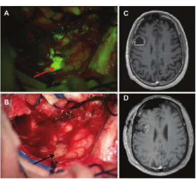
Figure 1.5 – Intraoperative view during resection of a right frontal glioblastoma; the 5-ALA fluorescent tumor
area to resect is pointed by a red arrow (A). The same view is shown with a white light illumination where tumor tissues are indicated by black arrows (B). Preoperative axial T1-weighted MR image with contrast agent (C). Postoperative (48 h) contrast MR image showing a complete tumor resection (D) [Acerbi et al. 2014].
1.2
Radio-guided cancer surgery
The concept of radioguided surgery was born in 1949, when Selverstone et al. used a Geiger-Muller tube to detect the β− radiation emitted from the 32P, intravenously injected to 33 patients suspected of brain tumor [Selverstone et al. 1949]. However, this first meta-bolic radiotracer was put aside due to high irradiation of the patient. From the sixties, the development of new nuclear imaging modalities, such as the gamma camera and the single photon emission tomography (SPECT), stimulated the development of new gamma-emitting radiotracers. The availability of these radiotracers led to the conception and development of dedicated intraoperative probes [Povoski et al. 2009, Tsuchimochi & Hayama 2013]. Along with the development of positron emission tomography (PET) and the associated β+ emit-ting radiopharmaceuticals in the 1970s, came also a renewed interest for the design of β intraoperative probes. More recently, research in target radionuclide therapy lead to the development of an increasing number of α, β− and Auger electrons emitting pharmaceutic-als. This had open the prospective of using β− radiotracers instead of β+ radiotracers for
intraoperative imaging [Collamati et al. 2015a]. Radioguided surgery of cancer and sentinel lymph nodes is now a well-established clinical practice. The efficiency of the intraoperative radio-guidance in terms of sensitivity and specificity relies on the association between the miniaturized detection device, the nuclides and the tumor-seeking agent. We present in the following sections (1.2.1and1.2.2) the main characteristics and diversity of radiotracers and intraoperative probes used in radioguided operative cancer surgery.
1.2.1 Radioactive tumor labelling
The radiopharmaceuticals generally used for radioguided surgery are a chemical combina-tion of a biological compound, which targets specific biochemical receptors or is metabolized by cells, with a radioisotope for radiolabeling. They exist as well radioactive nucleus that can be used alone as tracers, like204Tl, 123I or 125I. The great variety of molecules that can be synthesized allows to observe many different biological processes. We can distinguish two targeting mechanisms. The first one consists in the use of particular radiotracers that have negligible interaction with molecules in the body and whose non-specific uptake depends mainly on their diffusion properties (see Section1.2.1.2). The second method uses analogs of molecules that participate to the target biological process (peptide, amino acids, antibodies, neurotransmitter, etc.) (see Section1.2.1.3). These radiotracers can be used to label tumor cells that show specific physiological or metabolic anomalies.
1.2.1.1 Constraints on the radiotracer choice
The choice of the radioisotope associated to the ligand molecule is limited by constraints of different natures. It exists chemical constraints concerning the production of the couple tumor seeking agents labeled compound and physics constraints associated to the detection optimization and limitation of patient exposure.
Chemical constraints - First of all, the radiotracer uptake in the tumor must be as
spe-cific as possible. The spespe-cificity of a radiotracer is defined by the ratio between radiotracer uptake in tumor and heathy tissue. A high specificity improves the tumor detectability with external or intraoperative imaging devices. The radiotracer must also not interfere into the physiology or the metabolism of the observed process. In that context, the high sensitivity of radioisotopic imaging allows the injection of very low amount of radiotracer (nanomole con-centrations), thus minimizing any pharmacological effect. Furthermore, the chemical bound between tracer and radionuclide has to be stable in vivo in order to ensure a good uptake of the radionuclide in the target area while limiting its diffusion into the body, causing lost in sensitivity and specificity as well as potential toxicity. Finally, the rapid systemic clearance of unbounded radiotracers is also critical to achieve high contrast imaging.
Physic constraints - The radiations emitted by the radionuclide have to show specific characteristics in order to be detected without producing collateral damage for the patient. The energy of the radiation must be high enough to travel through the tissue and enter the detector, but in the meantime be low enough to be stopped inside it and detected. For γ emitters that has to ideally range between 100 and 300 keV. This energy range is optimal for external detection and it limits as well patient irradiation and distal contamination generated by scattered photons. For β+ emitters, the radionuclides must be selected with a low energy (less than 1 MeV) in order to limit the dose to the patient. The physical half-life of the radionuclide should be compatible with the in vivo pharmacokinetics of the tumor seeking agent [Zhou et al. 2013]. Half-life must be longer then the time required for the preparation of the pharmaceutical, its delivery to the clinic, its injection, the uptake in the tumor and the imaging protocol. For example, the short half-life of 11C (t1/2=20 min), 13N (t1/2=10