HAL Id: dumas-01176350
https://dumas.ccsd.cnrs.fr/dumas-01176350
Submitted on 15 Jul 2015HAL is a multi-disciplinary open access archive for the deposit and dissemination of sci-entific research documents, whether they are pub-lished or not. The documents may come from teaching and research institutions in France or abroad, or from public or private research centers.
L’archive ouverte pluridisciplinaire HAL, est destinée au dépôt et à la diffusion de documents scientifiques de niveau recherche, publiés ou non, émanant des établissements d’enseignement et de recherche français ou étrangers, des laboratoires publics ou privés.
Local early response to an MVA-mediated vaccination:
effects of an MVA infection on skin fibroblasts and
macrophages
Julie Jouffrey
To cite this version:
Julie Jouffrey. Local early response to an MVA-mediated vaccination: effects of an MVA infection on skin fibroblasts and macrophages. Life Sciences [q-bio]. 2015. �dumas-01176350�
Local early response to an MVA-mediated
vaccination: effects of an MVA infection on
skin fibroblasts and macrophages
Réponse locale précoce à une vaccination par MVA:
effets d’une infection par MVA sur des fibroblastes et
macrophages de la peau
Par : Julie JOUFFREY
Soutenu à Rennes le 15/06/2015
Devant le jury composé de :
Président :
Maître de stage : Dr Frédéric MARTINON Enseignant référent : Dr Jean-Marc FRASLIN
Autres membres du jury (Nom, Qualité)
Dr Hélène BOUVRAIS, Dr Anne CORLU, Dr Isabelle PELLERIN, Dr Claire PIQUET-PELLORCE
Les analyses et les conclusions de ce travail d'étudiant n'engagent que la responsabilité de son auteur et non celle d’AGROCAMPUS OUEST
AGROCAMPUS OUEST CFR Angers CFR Rennes Année universitaire :2014-2015 Spécialité : SCMV
Spécialisation (et option éventuelle) : Cancérologie-Immunologie
Mémoire de Fin d'Études
d’Ingénieur de l’Institut Supérieur des Sciences agronomiques, agroalimentaires, horticoles et du paysage
de Master de l’Institut Supérieur des Sciences agronomiques, agroalimentaires, horticoles et du paysage
Local early response to an
MVA-mediated vaccination: effects of an
MVA infection on skin fibroblasts and
macrophages
Commissariat à l'Energie Atomique et aux Energies Alternatives Julie JOUFFREY
M2 Sciences Cellulaires et Moléculaires du Vivant – Cancérologie et Immunologie 3e année Ingénieur agronome
Maître de stage : Dr Frédéric Martinon, INSERM
Université Paris Sud, UMR1184, France
CEA, DSV/iMETI, Division of Immuno-Virology, IDMIT, France INSERM, U1184, Center for Immunology of Viral Infections and Immune Diseases, France
CEA, 18 route du Panorama, 92265 Fontenay-Aux-Roses Cedex
AGROCAMPUS OUEST 65 rue de Saint-Brieuc 35000 Rennes
Université de Rennes 1 Campus de Villejean 2 av. du Pr. Léon Bernard 35000 Rennes
Acknowledgement
I would like to thank Frédéric MARTINON and Roger LE GRAND for allowing me to do this internship. I am grateful of the opportunity to be part of the ImVA team and to work on such an interesting subject.
I would also like to thank Pierre ROSEMBAUM for taking time in his last thesis year to train me and help me with my experiments and for helping with my presentations and the redaction.
I would like to thank the members of the IDMIT infrastructure: Pierre ROQUES, Delphine DESJARDINS, Martin SIGUIER, Gustavo MARTELLI PALOMINO, Sabine TRICOT and David PEJOSKI for helping me in my experiments and the redaction of my report.
I am grateful to the FlowCyTech team: Antonio COSMA, Christelle CASSAN, Sabrina GUENOUNOU for training me to use the cytometry platform. Thank you to Brice TARGAT for solving my many informatic problems at the beginning of my internship.
Finally, thank you to the TIPIV team: Benoît DELACHE, Naya SYLLA, Julie MORIN, Sébastien LANGLOIS, Nathalie DEREUDDRE-BOSQUET and Christophe JOUBERT for helping with the work on animals and the Multiplex® immunoassay.
Detailed summary
Acknowledgement ... Detailed summary ... List of annexes ... List of Figures and Tables ...
Introduction ... 1
Principles of vaccination ... 1
MVA and vaccination ... 2
Human immune responses to MVA ... 2
Study Models ... 4
Cynomolgus macaque ... 4
Fibroblasts and macrophages ... 4
Materials and Methods ... 4
Animals, skin biopsies and blood withdrawals ... 4
Fibroblasts cell culture from frozen cells or skin biopsies ... 5
Monocyte-derived macrophages cell culture from whole blood ... 6
MVA-eGFP virus and buffer ... 6
In vitro MVA Infection of fibroblasts and macrophages ... 6
Flow cytometry marking ... 7
Flow Cytometry analysis ... 7
Multiplex immunoassay analysis ... 9
Results ... 11
Fibroblasts – MVA infection, cell activation and induced cell death ... 11
Macrophages - MVA infection and cell activation ... 15
Supernatant multiplex analysis ... 17
Discussion ... 21
References ... 25 Annexe I - Résumé ... Annexe II - Abstract ...
List of abbreviations and commercial names
BluVid – LIVE/DEAD fixable blue dead, Life TechnologiesTM
CC Chemokines –Chemokines with two adjacent cysteines (C) near their amino terminus.
CPT - BD Vacutainer® CPTTM Cell preparation tube with Sodium Heparin CD – Cluster of differentiation
DC – Dendritic cells
DMEM – Dulbecco’s modified Eagle medium (Gibco®)
DPBS – Dulbecco’s phosphate-buffered saline
EDTA – Ethylenediaminetetraacetic acid eGFP – Enhanced green fluorescent protein
FBS – Fetal bovine serum
Fungizone ® - Amphotericin B, antifungal drug
G-CSF – Granulocyte colony-stimulating factor
GM-CSF – Granulocyte macrophage colony-stimulating factor
HeLa – Cervical cancer-derived cell line taken from Henrietta Lacks
HEPES – 4-(2-hydrowyethyl)-1-piperazineethanesulfonic acid
HLA-DR – MHC class II cell surface receptor, αβ heterodimer
HIV – Human Immunodeficiency Virus IFN-γ, IFN-α/β – Interferon (IFN) gamma (γ), alpha (α) and beta (β)
IL – Interleukin
IP – Interferon γ-inducible protein
MCP – Monocyte chemoattractant Protein MDA5 – Melanoma
differentiation-associated protein 5
MDDC – Monocyte-derived dendritic cell MHC – Major histocompatibility complex MOI – Multiplicity of infection
MyD88 – Myeloid differentiation primary response gene 88
MVA – Modified Vaccinia virus Ankara NALP3 – NACHT, LRR and PYD
domains-containing protein 3 or cryopyrin NHP – Non-human primate
NK – Natural killer
Pfu – Plaque-forming unit0
PSN – Penicillin, Streptomycin, Neomycin RIG-I – Retinoic acid-inducible gene 1 TLR – Toll-like receptor
TNF – Tumor necrosis factor
VEGF – Vascular endothelial growth factor
List of Annexes
Annexe I – Résumé Annexe II – Abstract
List of Figures and Tables
Figure 1 – Fibroblasts (left) and confluent fibroblasts (right)………p.8 Figure 2 – Monocyte-derived macrophages……….…p.8 Figure 3 – Fibroblasts gating strategy and living cell count after injection………..p.12 Figure 4 – Fibroblasts are infected by MVA-eGFP……….p.12 Figure 5 – Fibroblast markers’ expression………p.14 Figure 6 – Gating strategy for macrophages………..p.16 Figure 7 – Expression of GFP in macrophage-like cells……….….p.16 Figure 8 – Expression of activation markers in macrophages………p.18 Figure 9 – Fibroblasts cytokines concentrations……….p.20 Figure 10 – Macrophages cytokines mean concentrations……….…p.22 Table 1 – Flow cytometry, Fibroblast Panel………p.10 Table 2 – Flow cytometry, Macrophage Panel……….p.10 Table 3 – Preparation of marking mix for fibroblasts………..p.10 Table 4 – Preparation of marking mix for macrophages………p.10
1
Introduction
Principles of vaccination
Being the interface with the external environment and protecting the body from dehydration and infections, along with a wide variety of immune cells, skin is an important tissue for vaccine inoculation (Frenck Jr. et al., 2011; Fuchs and Raghavan, 2002; Haniffa et al., 2009; Stoitzner et al., 2008; Vogt et al., 2008). Skin is constituted of differently shaped layers. Epidermis is mainly made of keratinocytes and melanocytes, non-immune cells, and some immune cell types as Langerhans cells and lymphocytes. Underneath the epidermis is the dermis, a vascularized conjunctive tissue consisting in numerous and various cell populations including fibroblasts (non-immune cells) in great numbers but also immune cells as T, B and NK lymphocytes, macrophages, interstitial and plasmacytoid dendritic cells, neutrophils and mastocytes. The deeper layer is the hypodermis, mainly constituted of adipocytes (Zaba et al., 2008). Unlike the epidermis and more than the hypodermis, the dermis is rich in blood vessels and nerve endings. This team’s recent works show a high diversity of innate immune cells in the skin and their role in inflammatory responses to intradermal vaccination (Epaulard et al., 2014; Romain et al., 2012).
The immune system is able to keep track of its responses in the immunological memory. The aim of vaccination is to use this memory to simulate a first encounter with an antigen (from the target pathogen) and induce an immune response, stored in the immunological memory in prevention of a future encounter with the whole virulent pathogen. There are three kinds of vaccines. The first category is live attenuated vaccines, altered by passages in cells with genetic modifications; the second is inactivated vaccines, altered by heat or chemicals; the third category is subunits purified from pathogens or recombinant vectors. The term vaccination comes from the Latin for “cow” (vacca), as the first vaccine was made by Edward Jenner in 1796 from cowpox sore matter inoculated into human patients to prevent the smallpox illness (Downie, 1951). Although the major route of administration of a vaccine is intramuscular, recent methods and devices allow the development of skin routes’ administration. The skin inoculation of a vaccine can take several forms: scarification or puncture (e.g. the history of smallpox vaccine (variola)), subcutaneously or intradermally (e.g. Bacille de Calmette-Guérin used as vaccine against tuberculosis).
2
MVA and vaccination
Smallpox (variola virus) was eradicated using vaccinostyles and bifurcated needles, showing that the skin route of administration is effective (Liu et al., 2010). Vaccinia virus, another virus from the poxvirus family, is the active constituent of the vaccine used to eradicate smallpox in the late 20th century. If the original host of Vaccinia virus is unknown, the Choriollantois Vaccine Ankara strain was studied by Anton Mayr. After 516 passages of this virus in chicken embryo fibroblasts, an attenuated vaccinia virus was obtained, named MVA (Modified Vaccinia virus Ankara) (Verheust et al., 2012). Those passages damaged the MVA genome: 6 large genomic deletions totaling 31kb appear when compared with the parental virus genome(Blanchard et al., 1998). Amongst the damages, MVA mostly lost its replication capacity in mammalian cells and only has a limited cell-to-cell spread(Blanchard et al., 1998; Stittelaar et al., 2001). MVA genome, 178 kb long and fully sequenced, lacks the sequences for some of the known poxviral immune evasion and virulence factors (Verheust et al., 2012). MVA-based recombinant viruses (rMVA) show promises as vaccine candidates due to their capacity to integrate the sequences of the genes of interest into their genome. The good immunogenicity of MVA resides in its loss of virulence factors (Stittelaar et al., 2001). Safety data on the use of recombinant MVA were published concerning the frequent use of MVA as a viral vector basis for new vaccines in the domain of infectious diseases in humans, such as Malaria (Moorthy et al., 2003), AIDS (Howles et al., 2010), Tuberculosis (Brookes et al., 2008), Human Papilloma Virus associated cancer, Melanoma, and other cancers (Verheust et al., 2012).
Human immune responses to MVA
MVA is able to engage both innate and adaptive immunity (Burgers et al., 2008). Engaging a good adaptive immune response (i.e. specific to the pathogen, high affinity and long term memory) is the key that makes MVA such a good vaccine candidate.
As a vaccine candidate, MVA is able to induce the production of memory immune cells and specific antibodies so strongly that stabilized levels of those cells can be detected in humans up to 50 years after vaccination. Human vaccinia specific CD8+ T cells and CD4+ T cells responses are strong after a smallpox vaccination (Amanna et al., 2006). In Non-human primates (NHP), it was shown that MVA is effective against monkeypox challenges after vaccination (Artenstein, 2008). rMVA-HIV (i.e. rMVA expressing selected genes from HIV) successfully infected human primary cells in vitro such as macrophages, T cells, NK cells, monocytes and dendritic cells, but more interestingly, DCs expressing HIV antigens after rMVA infection could activate HIV-specific cytotoxic T lymphocytes and induced a
3 production of HIV-specific CD4+ cells. These results were confirmed in NHP and an MVA-SIV vaccination engages strong MVA-SIV-specific T cell response (Brandler et al., 2010). A strong and durable immunological memory is an advantage for a good vaccine candidate such as MVA.
The characteristics of adaptive immunity are determined by innate immunity through recruited cells, their activation states and secreted molecules since the beginning of the inflammatory response. DCs have a primordial role in the junction between innate and adaptive immunity, as T lymphocytes have to be activated by a DC to be able to then activate a B lymphocyte by direct contact. The selection of subpopulations of DCs targeting specific memory cells, the adaptive immunity can be set up by the innate immunity (Cohn and Delamarre, 2014; Coquerelle and Moser, 2010; Weaver et al., 2006).
An in vivo MVA infection triggers the rapid immigration of monocytes, neutrophils and CD4+ lymphocytes to the injection site (Altenburg et al., 2014). It has been shown that MVA activates two innate immunity pathways in the macrophage upon cell infection: one Toll-like Receptor (TLR)-dependent and the other TLR-independent. The first includes an activation of the TLR2-TLR6 heterodimer receptor, inducing an immune response including Th1 (T helper 1 cells) and Natural Killer (NK) cells through MyD88 interaction and inflammatory cytokines release (Delaloye et al., 2009; Reed et al., 2009; Zhu et al., 2007). The TLR-independent pathways involve the NALP3 inflammasome activation down to IL-1β production on the one hand and MDA-5, a RIG-I-like receptor activating a cellular feedback using type I IFN (Waibler et al., 2007). Both TLR-dependant and TLR-independent pathways are required to activate innate and adaptive immunity to Vaccinia Virus in vivo (Zhu et al., 2007). An MVA infection of whole human blood cells results in an abundant production of chemokines such as IL-8 or IP-10, but a less abundant production of pro-inflammatory cytokines (TNF, IL-1β and IL-6), which is believed to be due to the attenuation of MVA (Delaloye et al., 2009). More interestingly, RNA breakdown was demonstrated in Monocyte-Derived Dendritic Cells (MDDC) infected with MVA or NYVAC (another attenuated Vaccinia virus strain), showing an induced apoptosis in vitro (Guerra et al., 2007). Dendritic cells are known target cells of an MVA infection, as they were shown to be infected by MVA both in vivo and in vitro in mice (Liu, 2010). MDDC morphology, gene expression profile and maturation state are modified after an MVA infection, and their possible entrance in apoptosis demonstrates the high immunogenicity of MVA-vectored vaccines (Verheust et al., 2012). MVA also affects the migratory capacity of both immature and mature dendritic cells through viral binding proteins modulating the chemokine system of DC migration (Alcami, 2007).
4
Study Models
Cynomolgus macaque
Cynomolgus macaque (Macaca fascicularis) is the major Nonhuman Primate (NHP) used in biomedical research. The macaque’s immune system is very close to the human immune system, making it susceptible to human infectious diseases and pathologies. It is a uniquely valuable resource to study human infectious diseases in vaccinology, including influenza virus, flavivirus, arenaviruses, hepatitis E virus, papillomavirus, smallpox virus and Bacillus anthracis (Gardner and Luciw, 2008). The similarity of the two immune systems enables researches to explore the experimental conditions of a vaccine’s efficiency. Several studies (Barratt-Boyes et al., 2006; Kato et al., 2004; Lu et al., 2003; Querec, 2006; Teleshova et al., 2004; Wonderlich et al., 2011), including some from this team (Epaulard et al., 2014; Malleret et al., 2008a, 2008b; Romain et al., 2012), showed that some of the cynomolgus macaque’s cell populations involved in capturing and preparing antigens are similar to those in the human immune system and can be characterized in many ways. Finally, using NHP as animal models is well adapted to assess a vaccine’s feasibility and efficiency along with its safety. Those are essential preclinical trials before clinical tests on human patients.
Fibroblasts and macrophages
In vivo imaging of the injection site of an MVA-eGFP (recombinant MVA expressing a fluorescent protein) intradermal injection indicates an expression of GFP in cells whose morphology is compatible with that of macrophages and/or fibroblasts. The GFP expression appears during between approximatively 24 and 72 hours following the injection and the local inflammation is characterized by the recruitment of macrophages and neutrophils. In this context, this study aims to identify the immune role of fibroblasts and macrophages in response to MVA and characterize the effects of a possible infection of those cells by MVA on their physiology. To do so, fibroblasts and monocyte-derived macrophages are obtained and cultured from NHP skin biopsies and blood samples, then infected with injection of MVA in the culture medium. Physiological modifications induced by MVA are observed through flow cytometry staining, multiplex analysis of supernatant and culture wells pictures.
Materials and Methods
Animals, skin biopsies and blood withdrawals
Adult cynomolgus macaques (Macaca fascicularis) (and weighing 4-8 kg) were housed in Commissariat à l’Energie Atomique facilities. Animals were sedated with ketamine
5 chlorhydrate during handling. Three punch biopsies per animal (8 mm diameter) were performed on anesthetized animals. Biopsies were then cleaned removing fatty tissues, epidermis and dermis were separated using a dispase solution, and then the dermis of all three biopsies was mixed with the gentleMACSTM Dissociator technology (Miltenyi Biotec) to prepare a cell solution. Blood was drawn in CPT (Cell Preparation Tubes) (8 mL), up to 3 or 4 CPTs per animal in one time.
Fibroblasts cell culture from frozen cells or skin biopsies
Cell culture
Fibroblasts were obtained from skin biopsies prepared as described above and then cultured in 4 mL DMEM solution consisting of DMEM GlutaMAX (Gibco®) with 10% FBS (Eurobio), 1% PSN (Gibco®), 1% HEPES (Gibco®) and 1% Fungizone®(Gibco®). Splitting fibroblasts was performed when cells were approximately 80% confluent, and often overestimating the surface needed (Figure 1). Flasks used were 25, 75 and 175 cm². Splitting protocol implied use of 0,25% Trypsin-EDTA (Gibco®) (2 mL for 25cm² flasks, 5 mL for 75cm², 5 mL for 175cm²) during 10 min at 37°C, once the previous culture medium was emptied and the flasks were washed with DPBS (Gibco®) at 37°C. The action of Trypsin-EDTA was stopped by addition of DMEM solution (or DPBS 10% FBS) in excess.
Change of medium was performed every other day for fibroblasts. In case of holiday, more medium was added during the last medium change.
Cell flasks and plates were kept in an incubator set on 37°C, 7% CO2.
Passages
All splittings were recorded, to avoid reaching the maximum number of passages tolerated by fibroblasts, set around 25 by Hayflick in 1961 (Hayflick and Moorhead, 1961). In case of cells from different passage steps assembled in a new flask, the higher number of passage is recorded.
Frozen cells
Fibroblasts were frozen in Fetal Bovine Serum + 10% Dimethyl Sulfoxide (DMSO) solution in Nunc® cryogenic storage tubes and kept at -135°C. Tubes’ content was thawed gently and added to culture medium to prepare a cell solution.
6
Monocyte-derived macrophages cell culture from whole blood
Monocytes were isolated from whole blood drawn from macaques. Extracted Peripheral Blood Mononuclear Cells (PBMC) were washed and treated with red blood cell lysis solution if needed. PBMC were then set to differentiate following an adhesion protocol in DMEM solution containing 10% FBS 1% PSN and 1% HEPES, 10ng/mL M-CSF and 2ng/mL GM-CSF for six days and then moved to adequate wells for the experiment and changed to a DMEM 10% FBS 1% PSN 1% HEPES medium. On Day 8, well differentiated cells were harvested with DPBS 10mM EDTA (Figure 2). It appeared that lymphocytes did not survive a week in monoculture.
Change of medium was limited for macrophages, as differentiated macrophages are easily detached.
MVA-eGFP virus and buffer
MVA-eGFP (recombinant MVA expressing eGFP under the control of p11K7.5 promoter) was provided by Transgene (Illkirch-Graffenstaden, France) with L1 biosafety level. The clone is refered as MVATGN33.1. The viral was shipped in cryotubes of 1 mL of 5 x 10⁸ pfu/mL frozen in dry ice, and virus was kept at -80°C until use.
MVA Buffer is prepared following the virus manufacturer’s instruction. It contains 10mM of trizma hydrochloure, 5% sucrose, 10 mM of L-glumatic and monosodium salt monohydrate and 50mM of sodium chloride at pH 8.
In vitro MVA Infection of fibroblasts and macrophages
Fibroblasts previously harvested from culture flasks and counted were stimulated in 6-well culture plates, one well per condition containing 400 000 cells in 4 mL of culture medium. Macrophages were stimulated in 24 or 48-well culture plates depending on their number when harvested and counted as follows: 220 000 cells per well in 48-well culture plates in 150 µL of culture medium or 400 000 cells per well in 24-well culture plates in 400 µL of culture medium. Chosen MOI were set as 0.01, 0.1 and 1 for the different experiments. An aliquot of 100 µL MVA-eGFP at 5.108 PFU/mL kept at -135°C was thawed and diluted with MVA Buffer to obtain the selected MOI depending on the cell concentrations in wells. The virus particles were then injected in the culture medium of cells to be infected, according to MOI conditions (“Buffer only” wells received MVA Buffer in the same quantity and “Unstimulated” conditions received DPBS). Two hours after infection, the culture medium was changed to stop the viral infection.
7
Flow cytometry marking
On due time points (H6, H24 or H48), cells were harvested from the infection wells with Trypsin-EDTA 0.25% (if fibroblasts) or DPBS 10 mM EDTA (if macrophages). Cell mortality was assessed with a blue fluorescent reactive dye from the LIVE/DEAD Fixable Dead Cell Stain Kit “BluVid” (Life TechnologiesTM
). Cells were put 10 min at 70°C to induce cell death in BluVid and Annexin V (to assess cell apoptosis) control conditions only. Then all harvested cells were left 15 min to incubate with a BluVid solution (1 mL DPBS 1‰ BluVid) at 4°C in the dark. After washing with centrifugation (1800 rpm 4°C 5 min), nonspecific cell receptors were saturated with a Macaque Serum solution (50 µL DPBS 5% Macaque Serum) and incubated 20min at 4°C in the dark. An “Unstained” condition was also prepared with cells receiving only DPBS instead of BluVid, Macaque Serum and markers. The detailed antibodies’ list is presented in Table 1 and Table 2. Panels were prepared minimizing the overlapping of fluorochromes. Fluorochrome-free antibodies (“Purified”) were coupled with a secondary antibody coupled to an Alexa Fluor fluorochrome with the Zenon Kit (Life TechnologiesTM). Without washing, cells then received 90 µL of the panel mix or 90 µL of DPBS if unstained, as presented in Tables 3 and 4. In case of coupled markers with a Labeled Zenon (e.g. CMH I Purified + IgG2a Zenon PE Texas Red + Component B), cells received 80µL of the mix panel and 10µL of each labeled marker. Cells were incubated 20 min at 4°C in the dark with markers, then centrifugated (1800 rpm 4°C 4 min). Cells were finally fixed with 150 µL BD CellFIXTM solution (BD Biosciences) and analyzed with Flow Cytometry within hours (cells are kept at 4°C in the dark to preserve fluorochromes).
Flow Cytometry analysis
Flow cytometry acquisition was performed on an LSRFortessa cytometer (BD Biosciences, Le Pont de Claix, France) with five lasers (355 nm, 405 nm, 488 nm, 560 nm and 640 nm). Obtained data were then analyzed with FlowJo Single Cell Analysis software (Tree Star, Ashland, OR).
8 Figure 1 - Fibroblasts (left) and confluent fibroblasts (right). Photographs of dermis fibroblasts taken from usual in vitro culture conditions. Fibroblasts were isolated from dermis of skin biopsies. x 100
Figure 2 – Monocyte-derived macrophages. Pictures were taken in culture wells at H48 after infection. Differentiated monocytes with macrophages phenotype can be seen (black arrows). x 100
9
Multiplex immunoassay analysis
Supernatant samples (200 µL) taken from culture wells at interest time points were analyzed (25 µL per condition) using Milliplex® MAP Non-Human Primate Cytokine Panel coupled with Luminex xMAP® platform from EMD Millipore Corp., MA. 23 cytokines were tested: G-CSF, GM-CSF, IFNγ, 1β, 1ra, 2, 4, 5, 6, 8, 10, 12/23(p40), IL-13, IL-15, IL-17A, MCP-1, MIP-1α, MIP-1β, sCD40L, TGFα, TNFα, VEGF and IL-18.
During the fibroblast experiment, the medium volume (4 mL) was sufficient to allow sampling at every time point; on the contrary, macrophages were cultured in small amount of medium (150 or 400 µL depending on the number of cells per well), limiting the sampling at harvesting only. Supernatant was sampled from fibroblasts cultures at H0, H6, H24 and H48. Fibroblasts were counted and distributed in the experiment wells 24 hours before H0 (infection), therefore the “H0 pool” sample (sampled from a pool of supernatant from every wells belonging to the same individual) was really a H24 sample in normal culture condition.
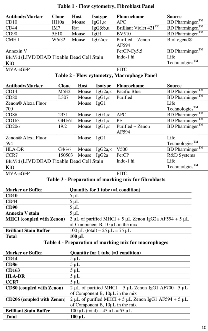
10 Table 1 - Flow cytometry, Fibroblast Panel
Antibody/Marker Clone Host Isotype Fluorochome Source
CD10 HI10a Mouse IgG1,κ APC BD PharmingenTM
CD44 IM7 Rat IgG&b,κ Brilliant Violet 421TM BD PharmingenTM
CD90 5E10 Mouse IgG1 BV510 BD PharmingenTM
CMH I W6/32 Mouse IgG2a,κ Purified + Zenon AF594
BioLegend®
Annexin V PerCP-Cy5.5 BD PharmingenTM
BluVid (LIVE/DEAD Fixable Dead Cell Stain Kit)
Indo-1 hi Life
TechonolgiesTM
MVA-eGFP FITC
Table 2 - Flow cytometry, Macrophage Panel
Antibody/Marker Clone Host Isotype Fluorochome Source
CD14 M5E2 Mouse IgG2a,κ Pacific Blue BD PharmingenTM
CD80 L307 Mouse IgG1,κ Purified BD PharmingenTM
Zenon® Alexa Fluor 700
Mouse IgG1 Life
TechnologiesTM
CD86 2331 Mouse IgG1,κ APC BD PharmingenTM
CD163 GHI/61 Mouse IgG1,κ PE BD PharmingenTM
CD206 19.2 Mouse IgG1,κ Purified + Zenon AF594
BD PharmingenTM Zenon® Alexa Fluor
594
Mouse IgG1 Life
TechnologiesTM
HLA-DR G46-6 Mouse IgG2a,κ V500 BD PharmingenTM
CCR7 150503 Mouse IgG2a PerCP R&D Systems
BluVid (LIVE/DEAD Fixable Dead Cell Stain Kit)
Indo-1 hi Life
TechonolgiesTM
MVA-eGFP FITC
Table 3 - Preparation of marking mix for fibroblasts
Marker or Buffer Quantity for 1 tube (=1 condition)
CD10 5 µL
CD44 5 µL
CD90 5 µL
Annexin V stain 5 µL
MHCI (coupled with Zenon) 2 µL of purified MHCI + 5 µL Zenon IgG2a AF594 + 5 µL of Component B, 10 µL in the mix
Brilliant Stain Buffer 100 µL (total) – 25 µL = 75 µL
Total 100 µL
Table 4 - Preparation of marking mix for macrophages
Marker or Buffer Quantity for 1 tube (=1 condition)
CD14 5 µL
CD86 5 µL
CD163 5 µL
HLA-DR 5 µL
CCR7 5 µL
CD80 (coupled with Zenon) 2 µL of purified MHCI + 5 µL Zenon IgG1 AF700+ 5 µL of Component B, 10µL in the mix
CD206 (coupled with Zenon) 2 µL of purified MHCI + 5 µL Zenon IgG1 AF594 + 5 µL of Component B, 10µL in the mix
Brilliant Stain Buffer 100 µL (total) – 45 µL = 55 µL
11
Results
Fibroblasts – MVA infection, cell activation and induced cell death
Cytometry analysis
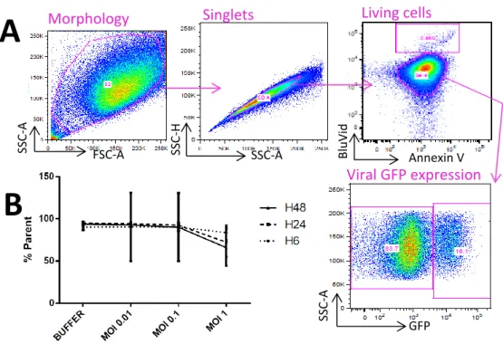
Harvested and stained cells were analyzed with flow cytometry. A gating strategy protocol was set up to define the recorded events as cells, based on the morphology of the events and selecting only singlet events (Figure 3A).
MVA-induced cell death
BluVid stains both living and dead cells by reacting with free amines, with a 50-fold more intense fluorescence for dead cells. Annexin V is a protein binding phospholipid phosphatidylserine exposed during cell apoptosis. Annexin V marking is used besides BluVid to confirm dead cells as double positive cells. The two populations observed in the “Living cells” dot plot (Figure 3A) were previously defined as Annexin V negative cells thanks to a control, and the upper population was verified as mostly cell debris. Figure 3B presents a living cell counts expressed in percentage of total cells according to the gating strategy. With an increasing MOI from 0.01 to 1, cell count seems to drop from 90% to about 60%. Without significance, living cells also appear to be fewer with a later time of harvest after the two-hour long infection.
Fibroblasts are infected by MVA
Although fibroblasts naturally show autofluorescence in the FITC channel (same flow cytometry channel as GFP), infected cells express GFP with significantly higher levels (Figure
12 Figure 3 - Fibroblasts gating strategy and living cell count after injection. A. Cytometry data were analyzed through the following gating strategy: adequate morphology and singlets were selected based upon flow cytometry parameters, then living cells were defined by being the cell population with the lowest BluVid staining fluorescence and Annexin V negative. The upper population was identified through backgating as not being cells. B. Living cells mean count. Negative BluVid-stained cells were expressed in percentage of all cells (singlet signal and with adequate morphology in flow cytometry). Data was obtained from four individuals’ fibroblasts
A
FSC-A SSC -A SSC-A SSC -H Annexin V BluVid SSC -A GFPB
Morphology Singlets Viral GFP expression Living cellsFigure 4 - Fibroblasts are infected by MVA-eGFP. A. GFP expression increased with viral load. Cells were separated into naturally GFP-expressing cells (left) and infected and expressing viral GFP cells (right). B. Fibroblasts express viral GFP. Viral GFP expression was determined after consideration of the natural fluorescence of fibroblasts using the “Buffer” condition results. GFP positive cell counts were obtained from the flow cytometry data and expressed in percentage of all cells (singlet signal and with adequate morphology). Data here was the mean from four individuals’ fibroblasts signal.
MVA Buffer MVA MOI 0,01
MVA MOI 0,1 MVA MOI 1
13 4A). The thus defined GFP+ population corresponds to infected cells expressing viral GFP. The number of cells in this population increases with the MOI from 0.01 to 1. Figure 4B also shows that the longer cells are cultured after infection, the more viral GFP they express. Fibroblasts markers
Although markers used are not exclusively expressed by fibroblasts, their association in dermis cell suspension allows to mainly characterize this cell population. Markers’ expression by long term in vitro culture fibroblasts is presented in Figure 5, depending on time points and MVA MOI if the cells were in presence of MVA-eGFP. Fibroblasts have been shown to be highly CD10 positive, also called gp100, a type II transmembrane glycoprotein (Kundrotas, 2012). CD10 expression seems to decrease lightly in MVA infected cells 6 hours after infection; but the expression is comparable with uninfected cells at 24 hours after infection. 48 hours after infection, infected cells’ expression of CD10 seems slightly higher than that of uninfected cells, especially at an MOI of 1. There appears to have a global decrease in CD10 expression when the MOI gets higher, from 0.01 to 1.
As matrix synthesizer cells, fibroblasts express proteins involved in matrix-cell adhesion, such as CD44, cell-surface glycoprotein involved in cell-cell interactions, cell adhesion and migration (Kundrotas, 2012) which ligand is hyaluronic acid, or CD90, or Thy-1 for Thymocyte antigen 1, a glycoprotein expressed on many cell types, including T cells, thymocytes, neurons, endothelial cells, and fibroblasts (Rege and Hagood, 2006). Thy-1 has widespread non immunologic functions, such as apoptotic signaling, cell adhesion and migration, and more interestingly fibroblast proliferation and migration. Thy-1 also modulates the fibroblast phenotype involved in wound healing and fibrosis. CD44 and CD90 expressions are similar when comparing uninfected and infected cells: infected cells seem to have a higher expression than uninfected cells at every time points. At 6 hours after infection, the higher the MOI, the higher the CD44 expression, both for infected and uninfected cells. This evolution is similar at 24 and 48 hours after infection but with lower MFI. MFI in buffer condition is also decreasing with time, which shows that this is more due to cell death therefore to a lower mean of fluorescence intensity. An opposite evolution can be observed at 48 hours after infection for CD90 expression: the buffer condition MFI is higher than that of H6 and H24 buffer conditions, but the pairs have decreasing expression with higher MOI. Time seems to increase CD90 expression by uninfected fibroblasts, but if infected by MVA, fibroblasts diminish their CD90 expression.
14 Figure 5 - Fibroblast markers expression. Markers’ Mean Fluorescence Intensity (MFI) was recorded from cytometry data. On every graph, black dots represent MFI of cells gated as unexpressing viral GFP or from the buffer condition. Red dots represent cells expressing viral GFP. A couple of black and red dots linked represent one paired cell populations obtained with one condition.
15 MHC 1 (symbol HLA), or Major Histocompatibility Complex class I, is expressed on nearly every nucleated cell, including fibroblasts. Its functions include intracellular proteins presentation to CTLs, cross-presentation, and inhibitory ligand for NK cells. MHC I expression doesn’t seem affected by MVA infection at 6 and 24 hours after infection, but at 48 hours after infection, infected cells tend to express more this marker and the global expression is increased with a higher MOI of MVA.
Macrophages - MVA infection and cell activation
Markers used on macrophages have two objectives: defining the cells as macrophages through a selection of phenotype markers and monitoring their activity via activation markers. Chosen phenotype markers are CD14, CD163 and CD206, macrophages being CD14+ CD163+ CD206+. Although few cells were harvested and analyzed, the majority of living cells are CD206+ HLA-DR+ CD14+ CD163+, confirming a macrophage phenotype as illustrated in Figure 6.
Differentiated monocytes are easily detached from the bottom of culture wells, and as presented in Figure 7, no cells expressing viral GFP was harvested 48 hours after infection. As for fibroblasts, a buffer condition helped to define the population in the left gate as expressing macrophages’ natural fluorescence in the FITC channel instead of viral GFP. The expected cell population expressing viral GFP should be in the right gate.
Chosen activity markers are CD80, CD86, CCR7 and HLA-DR. CD80 and CD86, or B7-1 and B7-2, are two molecules working in tandem to prime T cells by binding T cell CD28 and CTLA-4. CCR7 is a protein of the G protein-coupled receptor family involved in activation and chemotactism. HLA-DR is a Major Histocompatibility Complex class II molecule, working mainly as a peptide antigens presentation receptor and thus is expressed on antigen presenting cells such as macrophages, dendritic cells and B-cells. The expression of the activation markers is compared between the buffer condition and the infected condition. There is a strong variation of results between the four individuals that provided the PBMC (Figure 8). The lack of clear trend in the expression of activation markers (Figure 8) could be related to the absence of infection of these cells by the MVA-GFP (Figure 7).
16 Figure 6 - Gating strategy for macrophages. Macrophages were selecting following a gating strategy based on phenotype markers for macrophages. Adequate morphology and singlets were selected based upon flow cytometry parameters, then living cells were defined by being the cell population with the lowest BluVid staining fluorescence. Among living cells, differentiated monocytes were selected as HLA-DR+/CD206+/CD14+/CD163+ cells.
FSC-A SSC -A SSC-A SSC -H BluVid SSC -A CD163 CD 1 4 CD206 H LA -DR
Morphology Singlets Living cells
Macrophages phenotype SSC -A GFP SSC -A GFP SSC -A GFP SSC -A GFP Figure 7 – Expression of GFP in macrophage-like cells. The GFP gate separating non infected cells (left) from MVA-eGFP infected cells (right) was drawn in order not to have any “infected” cells in the buffer condition. Each graph stands for an individual. The colored dots represent the conditions of cells harvested at H48 after infection: buffer condition (black), MOI 0.01 (orange), MOI 0.1 (light blue) and MOI 1 (red).
17
Supernatant multiplex analysis
Fibroblasts
Out of the 23 cytokines assayed, the cytokines with the higher concentrations or more significant evolutions are presented in Figure 9. Exclusions were mostly due to out of range data. Even if fibroblasts are not primarily immune cells, they are able to secrete cytokines in vitro; indeed, after changing the medium at H2, the concentration of some cytokines drops near zero and then reach higher levels at H24 and H48 (e.g. IL-6, VEGF and IL-10).The concentration measured is an accumulation of the cytokine secreted by the cells. An increase or decrease of concentration between two conditions can be perceived as a faster or slower production of said cytokine. Concentration variation depending on time can be observed with the fibroblast supernatant analysis. IL-18, IL-10 and IL-6 have higher concentrations (ignoring conditions) at 24 hours after infection than at 6 hours after infection. IL-6 reaches high levels around 1500-2000 pg/mL at H24 and around 2500 pg/mLat H48; there seems to be an MVA-induced decrease of IL-6 secretion, as the concentration is slightly lower when the MOI rises. A difference between H24 and H48 seems to appear without significance between H24 and H48. There appear to be a very limited effect of MVA infection on cytokines’ secretion. As an example, MCP-1 concentration seems to be higher with MVA MOI at H48 compared to the MVA Buffer condition. On the contrary, the concentration of TNFα drops from 5-6 pg/mL at MOIs of 0.01 or 0.1 to about 2 pg/mL at an MOI of 1, without significance. The rate of TNFα at H48 MOI 1 is similar to that of H24 Unstimulated, which could imply that an MVA infection at MOI 1 slows the production of TNFα by fibroblasts.
18 Figure 8 – Expression of activation markers in macrophages. A. CD80 expression. B. CD86 expression. C. CCR7 expression. D. HLA-DR expression. Markers’ expression was measured by flow cytometry. The flow cytometry panel was set in order to avoid fluorochrome superposition in their absoption and expression channels.
B.
C.
D.
A.
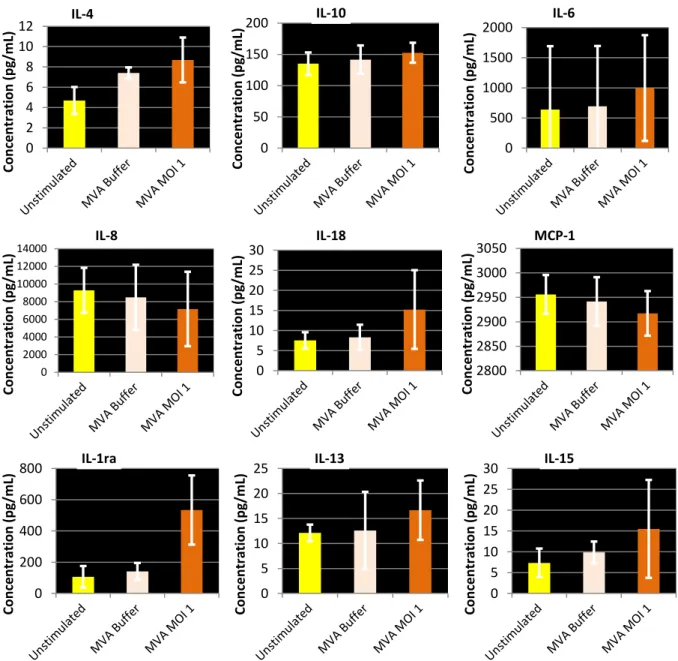
M FI M FI M FI M FI19 Macrophages
As for fibroblasts, because of out of range data, only 9 cytokine concentration graphs are presented in Figure 10. As macrophages were cultures in low amount of medium, supernatant could only be sampled when cells were harvested. Except for IL-8, MVA seems to induce a cytokine productions in macrophages
IL-8 is secreted by macrophages in vivo and is responsible for the recruitment of neutrophils and other granulocytes to the infection site (Harada et al., 1994). MVA seems to decrease this secretion by macrophages in vitro, as the concentration drops from about 10000 pg/mL in an unstimulated condition to about 7000 pg/mL in presence of an MOI 1 of MVA.
IL-13 is also a anti-inflammatory cytokine, and its secretion by macrophages in vitro appears to remain stable around 10-15 pg/mL when MVA is added to the culture medium.
The secretion of IL-4 appears higher when macrophages were in presence of MVA Buffer or MVA virus for two hours than when left unstimulated.
The concentration of IL-10 is mainly stable, around 140 pg/mL, unaffected by MVA or MVA Buffer.
IL-18, IL-1ra and IL-15 may increase in presence of an MOI 1 of MVA compared to an unstimulated condition or with MVA Buffer.
Finally, the MCP-1 concentration appears stable, slightly decreasing from 2950 to 2900 pg/mL from the unstimulated condition to the MVA MOI 1 condition.
20 Figure 9 - Fibroblasts cytokines concentrations. Cytokines with the higher concentrations and more significant evolution are presented as the mean on four individuals at every time points. Data was obtained with Multiplex® immunoassay and only the mean concentrations are presented here for the same four cells providing individuals. 0 2000 4000 6000 8000 10000 12000 H 24 U n st imu lated H 6 B u ff er H 6 M O I 0 ,0 1 H 6 - M O I 0 ,1 H 6 M O I 1 H 2 4 B u ff er H 2 4 M O I 0 ,0 1 H 2 4 M O I 0 ,1 H 2 4 M O I 1 H 4 8 B u ff er H 4 8 M O I 0 ,0 1 H 4 8 M O I 0 ,1 H 4 8 M O I 1 Co n ce n tr ation (p g/ m L) IL-8 0 5 10 15 H 2 4 U n st imu lated H 6 - B u ff er H 6 M O I 0 ,0 1 H 6 M O I 0 ,1 H 6 - M O I 1 H 2 4 B u ff er H 2 4 M O I 0 ,0 1 H 2 4 M O I 0 ,1 H 2 4 M O I 1 H 4 8 B u ff er H 4 8 M O I 0 ,0 1 H 4 8 M O I 0 ,1 H 4 8 M O I 1 Co n ce n tr ation (p g/ m L) IL-12/23 0 50 100 150 200 250 H 2 4 U n st imu lated H 6 B u ff er H 6 M O I 0 ,0 1 H 6 M O I 0 ,1 H 6 M O I 1 H 2 4 B u ff er H 2 4 M O I 0 ,0 1 H 2 4 M O I 0 ,1 H 2 4 M O I 1 H 4 8 B u ff er H 4 8 M O I 0 ,0 1 H 4 8 M O I 0 ,1 H 4 8 M O I 1 Co n ce n tr ation (p g/ m L) IL-10 0 2 4 6 8 10 12 14 H 2 4 U n st imu lated H 6 - B u ff er H 6 M O I 0 ,0 1 H 6 - M O I 0 ,1 H 6 - M O I 1 H 2 4 B u ff er H 2 4 M O I 0 ,0 1 H 2 4 M O I 0 ,1 H 2 4 M O I 1 H 4 8 B u ff er H 4 8 M O I 0 ,0 1 H 4 8 M O I 0 ,1 H 4 8 M O I 1 Co n ce n tr ation (p g/ m L) IL-1ra 0 10 20 30 40 50 60 H 24 U n st imu lated H 6 B u ff er H 6 M O I 0 ,0 1 H 6 M O I 0 ,1 H 6 M O I 1 H 2 4 B u ff er H 2 4 M O I 0 ,0 1 H 2 4 M O I 0 ,1 H 2 4 M O I 1 H 4 8 B u ff er H 4 8 M O I 0 ,0 1 H 4 8 M O I 0 ,1 H 4 8 M O I 1 Con ce n tr ati on (p g/m L) G-CSF 0 500 1000 1500 2000 2500 3000 3500 H 2 4 Un st imu lat ed H 6 B u ff er H 6 M OI 0 ,0 1 H 6 M OI 0 ,1 H 6 M OI 1 H 2 4 B u ff er H 2 4 M O I 0 ,0 1 H 2 4 M O I 0 ,1 H 2 4 M O I 1 H 4 8 B u ff er H 4 8 M O I 0 ,0 1 H 4 8 M O I 0 ,1 H 4 8 M O I 1 Co n ce n tr ation (p g/ m L) IL-6 0 500 1000 1500 2000 2500 3000 3500 H2 4 Un st im u la te d H6 B u ff e r H6 M OI 0,0 1 H6 M OI 0,1 H6 M OI 1 H2 4 - B u ff er H2 4 - M O I 0 ,01 H2 4 - M O I 0 ,1 H2 4 - M O I 1 H4 8 - B u ff er H4 8 - M O I 0 ,01 H4 8 - M O I 0 ,1 H4 8 - M O I 1 Co n ce n tr ation (p g/ m L) MCP-1 0 2 4 6 8 10 12 H 2 4 U n st imu lated H 6 B u ff er H 6 M O I 0 ,0 1 H 6 M O I 0 ,1 H 6 - M O I 1 H 2 4 B u ff er H 24 M O I 0 ,0 1 H 2 4 M O I 0 ,1 H 2 4 M O I 1 H 4 8 B u ff er H 48 M O I 0 ,0 1 H 4 8 M O I 0 ,1 H 4 8 M O I 1 Co n ce n tr ation (p g/ m L) TNFα 0 50 100 150 200 250 H 2 4 U n st imu lated H 6 B u ff er H 6 M O I 0 ,0 1 H 6 M O I 0 ,1 H 6 M O I 1 H 2 4 B u ff er H 2 4 M O I 0 ,0 1 H 2 4 M O I 0 ,1 H 2 4 M O I 1 H 4 8 B u ff er H 4 8 M O I 0 ,0 1 H 4 8 M O I 0 ,1 H 4 8 M O I 1 Co n ce n tr ation (p g/ m L) VEGF 0 2 4 6 8 10 H 2 4 Un st imu lat ed H 6 B u ff er H 6 M OI 0 ,0 1 H 6 M OI 0 ,1 H 6 M OI 1 H 2 4 B u ff er H 2 4 M O I 0 ,0 1 H 2 4 M O I 0 ,1 H 2 4 M O I 1 H 4 8 B u ff er H 4 8 M O I 0 ,0 1 H 4 8 M O I 0 ,1 H 4 8 M O I 1 Co n ce n tr ation (p g/ m L) IL-18
21
Discussion
It was shown here that macrophages and fibroblasts had different behavior to an MVA in vitro injection. Fibroblasts emerge as in vitro targets of MVA, as they express viral GFP after injection of MVA-eGFP in the culture medium. The infection is enhanced by time and viral load. The later cells are harvested after injection of virus and the higher MOI the well received, the more infected fibroblasts are counted. There seemed to have a dose-dependent effect of MVA on fibroblasts infection. In vivo, the encounter of MVA with fibroblasts may be limited by other cell types in dermis, whereas in vitro, the fibroblasts form an adherent layer of cells at the bottom of culture wells, improving the access of MVA to the membrane of fibroblasts. Markers used in flow cytometry to monitor the fibroblasts’ cell activation (CD10, CD44, CD90 and MHC I) underwent slight change in their expression depending on infection conditions. CD44 is ligand to the hyaluronic acid, component of the extracellular matrix: a higher expression of CD44 by fibroblasts increases the adhesion of those cells to the matrix. CD90 is involved in fibroblasts’ migration and proliferation, but its role in this field is vague. Following the idea that the increase observed in CD44 expression means an improved adhesion of fibroblasts to the local matrix, it would be logical to consider CD90 as a reducer of fibroblast migration. CD90 also follows a slight rise in expression by fibroblasts. Moreover, in other cell lines than fibroblasts, CD90 was also shown to be a marker of cell apoptosis, a fact coherent with the result of decreasing living cell numbers when infected (Rege and Hagood, 2006). Dermis fibroblasts infected by MVA would remain at their initial infection site and undergo MVA-induced cell-death; in the meantime their role as direct targets of MVA also improve their expression of viral antigens, triggering the immune response to MVA.
In vitro differentiated macrophages (monocyte-derived macrophages) do not possess the exact phenotype of macrophages differentiated in vivo. Moreover, monocyte-derived macrophages are already activated cells by definition, which questions the possibility to observe a real difference in “activation” markers’ expression induced by MVA. In spite of their low numbers, the harvested cells were defined as monocyte-derived macrophages through flow cytometry data analysis and thanks to the markers used, such as CD206 (a mannose receptor of the pattern recognition receptors’ family), CD14 (Pattern Recognition Receptor) and CD163 (a scavenger receptor cysteine-rich protein). Along with CD14 and
22 0 50 100 150 200 Co n ce n tr ation (p g/ m L) IL-10 0 5 10 15 20 25 30 Co n ce n tr ation (p g/ m L) IL-15 0 200 400 600 800 Co n ce n tr ation (p g/ m L) IL-1ra 0 5 10 15 20 25 Co n ce n tr ation (p g/ m L) IL-13 0 2 4 6 8 10 12 Co n ce n tr ation (p g/ m L) IL-4 0 500 1000 1500 2000 Co n ce n tr ation (p g/ m L) IL-6 0 2000 4000 6000 8000 10000 12000 14000 Co n ce n tr ation (p g/ m L) IL-8 2800 2850 2900 2950 3000 3050 Co n ce n tr ation (p g/ m L) MCP-1 0 5 10 15 20 25 30 Co n ce n tr ation (p g/ m L) IL-18
Figure 10 - Macrophages cytokines mean concentrations. Cytokines were assayed from culture medium samples. Cytokines with the higher concentrations and more significant evolution are presented as the mean on four individuals at 48 hours after infection. Data was obtained with Multiplex® immunoassay.
23 CD206, CD163 is used here as a positive phenotypic marker for monocytes and macrophage-like cells (Vogel et al., 2014). Among harvested cells, none expressed viral GFP, which suggests that either macrophages are not direct targets of MVA infect, or that infected macrophages were lost during the protocol. Indeed, numerous macrophages seemed to have been lost during well washings and medium changes, as differentiated monocytes lose their adhesion capacity to the bottom of the culture well. It is possible that the simple stress of injecting the virus in the culture medium induced a detachment of the macrophages during the first two hours of stimulation, thus leading to a massive loss of cells when washing. Those detached cells may or may not have been infected during those two hours by MVA. The remaining cells harvested at H48 were not infected but showed some slight changes in their activation markers and cytokine secretion. Either signals from infected and detached macrophages or detection of the MVA virus in the culture medium might generate a stress in uninfected macrophages. Previous results of our team showed GFP expression by cells with a phenotype close to that of macrophages. If macrophages are not infected directly by MVA, it is however sill possible that the cells identified as expressing GFP be macrophages after their phagocytosis of other infected cells or remains of infected and dead cells.
In vivo, the global effect of several cytokines depends on the balance between the concentrations of antagonist cytokines. The results from the fibroblast supernatants show probable time-dependent evolution of some cytokines, such as IL-10 and IL-6, anti-inflammatory cytokines (Conti et al., 2003) or IL-18, rather a pro-anti-inflammatory cytokine (Kanai et al., 2001). IL-6 however also seems to be affected by the MVA infection, as the concentration is slightly lower when the MOI rises, but overall there appears to be a very limited effect of MVA infection on cytokines’ secretion by fibroblasts. Fibroblasts’ secretion of cytokines went on during the experiment up to 48 hours after infection; the cytokines secreted by fibroblasts could have a consequence on the immune response, as fibroblasts are present in high numbers in the dermis.
However, the absence of antigen expression in macrophages may be due to the fast degradation of the antigen by those professional antigen presenting cells. On the other hand, macrophages had a higher cytokine secretion than fibroblasts. Unlike fibroblasts, macrophages are immune cells and as such, much more inclined to secrete cytokines. IL-8 secretion for example is responsible for recruiting T cells and neutrophils to the inflammation site, making IL-8 a pro-inflammatory cytokine. IL-8 appears reduced when MVA is added to the culture medium. It is difficult to conclude in vitro on isolated cytokines; moreover, there may be more influent and interesting cytokines in an MVA infection than the 23 cytokines
24 assayed in the Multiplex®. Furthermore, a slight difference in concentration may have important repercussions on the organism through complex domino effects, as explained earlier for fibroblasts.
In a recent study (Kurashima et al., 2014), skin fibroblasts appear to induce dermatitis by upregulating an ATP cellular receptor highly expressed by mast cells. This result shows the importance of the structural cells such as fibroblasts in their regulation of the homeostasis of the skin and the immune consequences of the disruption of this regulation. Targeting structural cells such as fibroblasts in a vaccine strategy may be a lead to explore if the results prove to be a long term adaptive response.
In vivo injections of MVA-eGFP have already been experienced by this team. Now that fibroblasts were identified as in vitro targets of MVA, verifying this fact in vivo demands a precise and exclusive marking strategy that has not yet been developed. In vivo analyzing of an MVA injection and infection includes a lot more information than an in vitro injection analysis. First, in vivo involves multiple cell types and a multiple-way cell communication between the local cells, the cells arriving on site through blood and those leaving through the lymph canals for example. An intradermal or subcutaneous injection, whatever the formula injected, causes an inflammation by itself, recruiting cells to the inflammation site, including macrophages.
25
References
Alcami, A. (2007). New insights into the subversion of the chemokine system by poxviruses. Eur. J. Immunol. 37, 880–883.
Altenburg, A., Kreijtz, J., de Vries, R., Song, F., Fux, R., Rimmelzwaan, G., Sutter, G., and Volz, A. (2014). Modified Vaccinia Virus Ankara (MVA) as Production Platform for Vaccines against Influenza and Other Viral Respiratory Diseases. Viruses 6, 2735–2761.
Amanna, I.J., Slifka, M.K., and Crotty, S. (2006). Immunity and immunological memory following smallpox vaccination. Immunol. Rev. 211, 320–337.
Artenstein, A.W. (2008). New generation smallpox vaccines: a review of preclinical and clinical data. Rev. Med. Virol. 18, 217–231.
Barratt-Boyes, S.M., Brown, K.N., Melhem, N., Soloff, A.C., and Gleason, S.M. (2006). Understanding and exploiting dendritic cells in human immunodeficiency virus infection using the nonhuman primate model. Immunol. Res. 265–274.
Blanchard, T.J., Alcami, A., Andrea, P., and Smith, G.L. (1998). Modified vaccinia virus Ankara undergoes limited replication in human cells and lacks several immunomodulatory proteins: implications for use as a human vaccine. J. Gen. Virol. 1159–1167.
Brandler, S., Lepelley, A., Desdouits, M., Guivel-Benhassine, F., Ceccaldi, P.-E., Lévy, Y., Schwartz, O., and Moris, A. (2010). Preclinical Studies of a Modified Vaccinia Virus Ankara-Based HIV Candidate Vaccine: Antigen Presentation and Antiviral Effect. J. Virol. 84, 5314– 5328.
Brookes, R.H., Hill, P.C., Owiafe, P.K., Ibanga, H.B., Jeffries, D.J., Donkor, S.A., Fletcher, H.A., Hammond, A.S., Lienhardt, C., Adegbola, R.A., et al. (2008). Safety and Immunogenicity of the Candidate Tuberculosis Vaccine MVA85A in West Africa. PLoS ONE 3, e2921.
Burgers, W.A., Shephard, E., Monroe, J.E., Greenhalgh, T., Binder, A., Hurter, E., Van Harmelen, J.H., Williamson, C., and Williamson, A.-L. (2008). Construction, Characterization, and Immunogenicity of a Multigene Modified Vaccinia Ankara (MVA) Vaccine Based on HIV Type 1 Subtype C. AIDS Res. Hum. Retroviruses 24, 195–206.
Cohn, L., and Delamarre, L. (2014). Dendritic Cell-Targeted Vaccines. Front. Immunol. 5.
Conti, P., Kempuraj, D., Kandere, K., Gioacchino, M.D., Barbacane, R.C., Castellani, M.L., Felaco, M., Boucher, W., Letourneau, R., and Theoharides, T.C. (2003). IL-10, an inflammatory/inhibitory cytokine, but not always. Immunol. Lett. 86, 123–129.
Coquerelle, C., and Moser, M. (2010). DC subsets in positive and negative regulation of immunity. Immunol. Rev. 234, 317–334.
Delaloye, J., Roger, T., Steiner-Tardivel, Q.-G., Le Roy, D., Knaup Reymond, M., Akira, S., Petrilli, V., Gomez, C.E., Perdiguero, B., Tschopp, J., et al. (2009). Innate Immune Sensing of Modified Vaccinia Virus Ankara (MVA) Is Mediated by TLR2-TLR6, MDA-5 and the NALP3 Inflammasome. PLoS Pathog 5, e1000480.
26 Epaulard, O., Adam, L., Poux, C., Zurawski, G., Salabert, N., Rosenbaum, P., Dereuddre-Bosquet, N., Zurawski, S., Flamar, A.-L., Oh, S., et al. (2014). Macrophage- and Neutrophil-Derived TNF-α Instructs Skin Langerhans Cells To Prime Antiviral Immune Responses. J. Immunol. 193, 2416–2426.
Frenck Jr., R.W., Belshe, R., Brady, R.C., Winokur, P.L., Campbell, J.D., Treanor, J., Hay, C.M., Dekker, C.L., Walter Jr., E.B., Cate, T.R., et al. (2011). Comparison of the immunogenicity and safety of a split-virion, inactivated, trivalent influenza vaccine (Fluzone®) administered by intradermal and intramuscular route in healthy adults. Vaccine 29, 5666–5674.
Fuchs, E., and Raghavan, S. (2002). Getting under the skin of epidermal morphogenesis. Nat. Rev. Genet. 3, 199–209.
Gardner, M.B., and Luciw, P.A. (2008). Macaque Models of Human Infectious Disease. ILAR J. 49, 220–255.
Guerra, S., Nájera, J.L., González, J.M., López-Fernández, L.A., Climent, N., Gatell, J.M., Gallart, T., and Esteban, M. (2007). Distinct Gene Expression Profiling after Infection of Immature Human Monocyte-Derived Dendritic Cells by the Attenuated Poxvirus Vectors MVA and NYVAC. J. Virol. 81, 8707–8721.
Haniffa, M., Ginhoux, F., Wang, X.-N., Bigley, V., Abel, M., Dimmick, I., Bullock, S., Grisotto, M., Booth, T., Taub, P., et al. (2009). Differential rates of replacement of human dermal dendritic cells and macrophages during hematopoietic stem cell transplantation. J. Exp. Med. 206, 371–385.
Harada, A., Sekido, N., Akahoshi, T., Wada, T., Mukaida, N., and Matsushima, K. (1994). Essential involvement of interleukin-8 (IL-8) in acute inflammation. J. Leukoc. Biol. 56, 559– 564.
Hayflick, L., and Moorhead, P.S. (1961). The serial cultivation of human diploid cell strains. Exp. Cell Res. 25, 585–621.
Howles, S., Guimarães-Walker, A., Yang, H., Hancock, G., di Gleria, K., Tarragona-Fiol, T., Hayes, P., Gilmour, J., Bridgeman, A., Hanke, T., et al. (2010). Vaccination with a modified vaccinia virus Ankara (MVA)-vectored HIV-1 immunogen induces modest vector-specific T cell responses in human subjects. Vaccine 28, 7306–7312.
Kanai, T., Watanabe, M., Okazawa, A., Sato, T., Yamazaki, M., Okamoto, S., Ishii, H., Totsuka, T., Iiyama, R., Okamoto, R., et al. (2001). Macrophage-derived IL-18–mediated intestinal inflammation in the murine model of Crohn’s disease. Gastroenterology 121, 875– 888.
Kato, M., Igarashi, H., Takeda, A., Horie, S., Higashihara, E., and Matano, T. (2004). Stimulation of Virus-Specific T Cell Responses by Dendritic Cell Vaccination in the Chronic Phase of Simian AIDS Models. Jpn. J. Infect. Dis. 57, 220–223.
Kundrotas, G. (2012). Surface markers distinguishing mesenchymal stem cells from fibroblasts. Acta Medica Litu. 19.
Kurashima, Y., Amiya, T., Fujisawa, K., Shibata, N., Suzuki, Y., Kogure, Y., Hashimoto, E., Otsuka, A., Kabashima, K., Sato, S., et al. (2014). The Enzyme Cyp26b1 Mediates Inhibition
27 of Mast Cell Activation by Fibroblasts to Maintain Skin-Barrier Homeostasis. Immunity 40, 530–541.
Liu, M.A. (2010). Immunologic Basis of Vaccine Vectors. Immunity 33, 504–515.
Liu, L., Zhong, Q., Tian, T., Dubin, K., Athale, S.K., and Kupper, T.S. (2010). Epidermal injury and infection during poxvirus immunization is crucial for the generation of highly protective T cell-mediated immunity. Nat. Med. 16, 224–227.
Lu, W., Wu, X., Lu, Y., Guo, W., and Andrieu, J.-M. (2003). Therapeutic dendritic-cell vaccine for simian AIDS. Nat. Med. 9, 27–32.
Malleret, B., Karlsson, I., Manéglier, B., Brochard, P., Delache, B., Andrieu, T., Muller-Trutwin, M., Beaumont, T., McCune, J.M., Banchereau, J., et al. (2008a). Effect of SIVmac infection on plasmacytoid and CD1c+ myeloid dendritic cells in cynomolgus macaques. Immunology 124, 223–233.
Malleret, B., Maneglier, B., Karlsson, I., Lebon, P., Nascimbeni, M., Perie, L., Brochard, P., Delache, B., Calvo, J., Andrieu, T., et al. (2008b). Primary infection with simian immunodeficiency virus: plasmacytoid dendritic cell homing to lymph nodes, type I interferon, and immune suppression. Blood 112, 4598–4608.
Moorthy, V.S., McConkey, S., Roberts, M., Gothard, P., Arulanantham, N., Degano, P., Schneider, J., Hannan, C., Roy, M., Gilbert, S.C., et al. (2003). Safety of DNA and modified vaccinia virus Ankara vaccines against liver-stage P. falciparum malaria in non-immune volunteers. Vaccine 21, 1995–2002.
Querec, T. (2006). Yellow fever vaccine YF-17D activates multiple dendritic cell subsets via TLR2, 7, 8, and 9 to stimulate polyvalent immunity. J. Exp. Med. 203, 413–424.
Reed, S.G., Bertholet, S., Coler, R.N., and Friede, M. (2009). New horizons in adjuvants for vaccine development. Trends Immunol. 30, 23–32.
Rege, T.A., and Hagood, J.S. (2006). Thy-1 as a regulator of cell-cell and cell-matrix interactions in axon regeneration, apoptosis, adhesion, migration, cancer, and fibrosis. FASEB J. 20, 1045–1054.
Romain, G., van Gulck, E., Epaulard, O., Oh, S., Li, D., Zurawski, G., Zurawski, S., Cosma, A., Adam, L., Chapon, C., et al. (2012). CD34-derived dendritic cells transfected ex vivo with HIV-Gag mRNA induce polyfunctional T-cell responses in nonhuman primates. Eur. J. Immunol. 42, 2019–2030.
Stittelaar, K.J., Kuiken, T., de Swart, R.L., van Amerongen, G., Vos, H.W., Niesters, H.G.M., van Schalkwijk, P., van der Kwast, T., Wyatt, L.S., Moss, B., et al. (2001). Safety of modified vaccinia virus Ankara (MVA) in immune-suppressed macaques. Vaccine 19, 3700–3709.
Stoitzner, P., Green, L.K., Jung, J.Y., Price, K.M., Tripp, C.H., Malissen, B., Kissenpfennig, A., Hermans, I.F., and Ronchese, F. (2008). Tumor Immunotherapy by Epicutaneous Immunization Requires Langerhans Cells. J. Immunol. 180, 1991–1998.
Teleshova, N., Jones, J., Kenney, J., Purcell, J., Bohm, R., Gettie, A., and Pope, M. (2004). Short-term Flt3L treatment effectively mobilizes functional macaque dendritic cells. J. Leukoc. Biol. 75, 1102–1110.
28 Verheust, C., Goossens, M., Pauwels, K., and Breyer, D. (2012). Biosafety aspects of modified vaccinia virus Ankara (MVA)-based vectors used for gene therapy or vaccination. Vaccine 30, 2623–2632.
Vogel, D.Y.S., Glim, J.E., Stavenuiter, A.W.D., Breur, M., Heijnen, P., Amor, S., Dijkstra, C.D., and Beelen, R.H.J. (2014). Human macrophage polarization in vitro: Maturation and activation methods compared. Immunobiology 219, 695–703.
Vogt, A., Mahé, B., Costagliola, D., Bonduelle, O., Hadam, S., Schaefer, G., Schaefer, H., Katlama, C., Sterry, W., Autran, B., et al. (2008). Transcutaneous Anti-Influenza Vaccination Promotes Both CD4 and CD8 T Cell Immune Responses in Humans. J. Immunol. 180, 1482– 1489.
Waibler, Z., Anzaghe, M., Ludwig, H., Akira, S., Weiss, S., Sutter, G., and Kalinke, U. (2007). Modified Vaccinia Virus Ankara Induces Toll-Like Receptor-Independent Type I Interferon Responses. J. Virol. 81, 12102–12110.
Weaver, C.T., Harrington, L.E., Mangan, P.R., Gavrieli, M., and Murphy, K.M. (2006). Th17: An Effector CD4 T Cell Lineage with Regulatory T Cell Ties. Immunity 24, 677–688.
Wonderlich, E.R., Kader, M., Wijewardana, V., and Barratt-Boyes, S.M. (2011). Dissecting the role of dendritic cells in simian immunodeficiency virus infection and AIDS. Immunol. Res. 50, 228–234.
Zaba, L.C., Krueger, J.G., and Lowes, M.A. (2008). Resident and “Inflammatory” Dendritic Cells in Human Skin. J. Invest. Dermatol. 129, 302–308.
Zhu, J., Martinez, J., Huang, X., and Yang, Y. (2007). Innate immunity against vaccinia virus is mediated by TLR2 and requires TLR-independent production of IFN-. Blood 109, 619– 625.