Université de Montréal
In vitro and in vivo effects of deoxynivalenol (DON) mycotoxin on porcine reproductive and respiratory syndrome virus (PRRSV) in piglets
par Vicente Pinilla
Département de biomédecine vétérinaire Faculté de médecine vétérinaire
Mémoire présenté à la Faculté de médecine vétérinaire en vue de l’obtention du grade de Maître ès sciences (M.Sc.)
en sciences vétérinaires option biomédecine
Avril 2015 Vicente Pinilla, 2015
i
RÉSUMÉ
Les récoltes de céréales sont souvent contaminées par des moisissures qui se développent pendant la récolte et l’entreposage et produisent des métabolites secondaires appelés mycotoxines. Le porc est reconnu pour être sensible au déoxynivalénol (DON). L’infection virale la plus importante chez le porc est causée par le virus du syndrome reproducteur et respiratoire porcin (VSRRP). Celui-ci provoque un syndrome grippal et des troubles de reproduction. L’objectif du présent projet était de déterminer l'effet in vitro de DON sur la réplication du VSRRP dans de lignées cellulaires permissives, MARC-145 et PAM, et déterminer in vivo l'impact de DON dans des aliments naturellement contaminés sur l’infection au VSRRP chez le porcelet. Tout d’abord, les cellules ont été incubées avec des doses croissantes de DON et ont été infectées avec du VSRRP pour évaluer la viabilité et la mortalité cellulaire, la réplication virale et l’expression de cytokines. Les résultats ont montré que les concentrations de DON de 560ng/ml et plus affectaient significativement la survie des cellules MARC-145 et PAM infectées par le VSRRP. En revanche, il y avait une augmentation significative de la viabilité et une réduction de la mortalité cellulaire à des concentrations de DON de 140 à 280 ng/ml pour les cellules PAM et de 70 à 280 ng/ml pour les cellules MARC-145 avec une réduction de l'effet cytopathique provoqué parle VSRRP. Au niveau in vivo, 30 porcelets divisés en 3 groupes de 10 porcelets et nourris pendant 2 semaines avec 3 différentes diètes naturellement ont été contaminées avec DON (0; 2,5 et 3,5 mg/kg). Les porcelets ont été subdivisés en 6 groupes, 3 groupes de 6 porcelets et ont été exposés au DON pendant 2 semaines et infectés par voie intratrachéale et intramusculaire avec le virus. Les 3 autres groupes de 4 porcelets servaient de contrôle non infectés. Les signes cliniques ont été enregistrés pendant 21 jours. La virémie a été évaluée par PCR. À la fin de l’expérimentation, les porcelets ont été euthanasiés et les lésions pulmonaires ont été évaluées. Les résultats ont montré que l’ingestion de DON à 3,5 mg/kg a augmenté l’effet du VSRRP sur la sévérité des signes cliniques, les lésions pulmonaires et la mortalité. L’ingestion de DON à 2,5 mg/kg a entrainé une augmentation de la virémie au jour 3 après l’infection mais sans impact sur les signes cliniques et les lésions pulmonaires.
ii
ABSTRACT
Cereal crops are often contaminated with moulds that grow during harvest and storage and produce secondary metabolites called mycotoxins. Pig is known to be sensitive to deoxynivalenol (DON). On the other hand, infection by porcine reproductive and respiratory syndrome virus (PRRSV) causes a flu-like syndrome and reproductive disorders. The objectives of this project were to determine the in vitro effect of DON on the replication of PRRSV in permissive cell lines, MARC-145 and PAM and the in vivo impact of DON-naturally contaminated feed on PRRSV infection in piglets. Firstly, cells were incubated with gradually increasing doses of DON and were infected with PRRSV to evaluate cytopathic effect and to assess cell viability, virus replication and cytokine mRNA expression on infected and uninfected cells. Results showed that DON concentrations of 560 ng/ml and higher were significantly detrimental to the survival of MARC-145 cells infected with PRRSV. In contrast, there was a significant increase of cell viability and decreased of cell mortality at DON concentrations within 140 to 280 ng/ml for PAM cells and 70 to 280 ng/ml ranges for MARC-145 showing a reduced cytopathic effect (CPE) caused by PRRSV.
In vivo study was carried out on 30 piglets divided into 3 groups of 10 piglets fed naturally contaminated diets with different levels of DON; 0, 2.5 and 3.5 mg/kg. After 2 weeks, pigs were further divided into 6 subgroups, 3 subgroups of 6 piglets were infected intra tracheally and intramuscularly with PRRSV. The other 3 subgroups of 4 piglets were used as uninfected controls. Clinical signs were recorded for 21 days post-infection (p.i.). Sera were evaluated for viremia by PCR. At the end of the experiment, piglets were euthanized and pulmonary lesions were evaluated. Results showed that ingestion of diet highly contaminated with DON at 3.5 mg/kg increased the effect of PRRSV infection on the severity of clinical signs, weight loss, lung lesions and mortality. Diet with DON at 2.5 mg/kg showed an increase of viremia at day 3 but had not significant impact on clinical signs and lung lesions.
iii
TABLE OF CONTENTS
RÉSUMÉ ... i
ABSTRACT ... ii
TABLE OF CONTENTS ...iii
LIST OF TABLES ... vii
LIST OF FIGURES ...viii
LIST OF ABBREVIATIONS ... x
ACKNOWLEDGEMENTS ... xv
I. INTRODUCTION ... 1
II. LITERATURE REVIEW ... 4
1. Mycotoxins ... 5
1.1 Introduction ... 5
1.2 Common mycotoxins in swine... 6
1.2.1 Aflatoxins ... 6
1.2.2 Ochratoxins ... 7
1.2.3 Trichothecenes ... 8
1.2.3.1 Group A T-2 and HT-2 toxins ... 9
1.2.3.2 Group A Diacetoxyscirpenol ... 10
1.2.3.3 Group B Deoxynivalenol ... 10
1.2.3.4 Group B Acetyl DON (3-ADON) and 15-acetyl DON (15-ADON) ... 11
1.2.4.5 Group B DON-glucoside ... 11 1.2.4.6 Group B Nivalenol ... 12 1.2.4 Zearalenone ... 12 1.2.5 Fumonisins ... 13 1.2.5.1 Fumonisin B1 ... 13 1.2.5.2 Fumonisin B2 ... 14
1.3 Deoxynivalenol and its toxicity ... 14
1.3.1 Absorption, metabolism and eliminaton of DON ... 14
1.4 Toxicological effects ... 17
iv
1.4.2 Chronic toxicity ... 17
1.5 Protein synthesis inhibition ... 18
1.6 Cytotoxicity... 21
1.7 Gene upregulation ... 22
1.8 Others animals affected by DON ... 23
1.8.1 Cows ... 23
1.8.2 Poultry ... 23
1.8.3 Rats/mice... 24
1.9 Control measures to reduce DON contamination ... 24
2. Porcine reproductive and respiratory syndrome ... 28
2.1 Generalities ... 28 2.2 Clinical disease ... 28 2.2.1Clinical signs ... 28 2.3 Etiology ... 29 2.3.1 Virus morphology. ... 29 2.3.2 Virus characteristics ... .30 2.3.3 Genotypes ... 30 2.3.4 Genome organization ... 31
2.3.5 The non-structural proteins ... 32
2.3.6 The structural proteins ... 34
2.3.6.1 Minor Structural proteins ... 34
2.3.6.2 Major structural proteins ... 35
2.4 Cell tropism of PRRSV ... 36
2.5 Entry and replication of PRRSV ... 37
2.6 PRRSV receptors ... 38 2.6.1 Heparin sulphate ... 38 2.6.2 Sialoadhesin ... 38 2.6.3 CD163 ... 38 2.6.4 Cellular proteases ... 39 2.7 Pathogenesis ... 41
v
2.8 Pathological lesions ... 42
2.9 PRRSV Effect on immune cells ... 43
2.9.1 Role of pulmonary alveolar macrophages ... 43
2.10 Immune response against PRRSV ... 44
2.10.1 Innate immune response against PRRSV ... 44
2.10.2 Adaptive immune response against PRRSV ... 46
2.10.2.1 Humoral immune response ... 46
2.10.2.2 Cell-mediated immune response ... 46
2.11 Diagnosis... 47
2.11.1 PCR assays ... 47
2.11.2 Serological assays ... 48
2.11.3 Virus isolation ... 49
2.12 Treatment and prevention ... 50
2.12.1 Biosecurity ... 50
2.12.2 Vaccines ... 52
3. Deoxynivalenol and Susceptibility to some infectious diseases to some ... 55
4. Hypothesis and objectives... 57
5. Author contribution ... 58
III. In vitro model ... 59
In vitro effect of deoxynivalenol (DON) mycotoxin on porcine reproductive and respiratory syndrome virus replication. Abstract ... 61
Introduction ... 62
Material and methods ... 64
Results ... 69
Discussion ... 72
Conflict of interest ... 75
Acknowledgements ... 76
References ... 85
vi
In vivo effect of deoxynivalenol (DON) naturally contaminated feed on porcine reproductive and respiratory syndrome virus (PRRSV) infection.
Abstract ... 92
Introduction ... 93
Material and methods ... 95
Results ... 98 Discussion ... 101 Acknowledgements ... 104 References ... 112 V. GENERAL DISCUSSION ... 117 VI. CONCLUSION... 125 VII. REFERENCES ... 127
vii
LIST OF TABLES
Table 1: Recommended tolerance levels (mg/kg) of several mycotoxins in Canada………….27
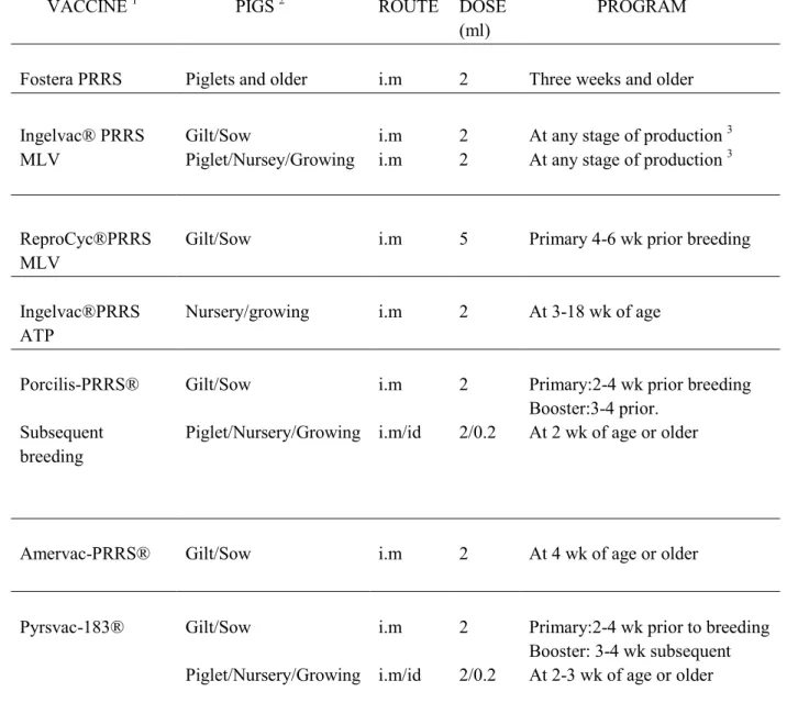
Table 2: List of Porcine reproductive and respiratory syndrome virus (PRRSV) serological assays that are used for specific antibodies detection…………...50 Table 3: Recommendation and vaccination schedule of commercial PRRS modified-live virus vaccines……….54 In vitro effect of deoxynivalenol (DON) mycotoxin on porcine reproductive and respiratory syndrome virus replication.
Table 1: Primers used for the evaluation of cytokine mRNA expressions……….84 In vivo effect of deoxynivalenol (DON) naturally contaminated feed on porcine reproductive and respiratory syndrome virus (PRRSV) infection.
Table 1: Diet composition ………110 Table 2: Mycotoxins content of the diets.……….111
viii
LIST OF FIGURES
Figure 1: Chemical structures of trichothecenes (Examples of groups A-D)………...9
Figure 2: Structure reaction of DON into DON-3-glucoside product……...………..12
Figure 3: Metabolism of DON in pig .……….17
Figure 4: Molecular mechanism of action of deoxynivalenol and others trichotheces .……….21
Figure 5: Schematic representation of the PRRSV genome organization ……...………...32
Figure 6: Schematic representation of the PRRSV non-structural proteins………33
Figure 7: Schematic representation of the PRRSV virion and the structural proteins………....36
Figure 8: Model of PRRSV entry into the porcine macrophage………..40
In vitro effect of deoxynivalenol (DON) mycotoxin on porcine reproductive and respiratory syndrome virus replication. Figure 1: Effect of DON on cell viability following PRRSV infection………...77
Figure 2: Effect of DON on cell mortality following PRRSV infection………...78
Figure 3: Effect of DON on PRRSV replication………...79
Figure 4: Cytokine mRNA expression by qPCR……….80
Figure 5: Caspase 3 activation……….81
Supplementary data 1: Morphology of noninfected and PRRSV infected MARC-145 cells underlight microscopy………..82
Supplementary data 1: Effect of DON on cell mortality following PRRSV infection: Effect of genotype………83
In vivo effect of deoxynivalenol (DON) naturally contaminated feed on porcine reproductive and respiratory syndrome virus (PRRSV) infection. Figure 1: Effect of DON naturally contaminated diets on piglet’s growth following PRRSV infection.………...105
ix
Figure 2: Time course of body temperature (°C) during experimental infection………..106 Figure 3: Effect of DON naturally contaminated diets on PRRSV viremia and viral load in the lungs………..107 Figure 4: Effect of DON naturally contaminated diets on macroscopic and microscopic lung
lesions in PRRSV infected animals………..108
Figure 5: Effect of DON naturally contaminated diets on PRRSV specific antibody response ……….………..109
x LIST OF ABBREVIATIONS AA Arachidonic acid aa Amino acids 3-ADON 3-acetyldeoxynivalenol 15-ADON 15-acetyldeoxynivalenol 3-15 ADON Acetyl-Deoxynivalenol ADWG Average daily weight gain AFB1 Aflatoxin B1
AM Alveolar macrophages
BAL Bronchoalveolar lavage
BALT Bronchiole-associated lymphoid tissue
CASP1 Active caspase-1
CMI Cell-mediated immunity CNS Central nervous system
CO2 Carbon dioxide
COX-2 Cyclooxygenase-2
CPE Cytopathic effect
DAS Diacetoxyscirpenol
DCs Dendritic cells
de-epoxy-DON Metabolite de-epoxy-deoxynivalenol
DON Deoxynivalenol
DOM-1 DON metabolite 1
dsRNA double strand RNA
EAV Equine arteritis virus
eIF2α Eukaryotic initiation factor 2α
ELISA Enzyme-linked immunosorbent assay
ERK1 Extracellular signal regulated protein kinase 1 ERK2 Extracellular signal regulated protein kinase 2 FB1 Fumonisin B1
xi
FBS Fetal bovine serum
FGSC Fusarium gramineum species complex FHB Fusarium head blight
GALT Gut-associated lymphoid tissue
GC-MS Gas chromatography–mass spectrometry GI Gastrointestinal
GPs Glycoproteins
GRMF Grain raw material for food 5-HT 5-hydroxy-typtamine Hck Hematopoietic cell kinase HIAA 5-hydroxyindole-3-acetic acid HSV-1 Herpes simplex virus type 1 IBDV Infectious bursal disease virus IBV Infectious bronchitis virus IECs Intestinal epithelial cells
IFA Indirect fluorescent antibody test IFN Interferon
Ig Immunoglobulin
IgAN Immunoglubolin A nephropathy IL Interleukin
i.m Intramuscularly i.n Intranasally IV Intravenous
IPS-1 IFN-β promoter stimulator 1 JAM Junctional Adhesion Molecule JNK 1/2 Jun N-terminal kinase 1 and 2
LD50 Lethal Dose 50
LDH Lactate dehydrogenase
LDV Lactate dehydrogenase-elevating virus LH Luteinizing hormone
xii
LV Lelystad virus
MA104 Monkey kidney cell line MAbs Monoclonal antibodies
MAPKs Mitogen-activated protein kinase MARC-145 Derived monkey kidney cells miRNAs MicroRNAs
MKK 1/2 Mitogen kinase kinases MLV Modified-live-virus MOI Multiplicity of infection NAbs Neutralizing antibodies NDV Newcastle disease virus NE Norepinephrine
N Nucleocapside
NNAbs Non-neutralizing antibodies NIV Nivalenol
NRTKs Non-receptor tyrosine kinases nsps Non-structural proteins
O2 Oxygen
OD Optical density ORFs Open reading frames OTA Ochratoxin A
P4 Progesterone
PAMs Porcine alveolar macrophages
PAMPs Pathogen-associated molecular patterns PBS Phosphate buffer saline solution PCR Polymerase chain reaction PCV2 Porcine Circovirus type-2
PGE2 Prostaglandin E2 PGH2 Prostaglandin H2 PLA2 Phospholipase A2 p.i. Post-infection
xiii
PIMs Pulmonary intravascular macrophages PKR Protein kinase R
PP Peyer’s patches pp Polyproteins
-1PRF -1 ribosomal frameshift
PRRSs Pathogen-recognition receptors
PRRSV Porcine reproductive and respiratory syndrome virus RdRp RNA-dependent RNA polymerase
RFU Relative fluorescence units or RFU RIG-1 Retinoic acid-induciblegene 1
RT-PCR Reverse-transcription polymerase chain reaction SFKs Src family kinases
SHFV Simian hemorrhagic fever virus S:P Sample-to-positive ratio
SPI-1 Salmonella pathogenicity island 1
SPJL St Jude porcine lung cells (epithelial cells line of the respiratory tract of swine)
SFKs Src family of protein kinases T4 Thyrosine 4
TGF-β Transforming growth factor beta TJs Tight junctions
TLRs Toll-like receptors
TNF-α Tumoral necrosis factor -alpha β-ZAL Beta zearalenol
ZEA Zearalenone α-ZOL Alpha zearalenol β-ZOL Beta zearalenol
xiv
To my parents, my little princess and my lovely wife
xv
ACKNOWLEDGEMENTS
I would like to thank all the people who were in contact with me during the course of my master degree, especially my director Younès Chorfi who trusted my abilities and conditions and gave me all the support, advices, patience and knowledge to have a critical views to be able to solve all the questions that were presented in this project.
During the project, the experience gained was very rewarding to increase my knowledge thanks to all members of the Veterinary Diagnostic laboratory of viral diseases especially to Dr. Chantale Provost and Dr. Christian Savard who supported me in the project. I would like to express my gratitude to Dr Carl A. Gagnon for the support and for giving me the opportunity to work in his laboratory to perform my project.
Moreover, I am grateful to Canadian Swine Research and Development Cluster (CSRDC) for funding my project. Swine and Poultry Infectious Disease Research Centre (CRIPA) and Innovagrains for the scholarship provided during the course of this project.
1
2
Mycotoxins are secondary metabolites synthesized by moulds, which colonize plants in fields and/or during storage. The Food and Agriculture Organization (FAO) estimates that as much as 25 % of the world’s agricultural commodities are contaminated with mycotoxins to a certain degree. Among these mycotoxins, deoxynivalenol (DON), also known as vomitoxin, is produced by Fusarium fungi and plays an important role as a toxin for humans and farm animals around the world because it is frequently found at toxicologically relevant concentrations. Deoxynivalenol can contaminate several types of grains such as wheat, barley and corn since Fusarium colonization is an increasingly common problem as a result of the expanded use of “no-till farming” and changing climate patterns (McMullen et al., 1997).
Pig diet is largely composed of grains, which represent the most important source of DON contamination. Reduced feed consumption and lower weight gain are the main clinical effects of the ingestion of DON-contaminated feeds. Moreover, DON has an immunomodulatory effect on pigs’ immune system depending on the concentration and duration of exposure; low concentrations of DON (1-2 mg/kg) are immunostimulatory while high concentrations (3-6 mg/kg) are immunosuppressive. While immunosuppression can be explained by the inhibition of the ribosome function and translation, immunostimulation can be related to interference with normal regulatory mechanisms such as suppression of normal immune response to pathogens apoptosis of macrophages and autoimmune-like effects which are similar to human IgA nephropathy (Pestka et al., 2004). Furthermore, DON rapidly activates mitogen-activated protein kinases (MAPKs) and induces cells’ apoptosis in a process known as ribotoxic stress response to cytotoxic interference (Iordanov et al., 1997). Consequently, feeding pigs with DON-contaminated diets may result in impaired immune function and decreased resistance to infectious diseases.
Swine industry faces many challenging diseases that have great financial impacts and pose a significant threat to the profitability. Among these diseases, porcine reproductive and respiratory syndrome (PRRS) is present worldwide. Since its emergence in late 1980s, PRRS has proven to be a persistent and insidious threat to the health and productivity of the US and Canadian swine herds (Neumann et al., 2005). The disease caused by the PRRS virus costs approximately $130 million per year to the Canadian swine industry (Mussell, 2010). This syndrome has many clinical manifestations in pigs at different physiological stages. In sows, it
3
is characterized by late-term abortions and an increased number of stillborn, mummified and weak born pigs. Piglets show respiratory problems and pneumonia (Rossow et al., 1994). In herds with multiple secondary diseases impacting production, co-infection with bacteria and viruses can exacerbate PRRS clinical signs. The virus of PRRS is known to infect a specific subset of pig macrophages that are mainly present in lungs; alveolar macrophages (Duan et al., 1997).
Previous studies have shown that the immunosuppressive effects of DON decrease host resistance to infectious viral diseases. In mice infected with reovirus, consumption of DON exacerbated bronchopneumonia clinical signs (Li et al., 2007) and interfered with immune response after enteric infection (Li et al., 2005). Since DON is frequently encountered in pig diets, its presence could exacerbate viral infection by PRRSV. On the other hand, to the author’s knowledge, there is no in vitro model that can determine the impact of DON on PRRS infection at the cellular level. Therefore, the objectives of this study were (1) to evaluate in vitro effects of DON on the replication of PRRSV in two permissive cell lines, cells derived from monkey kidney cells (MARC-145) and porcine alveolar macrophages (PAMs) and (2) to evaluate in vivo impact of DON-naturally contaminated feed on PRRSV infection in pigs.
4
5 1. Mycotoxins
1.1 Introduction
Mycotoxins are toxic and carcinogenic secondary metabolites produced by various fungal species that colonize plants in agricultural commodities and during storage. Forages and cereals represent the most important contamination source of mycotoxins and the level of contamination depends on processed stages before and after the harvest of raw materials (Devegowda and Murty, 2005). Temperature and humidity are important in mycotoxins’ production; fungi grow at temperatures between 20 °C and 30 °C with a high relative humidity (70 to 90%) (Nielsen et al., 2004). These temperatures are beneficial to deoxynivalenol (DON) production by promoting growth and development of Fusarium graminearum (Miller, 2001). It is important to note that if the grain is harvested at a high temperature, it can maintain this temperature for several days or even weeks in a storage facility unless cooling equipment can be provided to avoid fungus growth (Wegulo, 2012). Under field conditions, stress predisposes plants to infestation and colonization by toxigenic fungi. Some species of these fungi are able to produce more than one mycotoxin; these include species of Claviceps, Neoitphodium, Fusarium, Alternaria, Cladosporium, Diplodia, Gibberella and Helminthosporium (Devegowda and Murty, 2005).
Highest levels of mycotoxins are associated with insects and mites because they contribute to mould growth through physical damages of grain and by carrying mould spores. Their faecal material is an additional substrate for mould growth, which predisposes mould invasion of exposed endosperm (Munkvold and Desjardins, 1997). Also, metabolic activity produced by insects causes an increase in moisture migration, condensation, water leaks and fungal growth (Santin et al., 2005).
In stored grains, toxigenic fungal contamination and mycotoxin production result from a complex interaction between moisture, temperature and oxygen (O2) (Ominski et al., 1994). Although moulds are aerobic organisms that need O2 to grow normally, they can generally develop even when the O2 content is as low as ten percent of the normal atmospheric oxygen concentration. For example, Aspergillus flavus can grow in environments containing O2
6
Moisture content and ambient temperature are key determinants of fungal colonization and mycotoxin production. It is conventional to subdivide toxigenic fungi into “field” (plant-pathogenic) or “storage” (saprophytic/spoilage) organisms (Santino, 2005). Field fungi are those that invade seeds while crops are still in the field and require high moisture conditions (20-21%); these include species such as Claviceps, Fusarium, Cladosporium, Diplodia, Gibberella and Helminthosporium. Storage fungi species such as Aspergillus and Penicillium invade grains or seeds and need lower moisture conditions (13-18%); usually, they do not present any serious problem before harvest (Santino, 2005).
To avoid mycotoxin accumulation in stored grains and oilseeds, moisture has to be controlled. If the product is too dry to allow fungal growth and if it is kept dry, no further deterioration will occur. However, if there is insect or rodent activity, moisture migration, condensation, or water leaks, fungal growth could lead to mycotoxin contamination (Patience and Ensley, 2010).
1.2 Common mycotoxins in swine
1.2.1 Aflatoxins
Aflatoxins are produced primarily by a common fungus, Aspergillus flavus and closely related species A. parasiticus. These are well-defined species: A. flavus produces only B aflatoxins and A. parasiticus produces both B and G aflatoxins. Aflatoxin contamination is almost exclusively confined to tropical feeds in drought conditions; it is not produced in cold temperatures since high temperatures are needed for toxin synthesis (Sweeney & Dobson, 1998). Among the four major types of aflatoxins, aflatoxin B1 (AFB1) is the most toxic and carcinogenic to animals but outbreaks occur mostly in pigs and cattle. Piglets are extremely sensitive to aflatoxins and susceptibility decreases with age (Dhanasekarani et al., 2011). Therefore, aflatoxicosis clinical signs depend on the species, age, concentration and duration of the exposure. Normally, sows and boars tolerate concentrations over 0.5 mg/kg in feed for short periods (3 to 5 days), but when sows are fed for extended periods (1 to 3 weeks), aflatoxins concentrations in the feed should not exceed 0.1 mg/kg (Dhanasekarani et al., 2011).
7
In pigs, ingestion of 1 to 5 mg/kg of AFB1 reduced growth and feed efficiency and animals died within 12-20 hours (Drabeket al., 1979). Chronic exposure at concentrations of 0.1 to 0.5 mg/kg can result in suppressed immune response and decreased growth rate. The main target organ of aflatoxins is the liver. After aflatoxins’ invasion of the liver, lipids infiltrate hepatocytes which lead to hepatic centrilobular necrosis and bile duct hyperplasia (Weaveret al., 2013). The clinical syndrome in pigs includes depression, anorexia, decreased feed conversion and growth, weight loss, muscular weakness, bloody rectal discharge and icterus. At necropsy, livers of pigs that were fed toxic concentrations of AFB1 (0.5 mg/kg) vary in color from tan to pale yellow, are atrophied, and have abnormal amounts of fibrous connective tissue that is resistant to cutting (Dhanasekaran, 2011).
1.2.2 Ochratoxins
Ochratoxins are secondary metabolites of Aspergillus (mainly A. ochraceus) and
Penicillium (especially P. verrucosum and P. carbonations). Actually, the optimal temperature for ochratoxin synthesis by Aspergillus ochraceus is 28 °C, this synthesis being very low at 15 °C or at 37 °C. However, Penicillium viridicatum develops and can produce ochratoxins at a range of temperatures between 4 and 30 °C (Marin et al., 2009).
While ochratoxins B and C are less important, the most prevalent is ochratoxin A (OTA) which is found in cereal grains such as wheat, corn, rye and barley, but can also be found in rice, soybeans, coffee, cacao, beans, peas, peanuts and dried fruits (Marin et al., 2009). When pigs consume OTA over a long period of time, it can contaminate most of the edible tissues including organs, fat, muscle and blood. The main clinical symptoms of OTA include reduced growth rate and feed efficiency. Liver damage may occur, but the main effect is on the kidney, resulting in interstitial fibrosis. Increased water intake (polydipsia), and hence increased urine output (polyuria) are signs of this intoxication (Marin et al., 2009). Toxic effects of OTA in pigs depend on the concentration; for example, pigs fed 2.5 to 14 mg/kg of OTA are severely depressed with severe diarrhea, emesis and fever. Dehydration occurs rapidly following polydipsia and polyuria. The most obvious damage caused by OTA is observed in the kidneys with strong degenerative changes in proximal tubules and fibrosis (Stoev et al., 2012; Szczech et al., 1973).
8 1.2.3 Trichothecenes
Trichothecenes represent a very large family of chemically-related mycotoxins produced by various species of Fusarium, Myrothecium, Trichoderma, Trichothecium, Cephalosporium, Verticimonosporium, and Stachybotrys. Trichothecenes are classified into four groups: A, B, C, and D. All trichothecenes share a common tricyclic skeleton with an epoxide function. Group A trichothecenes include T-2 toxin, HT-2 toxin and diacetoxyscirpenol (DAS). Group B trichothecenes include DON and its four derivatives: 3-acetyl deoxynivalenol (3-ADON), 15-3-acetyl deoxynivalenol (15-ADON), DON-glucoside, and nivalenol (NIV). Group C includes compounds such as crotocine and group D includes compounds such as satratoxin G, but these two groups are less important than groups A and B (Figure 1 ) (Wu et al., 2010; Puri & Zhong, 2010).
The most essential structural features of trichothecenes affecting biological activities are: a) the double bond at C-9 and C-10 which is a basic requirement for toxic effects and biological activity for trichothecenes. The reduction of this double bond at C-9 and C-10 results in a substantial decrease in activity and causes inhibition in protein synthesis of eukaryotic cells; b) the presence of the 12, 13-epoxide ring as a typical structure for trichothecene toxicity. Since the epoxide ring is necessary for disruption of protein synthesis, this plays a significant role in the inhibitory mechanism; c) the 5 variable R groups, the structure and position of a side chain, the presence of a second epoxy ring in type C members such as crotocine that have another epoxy group at C-7 and C-8 position; and d) compounds in type D, such as satratoxins and roridins include a macrocyclic ring at C-4 and C-15 (Wu et al., 2013; Bräse et al., 2009).
9
Figure 1: Chemical structures of trichothecenes (Examples of groups A-D) adapted from Wu et al. (2010).
1.2.3.1 Group A T-2 and HT-2 toxins
T-2 and HT-2 toxins are produced by fungi F. acuminatum, F. equiseti, F. poae, and F. sporotrichoides. They often occur together in infected cereals, which are commonly found in various cereal crops (wheat, maize, barley, oats, and rye) and processed grains (malt, beer and
Group A: T-2 toxin Group B: Deoxynivalenol Group C: Crotocin Group D: Satratoxin 9 9 9 9 8 7 78 4 15 10 10 10 10
10
bread) (Balzer et al., 2004). T-2 toxin is rapidly metabolised in the intestine and liver to HT-2 toxin, which is the major in vivo metabolite. After ingestion by pigs and most other animal species, HT-2 toxin is rapidly absorbed and then distributed throughout the organism with very little accumulation in organs (Balzer et al., 2004). T-2 toxin at ≥ 0.6 mg/kg intravenous IV produced a large range of toxic effects in animals, especially in pigs, such as emesis, diarrhea, weight loss, lethargy, hemorrhage of lymph nodes and edema. Pathological changes in the stomach and liver have been reported along with necrosis and damage of cartilaginous tissues. (Li et al., 2011).
In pigs, acute effects of T-2 toxin occur after oral exposure at concentrations over 10-16 mg/kg and produce non-specific symptoms such as weight loss, feed refusal and vomiting. Sub-chronic concentrations of ≥ 1 mg/kg reduced body weight gain (European commission, 2001).
1.2.3.2 Group A Diacetoxyscirpenol
Diacetoxyscirpenol (DAS) is a potent mycotoxin produced by certain Fusarium strains (F. equiseti, F. poae, F. acuminatum, F. sambucinum and F. sporotrichoides). A small amount is sufficient to cause toxicosis in pigs. Diacetoxyscirpenol has been found in unharvested corn, silage, soybeans, as well as in finished feeds using corn and soybeans as ingredients (Jacobsen et al., 2007). All domestic animals, including pigs, are susceptible to dietary poisoning at low concentrations: 1 to 3.5 mg/kg. Clinical signs produced by DAS in pigs are established as feed refusal and decrease in weight gain. Diacetoxyscirpenol is especially known to produce severe inflammation of the gastrointestinal tract and possible hemorrhage, accompanied with bouts of diarrhea, lethargy and abdominal pain (Jacobsen et al., 2007).
1.2.3.3 Group B Deoxynivalenol (DON)
The occurrence of DON is associated primarily with Fusarium graminearum (Gibberella zeae) and F. culmorum. The exposure risk to animals is directly through foods of plant origin (cereal grains). It is found predominantly in grains such as wheat, barley, oats, rye, and maize. Deoxynivalenol plays an important role as a mycotoxin which affects farm animals in all parts of the world. Pigs are known to be especially susceptible to DON (Sobrova et al.,
11
2010; Placinta et al., 1999). It induces feed refusal at concentrations of ≥ 2 to 3 mg/kg and produces emesis at 7 to 10 mg/kg. These two effects are probably linked because DON is transported to the brain, where it runs dopaminergic receptors and alters brain neurochemicals and the serotoninergic system which play roles in the mediation of feeding behaviour and emetic response (Rotter et al., 1996).
1.2.3.4 Group B Acetyl DON (3-ADON) and 15-acetyl DON (15-ADON)
Fusarium graminearum strains produce two modified forms of DON called 3-ADON and 15-ADON. These trichothecenes are also detectable in cereals and differ by the presence of acetyl functions at C3 and C15 (Alexander et al., 2011). Surveys of naturally infected grain in the U.S. and Canada indicate that 3-ADON and 15-ADON are found only at low levels when high amounts of DON are present. However, it is not yet known if 3-ADON produces more DON in field-grown plants than ADON (Mirocha et al., 1989). Concentrations of 15-ADON as a DON precursor (50 to 75 mg/kg) in swine increased the emetic activity (Pestka et al., 1987).
1.2.3.5 Group B DON-glucoside
Detoxification reactions in plant tissue involve sequestration of compounds conjugated to glucose which are produced in the cell wall into the vacuole and are excreted by root exudation. This way, DON-glucoside can be stored in plant vacuoles, where they are protected against cleavage by beta-glucosidases. These are enzymes which catalyze the transfer of glycosyl group between oxygen nucleophiles resulting in the hydrolysis of beta glycosidic linkage present between carbohydrate residues in aryl-amino or alkyl-beta-D-glucosides, and cyanogenic glucosides under physiological conditions, while synthesis of glycosyl bond between different molecules can occur by hydrolysis and transglycosylation (Singhania et al., 2013; Berthiller et al., 2005). The trichothecene glucosides such as DON-3-glucoside and DON-15-glucoside have been obtained from Fusarium sulphureum cultures (Berthiller et al., 2005). The capacity of several plants to transform DON by conjugation to glucose was demonstrated by Poppenberger et al., (2003) who isolated and characterized a cytoplasmatic UDP-glycosyltransferase from model plant of Arabibidopis thaliana capable of transforming
12
DON into DON-3-β-D-glucopyranoside (DON-3-glucoside), where the hydroxyl group in position C13 of DON is replaced by a glucose unit (Figure 2).
Figure 2: Structure reaction of DON into 3-glucoside product (DOGT1: DON-glycosyltransferase). Taken from Poppenberger et al (2003).
1.2.3.6 Group B nivalenol
Nivalenol belongs to group B trichothecenes and is produced by Fusarium genus, Fusarium cerealis (F.crookwellence) and Fusarium poae and often co-occurs with DON. These fungi are abundant in various cereal crops such as wheat, maize, barley, oats and rye and processed grains like malt, beer and bread (Pettersson & Hedman, 1997). The capacity of NIV to produce clinical signs depends on the ingested concentration. Several countries have established regulation of NIV; concentrations between of 0.5 to 2 mg/kg produce no clinical signs in pigs. However NIV at 2.5 to 5 mg/kg of the diet caused vomiting and feed refusal. Pathological alterations in kidneys and the gastrointestinal tract have also been reported. Concentrations over 6 mg/kg have been described to cause feed refusal, decreased weight gain and increased serum alkaline phosphatase activity (Hedman et al., 1997).
1.2.4 Zearalenone
Zearalenone (ZEA) is a non-steroidal estrogenic mycotoxin biosynthesized by a variety of Fusarium fungi, including F. graminearum (Gibberella zeae), F. culmorum, F. cerealis, F. equiseti, F. crookwellense and F. semitectum (Bennett and Klich, 2003). In Europe, corn is the cereal at risk with high incidence and high levels of contamination with ZEA (EFSA, 2011).
Deoxynivalenol Deoxynivalenol-3-glucoside
10
9 9
13
In Canada, ZEA concentrations over 141 mg/kg were reported in corn and corn-based foods, as well as in wheat, barley and soybeans (Scott, 1997).
In pigs, ZEA is rapidly absorbed in the small intestine after oral administration. Indeed, over 85% of ZEA is absorbed by the intestinal lumen and can be found in the blood in less than 30 min. Once absorbed, enzymes such as CyP450 in small intestine epithelial cells and the liver can quickly convert ZEA into alpha zearalenol (α-ZOL), beta zearalenol (β-ZOL) and beta zearalanol (β-ZAL), the main metabolites which are subsequently conjugated with glucuronic acid (Kanora & Maes, 2013; Zinedine et al., 2007). In pigs, exposure to high concentrations of 20 mg/kg affects the prepubertal and cycling of young gilts which are more sensitive than pregnant sows. During pregnancy, ZEA reduces embryonic survival, decreases foetal weight, and may cause abortion. Moreover, ZEA causes alterations to the uterine environment by decreasing LH and progesterone secretion and by altering uterine morphology (Tiemann & Danicke, 2007). In gilts, concentration over 3 mg/kg induces low growth rate, alters nutrient availability and produces changes in genital organs, liver, as well as kidney functions. Histopathological changes suggest that ovaries are inflamed and hyperoestrogenized. Exposure to chronic concentration of 0.35 mg/kg for 35 days did not show any effect on uterus weight (EFSA, 2011; Doll et al., 2003).
1.2.5 Fumonisins
Fumonisins were first isolated in South Africa in 1988 from cultures of Fusarium verticillioides, formerly Fusarium moniliforme, and F. proliferatum. Fumonisins’ mechanism of action appears to involve disruption of sphingolipid biosynthesis by the inhibition of ceramide synthase, modification of cellular proliferation through changes in cell cycle regulators, and increased expression of cytokines such as tumoral necrosis factor alpha (TNFα). There are two prevalent isoforms of fumonisins: fumonisin B1 (FB1) and B2 (FB2) (Colvin et al., 1993).
1.2.5.1 Fumonisin B1
Fumonisin B1 commonly contaminates corn, wheat and other cereals worldwide. It is highly toxic for many farm animals including ducklings, horses, pigs, rats, and sheep. In pigs,
14
ingestion of dietary concentrations of 16 mg/kg results in pulmonary oedema with hydrothorax and concentrations over 200 mg/kg produce visible icterus by day 14 post-ingestion, with severe depression and anorexia. Lower concentrations of 1 to 10 mg/kg did not cause any clinical signs. At necropsy however, these concentrations produced evident pulmonary changes (Zomborszky-Kovacs et al., 2002). Histopathological examination demonstrated that FB1 is hepatotoxic and nephrotoxic in pigs. The earliest histological change to appear in either liver or kidney in fumonisin-treated animals is the increase of apoptosis followed by regenerative cell proliferation. In the liver, it produces hepatocellular degeneration and intermittent necrosis. Fumonisin B1 causes two diseases, which occur in horses and pigs with rapid onset: equine leukoencephalomalacia and porcine pulmonary oedema syndrome. Both of these diseases involve disturbed sphingolipid metabolism and cardiovascular dysfunction changes (Haschek et al., 2001; Fazekas et al., 1998).
1.2.5.2 Fumonisin B2
Fumonisin B2 is produced by Fusarium verticillioides and Fusarium moniliforme and is more cytotoxic than FB1. It has been demonstrated that FB2 is carcinogenic. FB2 causes lesions to the liver, kidney and heart of pigs at dietary concentrations ≥ 58 mg/kg (Voss et al., 2007).
1.3 Deoxynivalenol and its toxicity
1.3.1 Absorption, metabolism and elimination of DON
In pigs, after consumption of contaminated feed, DON is rapidly and efficiently absorbed in the stomach and intestine. The majority of DON ingested from the diet is conjugated and de-epoxidated in the proximal small intestine by microbes, which have a major effect on the bioavailability of DON and its metabolites, where it induces epithelial damage and inflammatory response causing inhibition of nutrient absorption and compromising intestinal barrier function (Wache et al., 2009). Between 54 and 89 % of the ingested toxin is absorbed through the jejunum in large amounts and can cross the intestinal epithelium and reach the blood compartment. Deoxynivalenol is observed in the plasma after 30 min and its
15
serum concentration reaches a peak value within three to four hours post-ingestion (Goyarts & Danicke, 2006; Eriksen et al., 2003). Deoxynivalenol can be detected very rapidly in other organs; for example, in the cerebral spinal fluid, DON is detected in less than 2.5 min following IV administration in pigs. When DON is administered IV, the affected organs are those that are the most perfused such as the lungs, myocardial muscle, kidneys and brain (Eriksen et al., 2003; Prelusky et al., 1990).
To characterize the pathophysiologic effect of DON, it is important to understand the integrity of the intestinal mucosa, which is the border between the organism and the environment. This is the first barrier for nutrients and potentially harmful substances such as mycotoxins (Fanning et al., 1999). Epithelial cell architecture must exhibit polarity for them to meet these functional demands. Intestinal epithelial cells (IECs) are in charge of two crucial processes: transporting nutrients and fluids and the restricting the access for luminal antigens to the internal milieu (Madara et al., 1987). These cells express intercellular connections known as tight junctions (TJs), which form a largely impermeable barrier to prevent unspecific penetration of molecules and microorganisms into the body, and possess different types of transmembrane proteins such as occludin, claudins and junctional adhesion molecule (JAM) for regulating TJ assembly and function (Fanning et al., 1999). These molecules extend into the intercellular space and determine the permeability characteristics of TJs in terms of specificity and tightness (Harhaj & Antonetti, 2004). Barrier disruption is an important etiologic factor of intestinal inflammation, causing not only defective nutrient uptake and retention, but also increased permeability to luminal antigens with subsequent contact and activation of underlying immune cells (Harhaj & Antonetti, 2004).
Metabolism of DON is used to describe conversion of the native toxin to various degradation derivatives occurring mainly in the digestive tract with high concentrations. Substantially lower levels were detected in organs such as kidney, liver, heart, lung, spleen and brain. Derivatives of DON include its microbial metabolised de-epoxidized form, called de-epoxy-DON, DOM-1 (DON metabolite 1) or simply de-DON (Figure 3), and their conjugated forms as a result of the phase I and II metabolism (Danicke & Brezina, 2013). The only metabolite in pigs, DOM-1 or de-epoxy-DON, was identified by gas chromatography– mass spectrometry (GC-MS). Several microorganisms in the caudal segments of the gut,
16
particularly the colon content, showed the strongest metabolic activity for reduction of 12-13 epoxide group of DON which is responsible for its toxicity. Also, DON and DOM-1 were present in the kidneys and were found to be higher in muscles and liver (Doll et al., 2008).
The first effect is mediated by different gastrointestinal and hepatic enzymes that result in the oxidation, reduction or hydrolysis (phase I reactions). Phase I, also called transformation phase, includes reactive groups which are introduced into the molecules by oxidation or dealkylation for instance. In many cases, activation of the molecules through phase I biotransformation is necessary for conjugation, but if the reactive groups are already present in the parent molecule, they may undergo direct conjugation (Berthiller et al., 2007).
Phase II includes sulfation and glucuronidation reactions, along with glutathione conjugation, methylation, amino acid conjugation, and acetylation of DON considered as detoxification pathways, which increase the water solubility. Phase II detoxification typically involves biochemical conjugation by which various enzymes in the liver attach small chemical moieties of the toxin. Phase II enzymes are also widespread in extra-hepatic tissues, especially in the lungs, kidneys, skin and gastrointestinal tract (Goyarts & Danicke, 2006). Glucuronidation is a significant pathway in the detoxification mechanism of Phase II. Glucuronidation requires the enzyme UDP glucuronyltransferase to combine glucuronic acid with the toxin. The degree of conjugation varies markedly and depends on tissue-specific activities of the UDP-glucuronosyltransferases and sulfotransferases (Goyarts & Danicke, 2006).
Following oral intoxication of DON, 68% of the toxin is excreted in the urine as DON, DOM-1 and glucuronide-DON. The remaining DON (20%) is eliminated in faeces as DON, DOM-1 and low amounts of 3-ADON. Excretion mechanisms of DON/DOM-1 and glucuronide-DON/DOM-1 are unknown; however, they could involve both glomerular filtration of the metabolites present in the blood and their excretion though P-glycoproteins expressed by intestinal, renal or hepatic epithelial cells (Goyarts & Danicke, 2006; Eriksen et al., 2003). IV and intragastric administration of high concentrations of DON (3000-1000ppb) suggested that DON was very rapidly absorbed and eliminated. However, residues remain in
17
pigs because of the extensive systemic distribution and the poor metabolism of DON by different organs (Prelusky et al., 1988).
Figure 3: Metabolism of DON in pig. Modified from Danicke and Brezina (2013). 1.4 Toxicological effects
1.4.1 Acute toxicity
The common toxicity symptoms following acute DON poisoning in sensitive species such as pigs include abdominal distress, increased salivation, discomfort, anorexia, diarrhea and emesis. Also, a complete feed refusal is evident at 12 mg/kg (Rotter et al., 1994). Although DON is less toxic than T-2 toxin, extremely high DON concentrations (≥27 mg/kg) can cause shock and death in pigs (Pestka, 2007). Lethal DON concentrations (≥ 27 to 30 mg/kg) evoke histopathologic effects ranging from hemorrhage/necrosis of intestinal tract, necrosis of bone marrow and lymphoid tissues as well as kidney and heart lesions (Forsyth et al., 1977; Pestka et al., 1987).
1.4.2 Chronic toxicity
Prolonged dietary exposure to DON causes decreased weight gain, anorexia, and altered nutritional efficiency. It was suggested that monogastric species such as mice and pigs,
D IGES TIVE T RACT ORAL INTAKE DON DON FAECAL EXCRETION DOM-1 D ECONJ UGA TI ON INTESTINAL MUCOSA DON CONJUGATE DON DOM-1 CONJUGATE DOM-1 BILARY EXCRETION ENTERO-HEPATIC CYCLING LIVER SYSTEMIC CIRCULATION URINARY EXCRETION
18
experience severe growth and weight gain suppression during chronic exposure to DON (Pestka, 2007). Deoxynivalenol concentrations at 1-2 mg/kg caused partial feed refusal in pigs ingesting naturally contaminated feed (Pestka et al., 1987). However, pigs fed with DON concentrations at 2 mg/kg showed loss of weight gain during the first 8 weeks and DON concentrations at 4 mg/kg decreased feed intake, weight gain and efficiency of feed utilization (Bergsjo et al., 1992). In conclusion, pigs are very sensitive to growth performance and weight suppression following a DON diet contamination exceeding 1 mg/kg.
1.5 Protein synthesis inhibition
Deoxynivalenol binds to ribosomes (40S and 60S) in eukaryotic cells and produces protein synthesis inhibition (Ueno, 1984). Most of the trichothecenes predominantly inhibit initiation and DON is an inhibitor of elongation of protein synthesis which requires the presence of unsaturated bond at the C9-C10 position, integrity of the 12, 13 epoxy ring, substitution of the hydroxyl of other groups at appropriate positions on the trichothecene nucleus, and the structure and position of a side chain. (Dänicke et al., 2000; Ueno, 1984). Therefore, these features can all influence the extent of cytotoxicity and protein synthesis inhibition. The opening of the 12-13- epoxide ring results in loss of any apparent toxicity. The effect of a particular trichothecene on protein synthesis depends on the number and position of the hydroxyl groups and on the type of esterifying acids (Betina, 1989). Deoxynivalenol with substituents at both C-3 and C-4 predominantly inhibit polypeptide chain initiation. The cytotoxicity of DON, a trichothecene with a keto group at C-8 and a hydroxyl group at C-7, results from protein synthesis inhibition at the ribosomal level during the elongation and termination step in mammalian cells (Ueno, 1983; Ehrlich & Daigle, 1987).
As we know, the most prominent molecular target of DON is the mentioned 60S ribosomal subunit. Deoxynivalenol enters cells and binds to active ribosomes, which transduce a signal to protein kinase R (PKR) and hemoitopoeitic cell kinase (Hck) by different mechanisms involved in this inhibition of translation. First, in phagocytes, DON can induce activation of ribosome-associated kinase known as double-strand RNA associated PKR. When activated, PKR can phosphorylate eukaryotic initiation factor 2α (eIF2α) impeding translation (Zhou et al., 2003). Second, DON can promote intracellular 28S rRNA cleavage, potentially
19
by facilitating the action of endogenous RNases or by upregulating RNase expression, which could impede ribosome function and translation (Li & Pestka, 2008). A fundamental issue relates to the nature of the linkage between ribosomal RNA damage and induction of MAPKs (Mitogen-activated protein kinase) signaling cascades. Unidentified intermediate signal transduction steps, possibly protein-mediated, might occur between toxicant-damaged 28S rRNA and MAPKs (Iordanov et al., 1997). Third, DON can up-regulate a large number of micro RNAs (miRNAs), with selected gene downregulation. Thus, it might be speculated that DON-exposed cells employ miRNA downregulation of ribosome synthesis to conserve and redistribute resources needed for survival (Pestka, 2010). It is also known that DON and other translation inhibitors which bind to ribosomes can rapidly transduce a signal to PKR and Hck to activate MAPKs and induce apoptosis, a process known as the “ribotoxic stress response”. MAPKs module physiological processes including cell growth, differentiation and apoptosis which are critical to the signal transduction of immune response (Figure 4) (Zhou et al., 2005). Mitogen-activated protein kinase subfamilies include p44 and p42 also known as extracellular signal-regulated protein kinase 1 and 2 (ERK1 and 2), p54 and p46 c-Jun N-terminal kinase 1 and 2 (JNK 1/2) and p38 MAPK. MAPKs are activated by dual phosphorylation at the tripeptide motif Thr-Xaa-Tyr (Dong et al., 2002). The sequence of this tripeptide motif is different in each group of MAPKs: ERK (Thr-Glu-Tyr); p38 (Thr-Gly-Tyr); and JNK (Thr-Pro-Tyr). The dual phosphorylation of Thr and Tyr is mediated by a conserved protein kinase cascade. The ERK MAPKs are activated by the MAP kinase kinases (MKK) MKK1 and MKK2; the p38 MAPKs are activated by MKK3, MKK4 and MKK6; and the JNK pathway is activated by MKK4 and MKK7 (Casteel et al., 2010; Pestka, 2008; Zhou et al., 2005).
In macrophages, ERK and p38 are involved in DON induced transactivation of TNF-α and cyclooxygenase-2 (COX 2), whereas p38 appears to be involved in DON mediated mRNA stability (Moon & Pestka, 2003). This activity is possibly restricted to selected stages of the ribosomal cycle. These two kinases (PKR and Hck) potentially mediate signalling of DON to induced MAPK activation resulting in ribotoxic stress response (Zhou et al., 2003; Iordanov, 1997).
20
Protein kinase R is a member of a small family of kinases, which are expressed seritine/threonine protein kinases activated by extracellular stress and phosphorylate the α-subunit of protein synthesis eukaryotic initiation factor 2 (eIF-2), thereby inhibiting protein synthesis. The activation of PKR during infection by viral double strand RNA (dsRNA) intermediates results in the inhibition of viral replication. Protein Kinase R also mediates the activation of signal transduction pathways by pro-inflammatory stimuli, including lipopolysaccharide (LPS), TNF-α, and interleukin (IL)-1 (Williams, 2001). Moreover, it is known that PKR is a critical upstream mediator of the ribotoxic stress response induced by DON resulting in a critical upstream role in the ribotoxic stress response inducible by translational inhibitors (Zhou et al., 2003).
One distinct family of protein tyrosine kinases consists of Src non-receptor tyrosine kinases (NRTKs) which have been called “rheostats for immune signalling”. One member of this family is Hck which transduces a variety of extracellular signals capable of impacting cellular processes related to proliferation, differentiation and migration. The Src family of protein kinases (SFKs) can be divided into two groups: those that exhibit a relatively ubiquitous pattern of expression (Src, Yes, Fyn) and those whose expression is restricted to the hematopoietic system, such as Lck in T lymphocytes and Hck in myelomonocytic cell lineages (Ernst et al., 2002; Moarefi et al., 1997). The Src-family-selective tyrosine kinase inhibitors PP1 and PP2 effectively block DON to activate MAPKs, which suggest involvement of the Hck. The PP1 reduces DON-induced increases in nuclear levels and binding activities of several transcription factors (NF-κB, AP-1, and C/EBP), which corresponded to decreases in TNF-α production, caspase-3 activation, and apoptosis. As expected for a signalling event upstream of MAPK activation, tyrosine phosphorylation of the Hck was detectable as early as 1 min, maximized at 2.5 min then declined within 30 min after DON addition (Zhou et al., 2005; Ernst et al., 2002).
21
Figure 4: Molecular mechanism of action of deoxynivalenol and other trichothecenes. Reproduced from Pestka et al., (2007).
1.6 Cytotoxicity
Numerous studies of DON’s cytotoxicity have been conducted because of DON’s capacities to alter host resistance, humoral and cell-mediated responses, cytokine expression, and apoptosis of lymphoid tissue in humans and animals (Sobrova et al., 2010). The common theme is that DON can be immunostimulatory or immunosuppressive depending on the dose and exposure frequency (Bondy & Pestka, 2000). Immunosuppression can be explained by the inhibition of translation when DON binds to the active ribosomes and immunostimulation can be related to interference with normal regulatory mechanisms (Bondy & Pestka, 2000; Rotter
22
et al., 1996). Stimulation of macrophages by DON at the concentration of 1 to 2 mg/kg could upregulate the expression of inflammation-related genes in vivo and in vitro. These include COX-2 and pro-inflammatory cytokines (Zhou et al., 2005; Moon & Pestka, 2002). Exposure to DON concentrations at 3 to 5 mg/kg upregulates the expression of many immune-related proteins, including Th1 and Th2 cytokines as well as chemokines. In contrast, it appears that the suppressive effects of DON on leukocyte function are intimately linked to the induction of apoptosis of macrophages, T cells and B cells isolated from peyer’s patches (PP), thymus, and bone marrow, which represent the functional cell repertoire of the immune system. Thus, DON immune modulation might be considered as a spectrum whereby low doses stimulate immune function, while high doses increase leukocyte apoptosis leading to immunosuppression (Pestka et al., 2004; Pestka et al., 1994).
1.7 Gene upregulation
Selective elevation of cytokines, chemokines and other immune-related proteins by DON is immediately preceded by increased expression of their mRNAs. For example, DON upregulates mRNAs of TNF-α by increasing transcription and mRNA stability through activation of p38 and interleukin (IL)-6 in macrophages (Chung et al., 2003).
Mice fed with a diet containing DON concentrations at 10 mg/kg dysregulates immunoglobulin (Ig) A production in serum and results in immunopathology. The mechanism by which DON induces IgA dysregulation is unclear. However, the gut mucosal compartment may be a primary target of DON to produce the aberrant stimulation of the gut mucosal immune system (Jia & Pestka, 2005). Deoxynivalenol can induce proinflammatory cytokines, particularly IL-6 which could impair oral tolerance and with subsequent polyclonal activation of IgA-committed B cells to terminal differential (Pestka & Zhou, 2000). This appears to contribute to high concentrations of serum IgA as well as IgA deposition in the kidney and DON could be an etiological factor in IgA nephropathy (IgAN).
The capacity of DON to expand polyclonal IgA-secreting cells in mice is mediated by increased cytokine, mainly IL-6 production by macrophages and T cells. These lymphocytes can be involved in the polyclonal expansion of IgA secreting cells which is observed in mice
23
fed with DON (Pestka, 2003; Pestka et al., 1989). A study provided evidence that dietary DON enhanced terminal differentiation of IgA secreting cells in PP. The migration of IgA secreting cells into the systemic compartment favours a shift from IgG to IgA as the primary serum isotype. At the same time, there is an increased help for terminal differentiation of control PP B cells upon addition of T cells from PP of DON-fed mice (Pestka, 2003).
On the other hand, DON-mediated elevations in cytokines, chemokines, immune related proteins and enzymes induce of COX-2. Selective inhibitor COX-2 is an enzyme that catalyzes oxygenation of arachidonic acid to transform prostaglandin H2 (PGH2) endoperoxides which are subsequently converted to prostaglandins and thromboxane A2. Also COX-2 stimulates cancer cell proliferation and promotes angiogenesis (Moon & Pestka, 2002; Tsuji et al., 2001; Smith et al., 2000). Selective inhibitor COX-2 induced production of prostanoids that are often involved in inflammatory diseases, characterized by edema and tissue injury due to the release of many inflammatory cytokines and chemotactic factors, prostanoids, leukotrienes, and phospholipase (Arslan & Zingg, 1996).
1.8 Other animals affected by DON
1.8.1 Cows
The impact of DON on dairy cattle is not established and the susceptibility of ruminants to DON is low, as DON is converted almost completely into a less toxic metabolite, DOM-1 by the rumen flora (Fink-Gremmels, 2008). No effects were detectable in rumen and duodenal flow in dairy cows even if they were fed DON at 7 mg/kg. The toxin was nearly completely metabolized to DOM-1 indicating a complete degradation (Danicke et al., 2005). However, other clinical data showed an association between DON and poor performance in dairy herds. Cows fed DON at 2.6 to 6.5 mg/kg produced less milk (13% or 1.4 kg) compared to cows on uncontaminated feed (Charmley et al., 1993).
1.8.2 Poultry
In general, poultry have a tolerant nature to the presence of dietary DON with feeds containing low concentrations at 1 to 5 mg/kg. Reasons for poultry tolerance to DON probably
24
include poor toxin bioavailability and rapid elimination from the body (Danicke et al., 2001). It has been suggested that dietary DON concentration at 5 mg/kg was necessary to negatively influence the growth performance of broilers. It was also demonstrated that DON has the capacity to decrease absorption of nutrients such as glucose and amino acid in the small intestine and to impair immune functions in broilers by inducing changes in the haematopoietic system and by altering lymphocytes’ proliferation (Awad et al., 2008).
1.8.3 Rats/mice
Absorption and clearance of DON in rodents is also rapid: mice and rats are sensitive to DON but there seems to be differences in the kinetic, metabolism and tissue distribution of DON. Studies have demonstrated that DON reduces weight gain. For example, female mice B6C3F1 were fed with a high fat diet for 8 weeks to induce obesity. Then mice were fed DON concentrations at 0, 2, 5, 10 mg/kg for an additional 8 weeks. Animal fed with higher DON concentrations, i.e. 5 and 10 mg/kg, showed a decrease of 16 and 23% in weight gain respectively (Flannery et al., 2010).
1.9 Control measures to reduce DON contamination
The proliferation of Fusarium in cereal crops may occur when the development of mycelia is followed by ascospores’ production in sexual fruiting structures located on the surface of the pathogen-infested residue. The incidence is most closely related to the presence of moisture at the time of flowering and during and after rainfall, rather than to the amount of rain or the distribution of [14C] DON on crop debris (Miller, 1994).
Consequently, measures taken to control or minimize Fusarium infection will also reduce DON formation. Such measures include agronomic practices, cultivars, use of fungicides, biocontrol and genetic resistance, which are the most dependable and cost-effective ways to control Fusarium head blight (FSB). Agronomic practices include suitable crop rotation, appropriate use of fertilizers, irrigation, and weed control (Milus & Parsons, 1994) Maize-wheat rotation increases the incidence of FHB and should be avoided, whereas removal or ploughing of crop debris reduces the incidence in wheat. Rotation with non-graminaceous crops such as soybean, burial or burning of corn residue appears to be very
25
effective approaches for removing the substrate of Fusarium proliferation (Bai & Shaner, 1994).
High concentrations of nitrogen fertilizer may increase plant water stress. Effective weed control may be useful in reducing Fusarium inoculum, but the efficacy of weed control in reducing FHB is debated. Irrigation may avoid water stress and reduce the severity of Fusarium foot rot in wheat, which may serve as an inoculum for the development of FHB (Milus & Parsons, 1994). Experimental work in plots revealed significant differences in the activity of fungicides against the FHB pathogens. Those that effectively controlled the mycotoxin-producing pathogens also decreased the concentrations of DON in grain (Milus & Parsons, 1994).
Biocontrol measures exist to inhibit DON production by the pathogen and directly reducing DON concentration in grains. One of these strategies is to use endophytic microorganisms which inhabit plant tissues without causing disease or other effects, as an antagonist against the pathogen (Schardl et al., 2004). Endophytic fungi and bacteria have been found in graminaceous plants producing ecological benefits to the grass host. The ability for these fungi to be active within plant tissues such as seed and to be vertically transmitted, confers a distinct advantage to this group over the conventional biocontrol agents (Schardl et al., 2004).
Another strategy to inhibit Fusarium is to apply isolated microorganisms to prevent saprophytic growth and perithecial development by the pathogen. Research in Canada using Microshaeropsis sp demonstrated its capacity to decrease perithecia production by G. zeae; however, the size of inoculum reductions required for appropriate control of Fusarium is too high to consider it alone (Bujold et al., 2001). A study of communities of reduced numbers of residue saprophytic fungi including Fusarium spp showed that residue decomposition rates had been slowed by 23%. Thus, it appears that while fungicides might directly inhibit the growth of the pathogen in residue, they might have a negative effect on colonization by competitive, decomposing microbes (Beare et al., 1993).
26
Other methods of reducing toxicity to minimize risk factors which influence the contamination of cereals with DON included physical methods such as cleaning, mechanical sorting and separation, thermal inactivation, irradiation, ultrasound, and chemical procedures such as solvent extraction, treatment with acid/base solutions or ammonization and ozonation (Kabak & Dobson 2009). Numerous chemicals have been tested for their ability to decontaminate trichothecene-contaminated grain or feed. Sodium bisulfite is a common food additive and is used as treatment of grains contaminated with DON. The DON concentration was reduced with increasing amounts of sodium bisulfite reacting with DON to form DON– sulfur adducts, which are unstable at high temperatures and basic pH (Young, 1986).
Another approach for reducing toxicity in feedstuffs is the use of adsorbent materials. Adsorbent materials present in animal feed are compounds that should be able to bind mycotoxins during passage through the gastrointestinal tract under all pH, temperature and moisture conditions. These materials could reduce absorption and systemic toxicity (Döll & Dänicke, 2004). On the other hand, the addition of mycotoxin binders to contaminated diets has been considered the most promising dietary approach to reduce effects of mycotoxins. Mycotoxin binders have been evaluated using both in vitro and in vivo systems. In vitro evaluations are used for potential binder products, providing an idea of binding affinity and capacity. In vivo studies have generally used performance responses or biological markers such as tissue residues or changes in biochemical parameters to determine effectiveness of binders (Whitlow, 2006). Among the mycotoxin binders are the activated carbon which can increase binding to mycotoxins such as DON. Complex indigestible carbohydrate polymers derived from yeast cell walls are shown effective in binding aflatoxin and restoring performance to animals consuming multiple mycotoxins (generally Fusarium produced) (Whitlow, 2006).
On the other hand, legislation exists to prevent the sale of any feed containing toxicological levels or unacceptable amounts of contaminants from an animal and human health point of view. Contaminant levels are required to be kept as low as can reasonably be achieved by good practice. In most countries, regulations are established to control the contaminants in foodstuffs to protect animal health; they may include specific maximum limits
27
for several contaminants in various foods and a reference to the sampling methods and performance criteria of analysis to be used (Table 1) (Mycotoxins Legislation Worldwide, 2014).
Table 1: Recommended tolerance levels (mg/kg) of several mycotoxins in Canada. Reproduced from Center for Veterinary Medicine/Food and Drug Administration Draft report, 2015.
Mycotoxin Feedstuff Tolerance levels (mg/kg)
T-2 toxin Compound feed for pigs 0.5-1
Deoxynivalenol Complementary and complete
feedingstuffs for pigs 0.5-1 Zearalenone
Complementary and complete feedingstuffs for piglets and gilts
0.1
Fumonisin B1 + B2 Complementary and complete feedingstuffs for pigs 5-10
Ochratoxin A Complementary and complete
feedingstuffs for pigs 0.05