ISSN: 2165-7025
Journal of Novel Physiotherapies
The International Open Access
Journal of Novel Physiotherapies
Executive Editors
Raymond Chong
Georgia Health Sciences University, USA
Yasser Salem
University of North Texas Health Science Center, USA
Antonios G. Angoules
Technological Educational Institute of Athens, Greece
Steven L Wolf
Emory University, USA
Alexandre Evangelista
University of Matanzas, Cuba
T
his article was originally published in a journal by OMICS
Publishing Group, and the attached copy is provided by OMICS
Publishing Group for the author’s benefit and for the benefit of
the author’s institution, for commercial/research/educational use
including without limitation use in instruction at your institution,
sending it to specific colleagues that you know, and providing a copy
to your institution’s administrator.
All other uses, reproduction and distribution, including without
limitation commercial reprints, selling or licensing copies or access,
or posting on open internet sites, your personal or institution’s
website or repository, are requested to cite properly.
*Corresponding author: Jean-François Kaux, Physical Medicine and Sport
Traumatology Service, University Hospital of Liège, Avenue de l’Hôpital, B35, 4000 Liège, Belgium, Tel: 32-4366-8241; Fax: 32-4366-7230; E-mail: jfkaux@chu.ulg.ac.be Received July 23, 2013; Accepted August 23, 2013; Published August 26, 2013 Citation: Kaux JF, Forthomme B, Foidart-Dessalle M, Debray FG, Crielaard JM ,
et al. (2013) Eccentric Training for Elbow Hypermobility. J Nov Physiother 3: 180. doi:10.4172/2165-7025.1000180
Copyright: © 2013 Kaux JF, et al. This is an open-access article distributed under
the terms of the Creative Commons Attribution License, which permits unrestricted use, distribution, and reproduction in any medium, provided the original author and source are credited.
Eccentric Training for Elbow Hypermobility
Kaux JF1*, Forthomme B1, Foidart-Dessalle M1, Delvaux F1, Debray FG2, Crielaard JM1 and Croisier JL1
1Physical Medicine and Rehabilitation Department, University Hospital of Liège and Motor Science Department, University of Liège, 4000 Liège, Belgium 2Human Genetics Department, University Hospital of Liège, University of Liège, Belgium
Abstract
Introduction: Joint hypermobility involves an increased range of motion compared to normal amplitudes for the
same age, sex and ethnic group. Patients with hypermobility suffer from joints problems and chronic pain is the most frequently reported symptom. Eccentric muscle strengthening could be very important to protect hypermobile joints.
Case report: An Ehler-Danlos syndrome patient presented pain in the right elbow and the right wrist after a season
of tennis. Her physiotherapy (18 sessions, 3 times a week) consisted of wrist prono-supination and flexion-extension muscle group reinforcement and proprioceptive training. To protect the wrist against excessive load, the eccentric strengthening exercises of prono-supinator and flexor-extensor muscles of elbow and wrist were undertaken gradually, at increasing speeds [30°/s, 60°/s, and 90°/s] within a limited range of motion in flexion and extension, on an isokinetic device after an evaluation. She was also given an ortheosis restricting the joint range of motion of the wrist. The patient rapidly noted a decrease in pain and an increase in the stability of her right arm even when playing tennis. Isokinetic evaluation objectified an improvement in maximal torque of 20 to 25% in flexion-extension muscles of the right elbow. She was also given individualized home exercises.
Conclusion: The goal of rehabilitation is to avoid hypermobility by using the muscles as a protective brake in the
control of joint positioning. Muscles can be reinforced in eccentric mode with starting position at the maximum length of these muscles when unstreched. The exercises can be carried out safely on an isokinetic device, at slow speed and limited range of joint motion to avoid risk of luxation. Thus, in this case report, the eccentric exercises using an isokinetic device were effective to safely reinforce the muscles as a protective brake for joint hypermobility.
Introduction
The hypermobility can be defined as an increase in range of motion compared to normal amplitudes for the same age, sex and ethnic group [1]. It is also due to ligament laxity determined by coding genes for fibrous proteins such as collagen, elastin, fibrillin and tenascin and can be affected by mutations [2,3].
Conventionally, these people with an exclusively joint hypermobility are considered solely hypermobile and those with musculoskeletal symptoms due to their abnormal joint mobility associated do fall into are contained in the “hypermobility syndrome” [3].
Benign Joint Hypermobility Syndrome (BJHS) affects between 5 and 10% of the Caucasian population (women predominantly) but it also concerns rare hereditary dystrophies with abnormal collagen
Major criteria Beighton Score higher than 4/9
Arthralgia for over 3 months at 4 or more joints
Minor criteria
Beighton score of 1, 2 or 3/9
Arthralgia at 1 to 3 joints or lower back pain or spondylosis or spondylolisthesis
Luxation of more than one joint or at one joint several time
3 or more injuries of soft tissues (for example, epicondylitis, tenosynovitis, bursitis)
Skin: streaked or hyperextension or fine skin or abnormal scarring
Marfanoid habitus
Ocular symptoms: ptosis or myopia
Varicose veins or hernias or uterine/rectal prolapse Prolapse of the mitral valve
Hypermobility syndrome if:
Two major criteria
OR 1 major criteria + 2 minor criteria OR four minor criteria
Table 1: Brighton Criteria [7].
structure or metabolism (i.e.: Ehlers-Danlos Syndrome (EDS) and Marfan or Osteogenesis imperfecta) [4]. The hypermobility also can be hardly and reversibly acquired after repeated stretching just as for ballet dancers and gymnasts. The acquired generalized hypermobility is also found sometimes in certain pathologies such as acromegaly, hyperparathyroidism, chronic alcoholism, or systemic lupus erythematosus [5].
Beighton criteria [6], used clinically to detect a joint hypermobility in a patient include:
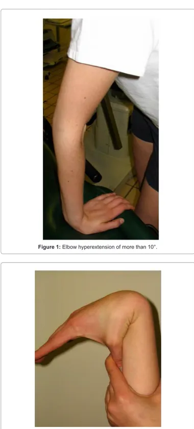
- Passive dorsiflexion of little fingers up to 90°. - Passive opposition of thumbs on the forearm. - Both hands flat on the ground with stretched legs. - Hyperextension of the elbow up to 10°.
- A recurvatum of the knees up to 10°.
More recently, these criteria have been included into a more complex classification that of Brighton [7], divided into major and minor criteria (Table 1).
Patients with joint hypermobility complain regularly of acute osteoarticular such as shoulder luxation or ankle sprains but also
Page 2 of 5
Figure 2: Hypermobility of the right wrist with the thumb under the forearm.
Figure 1: Elbow hyperextension of more than 10°.
suppressive and protective role of the trained muscle group, as already used in the rehabilitation of instabilities of traumatic origin [13]. The use of an isokinetic dynamometer allows to train safely in eccentric mode. Indeed, this device permits us working gradually, at controlled speed, within a limited range of joint motion in flexion and extension to avoid risk of luxation.
We illustrate this concept by the description of a clinical case.
Clinical Case
A 16 year old girl, diagnosed as EDS type III, consulted us for pain in the elbow and right wrist (dominant side) when playing tennis [14].
She had particularly:
- A significant known hypermobility (1 major criteria and 2 minor criteria according Brighton) (Figures 1 and 2).
- A hypermobile type EDS according to clinical and immunohistochemical studies.
- A myopia. - Regular epistaxis.
- Multiple ankle and knee sprains when playing soccer.
Her bone density was within the standards for her age. At the family level, several subjects had hypermobility, heart problems or significant scoliosis. Her twin brother died prematurely, after 4 months of gestation (recognized sign in certain types of EDS) [15].
You may recall that the EDS is a rare hereditary dystrophy affecting the synthesis or metabolism of collagen (I, III or V) or due to an enzymatic deficiency (lysyl hydroxylase and procollagen peptidase) involved in the synthesis of connective tissue [2,11,15]. The last classification called Villefranche classification, based on partially identified genetic and biochemical alterations and clinical presentations, recognizes 6 types [5,11,12,15] (Table 2). All have two major criteria, expressed at different levels: joint hyper mobility and skin hyper elasticity (+ possibly dystrophic scars) [5,16].
The immuno-histological analyzes make it possible to detect pathologies of the connective tissue but in all cases are not capable of a precise diagnosis. The appropriate course of action is, in the case of strong suspicion of EDS (mainly classic and vascular forms), to resort to the research of biochemical abnormalities of collagen produced by a skin fibroblast lines under culture: quantitative or qualitative (electrophoretic) abnormalities of type I and V collagens (classic or previous I and II type EDS) or of type III procollagen (EDS vascular or previous IV) [15]. Based on this analysis, we can afterwards try to find a direct mutation in the corresponding gene (Table 2). However, in the hypermobile form (previous type III), the incriminated gene remains unknown at the present time and the electrophoretic analysis is generally normal in most cases [9,15,17]. The diagnosis of hypermobile type EDS is still clinical at present and can be confused with SHB [12].
A rehabilitation program, 3 times a week, during 6 weeks, aimed in particular at eccentric strengthening of flexor and extensor muscles of the elbow together with prosupinators. Indeed, a prior isokinetic test had noted a deficit in muscle strength on the affected side compared to the opposite side of -15 to -35% at slow (60°/s) and fast speed (180°/s) speeds, with a prevailing impairment on flexors (Table 3). In order to protect joints and maintain movements within a reasonable range of motion (avoiding the hyperextension of the chronic joint pain from many causes: high frequency of subluxation,
repeated tendon or ligament pathologies, peripheral neurological irritation, multiple operations [1,8-12]. Generally, a classic rehabilitation causes little positive effects on both pain and quality of life. The rehabilitative content should be adapted individually by prioritizing techniques directly focused on joint hypermobility [3]. Muscle reinforcement for active stabilization of the involved joint is therefore preferred, especially in eccentric mode to develop the
Citation:Kaux JF, Forthomme B, Foidart-Dessalle M, Delvaux F, Debray FG, et al. (2013) Eccentric Training for Elbow Hypermobility. J Nov Physiother 3: 180. doi:10.4172/2165-7025.1000180
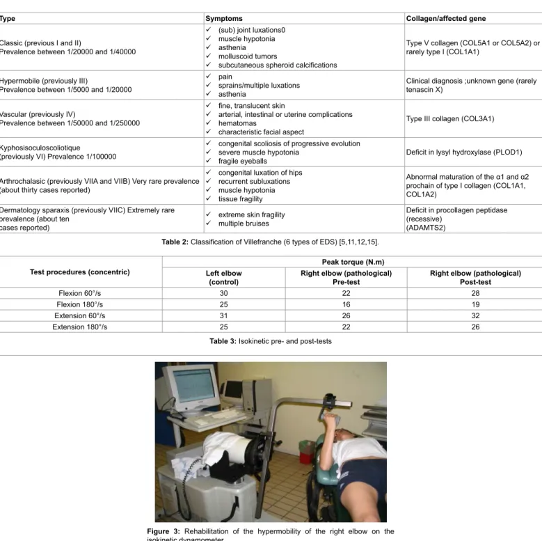
elbow), eccentric strengthening exercises at slow speeds, were carried out on an isokinetic dynamometer (Cybex Norm®) (Figure 3). Thus, she was trained to move her wrist at increasing speeds [30°/s, 60°/s, and 90°/s] within a limited range of motion in flexion and extension. All the more, as our purpose was to improve the gesture control, she started a submaximal eccentric program, using slow speeds, very gradually intensified (3 to 5 sets of 15 repetitions), in a safe range of motion in order to promote protective braking action of the joint.
A proprioceptive rehabilitation has also been undertaken in order to improve voluntary control of extreme ranges of motion. The wearing of a semi-rigid ortheosis to limit the wrist range of motion when playing tennis was also recommended. Rapidly, the patient
noted a significant decrease in pain (assessed using a 10-point visual analogue scale which dropped from 7/10 to 1/10, and quality of life scale MOS SF-36 which went from 70 to 100) and an increase in the stability of the elbow when playing tennis, as reported by the patient and the objectivation of a better control of the positioning of the elbow in space. An isokinetic control test at the end of 18 sessions noted a 20 to 25% increase in the time of maximum strength in the right elbow muscles and a recovery of the equilibrium of studied values in relation to the left elbow, with the exception of high speed concentric flexors. It would have been appropriate to continue with the isokinetic strengthening in order to hope for the complete normalization of muscular profile, however, the young patient, satisfied, could
Type Symptoms Collagen/affected gene
Classic (previous I and II)
Prevalence between 1/20000 and 1/40000
(sub) joint luxations0 muscle hypotonia asthenia molluscoid tumors
subcutaneous spheroid calcifications
Type V collagen (COL5A1 or COL5A2) or rarely type I (COL1A1)
Hypermobile (previously III)
Prevalence between 1/5000 and 1/20000
pain
sprains/multiple luxations asthenia
Clinical diagnosis ;unknown gene (rarely tenascin X)
Vascular (previously IV)
Prevalence between 1/50000 and 1/250000
fine, translucent skin
arterial, intestinal or uterine complications hematomas
characteristic facial aspect
Type III collagen (COL3A1) Kyphosisoculoscoliotique
(previously VI) Prevalence 1/100000
congenital scoliosis of progressive evolution severe muscle hypotonia
fragile eyeballs Deficit in lysyl hydroxylase (PLOD1) Arthrochalasic (previously VIIA and VIIB) Very rare prevalence
(about thirty cases reported)
congenital luxation of hips recurrent subluxations muscle hypotonia tissue fragility
Abnormal maturation of the α1 and α2 prochain of type I collagen (COL1A1, COL1A2)
Dermatology sparaxis (previously VIIC) Extremely rare prevalence (about ten
cases reported)
extreme skin fragility multiple bruises
Deficit in procollagen peptidase (recessive)
(ADAMTS2)
Table 2: Classification of Villefranche (6 types of EDS) [5,11,12,15].
Test procedures (concentric) Left elbow Peak torque (N.m)
(control) Right elbow (pathological)Pre-test Right elbow (pathological)Post-test
Flexion 60°/s 30 22 28
Flexion 180°/s 25 16 19
Extension 60°/s 31 26 32
Extension 180°/s 25 22 26
Table 3: Isokinetic pre- and post-tests
Figure 3: Rehabilitation of the hypermobility of the right elbow on the
Page 4 of 5
not continue with the program treatment given the remote home location. At the end of the sessions in rehabilitation center, the patient was encouraged to continue with proprioception and isometric strengthening exercises at home as instructed in physiotherapy (for example, self-mobilization of the wrist and elbow in predefined range of motion, with eyes open and then closed; isometric contractions in different positions of the wrist and elbow, especially close to extreme positions).
Discussion
The patients suffering from hypermobility regularly develop associated proprioception disorders. However, it is still not clearly defined whether or not this deficit is present from birth or developed during childhood. Indeed, some hypermobile patients develop symptoms only after puberty or in adulthood. These proprioceptive disorders may therefore appear gradually (over several years) due to repeated (micro) traumas related to this joint hypermobility and potentially magnified by a specific sporting activity [18].
The Panjabi model [19] shows that joint stability depends on three subsystems which are functionally interdependent:
- The passive musculoskeletal system, including bones, joint surfaces, ligaments, joint capsules and passive mechanical properties of muscles;
- The active musculoskeletal system, including muscles and tendons surrounding the joint;
- The control (or neural) system and the feedback, which include transducers of strength and movement variations located in the ligaments, tendons, muscles and the peripheral and central nervous system.
These concepts provide an explanation to some symptoms in hypermobile patients and can form the basis of their rehabilitation to increase the joint stability by muscle control.
In addition, these hypermobile patients frequently have specific muscle weakness compared to healthy subjects. The origin of this weakness would probably be multifactorial: increased muscle and ligament elasticity, lack of proprioception, joint instability, associated pain [20]. It is therefore important to undertake a specific rehabilitation for muscle strengthening (concentric motor role and protective suppressive eccentric function) and fresh proprioceptive training.
The goal of rehabilitation, based on the Panjabi model [19], entailed avoiding the joints hypermobility of the elbow and wrist, painful when playing tennis, by using the suppressive and stabilizer role of this arm’s muscles. We thought appropriate to emphasize the importance of measuring muscle performances, in order to quantify possible shortfalls and objectivize the benefits of rehabilitation [20,21]. Regarding the selected procedures in following our patient progress, only the concentric mode was appraised. Indeed, by definition, the purpose of the appraisal is to obtain the maximum intensities that can be developed by the tested muscle group. Knowing that the tension developed in the eccentric mode is higher compared to the concentric mode, we preferred to avoid the taking of risk, especially in a young patient.
However, the eccentric rehabilitation began in accordance with submaximal procedures, very gradually intensified [21]. The objective here clearly aimed to “motion control” and proprioceptive in relation to a possible objective of pure muscle strengthening.
Although eccentric exercises against manual resistance are possible, delivering the eccentric exercise using an isokinetic dynamometer has some advantages in terms of security that cannot be possible with conventional strengthening techniques: the speed control, a fixed amplitude managed by electronic stops, the control of the developed force level (graphics and display values). In addition, during the exercises, in case of pain and stoppage of muscle contraction, the isokinetic device stops, the movement is imposed on the basis of a minimum tension to develop and not as an “blind” motor, if the patient reaches a level of strength above the fixed limit, the device stops (“torque limiter”) [21].
Stretching exercises have of course been banned because in one hand, the patient is already hypermobile and in the other, risks of fracture have been reported [22]. In addition, a semiflexible ortheosis should have been worn when playing tennis [23].
In conclusion, the rehabilitation of hypermobile patients with joints pain should be individually rehabilitated. Exercises must include muscle eccentric strengthening in order to obtain a voluntary limitation of the involved joint range and in the other hand, a proprioceptive retraining compensating the deficit in proprioception in these patients [18,20]. The contribution of isokinetic dynamometry acknowledges two axes: at the evaluative and rehabilitative level [21]. Indeed, this device allows directing the management of the patient according to deficits objectivized by a pre-test, to ensure the follow-up and measure the effectiveness of the treatment by a post-test. In addition, the rehabilitation can be performing on it safely by avoiding excessive loads and greater range of motion.
References
1. Grahame R (1999) Joint hypermobility and genetic collagen disorders: are they related? Arch Dis Child 80: 188-191.
2. De Paepe A, Nicholls A, Narcisi P, De Keyser F, Quatacker J, et al. (1987) Ehlers-Danlos syndrome type I: a clinical and ultrastructural study of a family with reduced amounts of collagen type III. Br J Dermatol 117: 89-97. 3. Keer R, Grahame R (2003) Hypermobility Syndrome: Recognition and
management for physiotherapists. London: Butterworth Heinemann.
4. Maroteaux P, Frézal J, Cohen-Solal L (1986) The differential symptomatology of errors of collagen metabolism: a tentative classification. Am J Med Genet 24: 219-230.
5. Beighton P, De Paepe A, Steinmann B, Tsipouras P, Wenstrup RJ (1998) Ehlers-Danlos syndromes: revised nosology, Villefranche, 1997. Ehlers-Danlos National Foundation (USA) and Ehlers-Danlos Support Group (UK). Am J Med Genet 77: 31-37.
6. Beighton P, Solomon L, Soskolne CL (1973) Articular mobility in an African population. Ann Rheum Dis 32: 413-418.
7. Grahame R, Bird HA, Child A (2000) The revised (Brighton 1998) criteria for the diagnosis of benign joint hypermobility syndrome (BJHS). J Rheumatol 27: 1777-1779.
8. Chattopadhyay AK, Kandler RH, Sharrack B (1995) The association of hereditary neuropathies and heritable skeletal disorders. Postgrad Med J 71: 245-246.
9. De Coster PJ, Van den Berghe LI, Martens LC (2005) Generalized joint hypermobility and temporomandibular disorders: inherited connective tissue disease as a model with maximum expression. J Orofac Pain 19: 47-57. 10. Sacheti A, Szemere J, Bernstein B, Tafas T, Schechter N, et al. (1997) Chronic
pain is a manifestation of the Ehlers-Danlos syndrome. J Pain Symptom Manage 14: 88-93.
11. Stanitski DF, Nadjarian R, Stanitski CL, Bawle E, Tsipouras P (2000) Orthopaedic manifestations of Ehlers-Danlos syndrome. Clin Orthop Relat Res : 213-221.
12. Weinberg J, Doering C, McFarland EG (1999) Joint surgery in Ehlers-Danlos patients: results of a survey. Am J Orthop (Belle Mead NJ) 28: 406-409.
Citation:Kaux JF, Forthomme B, Foidart-Dessalle M, Delvaux F, Debray FG, et al. (2013) Eccentric Training for Elbow Hypermobility. J Nov Physiother 3: 180. doi:10.4172/2165-7025.1000180
13. Croisier JL, Crielaard JM (2001) [Isokinetic exercise and sports injuries]. Rev Med Liege 56: 360-368.
14. Kaux JF, Foidart-Dessalle M, Croisier JL, Toussaint G, Forthomme B, et al. (2010) Physiotherapy intervention for joint hypermobility in three cases with heritable connective tissue disorders. Journal of Musculoskeletal Pain 18: 254-260.
15. Quatresooz P, Hermanns-Lê T, Piérard GE (2008) Le syndrome d’Ehlers-Danlos. Qu’y a-t-il sous la pointe émergée de l’iceberg ? Rev. Med Liège 63 (supplément 1): 60-65.
16. Adib N, Davies K, Grahame R, Woo P, Murray KJ (2005) Joint hypermobility syndrome in childhood. A not so benign multisystem disorder? Rheumatology (Oxford) 44: 744-750.
17. Burrows NP, Nicholls AC, Yates JR, Gatward G, Sarathachandra P, et al. (1996) The gene encoding collagen alpha1(V)(COL5A1) is linked to mixed Ehlers-Danlos syndrome type I/II. J Invest Dermatol 106: 1273-1276.
18. Ferrell WR, Tennant N, Sturrock RD, Ashton L, Creed G, et al. (2004) Amelioration of symptoms by enhancement of proprioception in patients with joint hypermobility syndrome. Arthritis Rheum 50: 3323-3328.
19. Panjabi MM (1992) The stabilizing system of the spine. Part I. Function, dysfunction, adaptation, and enhancement. J Spinal Disord 5: 383-389. 20. Sahin N, Baskent A, Ugurlu H, Berker E (2008) Isokinetic evaluation of knee
extensor/flexor muscle strength in patients with hypermobility syndrome. Rheumatol Int 28: 643-648.
21. Croisier JL, Maquet D, Codine P, Forthomme B (2008) Renforcement musculaire et rééducation : apport de l’isocinétisme. Renforcement musculaire et reprogrammation motrice. Paris: Masson.
22. Kharrazi FD, Rodgers WB, Coran DL, Kasser JR, Hall JE (1997) Protrusio acetabuli and bilateral basicervical femoral neck fractures in a patient with Marfan syndrome. Am J Orthop (Belle Mead NJ) 26: 689-691.
23.
Submit your next manuscript and get advantages of OMICS Group submissions
Unique features:
• User friendly/feasible website-translation of your paper to 50 world’s leading languages • Audio Version of published paper
• Digital articles to share and explore
Special features:
• 250 Open Access Journals • 20,000 editorial team • 21 days rapid review process
• Quality and quick editorial, review and publication processing
• Indexing at PubMed (partial), Scopus, EBSCO, Index Copernicus and Google Scholar etc • Sharing Option: Social Networking Enabled
• Authors, Reviewers and Editors rewarded with online Scientific Credits • Better discount for your subsequent articles
Submit your manuscript at: http://www.omicsonline.org/submission/
Citation: Kaux JF, Forthomme B, Foidart-Dessalle M, Delvaux F, Debray FG, et al. (2013) Eccentric Training for Elbow Hypermobility. J Nov Physiother 3: 180. doi:10.4172/2165-7025.1000180
Collier SE, Thomas JJ (2002) Range of motion at the wrist: a comparison study of four wrist extension orthoses and the free hand. Am J Occup Ther 56: 180-184.
![Table 1: Brighton Criteria [7].](https://thumb-eu.123doks.com/thumbv2/123doknet/6827006.190233/2.892.77.453.823.1169/table-brighton-criteria.webp)