COMMUNAUTÉ FRANÇAISE DE BELGIQUE
UNIVERSITÉ DE LIÈGE – GEMBLOUX AGRO-BIO TECH
EARLY LIFE PROGRAMMING OF PIGLETS’
MICROBIOTA AND GUT HEALTH BY
MATERNAL DIETARY FIBRE
SUPPLEMENTATION
Julie LEBLOIS
Dissertation originale présentée en vue de l’obtention du grade de docteur en sciences agronomiques et ingénierie biologique
Promoteurs : Nadia Everaert, Jérôme Bindelle Année civile : 2018
Copyright
Abstract
Post-weaning diarrhoea (PWD) is a widespread disease causing loss of weight and mortality of the piglets. To cure or prevent PWD, the treatment of pigs with antibiotics is frequent. The overuse of these substances led to the appearance of multi-resistant bacteria, raising public health issues. Thus, finding sustainable alternatives to antibiotics for PWD curation is of major importance. Most research focusses on the use of substances like prebiotics able to affect the microbiota of the piglets, as gut microbiota is responsible for the maturation of the intestinal immune system. Promoting a beneficial microbiota as early in life as possible is a good strategy for a better future health and a lower prevalence of PWD. Our hypothesis was that using dietary fibres (wheat bran and resistant starch) in the diet of sows would alter their microbiota and in turn affect their piglets’ microbiota and future health. In addition, the ability of the two fibre sources to alter milk composition, also affecting piglets’ performances and health, was tested. This hypothesis was challenged with two animal experiments.
Results indicated that wheat bran (WB) and resistant starch (RS) had the ability to alter sows’ microbiota during gestation but not anymore during lactation, possibly limiting a differential microbial transfer to their offspring. These two dietary fibre slightly altered milk composition. Maternal wheat bran had the ability to increase the villus height and villus to crypt ratio in the small intestine of the progeny, while resistant starch increased the gene expression of tight junction proteins at weaning. These two fibre sources included in a high level in sows’ diets did not affect their performance or their piglets’, making their use in animal diets realistic.
A second objective of the thesis was to unravel whether the diet of sows could program the metabolism of piglets for later life, using them as model for human. For this, piglets were challenged with a high fat diet in order to induce low-grade inflammation and/or obesity symptoms. After 7 weeks on a high fat diet, piglets had an increased backfat thickness and higher serum cholesterol levels. The main findings are that feeding sows resistant starch increased the total sum of short-chain fatty acids (SCFA) production in the caecum and colon of their progeny, which is beneficial but did not affect the microbiota of the pigs. Moreover, maternal RS diet seemed to increase the barrier function of the colon due to a higher gene expression of tight junction proteins while the maternal effects on intestinal inflammation were contradictory for TNF-α and IFN-γ. It seems thus that the maternal diet had the ability to decrease gut permeability. However, the high fat diet did not alter the microbiota of the pigs, nor was it affected by the maternal diet.
In conclusion, using dietary fibre in sows’ diet had the ability to alter their own microbiota during gestation and milk composition, but the impact on the piglet’s microbiota was rather limited. It could be thus interesting to use these diets on piglets’ themselves after birth to promote the establishment of beneficial bacteria. Although
effects on the microbiota were limited, the maternal diet seemed to affect some aspects of the health of their progeny in later life.
Keywords: maternal transfer, microbiota, piglet, sow, gut health, dietary fibre, wheat bran, resistant starch
Résumé
La diarrhée post-sevrage est une maladie très répandue dans les élevages porcins, causant des pertes de poids et la mortalité des porcelets. La solution la plus répandue pour pallier au problème est l’utilisation d’antibiotiques, dont l’utilisation abusive est à la source de l’apparition de souches bactériennes multi-résistantes, représentant un problème de santé publique. Trouver des alternatives durables à l’utilisation de ces antibiotiques pour traiter ou prévenir la diarrhée post-sevrage est donc d’une importance capitale. De nombreuses recherches se concentrent sur l’utilisation de substances comme des prébiotiques, qui sont capables d’impacter le microbiote intestinal des porcelets. Le microbiote étant responsable de la maturation du système immunitaire intestinal, promouvoir des bactéries bénéfiques permettrait une meilleure immunité au moment du sevrage, réduisant ainsi la fréquence des diarrhées. Notre hypothèse était qu’utiliser des fibres alimentaires (du son de blé et de l’amidon résistant) dans l’alimentation des truies pourrait impacter leur microbiote intestinal, qui pourrait à son tour affecter celui de leurs porcelets ainsi que leur santé future. De plus, l’aptitude de ces deux sources de fibres à modifier la composition du lait des truies a également été testée, étant donné que celle-ci peut affecter les performances et la santé des porcelets. Pour tester cette hypothèse, deux expérimentations animales ont été réalisées.
Les résultats montrent qu’à la fois le son de blé et l’amidon résistant peuvent modifier le microbiote intestinal des truies durant la période de gestation, mais que ces modifications ne perdurent pas pendant la lactation, ce qui peut limiter le transfert à la descendance. Les deux fibres alimentaires ont impacté la composition du lait des truies, augmentant principalement la quantité de lactose. De plus, la supplémentation avec du son de blé a résulté en de plus hautes villosités intestinales et un ratio villosités/cryptes plus élevé dans l’intestin des porcelets avant sevrage. L’amidon résistant a, quant à lui, mené à une augmentation de l’expression des gènes des protéines de jonction serrées dans l’intestin des porcelets avant sevrage. De plus, aucune de ces sources de fibres à haute dose dans l’alimentation des truies n’a détérioré les performances des truies ou porcelets, ce qui rend leur utilisation en routine réaliste.
Un deuxième objectif de cette thèse était de déterminer si l’alimentation des truies pouvait programmer le métabolisme des porcelets sur du long terme. Lors de cet essai, le porc a été utilisé comme modèle pour l’humain. L’hypothèse a été testée en soumettant les porcs à un régime alimentaire contenant une haute teneur en graisses saturées afin d’induire une inflammation chronique et/ou des symptômes liés à l’obésité. Les conclusions sont qu’alimenter les truies avec de l’amidon résistant permet d’augmenter la production totale d’acides gras volatils dans l’intestin de leur descendance sur le long terme. Cette augmentation peut être considérée comme bénéfique pour la santé même si le microbiote intestinal n’a pas été impacté par le régime maternel. De plus, le régime alimentaire maternel avec de l’amidon résistant
semble augmenter la fonction barrière du colon de la descendance, étant donné l’augmentation de l’expression des gènes codant pour les protéines de jonction serrées. L’effet maternel sur l’inflammation du colon est quant à lui contradictoire, étant donné que l’expression de TNF-α et IFN-γ ont été affectées de façon opposée. Une autre conclusion qui peut être tirée de cette expérimentation à long terme est que le challenge métabolique de 7 semaines a permis d’induire chez les porcs les premiers symptômes de l’obésité, à savoir une augmentation du cholestérol sanguin et du gras dorsal. Cependant, le microbiote intestinal des porcs n’a pas été impacté par le challenge métabolique ni par la supplémentation alimentaire des mères.
Pour résumer, l’utilisation de fibres alimentaires dans la ration des truies a permis de modifier la composition de leur lait, mais l’effet sur le microbiote intestinal de leurs porcelets est très limité. Une perspective serait l’utilisation de ces mêmes fibres dans le régime alimentaire des porcelets après la naissance en vue de promouvoir l’établissement précoce d’un microbiote intestinal bénéfique. Bien que les effets du régime alimentaire maternel sur le microbiote des porcelets soient limités, certains aspects de leur santé intestinale semblent impactés.
Mots-clés: transfert maternel, microbiote, porcelet, truie santé intestinale, fibre alimentaire, son de blé, amidon résistant.
Acknowledgments
I would first like to acknowledge my supervisor, Nadia Everaert for giving me the opportunity to perform this PhD thesis. She’d been with me the whole journey, pushing me (not too hard) when it was needed, supporting me, congratulating me and finally letting me fly by myself. For all the time passed, especially during her holidays and/or weekends lately, I sincerely want to thank her.
Then, I would like to thank Jérôme Bindelle, my co-supervisor, who was always there when I needed him, to understand protocols, correct my papers or just to give advices concerning this topic he knows so well. And thank you for bringing colours in the lab with your incredible Congolese shirts!
My acknowledgments also go to Yves Beckers, head of the Precision Livestock and Nutrition unit, for his support and good advices and to José Wavreille and Sébastien Massart from my thesis committee, whose help for the animal experiments (merci José!) and the bioinformatics analyses was very precious.
I would like to thank the members of the jury, especially Isabelle Le Huërou-Luron, whose work has been inspiring and who accepted to be part of my thesis jury. Moreover, my acknowledgments go to the co-authors of my papers, for their help for statistics and reviewing of the papers.
I’m grateful to the COST action PiGutNet for giving the opportunity to meet lots of great scientists from all around Europe (great training school!) and supporting my oral presentation at EAAP 2016. I’m also thankful to the COST action SALAAM and to the laboratory of Prof. Oswald for giving the opportunity to learn the ex vivo technique in Toulouse. I would also like to thank the feed company Dumoulin for helping me in the formulation of sows’ diets that were complicated (merci Nicolas) and for their efficiency.
The help of my master thesis student, Anthony and Manon, was also very valuable and appreciated. Thank you Anthony for the nights spent at the farm and my birthday cake (!), and thank you Manon for the amazing job in the lab and your enthusiasm to learn.
J’aimerais remercier mes collègues de l’unité de zootechnie pour leur aide/conseils : Emilie, Cécile, Martine, Sylvie, Ester et tous les autres. Un merci particulier à mes collègues et anciens collègues de la bibliothèque, et en particulier Emilie, Lauriane, Yannick, Sophie, Julie, Bienvenu, Gaëtan, Hajer, Li et tous les autres pour ces moments géniaux passés ensemble. Au-delà des collègues, j’ai eu la chance inouïe de me faire des amis.
Je voudrais dire un grand merci également à tout le personnel du CRA-w pour leur aide précieuse pendant mes essais, leur bienveillance et leur gentillesse : Vincent, Yvon, Marc, Elise, René, Maxence, Marion, les stagiaires et tous les autres. La traite des truies aurait été bien moins sympa sans vous !
J’aimerais remercier mes amis, qui m’ont toujours soutenue et écoutée. Un merci particulier à Yael, la meilleure des meilleures, qui a toujours été là pour moi inconditionnellement. Un merci tout spécial également à mes amis proches, sur lesquels j’ai toujours pu compter : Alexandra, Pauline, Sophie (petite) & Alex, Anne-Catherine, Sophie (Vdh), Sophie (Maloteau), Céline, Sabri, Romain & Camille.
Comme on dit ‘last but not least’, j’aimerais remercier ma famille. Mes parents, qui m’ont toujours soutenue dans mes choix, même si je m’oriente vers une carrière scientifique à mille lieues de leurs métiers. Une fille ne peut pas rêver de plus de soutien et d’autant d’amour. Mamy, tu as en grande partie construit la personne que je suis devenue et je ne pourrai jamais assez te remercier. Fab, merci d’être là aujourd’hui et j’espère vraiment que tu comprendras maintenant que je suis autre chose que « mi-ingénieure, mi-fermière ! »
William, tu as été mon ancrage pendant ces 4 ans, à me remonter quand j’en avais besoin et m’aidant à tout relativiser. Travaillant dans notre super maison quand je n’avais du temps à consacrer qu’à ma thèse… Merci pour ces merveilleuses années ensemble et les prochaines, tes encouragements, ton soutien et ton amour.
Table of contents
Chapter 1: General introduction
1.
Gut function and maturation ... 3
1.1.
Gut functions... 4
1.1.1. Digestive function & fermentation (Sherwood et al. 2016) ... 4
1.1.2. Protective function ... 5
1.2.
Development and maturation of the digestive tract ... 12
2.
The microbiota ... 14
2.1.
Development of the microbiota and early colonization ... 14
2.2.
Roles for host ... 15
2.3.
Composition of the young and adult microbiota in the hindgut .. 16
3.
Post-weaning diarrhoea ... 17
3.1.
Weaning ... 17
3.2.
Adverse consequences ... 18
3.3.
Allopathic management: antibiotics and zinc oxide ... 20
3.4.
Alternatives to antibiotics and zinc oxide ... 20
4.
Dietary fibre: wheat bran and resistant starch ... 23
5.
The pig as model for human gastrointestinal tract ... 26
Chapter 2: Objectives, methods and articles related to the thesis
1.
Objectives ... 30
2.
Methods, animal experiments and publications ... 32
Chapter 3: Article 1
1.
Introduction ... 37
2.
Methods ... 38
2.1.
Animals ... 38
2.3.
Diets and feeding ... 38
2.4.
Sample collection ... 39
2.5.
DNA extraction and sequencing ... 39
2.6.
Short-chain fatty acids (SCFA) and branched-chain fatty acids
(BCFA) determination ... 40
2.7.
Bioinformatics and statistical analyses ... 40
3.
Results ... 41
3.1.
Sows ... 43
Continutation of Table 3. ... 46
3.2.
Umbilical cord blood and meconium ... 47
3.3.
Piglets ... 47
4.
Discussion ... 52
5.
Conclusion ... 55
Chapter 4: Article 2
1.
Abstract ... 67
2.
Introduction ... 68
3.
Materials and methods ... 69
3.1.
Animals, diet and housing ... 69
3.2.
Zootechnical performances ... 70
3.3.
Milk ... 70
3.4.
Sampling of intestinal tissues and rectum content ... 72
3.5.
Histomorphological analyses... 72
3.6.
Ex vivo experiment and calprotectin concentration ... 72
3.7.
Relative gene expression ... 73
4.
Results ... 74
4.1.
Zootechnical parameters ... 74
4.2.
Milk composition... 76
4.3.
Intestinal parameters ... 77
5.
Discussion ... 81
5.1.
Sows and piglets’ performances ... 81
5.2.
Milk composition and gut morphology ... 81
5.3.
Intestinal fermentation and intestinal health ... 82
6.
Conclusion ... 83
Chapter 5: In vitro characterization of different resistant starch
sources
1.
Introduction ... 86
2.
Materials and methods ... 86
3.
Results and discussion ... 86
4.
Conclusion ... 89
Chapter 6: Article 3
1.
Abstract ... 93
2.
Introduction ... 93
3.
Materials and methods ... 95
3.1. Animals, diets and housing ... 95
3.2. Zootechnical performances ... 98
3.3. Feed chemical analyses ... 98
3.4. Milk ... 98
3.5. Sampling of intestinal tissues and contents ... 99
3.6. Microbiota composition ... 99
3.7. Bioinformatics analysis ... 100
3.8.
Short-chain
fatty
acids
determination
and
calprotectin
concentration ... 100
3.9. Gene expression analysis ... 101
3.10. Statistical analyses ... 101
4.1.
Zootechnical parameters ... 103
4.2.
Colostrum and milk ... 103
4.3.
Microbiota of sows’ faeces ... 106
4.4.
Microbiota of the colonic content of piglets at 26 days of age . 110
4.5.
SCFA,
calprotectin
concentration
in
digesta
and
gut
morphology ... 110
4.6.
Gene expression... 111
4.7.
Performances of piglets after weaning ... 112
5.
Discussion ... 113
Chapter 7: Article 4
1.
Introduction ... 126
2.
Materials and methods ... 127
2.1.
Animals, diets and housing... 127
2.2.
Piglets’ performances ... 128
2.3.
Blood sampling and analysis ... 128
2.4.
Sampling of intestinal tissue and content ... 128
2.5.
Short-chain fatty acids (SCFA) determination ... 130
2.6.
Microbiota composition and calprotectin concentration ... 130
2.7.
Gene expression... 130
2.8.
Bioinformatics and statistical analyses ... 131
3.
Results ... 133
3.1.
Performances ... 133
3.2.
Cholesterol ... 134
3.3.
Microbiota and SCFA production ... 134
3.4.
Intestinal inflammation and permeability of the colon ... 138
4.
Discussion ... 139
Chapter 8: General discussion
Overview ... 144
Choice of ingredients and percentage of inclusion in the
diet ... 146
Microbiota of the sow and offspring ... 146
Milk composition ... 150
Gut morphology ... 152
Inflammatory status and mucosal integrity... 152
High fat challenge ... 154
General conclusion and highlights of the thesis ... 155
References ... 157
List of figures
Figure 1. Structure of the gastrointestinal tract (Walthall et al. 2005). ... 3 Figure 2. Schematic view of the components of the tight junctions (Ulluwishewa et al. 2011). ... 7
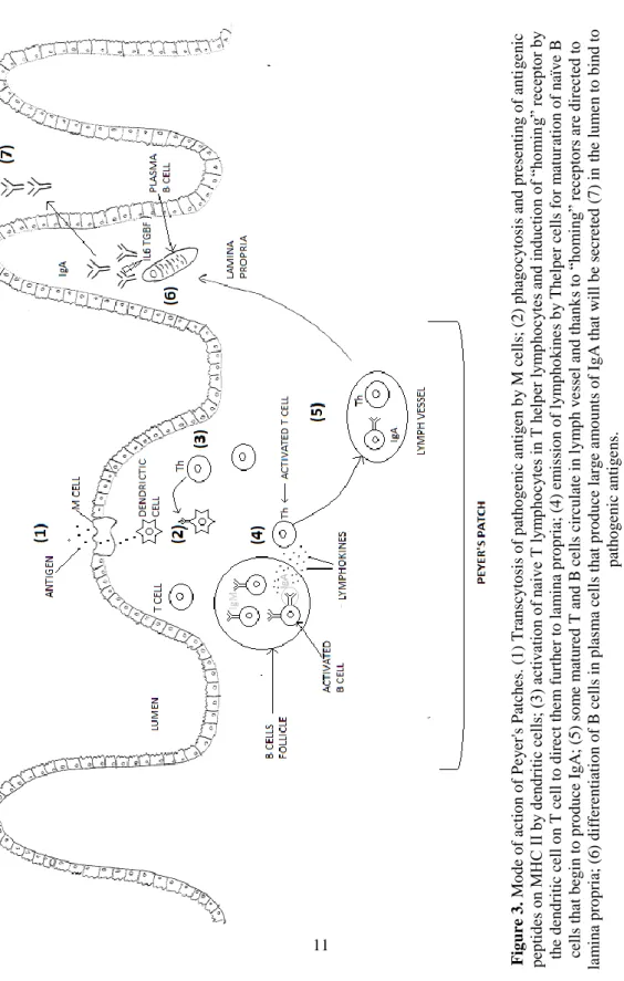
Figure 3. Mode of action of Peyer's Patches. (1) Transcytosis of pathogenic antigen by M cells; (2) phagocytosis and presenting of antigenic peptides on MHC II by dendritic cells; (3) activation of naive T lymphocytes in T helper lymphocytes and induction of “homing” receptor by the dendritic cell on T cell to direct them further to lamina propria; (4) emission of lymphokines by Thelper cells for maturation of naïve B cells that begin to produce IgA; (5) some matured T and B cells circulate in lymph vessel and thanks to “homing” receptors are directed to lamina propria; (6) differentiation of B cells in plasma cells that produce large amounts of IgA that will be secreted (7) in the lumen to bind to pathogenic antigens. ... 11
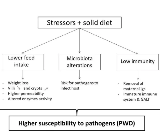
Figure 4. Main mechanisms underlying the susceptibility of piglets to PWD. ... 19 Figure 5. Hypothesis underlying the research question of the thesis. ... 31 Figure 6. Time line and different sampling points of the two animal experiments performed. ... 32
Figure 7. Individual composition of sows’ (a) and piglets’ (b) microbiota at the genus level. The Y-axis represents the relative abundances of the different genera (expressed as % of the total microbiota) and the X-axis represents the individuals (ID number). ... 42
Figure 8. Distribution of alpha diversity as measured by Shannon index, box plots represent the calculated Shannon index for microbiota samples of sows fed the control diet (CON, N=6) and the wheat bran-enriched diet (WB, N=6) at three different stages: 21 d (G21) and 98 d (G98+) of gestation, respectively before and after the experimental diets were distributed, and 20 d of lactation (L). ... 43
Figure 9. Percentage of target ingestion curve reached for sows in 4-days periods from farrowing until weaning. CON sows (N=7) are represented by the black dot and WB sows (N=8) by the white dot. Values are mean ± SEM. *** represent a p-value<0.001. ... 75
Figure 10. Weekly bodyweight of piglets born from WB and CON sows, from birth (week 0) until weaning (week 4). CON piglets are represented by dots and WB piglets by triangles; black symbols are males and white ones are females. Results are shown as mean ± SEM, n=54 for CON piglets and n=71 for WB piglets ... 76
Figure 11. Villus height (a), crypt depth (b) and ratio V:C (c) of piglets botn from CON (black bar, N=8) or WB (grey bars, N=8) sows. Values are mean ± SEM. P-values <0.05 and <0.01 are indicated with the symbols * and **, P<0.10 are indicated with the symbol +. ... 79
Figure 12. Relative gene expression for TNF-α, TLR4, ZO-1 and CLDN2 in piglets’ ileum tissue challenged or not with LPS. No-LPS CON is set to 1 to serve as reference for comparison; n=8 piglets/maternal treatment. ... 80
Figure 13. Total gas production curve of the different RS substrates over time. . 88 Figure 14. IgA concentration (mg/ml) in colostrum of sow. DS sows are represented with the black bar (N=12) and RS sows with grey bars (N=11) sows. Results are expressed as mean+SEM. ... 105
Figure 15. PCoA discriminating periods. Individual red dots are the fecal samples of sows during gestation (N=20) while blue squares are individual fecal samples of lactating sows (N=20). ... 106
Figure 16. PCoA discriminating dietary treatments during gestation. Red squares represent the faecal microbiota composition of sows fed DS during gestation (N=10) while blue dots represent microbiota of sows fed RS diet (N=10). ... 107
Figure 17. Piglets' faecal score during 2 weeks post-weaning. Score was assessed daily for 15 days. ... 113
Figure 18. Principal component analysis of the microbial composition of the colonic contents. Red dots are DS CON pigs, blue dots are DS HF pigs, orange dots are RS CON pigs, green dots are RS HF pigs (n=8/treatment). ... 135
Figure 19. Proportion of the main abundant phyla in the different pigs groups (n=8/treatment). ... 136
Figure 20. Gene expression of inflammatory cytokines and tight junction proteins in the proximal colon of pigs (n=8/treatment). P<0.05 is represented by * and P<0.001 is represented by ***. ... 138
Figure 21. Main results of the two animal experiments led during this PhD thesis. Green V signs indicate a change observed for the studied parameter while the red X sign indicate no change. ... 144
List of tables
Table 1. Overview of studies using maternal nutrition to impact piglets' microbiota, gut morphology and inflammation and passive immunity provided. G=gestation. .. 22
Table 2. Summary of the effects of wheat bran (WB) and resistant starch (RS) on several parameters of the intestinal content, mucus or faeces. #Microbiota changes
comprise changes at relative abundance of beneficial genera and overall changes of microbiota (composition, diversity); * Gut health comprises effects on mucus production, tight junctions and/or inflammation of the intestine. 1: Bird et al. (2007) ; 2 : Chen et al. (2013); 3: Chen et al. (2015); 4: Haenen et al. (2013); 5: Ivarsson et al. (2015); 6: Iyayi & Adeola (2015); 7: Martin-Pelaez et al. (2009); 8: Molist Gasa et al. (2010); 9: Molist et al. (2012); 10: Nielsen et al. (2014); 11: Nofrarias et al. (2007) ; 12 : Regmi et al. (2011) ; 13 : Sun et al. (2015) ; 14 : Yu et al. (2016) ; 15 : Yan et al. (2017); 16 : Zhang et al. (2016) ; 17 : Zhou et al. (2016). ... 25
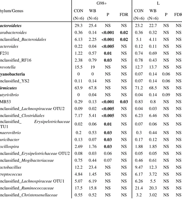
Table 3. Composition of the faecal microbiota of sows fed the control diet (CON, N=6) and the wheat bran-enriched diet (WB, N=6) at two different stages: 98 d into gestation (G98+) and 20 d into lactation (L), expressed as a percentage (%) of the total mi ... 45
Table 4. Microbial composition of the umbilical cord blood of piglets born from sows fed the control diet (CON, N=6) and the wheat bran-enriched diet (WB, N=8), expressed as the percentage (%) of the total microbiota. Only top ten genera and those with a consistent p-value (<0.1) were included in the table. ... 48
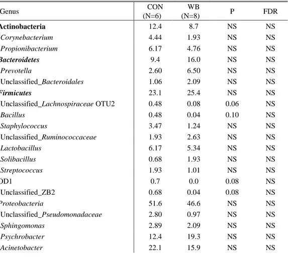
Table 5. Relative abundance of bacterial genera sampled in the colon of piglets born from sows fed the control diet (CON) and the wheat bran-enriched diet (WB), only genera with abundance >0.01% are displayed in this table. P-values and FDR are considered as significant <0.05 and numerically different with a value <0.10. .. 49
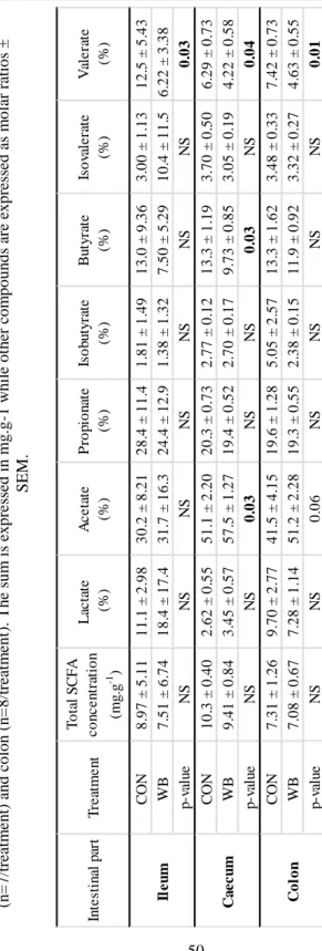
Table 6. SCFA concentrations and molar ratios of piglets' digesta in the terminal ileum (n=5 for CON, n=7 for WB), caecum (n=7/treatment) and colon (n=8/treatment). The sum is expressed in mg.g-1 while other compounds are expressed as molar ratios ± SEM. ... 50
Table 7. Pearson's correlations between SCFA molar ratios and genera of the microbial community in the colon of piglets from sows fed a control and a wheat bran-enriched diet (N=14). Only the results with a p-value<0.05 and r>0.70 were included in this table. Negative correlations are expressed in the table with the symbol “-“. ... 51
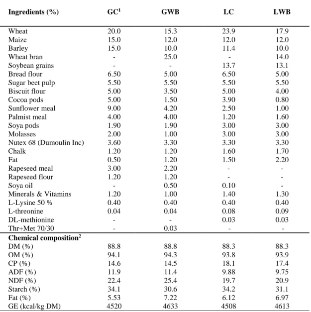
Table 8. Ingredients proportions and analysed chemical composition of gestation and lactation diets of the sows. ... 71
Table 9. Primers used for qPCR analysis. Genes analyzed are peptidylprolyl isomerase A (PPIA), glyceraldehyde-3-phosphate dehydrogenase (GAPDH), Tumor necrosis factor α (TNF-α), Toll-like receptor 4 (TLR4), Zonula occludens protein 1 (ZO-1), Claudin 2 (CLDN2). Primers were tested on a pool of cDNA from the explants. ... 74
Table 10. Fat, protein, and lactose percentage, IgA, IgG and IgM concentrations in milk samples collected on a weekly basis after farrowing for piglets born from control (CON) or wheat bran (WB) sows. Values are presented as mean and SEM are given for each period, n=3 or 4 for each parity within a treatment. ... 78
Table 11. Fermentation kinetics parameters for the different RS sources fermented
in vitro. N=3 per substrate. A= maximal gas volume; B= time to reach A/2; Rmax=
maximal fermentation rate; Tmax= time to reach Rmax. ... 87 Table 12. Total SCFA production and individual molar ratios of acetate, propionate and butyrate produced by the 5 types of RS at 3 time points. ... 89
Table 13. Composition of sows’ diets during gestation for digestible starch (GDS) and resistant starch (RDS) and during lactation (LDS and LRS) and analyzed chemical composition. ... 97
Table 14. Primers used for gene expression analysis. ... 102 Table 15. P-values of the treatment, time, parity and interactions for protein, fat and lactose content of the milk. ... 103
Table 16. Composition (protein, fat and lactose) of colostrum and milk of sows fed digestible starch (DS) and resistant starch (RS) in function of parity. N=12 for the DS group and N=11 for the RS group after because of removal of one sow from the experiment. ... 104
Table 17. Relative abundances of the phyla and genera in sows’ faeces. Only genera present at >0.01% in the faecal microbiota of the sows fed either digestible starch (DS, n=10) or resistant starch (RS, n=10) -based diets during gestation and lactation were considered. ... 109
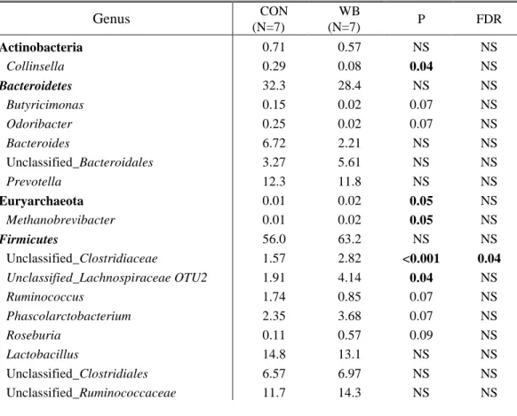
Table 18. Relative abundances in piglets' colonic contents (N=7 for DS piglets, N=8 for RS piglets). Results showed are only for the top 10 genera and the genera with p<0.10 and relative abundance >0.01%... 111
Table 19. Relative gene expression in the ileum and colon of piglets at weaning. The 2-ΔΔCt value of DS piglets is set at for each gene 1 to allow comparisons. ... 112
Table 20. Ingredients and analyzed chemical composition of grower diets fed to pigs. ... 129
Table 21. Primers used for the qPCR analysis. ... 132 Table 22. Backfat thickness, muscle thickness, meat percentage and bodyweight of pigs after the high fate challenge. (n=9 for DS CON, DS HF, RS CON and n=11 for RS HF) ... 133
Table 23. Total cholesterol (mg/dl), triglycerides (mg/dl), high density lipoprotein (mg/dl), low density lipoprotein (mg/dl) and ratio in pigs' fasted plasma after 6 weeks of HF challenge (n=8/treatment). ... 134
Table 24. Total SCFA production and molar ratios of acetate, propionate and butyrate in the large intestinal contents of the pigs born from DS or RS sows and fed CON or HF diets. ... 137
Table 25. Main results of the PhD thesis with the two animal experiments using either wheat bran (WB) or resistant starch (RS) until weaning ... 145
List of abbreviations
ADF Acid detergent fibre
AI Artificial insemination
APC Antigen presenting cell
BCFA Branched-chain fatty acids
BW Bodyweight CLDN Claudin CON Control CP Crude protein DM Dry matter DS Digestible starch EE Ether extract
ETEC Enterotoxigenic E.coli
FI Feed intake
FOS Fructo-oligosaccharide
GALT Gut-associated lymphoid tissue
GE Gross energy
GIT Gastrointestinal tract
GLUT Glucose transporter
HPLC High performance liquid chromatography
Ig Immunoglobulin
JAM Junctional adhesion molecules
LPS Lipopolysaccharide
MHC Major histocompatibility complex
MLN Mesenteric lymph node
NDF Neutral detergent fibre
NGS Next generation sequencing
NK Natural Killer cells
OCLN Occludin
OM Organic matter
OTU Operational taxonomic unit
PAMPs Pathogen associated molecule patterns PCoA Principal coordinate analysis
PP Peyers’ Patches
PRR Pattern recognition receptor
PW Post-weaning
PWD Post-weaning diarrhoea
SCFA Short-chain fatty acids
SGLT Sodium glucose linked transporter
TJ Tight junction
TLR Toll-like receptor
WB Wheat bran
WP Work package
1
Early life programming of piglets’ microbiota and gut health by maternal dietary fibre
2
Pig production is the first animal production worldwide, with China being the first producer and the European Union following (FAO data 2016). As the total population worldwide is continuously increasing (an increase of 32% is planned by 2050, leading to a world population of 9.8 billion according to the United Nations), so does the demand for animal products and in particular pig meat. It is thus necessary to reach high yields in a short period. In practice, several strategies are applied to improve efficiency, i.e. the selection of sows with high prolificity, an increased carcass weight and a reduced time for weaning that is very low compared to natural rearing (3-4 weeks compared to 8-12 weeks). This intensification of production in terms of abrupt early weaning was concomitant with the spread of post-weaning diarrhoea worldwide (Fairbrother et al. 2005). This disease, which is mainly caused by pathogenic E. coli infections (Melin et al. 2004), is characterized by lower feed intake, loss of weight, infections and mortality resulting in economic losses for the farmer. To counteract this disease, antibiotics have been widely used but their sustainability poses more and more questions nowadays. Therefore, solutions are required to prevent infections at weaning and this thesis aimed at investigating a possible strategy, by acting on the diet of the sows. In this introduction, the function and development of the gastrointestinal tract and intestinal microbiota of pigs will be described followed by a description of the weaning problems. In addition, a brief introduction to the use of the pig as a model for human disease and in particular metabolic troubles will be presented, as part of this work focussed on metabolic disorders related to ingestion of a high fat diet.
Chapter 1. General introduction
3
1. Gut function and maturation
The small intestine is composed of 4 layers (from the inside to the outside): the tunica mucosa, the tela mucosa, the tunica muscularis and the tunica serosa (Figure 1). Within the tunica mucosa, 3 layers are distinguished: (a) the epithelial layer, covered with exocrine (goblet cells), endocrine cells (secreting hormones) and epithelial cells (nutrients absorption); (b) the lamina propria (containing the blood and lymphatic vessels and gut-associated lymphoid tissue) and (c) the muscularis mucosa. The small intestine is very efficient in absorbing nutrients thanks to its impressive absorptive surface possible by the presence of villi and microvilli on the epithelial cells. Lieberkühn crypts, located between the villi mainly serve as a nursing for new enterocytes, having a high mitosis rate. Enterocytes will then migrate from the crypt to the villus and will replace damaged or older enterocytes cleared by apoptosis. Intestinal crypts also contain Paneth cells that produce lysozyme and defensins, protecting the intestine against pathogens. In this introduction, the emphasis lies on the small and large intestines.
Early life programming of piglets’ microbiota and gut health by maternal dietary fibre
4
1.1. Gut functions
1.1.1. Digestive function & fermentation (Sherwood et al. 2016)
The primary function of the digestive tract is to digest carbohydrates, proteins and lipids and absorb them for a release in the systemic blood flow. The first part of the digestion occurs in the stomach, where the digestion of carbohydrates and proteins begins with pepsin and salivary amylase enzymes and is continued in the duodenum. In the duodenum, the chime (pre-digested food) is mixed with pancreatic juice, containing proteases, pancreatic amylase and lipase that will convert complex proteins, carbohydrates and lipids in amino acids and short-chain peptides, disaccharides and monosaccharides, monoglycerides and free fatty acids. The digestion of lipids is complete after the release of pancreatic lipase and bile salts, while the digestion of disaccharides and small peptides needs to be terminated in the small intestine. The brush border of the epithelial layer secretes disaccharidases (lactase, maltase, sucrase and trehalase) and aminopeptidases that will allow the final digestion step of those molecules in monosaccharides (glucose, galactose, fructose) and amino acids. Glucose and galactose absorption in the enterocytes is possible by an active transport with sodium glucose linked transporters (SGLT) while fructose enters the enterocytes by a facilitated diffusion with glucose transporters (GLUT); these monosaccharides are further transported to the capillaries by GLUT2 transporter. Another way of glucose transportation is the paracellular transportation by the tight junctions between epithelial cells. The absorption of amino acids and small peptides follows the same pattern as monosaccharides, with intraepithelial peptidases able to fulfil the digestion of the di- or tripeptides (Le Huërou-Luron 2003). One exception is noteworthy for the protein passage. Indeed, the new-born piglets are equipped within the first hours of life (48h) with foetal-type enterocytes that are able to directly absorb entire proteins, like immunoglobulins from the colostrum which is necessary as the piglets are born agammaglobulinemic due to epitheliochorial placentation. The replacement of foetal-type enterocytes to adult-type enterocytes occurs within 2 days of life and is responsible for the so-called “gut closure” (Zabielski et al. 2008).
The hindgut (large intestine, comprising the caecum, colon and rectum) is important for the mineral balance as reabsorption of biliary salts, minerals, vitamins and water will happen. Some undigested carbohydrates and proteins (due to their size or their cell structure) reach the large intestine (caecum and colon) nearly intact. In the hindgut, they will be fermented by the microbiota and the end-products of their fermentation will be absorbed. This point will be discussed in another part (see section 2 “The microbiota”).
Chapter 1. General introduction
5
1.1.2. Protective function
The gut epithelium represents a physical barrier against harmful bacteria, viruses and antigens1 as epithelial goblet cells produce mucins that will allow the formation
of a viscous gel on the surface of the epithelium (King et al. 2003). Moreover, the epithelium is sealed by proteins called tight junctions that will prevent the paracelullar transduction of macromolecules as well as bacteria; the acidity of the gastric juice and the motility of the chime are also physical obstacles for pathogens survival. Moreover, the digestive tract harbours a highly condensed and developed immune system, often referred to as the “gut associated lymphoid tissue” (GALT), composed of immune cells in the lamina propria and epithelium (like intraepithelial T lymphocytes), highly organized lymphoid follicles (of which Peyers’ patches is a major representative) and small aggregates of lymphoid follicles, as summarized by King et al. (2003). Every mechanism involved in the animal protection is detailed hereunder.
1.1.2.1. Mucus layer
The mucus layer is formed after secretion of mucin glycoproteins (MUC2) and trefoil peptides (protease resistant) by goblet cells (Bourlioux et al. 2003) and has several roles s.a. the lubrication of the lumen but also a mechanical barrier against pathogens, as the tight mucus layer forms a network with a mean pore size of 100nm (Mackie et al. 2016) and exerts hydrophobic properties thanks to surfactant lipids produced by epithelial cells, preventing the passage of water-soluble toxins (Bourlioux et al. 2003). Moreover, the mucus layer serves as an habitat for commensal bacteria, representing a nutrient source (Sommer & Bäckhed 2013). Goblet cells can produce more mucins in presence of some stimuli, like toxins and bacterial infection but there is no change with aging for the piglet from day 7 to 18 of life (Brown et al. 2006) while the thickness of the mucus layer increases when going from the proximal to the distal intestine (Bourlioux et al. 2003; Mackie et al. 2016). The intestinal microbiota plays a role on the mucus layer as microbial colonization is related to higher goblet cells number and thicker mucus layer, (Sommer & Bäckhed 2013). The underlying mechanisms are so far unknown but these conclusions could be drawn from the use of germ-free animals.
1.1.2.2. Tight junctions
To connect epithelial cells (absorptive enterocytes, endocrine, Paneth and goblet cells) together and thus act as a barrier against pathogens, intracellular junctional complexes are necessary to obtain a semi-permeable dynamic and selective membrane allowing the passage of small molecules, ions and water but preventing
1 An antigen is a molecule (often proteins) interacting with the immunoglobulin receptor of
Early life programming of piglets’ microbiota and gut health by maternal dietary fibre
6
the passage of pathogens, toxins and harmful antigens (Niessen 2007). One part of this complex is formed by “tight junctions” (TJ) composed of more than 50 proteins (Ulluwishewa et al. 2011).
TJ are composed of different proteins and interface with several molecules. Tight junctions are transmembrane proteins (Figure 2), having 4 transmembrane domains and 2 extracellular loops (tetra-span tight junction, including occludin, claudin and tricelullin) or one transmembrane domain (single span, junctional adhesion molecules, abbreviated as “JAM”). Claudin proteins are involved in the tightening (CLDN 1, 3, 4, 5, 8) or the opening (CLDN2) of the paracelullar pores, while occludin (OCLN) is recognized to be involved in the regulation of intermembrane and paracelullar diffusion of small molecules (Ulluwishewa et al. 2011). Tricelullin is a protein that will tighten the closure of the membrane between 3 adjacent enterocytes, reinforcing the effect of claudins. Those tight junctions interact with plaque proteins, which are responsible for the anchoring of tight junctions into the cytoplasm of enterocytes. Important plaque proteins are zonula occludens (ZO1, 2, 3) that are composed of 3 PSD95–DlgA–ZO-1 homology (PDZ) domains that can bind to claudins, to JAM-A and to another ZO in order to form dimers. ZO-1 is thought to be very important in TJ regulation as they can interact with F-actin that are projected by the actin and myosin belt that surrounds the apical pole of the enterocytes and can thus regulate the tightening or smoothing of the TJ (Niessen 2007; Ulluwishewa et al. 2011). Moreover, ZO1- and ZO-2 have been demonstrated to determine the polymerization and extent of polymerization of claudin proteins (Umeda et al. 2006). Tight junction expression has been shown in humans to interact both with microbiota and with dietary components (Ulluwishewa et al. 2011).
1.1.2.3. The gut immune system / Gut-associated lymphoid tissue (GALT)
The immune system provides a continuous surveillance and protects the host against pathogenic infections. It is composed of primary and secondary lymphoid organs together with a collection of cells circulating in the blood and lymphatic vessels. Immunity is innate but also acquired from the contact with different pathogens; immune cells are produced and matured in the bone marrow and/or thymus and stored in secondary lymphoid organs, including the GALT. A short review of the functioning and composition of both innate and adaptive immunity is given below, before characterization of the GALT.
Chapter 1. General introduction
7
a. Innate immune system
The innate immune system is a very efficient defence line of the organism against pathogenic bacteria, viruses or antigens and constitutes a rapid response (Sherwood et al. 2016). This response is non-specific as it will target any pathogenic antigens and does not rely on previous contact with a specific antigen. In addition to immune cells, the innate immunity also relies on physical characteristics of the intestine, e.g. the barrier function and intestinal motility (King et al. 2003). Below is a brief summary of the main cells involved in the innate immune response. Enterocytes are able to produce cytokines after recognition of pathogen-associated molecule patterns (PAMPs) on their receptors (pattern recognition receptors, PRR including toll-like receptors, TLR) like TNFα and interferon (IFN) γ to block pathogen replication. Enterocytes have also the ability to directly produce anti-microbial peptides and pigs
Figure 2. Schematic view of the components of the tight
Early life programming of piglets’ microbiota and gut health by maternal dietary fibre
8
do not express major histocompatibility complex (MHC) class II2 on their
enterocytes, unlike humans (Mair et al. 2014). Neutrophils represent the first defence against a pathogenic infection and have different roles, including phagocytosis of pathogens, production of anti-microbial peptides and signals for maturation, activation and attraction of macrophages and dendritic cells to the site of infection, and regulation of T-cells response (Kumar & Sharma 2010). Monocytes are differentiated in macrophages after IFNγ stimulation. Macrophages then destroy pathogens by phagocytosis and present the pathogenic antigens on MHC II together with the release of inflammatory cytokines (e.g. IL1β) that are the main communication sources between immune cells and are responsible for the recruitment of T helper cells (Mair et al. 2014). Natural killer cells have a lysis activity against pathogens and produce various cytokines including IFNγ (Mair et al. 2014). Dendritic cells are antigen presenting cells (APC) and interact with the adaptive immune system by stimulating cytotoxic and helper T cells. APC are thus essential as they will recruit the adaptive immune system; these APC include macrophages and dendritic cells. The release of cytokines related to these cells is also of major importance as they are the mediator between innate and adaptive immune responses.
b. Adaptive immune system
The adaptive immunity can be divided in two components, the humoral immunity (relying on antibodies and B cells) and the cellular immunity (mediated by T cells) (Sherwood et al. 2016). The B and T cells are produced in bone marrow while maturation occurs also there for B lymphocytes and in the thymus for T lymphocytes. The matured lymphocytes then migrate to lymphoid tissues where they establish and can proliferate (Sommer & Bäckhed 2013). Those lymphoid tissues comprise the GALT that will be described below.
The adaptive immunity relies on the detection of antigens (foreign large sized molecules that will induce a specific immune response), including dietary antigens (for which a tolerance is developed). One example of pathogenic antigen is the protein lipopolysaccharide (LPS) that is present on the cell wall of gram-negative pathogenic bacteria (King et al. 2003). Those antigens are presented by APC from the innate immunity and this presentation constitutes the first step in the activation of the adaptive immunity. Antigenic peptides are presented on MHC II.
2 MHC are divided in two classes. MHC I is involved in the recognition of infected
cells and recruits cytotoxic T cells while MHC II function is to present antigenic peptides deriving from phagocytosis to the immune system.
Chapter 1. General introduction
9 Humoral immunity
Specific receptors on the B cells membrane allow them to bind to a specific site of the antigen. This binding leads to the mitosis of B cells that will differentiate either in plasma cells or in memory B cells. Plasma cells will massively produce antibodies specific for the antigens that induced the mitosis. Those specific antibodies, also called immunoglobulins (Igs), are released and circulate in the blood. The recognition of the antigen by T helper lymphocytes also leads to the differentiation of B cells in plasma cells (King et al. 2003). Immunoglobulins are categorized in 5 types (IgG, IgA, IgM, IgE, IgD) even though in each type, there are millions of specific antibodies. IgG is the most abundant immunoglobulin which is present in high amounts within the blood and the colostrum of sows; IgM has a role of receptor on B cells membrane and IgA are mainly produced in the digestive tract. The effects of Igs are mainly indirect and they do not have any destructive capacity. Indeed, Igs bind to antigens, preventing them to bind to other cells, agglutinate to form insoluble complexes and act indirectly by allowing a more effective protection by activating the complement and amplifying the phagocytosis of innate immune cells (King et al. 2003; Sherwood et al. 2016). The memory B cells exist to allow a faster and long-lasting effect after a second contact with the same antigen, even years after the first attack.
Cellular immunity
In addition to threats related to the presence of bacterial antigens, viruses are able to penetrate cells and to replicate inside it. To warn the immune system of this attack or mutation, most cells can present antigenic peptides after the degradation of the viral antigens within the cells and present them on their surface thanks to MHC I. This complement allows the recognition of the presented antigen by cytotoxic (also called CD8+) T cells. Cytotoxic T cells (Tc) are thus activated, multiply and secrete factors that destroy the infected cells; these T cells are also antigen-specific and memory T cells are produced for a longer lasting protection.
In addition to Tc, two other types of T cells exist. As mentioned above, T helper (Th) cells are not killing cells but allow the modulation of other immune cells, as the proliferation of B lymphocytes and macrophages. Finally, T regulatory cells (Treg) suppress the immune response and regulate the expression of both innate and adaptive immune cells.
Early life programming of piglets’ microbiota and gut health by maternal dietary fibre
10
c. The Gut-associated lymphoid tissue (GALT)
The GALT is considered to be the largest immune organ of the porcine body, a fact that can be explained by its large surface and continuous contact with pathogenic and commensal bacteria as well as dietary antigens, for which a tolerance mechanism (“oral tolerance”) has been developed (Burkey et al. 2009; Le Huërou-Luron & Ferret-Bernard 2014). The GALT is composed as any other immune organ by innate immunity (mucus layer, tight junctions, intestinal motility, phagocytic cells) and an adaptive immune system. The adaptive immune system of the gut is mainly located within the Peyer’ Patches (PP), the mesenteric lymph nodes (MLN) and the lamina propria and intraepithelial lymphocytes (Le Huërou-Luron & Ferret-Bernard 2014). PP and MLN are considered as the inductive sites of the immune response, detecting pathogenic antigens and triggering the inflammatory response, while intraepithelial lymphocytes and the lamina propria are considered as the effector sites of inflammation as they will directly be able to kill the pathogens and protect the organism (Burkey et al. 2009). Peyer’s Patches are present from the jejunum (as discrete PP) to the end of the ileum, where it forms a unique large PP (Everaert et al. 2017). PP surface is composed of epithelial cells without absorptive capacity and contain M cells specialized in the uptake of antigens from the intestinal lumen (containing TLR on their membrane) and thus in surveillance of the gut health. Large B-cells follicles, T cells and follicular dendritic cells are present underneath the epithelial layer (Le Huërou-Luron & Ferret-Bernard 2014; Burkey et al. 2009). M cells have the ability to transport pathogenic antigens transcellularly; these antigens will interact with the underlying APC that will present the immunogenic peptides on MHC II. T-helper cells are activated by the APC presenting pathogenic peptides and dendritic cells induce “homing receptors” on T cells that will enable them to go to the lamina propria later on. Activated T cells secrete substances (lymphokines) that induce B cells to produce antibodies. Some T and B activated cells then migrate to the lamina propria by lymphatic circulation where B cells differentiate in plasma B cells that will secrete large amounts of specific IgA, a process induced i.a. by IL-6 and TGF-β (King et al. 2003). These IgA are then released in the lumen to bind pathogenic antigens. A summary of this mechanism is shown in Figure 3.
Chapter 1. General introduction 11 F ig ure 3 . Mo d e o f ac tio n o f P e y er 's P atch es. (1 ) T ran scy to si s o f p ath o g e n ic a n ti g en b y M ce lls ; ( 2 ) p h ag o c y to si s an d p resen ti n g o f an ti g e n ic p ep tid es o n MH C I I by d en dr itic ce lls ; ( 3) ac tiv atio n of n ai ve T ly m ph oc ytes i n T h elp er ly m ph oc yte s an d in du ctio n of “h om in g” rec ep to r by th e d en d ritic c ell o n T ce ll to d ir ec t th e m f u rt h er to la m in a p ro p ria; (4 ) em is sio n o f ly m p h o k in e s b y T h elp er ce lls f o r m at u ratio n o f n aï v e B cells th a t b eg in to p ro d u ce Ig A ; ( 5 ) so m e m at u red T a n d B cells c ircu late i n l y m p h v e ss el a n d th an k s to “ ho m in g” recep to rs a re direc ted to la m in a p ro p ria; (6 ) d if fer en tiat io n o f B ce lls in p las m a ce ll s th at p ro d u ce lar g e a m o u n ts o f Ig A t h at w ill b e sec reted ( 7 ) in th e lu m e n to b in d to p ath o g en ic an ti g e n s.
Early life programming of piglets’ microbiota and gut health by maternal dietary fibre
12
1.2. Development and maturation of the digestive tract
The gut development begins during early gestation and Buddington & Malo (1996) categorized the ontogenetic development of the gut in 5 phases. The first three phases occur during gestation (organogenesis, differentiation, growth & maturation), the fourth phase is related to ingestion and processing of milk and the switch from the neonatal intestine to a fully developed and functional adult-like intestine at weaning characterizes the fifth phase.
Everaert et al. (2017) summarized the first steps of development and differentiation of intestinal tissues and cells in a recent review. The villi present on the small intestine surface are already present from the 40th day of gestation, while the muscularis
mucosae and differentiation of cells in enterocytes, goblet cells and secretory cells are achieved by the third month of gestation (Zabielski et al. 2008). It has been reported that during late gestation, the small intestine is growing faster than the body itself, probably to support the growth and need of arginine of the foetus (Sangild et al. 2000; McPherson et al. 2004; Everaert et al. 2017). The intestinal architecture is also evolving during the first days of life, as crypts depth and villi heights increase to allow a higher absorptive capacity of the intestine, together with thickening of the mucosa (Skrzypek et al. 2010). These observations also highlight the increase in absorptive area of duodenum and jejunum when piglets age, translating the maturation of the digestive and absorptive system of the piglet which is a consequence of colostrum and milk intake. Not only the architecture of the gut is modified during the first days of life, but the overall weight of the small intestine doubles during the first days of life, due to an increase of blood flow, accumulation of the colostral proteins in epithelial cells that are capable of engulfing Igs and other macromolecules, and a high mitosis rate accompanied with lower apoptosis for epithelial cells, resulting in more differentiation into goblet, immune, endocrine and epithelial cells (Zabielski et al. 2008; Le Huërou-Luron & Ferret-Bernard 2014). Epithelial cells also encounter profound changes during the first postnatal days as adult-type cells will be reached, characterized by an altered composition of transporter proteins, brush border enzymes and membrane receptors (Zabielski et al. 2008) and no ability of forming large lysosomial vacuoles like foetal-type enterocytes, leading to gut closure.
Concerning the large intestine of the piglets, the colon is not fully developed at birth and has similar functions as the small intestine (presence of villi and ability to transport amino acids) but these foetal-types colonocytes are progressively replaced by adult-type and thus loose this ability within the first two weeks after birth (Everaert et al. 2017). The replacement of foetal colonic enterocytes to mature-type enterocytes and the colonization of micro-organisms will provide the fermentative capacity that is observed in the hindgut when the pig ages.
Chapter 1. General introduction
13
Digestive enzymes are not fully efficient at birth. Indeed, only lactase and peptidase are already active in utero while glucosidase (sucrase-isomaltase and maltase-glucoamylase) activity remains low until birth (Manners & Stevens 1972; Sangild et al. 2000). Lactase expression is lower from the proximal to the distal part of the small intestine and decreases over time while sucrase-isolmaltase activity increases after birth to reach a plateau at 2 or 3 weeks (Le Huërou-Luron 2003). Aminopeptidases N and A and dipeptidyl peptidase IV are high at birth and decrease over time (Le Huërou-Luron 2003). It is reasonable to relate this lower activity over time to a lower protein content of milk compared to colostrum (Loisel et al. 2013; Krogh et al. 2017). Due to the epitheliochorial3 placentation of the pigs, no transfer of immunoglobulin
is possible during gestation, rendering the piglet agammaglobulinemic at birth (Rooke & Bland 2002; King et al. 2003). The piglet thus relies on the intake of colostrum containing large amount of IgG and IgA and hormones within the first hours of life but also on milk during the whole lactation period as the piglet does not harbour a totally developed and mature immune system before weeks (Salmon et al. 2009; Le Huërou-Luron & Ferret-Bernard 2014). As mentioned above, this macromolecules absorption is possible by foetal-type enterocytes before the gut closure of the piglets that is complete 48h after birth (Sangild et al. 2000; Everaert et al. 2017).
Milk ingestion will trigger the absorptive and digestive functions of the small intestine of the piglets, while the microbiota colonization is involved in different functions, including the maturation of the gut-associated lymphoid tissue and fermentative ability of the large intestine (more details in 2.2).
The GALT maturation relies both on the contact with dietary antigens, occurring firstly at birth and then at weaning, and on the colonization of the gut by commensal bacteria, that will induce a tolerance of the immune system, mediated by the intervention of regulatory T cells and production of anti-inflammatory cytokines. The PP will expand within the first two weeks of life (Le Huërou-Luron & Ferret-Bernard 2014) but will not be totally mature due to the lack of IgA follicles until week 6 (Everaert et al. 2017). The effector sites of the immune response is not mature either as intraepithelial cytotoxic T cells do not populate densely the paracellular space between enterocytes until 7 weeks of age (Vega-Lopez et al. 2001) while the APC will develop in the first two weeks of life (Everaert et al. 2017). Brown et al. (2006)
3 Diffuse epitheliochorial placentation in swine is made of 6 distinct layers
separating blood from the dam and the fœtus. This means no invasion between tissues, impairing the passage of large molecules. Thus, fetal nutrition occurs by hemotrophic (diffusion or active transport) but also histotrophic (nutrition by uterine milk) nutrition (Carter & Enders 2013).
Early life programming of piglets’ microbiota and gut health by maternal dietary fibre
14
observed an increased proportion of activated Tc and activated T and B cells while ageing.
All this information together poses questions about the abrupt weaning occurring at 21 to 28 days of age in the current production systems; this point will be discussed below (see 3.1).
2. The microbiota
The gut is densely populated (1014 bacteria) with beneficial microbes (Isaacson &
Kim 2012) that have a mutualistic relationship with the host (Sommer & Bäckhed 2013). Indeed, microorganisms populating the gut benefit from the environmental and trophic condition (lack of oxygen, temperature and substrate for growth) while the bacteria provide the host with important functions, such as the protection against pathogens and production of energy from the fermentation processes (Kamada et al. 2013; Sassone-Corsi & Raffatellu 2015).
2.1. Development of the microbiota and early colonization
The bacterial colonization of the gut begins early in life (Lawley & Walker 2013) and will be acquired by different sources, mainly maternal. Indeed, the first colonization step begins at birth when the neonate passes through the genital tract: it will ingest bacteria present in the cervix, vagina and skin (Mackie et al. 1999; Dominguez-bello et al. 2011). After birth, loads of bacteria will be ingested when suckling: bacteria present on the skin of sows will be acquired as well as bacteria and oligosaccharides (considered as probiotics) present in milk (Mackie et al. 1999; Chen et al. 2018). Moreover, bacteria from the mouth can also be transmitted by licking from the mother (Mackie et al. 1999). Another way of important bacterial transmission from the mother to the progeny is by the contact with sows’ faeces, as piglets will explore the pen and ingest some (O’Doherty et al. 2017). Actually, the colonization of the progeny’s gut at 3 days of age has been shown to be more related to faecal microbiota of the mother than to colonization by vaginal bacteria in humans (Sakwinska et al. 2017).It is still under contest whether the foetus is completely sterile. In humans, bacteria have been found in the umbilical cord blood, amniotic fluid, placenta and neonate’s meconium (Jiménez et al. 2005; Jiménez et al. 2008; Aagaard et al. 2014). It can be argued that the type of placentation in humans and pigs is different and this is a valid argument. However, there is evidence of trans-placental transmission of viruses (swine fever) during pregnancy as well as pathogenic bacteria (Leptospira
interrogans) and parasites (Schistosoma japonicum and Toxoplasma gondii) from
sow to foetus (Iburg et al. 2002; Soto et al. 2006; Basso et al. 2015; Girotto-soares et al. 2016) as well as evidence of pathogenic bacteria transfer in cows to the calf
Chapter 1. General introduction
15
foetuses (Girotto-soares et al. 2016; Abreu et al. 2017). Thus, a bacterial transfer during pregnancy from the sow to the piglets is possible despite the epitheliochorial placentation, separating the foetal blood from the maternal blood by six layers.
These bacteria will have different roles in early life, the most important being related to the development of the GALT (see 2.2). Bacteria populate the gut from the stomach to the distal colon but the bacterial composition of the different segments varies widely, as does the load of bacteria (Fava et al. 2007; Sommer & Bäckhed 2013; Yang et al. 2016). Indeed, very few bacteria are present in the stomach and duodenum due to acidic conditions and peristalsis; an increase in the terminal ileum is observed and the maximal load and diversity of bacteria is present in the caecum and colon of the animals (Lawley & Walker 2013). The relative abundance of the different phyla are also gut compartment-dependent. The jejunum has been demonstrated to be dominated by Firmicutes (>90%), Proteobacteria, Cyanobacteria and Actinobacteria; the ileum was dominated by Firmicutes and Proteobacteria (accounting for 5-40%) and caecum and colon by Firmicutes and Bacteroidetes, those two phyla accounting for more than 90% of the total microbiota (Isaacson & Kim 2012).
The microbiota of the piglets at the first day of life is similar between piglets at the phylum level (see 2.3) and then differentiates with ageing until reaching a stable community (Isaacson & Kim (2012) reported the age of 22 weeks for stabilization while Bian et al. (2016) reported 49 days). Microbiota composition is influenced by several factors including thus age, but also diet, environment, genetics and physiological state (Dominguez-bello et al. 2011). As a consequence, a high inter-individuals variation is reported in several studies (Kim et al. 2011). As microbiota of the piglets is partly acquired from the sows’ faecal microbiota, several studies have been performed with the purpose of modifying the microbiota and immune competence of the piglets by changing sows’ microbiota with pre- or probiotics. Even though it is known that microbiota is important for the proper development of the intestinal immune system and oral tolerance, the exact mechanisms still have to be elucidated.
2.2. Roles for host
Microbiota plays several roles for the host. The first important role is the priming of the gut immune system. Indeed, the contact with antigens will allow the proper development of the adaptive immune cells like T regulatory cells, T helper cells and plasma B cells (Kamada et al. 2013) and promote the extension of the lamina propria (Isaacson & Kim 2012) together with promoting the differentiation of immune cells like NK (Sommer & Bäckhed 2013); the exact mechanisms are not known but the interplay between microbiota colonization and GALT priming has been demonstrated with germ-free mice and piglets (Kamada et al. 2013; Everaert et al. 2017). It seems that the maturation of secondary lymphoid tissues rely on the early microbial
Early life programming of piglets’ microbiota and gut health by maternal dietary fibre
16
exposure, leading to the development of the GALT and in particular PP and MLN (Sommer & Bäckhed 2013).
A second very important role of the microbiota is the fermentation of undigested carbohydrates, including dietary fibres and resistant starch, the portion of starch that escaped enzymatic digestion due to the unavailability of this polysaccharide (due to cellulose or physical structure of starch granules). The end-products of carbohydrate fermentation are short-chain fatty acids (SCFA), namely propionic, acetic and butyric acid that constitute energy sources for the host. The end-products of protein fermentation are branched-chain fatty acids (valeric, isovaleric and isobutyric acids) and are not desirable as they will induce the formation of inflammatory compounds. From the SCFA, butyric acid is considered as the most beneficial as it constitutes the main energy source of colonocytes and can increase the expression of intestinal alkaline phosphatase, an enzyme involved in the detoxification of LPS. Other abilities of the microbiota are the physical protection against pathogens by competition for food and space, involvement in mucus production, direct production of anti-pathogenic compounds, stimulation of innate and adaptive immune responses against pathogens and production of vitamin K (Isaacson & Kim 2012; Kamada et al. 2013; Sassone-Corsi & Raffatellu 2015).
2.3. Composition of the young and adult microbiota in the
hindgut
As mentioned previously, the microbiota of the neonate and the young differ from each other and differ from the complex and stable community reached in adult animals. It has been shown that within the first days of life (1-3 days of age), all piglets harbour the same microbiota type, with no influence of their breed or nursing mother, these factors driving microbiota later on (Bian et al. 2016). At the Phylum level, the microbiota of the young pig is dominated by Firmicutes, Proteobacteria and Fusobacteria at the first day of life, while Bacteroidetes begin to be dominant after 3 days of age (Bian et al. 2016), while for sows it is composed mainly of
Fimicutes, Bacteroidetes and Spirochaetes (Larivière-Gauthier et al. 2017). A
comprehensive study of the microbiota changes has been led on 10-weeks old pigs whose faeces were then collected a 5 different time points every 3 weeks (Kim et al. 2011). These authors found out that at every time point, Firmicutes and Bacteroidetes accounted for >90% of the total microbiota and that Firmicutes and Spirochaetes proportions in the microbiota increased when the pigs aged while Bacteroidetes decreased. Their study drew the conclusion that age and related dietary changes are the most important factors in driving microbial composition. As said previously, microbiota is not fixed and will vary in some conditions, including stress and disease (Isaacson & Kim 2012), before reaching again the climax community when the trouble ends. Microbiota of the sow or growing pig can thus be modified by the inclusion of fermentable materials in the diet; this will be discussed in section 4.4. It