Oxylipin Basics
Almost 30 years ago, the term ‘oxylipin’ (see Glossary) appeared in the literature and since then, publications on the topic have increased steadily. Oxylipins are found in almost all organisms and are present in free forms, esterified to phospholipids or galactolipids, or combined with other compounds (e.g., methyl groups, isoleucine) [1–10]. The precursors of oxylipin synthesis vary among organisms, as do the enzymes that will oxidise them. Because aerobic biological systems are continuously subject to autooxidation, oxylipins (e.g., phytoprostanes) are also produced through nonenzymatic routes in the presence of singlet oxygen or reactive oxygen species (ROS) [3,11–15]. Both pathways have been extensively reviewed. Figure 1 summarises the enzymatic production pathways of oxylipins (free forms) in mammals, fungi, and flowering plants (for detailed information see [8,13–21]).
Opinion
Plant
–
Pathogen Interactions: Underestimated Roles of Phyto-oxylipins
Estelle Deboever1, 2, ∗ estelle.deboever@doct.uliege.be Magali Deleu1 Sébastien Mongrand3 Laurence Lins1, 4 Marie-Laure Fauconnier2, 4 1Molecular Biophysics at Interface Laboratory (LBMI), Gembloux Agro-Bio Tech, University of Liège, 2, Passage des Déportés, B-5030 Gembloux, Belgium 2Laboratory of Natural Molecules Chemistry (LCMN), Gembloux Agro-Bio Tech, University of Liège, 2, Passage des Déportés, B-5030 Gembloux, Belgium 3Laboratory of Membrane Biogenesis (LBM), Research Mix Unity (UMR) 5200, National Scientific Research Center (CNRS), University of Bordeaux, Bordeaux, France ∗Correspondence 4These authors contributed equally to this work and must be considered as co-last authors Plant (or phyto-) oxylipins (POs) are produced under a wide range of stress conditions and although they are well known to activate stress-related signalling pathways, the nonsignalling roles of POs are poorly understood. We describe oxylipins as direct biocidal agents and propose that structure–function relationships play here a pivotal role. Based on their chemical configuration, POs, such as reactive oxygen and electrophile species, activate defence-related gene expression. We also propose that their ability to interact with pathogen membranes is important, but still misunderstood, and that they are involved in cross-kingdom communication. Taken as a whole, the current literature suggests that POs have a high potential as biocontrol agents. However, the mechanisms underlying these multifaceted compounds remain largely unknown.PO pathways result in structurally diverse metabolites with key biological activities. POs, especially jasmonic acid (JA), function as vital signalling molecules in plant growth and development (e.g., flower and pollen development, seed maturation) and in plant stress responses [4,7,22–29]. Through its precursor [12-oxo-phytodienoic acid (OPDA)] and its derivatives, JA also plays key roles in plant defences against herbivores and certain pathogens, mainly necrotrophic [25,29,30]. Accordingly, this may be extended to all POs playing crucial roles in early defence reactions against insect or pathogen attacks.
Shaping the PO Signature
PO signatures are influenced by the type of stressor, the plant species being stressed, and the affected organ(s), as well as by the pathogen lifestyle (for reviews see [4,18,31–38]). For instance, tobacco (Nicotiana tabacum) leaves infected by the hemibiotroph Pseudomonas syringae accumulate high levels of α-dioxygenase (α-DOX) and 9-lipoxygenase (LOX) products [39], whereas certain potato cultivars infected by Phytophthora infestans and tomato leaves infected by necrotrophic Botrytis cinerea accumulate 9(S)- and 13(S)-polyunsaturated fatty acid (FA) (PUFA) hydroperoxides (HPOs) [40–42].
Considering the context of this review and the extensive diversity among oxylipins and pathogens, we have decided to focus on the role of POs in the interactions between host plants and their pathogens.
Effectiveness of POs as Signalling Molecules in Plant Defence Mechanisms
Like animals, plants have developed a sophisticated ‘immune system’, called innate immunity [43]. The starting phase is the pathogen-associated molecular pattern (PAMP)-triggered immunity (PTI) that acts as a basal resistance. Then, pathogens release effectors to thwart PTI. Effectors are recognised and induce a much stronger disease resistance, effector-triggered immunity (ETI). A broad-spectrum resistance called systemic acquired resistance (SAR) is finally activated, which will keep the plant alarmed and prepared for other attacks for weeks to months [44]. SAR can also be activated by elicitors [45–48].
Literature is replete with publications on oxylipins as signalling molecules in plant defence mechanisms and many studies provide evidence for a strong interplay between phytohormones [3,26,39,49–57]. JA and its derivatives, known as jasmonates, are the best-characterised LOX-derived metabolites as they accumulate quickly in damaged plants [2,36,58]. JA has a key role in systemic wound signalling [59]. Methyl-JA can be released as a volatile organic compound for anticipation of mutual danger between plants (i.e., intra- and interspecies communication) [60]. In arabidopsis (Arabidopsis thaliana) leaves, the bioactive form of JA, (+)-7-iso-jasmonoyl-L-isoleucine (JA-Ile), accumulates
Figure 1 Oxidative Metabolism of Fatty Acids in Mammals, Flowering Plants, and Fungi.
Starting mainly from arachidonic acid in the mammalian system or from C18 polyunsaturated fatty acids (PUFAs) in flowering plants and C18/20 in fungi, both pathways involve oxygenation of fatty acids by one, two, or four oxygen atoms. Moreover, linoleic acid (C18:2) can be a substrate in mammals. 16:3 is also found as a starting substrate in flowering plants. It is important to note that, even if they are not presented in this figure, oxylipin pathways exist in other members of the Plantae reign (mosses, algae, etc.) [1,8,9]. FA, fatty acid; LOX, lipoxygenase; α-DOX, α-dioxygenase; AOC, allene oxide cyclase; AOS, allene oxide synthase; DES, divinyl ether synthase; EAS, epoxy alcohol synthase; HPL, hydroperoxide lyase. Adapted from [4,16,17].
rapidly (<5 min) in leaves distal to wounding sites [30,50,61]. Interestingly, this long-distance signalling does not seem to involve the direct movement of hormones and could involve much faster systems: electrical coupling or ROS and Ca2+-mediated waves, ‘trio signalling’ [62,63]. Also, many studies have reported that JA signalling involves ion channel- or pump-coding genes, such as GLUTAMATE RECEPTOR-LIKE, which supports long-distance Ca2+ signalling [64–66]. Recent studies have also supported the idea that long-distance trafficking of lipid-based signals is involved in the control of SAR [59,63]. Because pest attacks are linked with the release of various PAMPs, herbivore-associated molecular patterns (HAMPs), and/or damage-associated molecular patterns (DAMPs) [44,45], it remains unclear how PAMPs, HAMPs, DAMPs, and, ultimately, POs interconnect leading to JA biosynthesis. Such connections could drastically influence the way plants defend themselves against pathogens. POs may be involved in SAR and act as long-distance signals, like salicylic acid (SA) does on pathogen infection [48]. Usually, this is the role played by elicitors, suggesting that POs may represent a new source of eliciting molecules.
Study of PO: Mind the Gap
Although the roles of POs in plant defence signalling pathways are described extensively in the literature, a direct interaction of POs with pathogens is also possible. Studies regarding the potential antimicrobial activities of POs have focused on the in vitro effects of POs against biotrophic, necrotrophic, or hemibiotrophic pathogens. Oxylipins derived from the 9-LOX and α-DOX pathways exhibit strong action against bacterial infections [67] . 9-Keto-10(E),12(Z),15(Z)-octadecatrienoic acid (9-KOT), a 9-LOX major product from linoleic acid (LA), is highly active against P. syringae pv. tomato (Pst) DC3000 [38]. Accordingly, the in vitro activity of ∼50 POs against 13 agronomically relevant pathogens (bacteria, fungi, and oomycetes) have been examined, as well as their stability [35,68]. As shown in Table 1, all pathogen growth was inhibited by one or more oxylipins. The oxylipins were even more effective on fungal spore germination inhibition, with a very high effectiveness of (ω-5-Z)-etherolenic, colneleic, and colnelenic acids [35,68,69]. It is generally accepted that divinyl-, keto-, and hydroxy-FAs and HPOs exhibit strong direct antimicrobial activities while others, JA and some volatile aldehydes, seem to be only implied in signalling.
Table 1 Summary of the Most Effective Antimicrobial Free POs (Based on [35,67], In
Vitro Assays)a,b
PO Strain
Bacteria Fungi Oomycetes
Pc Ps Xc Ab Bc Ch Fo Lm Rsp Ss Vl Pi Pp
ω
-5(Z)-Etherolenic acid − ++++ − +++ − + − ++ − − − + − (±
)-cis-12,13-Epoxy-9(Z )-octadecenoic acid − − − ++ − +++ − − + ++ − − +++ (±
)-cis-9,10-Epoxy-12(Z )-octadecenoic acid − ++ − − − +++ − ++ + − − − ++ (±
)-threo-12,13-Dihydroxy-9(Z )-octadecenoic acid − − − ++ − +++ − +++ − + − − − (±
)-threo-9,10-Dihydroxy-12(Z )-octadecenoic acid − − + ++ − − − − − + − − − 10(S),11(S)-Epoxy-9(S )-hydroxy-12(Z),15(Z)-octadecadienoate − − − x − − − x − x x − − 11(S),12(S)-Epoxy-13(S )-hydroxy-9(Z),15(Z)-octadecadienoate − ++ − x + − − x − x x + − 13(S )-Hydroperoxy-9(Z),11(E),15(Z)-octadecatrienoic acid (13-HPOT) − − − ++ ++++ ++++ + ++ − − − − ++++ 13(S)-Hydroperoxy-9(Z),11(E )-octadecadienoic acid (13-HPOD) − + − − − ++++ − − − − − − ++++ 13(S)-Hydroxy-9(Z),11(E),15(Z )-octadecatrienoic acid (13-HOT) − − − + ++++ ++++ − +++ − − − − ++++ 13(S)-Hydroxy-9(Z),11(E )-octadecadienoic acid (13-HOD) − + − − + ++++ + − − − − + ++++13-Keto-9(Z),11(E )-octadecadienoic acid (13-KOD) − − − − − − − x − x x − + 13-Keto-9(Z),11(E),15(Z )-octadecatrienoic acid (13-KOT) − + − − ++ +++ − x + x x + ++++ 9(S )-Hydroperoxy-10(E),12(Z),15(Z )-octadecatrienoic acid (9-HPOT) − − − − +++ ++++ − − − − − − ++++ 9(S)-Hydroperoxy-10(E),12(Z )-octadecadienoic acid (9-HPOD) − + − − ++ ++++ − x − x x − ++++ 9(S)-Hydroxy-10(E),12(Z),15(Z )-octadecatrienoic acid (9-HOT) − − − − ++ +++ ++ x + x x − ++++ 9(S)-Hydroxy-10(E),12(Z )-octadecadienoic acid (9-HOD) − − − − − ++++ + x − x x − ++++ 9-Keto-10(E),12(Z )-octadecadienoic acid (9-KOD) − ++ +++ − − +++ − x − x x − +++ 9-Keto-10(E),12(Z),15(Z )-octadecatrienoic acid (9-KOT) − + − − − ++++ − x − x x − ++++ Colneleic acid − ++++ − − − ++++ + x − x x − − Colnelenic acid − ++ − − + ++++ − x − x x + + Anacardic acid − − ++++ − − − + x − x x +++ − 12-Oxo-10,15(Z)-phytodienoic acid (OPDA)
–
+–
+ +++ ++++ + x + x x + ++++ 2(E)-Nonenal − ++ − − − − − x − x x + − 3(Z)-Nonenal − ++ − − − − + x ++ x x − − 2(E)-Hexenal x ++++ ++++ ++++ ++++ ++++ ++++ x ++++ x x ++++ ++++ 3(Z)-Hexenal − ++ − − − − − x − x x − − aThe oxylipins were tested at concentrations of around 100μ
M and measurements were taken after 24-h incubation. bThe bacteria tested were Pectobacterium carotovorum (Pc), Pseudomonas syringae (Ps), and Xanthomonas campestris (Xc). The fungi tested were Alternaria brassicae (Ab), Botrytis cinerea (Bc), Cladosporium herbarum (Ch), Fusarium oxysporum (Fo), Leptosphaeria maculans (Lm), Rhizopus sp. (Rsp), Sclerotinia sclerotiorum (Ss), and Verticillium longisporum (Vl). The oomycetes tested were Phytophthora infestans (Pi) and Phytophthora parasitica (Pp).++++, very highly effective;
+++, highly effective;
++, moderately effective;
+, effective; −, not effective; x, not tested.
However, because the studies above were conducted in vitro, it remains unclear whether oxylipins can induce such defence mechanisms under field conditions. In nature, plants never face an isolated stress and responses are controlled by various pathways that may interact and inhibit each other [52]. To partially explore this issue, arabidopsis plants grown in a conditioned culture chamber were pretreated with 9-LOX-derived oxylipins and then challenged using P. syringae DC3000. A maximal effect was observed in local tissues and a significant reduction of bacterial growth occurred in distal leaves, mostly with 9-KOT as predicted by in vitro assays. Further genetic evaluations have also determined that 9-LOX affects Pseudomonas-responsive genes that are linked to oxidative stress and hormonal responses [38]. Therefore, 9-LOX and other active PO compounds may help to establish plant innate immunity, as elicitors and/or direct biocides. Since only 50 POs have been tested in vitro, the ability of the others as direct biocides or elicitors can be questioned. Concomitantly, the molecular mechanisms of this biocidal effect must be elucidated. Multidisciplinary and complementary approaches (e.g., transcriptomics, proteomics, metabolomics) will lead to a better understanding of the action modes of oxylipins in plant stress responses. Also, the subcellular localisation of oxylipins during pathogen attack must be elucidated because this is the key element in the development of new biocontrol agents. Moreover, an integrated biological approach should be adopted whereby researchers aim to mimic real field stresses (culture substrate, choice of strain, mode of application, duration and timing of stress, growing conditions, etc.).
The Link Between Structure and Biocidal Activity
The biocidal properties of POs could be linked to various lethal mechanisms such as membrane pore formation, membrane destabilisation, protein or nucleic acid denaturation, or oxidative bursts. In vitro assays notably showed that 2(E)-hexenal exhibits the highest efficacy among POs (Table 1), with detergent-like action that severely damages membranes and cell walls [70]. Such behaviour is typically observed with reactive electrophiles species (RES). By analogy with ROS, the term RES was given to molecules containing an α,β-unsaturated carbonyl moiety that accumulate in diseased and wounded plant tissues. Nowadays, this term is also used for molecules like FA ketodienes, ketotrienes, and OPDA, as other chemical configurations can confer electrophilic properties on molecules [13,71]. In pathogen-infected plants, RES have been reported to stimulate the expression of defensive genes and to directly modify proteins [71]. For example, OPDA has been reported to bind cyclophilin 20-3 (CYP20-3), a binding protein that regulates stress-responsive cellular redox homeostasis [72,73]. Regardless of the defence route, it is well known that the excessive production of electrophilic molecules can disrupt natural cellular functions and, eventually, cause cell collapse [74].
Perhaps less well-known is the role that chemical structure might play in this biological machinery. PUFAs, starting points of oxylipin synthesis pathways, owe their antibacterial properties to their structure and shape (i.e., carbon or chain length and the presence, number, position, and orientation of double bonds) [75]. FAs with cis double bonds are stronger antibacterial agents than FAs with trans double bonds [75,76], owing to the lower thermodynamic stability of cis double bonds compared with trans double bonds. Also, cis forms may induce greater membrane deformation than trans forms. In addition, conjugated cis bonds activation energy is lower and, apparently, more active against pathogens than monounsaturated FA [77].
The PO family is versatile in terms of chemical structure, with many geometric isomers. Regioisomers of the same oxylipin structure can have different biological effects [35]. Modifications in the hydroxy or epoxy groups change drastically the biological activity of the corresponding oxylipins. Even HPOD and HPOT, which differ structurally by only a simple double bond, exhibit different antimicrobial efficacies. Conversely, the difference between 9- and 13-forms was unclear for the inhibition of pathogen growth, whereas linolenic acid (LnA) products seem more effective than LA products [68]. In addition, epimerisation is a simple mechanism that regulates hormone activity by converting bioactive (+)-7-iso-jasmonoyl-L-isoleucine into the inactive form, JA-Ile [30].
Further investigations are needed to determine whether all isomers exhibit growth-inhibitory activity against various pathogens, ultimately to elucidate potential structure–activity relationships, and to determine how much those mechanisms, or still-unknown mechanisms, contribute to the biocidal effect of POs. As they might still cause detergent-like disruption of membranes, the effects of selected oxylipins on membrane permeability should be investigated in the future. Nevertheless, they presumably depend on the chemical nature of the oxylipin, the damage exerted, and the molecular mechanisms involved.
Are Membranes Active Actors or Passive Filters in PO Biocidal Activity?
Although hundreds of studies have investigated PO biosynthesis, to the best of our knowledge no studies have investigated the trafficking of POs at the subcellular level during pathogen attack, and it remains unclear how POs contact and interact with plant cells or pathogens after being released into cellular compartments. Recently, a study reported that HPOs can interact with biomimetic plant plasma membranes (PM) by modifying domains’ lateral organisation, in a lipid-dependent manner [78]. The chemical structure and subsequent conformation of HPOs seems to be involved, thereby implicating a structure–activity relationship. These results raise the question of the origin of PO antimicrobial activities and whether/how POs interact with the pathogen PM (Figure 2).
The amphiphilic nature of HPOs may also explain their central role in plant defence, allowing them to interact with PM lipids. Other amphiphilic molecules, such as lipopeptides, exhibit direct biocidal activity, which seems directly linked to their ability to interact with the lipid part of biological membranes and, therefore, to modify membrane properties [79,80]. Biomimetic membranes are reportedly sensitive to lipopeptides in a lipid composition- and organisation-dependent manner, confirming the essential role of lipids in these interactions [81]. By analogy with chemical structure and amphiphilic properties, we can assume the same behaviour for HPOs along with other POs that interact with bacterial or fungal membranes.
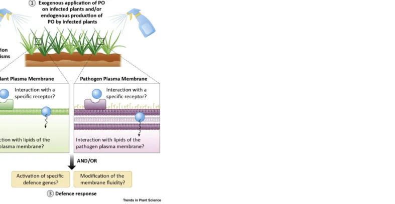
The composition and organisation of the PM are remarkably complex, with strong asymmetrical distribution, and it is evident that its organisation and dynamics ensure good communication that regulates key physiological processes. However, although the role of lipids as important regulators remains indisputable, the mechanisms by which lipids are assembled in the PM remain under investigation [82]. Lipids have frequently been implicated as signals regulating reproductive development, secondary metabolism, and pathogen growth [83,84] or the mitigation of stress responses, immune signalling, and inflammatory processes [75,85–88] in plants and mammals. Emerging evidence showed that FA functions were related to membrane lipid composition changes and adjustment of fluidity [89]. By analogy, it can be assumed that HPOs exhibit similar behaviours, since they are differentiated only by an additional highly reactive hydroperoxide function. Further studies are required to identify additional lipids involved in those interactions and to reveal lipid patterns that are common or distinct among infections with pathogens. Membrane interactions are also suspected to differ between monocotyledons and dicotyledons, since the lipid compositions are extremely variable from one plant species to another. Furthermore, the involvement of membrane receptors, proteic or not, has not been ruled out. Recent reports revealed oxylipin plant transporters such as the AtJAT1/AtABCG16 transporter in arabidopsis, which exhibits an unexpected dual localisation in both the nuclear envelope and the PM [6]. It controls the cytoplasmic and nuclear partitioning of jasmonate phytohormones by mediating both cellular efflux of JA and nuclear influx of JA-Ile. It is thus essential for the maintenance of a critical nuclear JA-Ile concentration to activate JA signalling. Meanwhile, additional studies have reported that GLUCOSINOLATE TRANSPORTER-1 (GTR1) is another JA and JA-Ile transporter in arabidopsis [90,91]. Those studies highlight new mechanisms of signalling hormone regulation, and many other transporters may be described in coming years. Figure 2 Hypothetical Schematic Diagram Showing the Roles of the Plasma Membrane (PM) in Plant Oxylipin (PO)–Pathogen Interactions. After being sprayed on infected plants, POs meet the PM of plants and pathogens. Also, POs may be produced by infected plants and encounter the PM of the cell or a neighbour. They can interact directly with the lipid part of the membranes or with specific receptors present at the surface. This may lead to a series of membrane modifications (stiffening, thinning, pore formation, etc.). POs exogenously applied or endogenously produced by infected plants can result in the activation of various cascades and lead to different responses.
The Parallelism with Eicosanoids
Plants and mammals are not so different, especially when considering their analogous signalling systems. In both groups of organisms, PUFA oxidation products (e.g., oxylipins in plants, eicosanoids in mammals) are crucial stress signalling mediators. Eicosanoids are frequently cited for their multiple biological roles (i.e., in regulating wound responses, inflammation, and cancer and immune responses) and are involved in many enzymatic pathways (Figure 1).
Interplay between eicosanoids is well known, although incompletely understood [92]. For example, transcellular leukotriene (LT) biosynthesis is frequently observed and changes in levels of eicosanoids regulate each other [17,20,87,93]. In mammals, eicosanoids are recognised by various G protein-coupled receptors (GPCRs) at the cell membrane [94]. LA-derived oxylipins, including 9(S)- and 13(S)-HOD, are also produced by mammalian cells and the human GPCR G2A functions as their specific receptor [17,95–97]. This suggests a possible involvement of GPCRs in HPO recognition in plant cells.
Based on their biosynthesis pathways, jasmonates exhibit many similarities to eicosanoids, particularly LTs. Their functional similarities and capacity for being synthesised and released for immediate, local, and systemic responses to stress reinforce this parallelism [3,98,99]. Furthermore, the use of JA as an anticancer agent has been investigated by several in vitro and in vivo experimental systems [98]. This supports the potential for interkingdom communication and applications.
Oxylipin Interkingdom ‘Communication’: An Even More Complicated Interplay?
Given that oxylipins are involved in plant–plant, plant–pathogen, and plant–insect interactions, research on lipid-mediated cross-kingdom communication between hosts and pathogens has emerged in the past years [100]. It is becoming increasingly clear that organisms commonly use oxylipin pathways as a means of communication to elicit biological responses.
During the past decade, studies on fungi and their ability to produce oxylipins for their own development have exploded. Fungal oxylipins (FOs) have appeared to modify plant and mammalian host responses [17,37]. The possibility of crosstalk has also been proposed, given the discovery that many microbes, including fungi, produce eicosanoids. Fungal eicosanoids may mediate host–pathogen crosstalk by downregulating host local defence responses and increase their virulence [95]. In general, oxylipins could also have this potential. It is suggested that plants and fungi communicate through an oxylipin ‘language’, mostly mechanisms that resemble quorum sensing [96]. Thus, a clear response to exogenous application of PUFAs derivates purified from the interacting partners should be observed. Previous studies have shown that C18:2 products like 9(S)- and 13(S)-HPOD, and even compounds like green leaf volatiles, regulate fungal growth, spore development, and mycotoxin production in Aspergillus sp. grown on diverse plant species [84,101,102]. This supports the hypothesis that FOs and POs could be involved in quorum sensing. Because precocious sexual inducer (psi) factors are similar to POs, especially 9(S)- and 13(S)-HPOD, they may affect physiological processes in fungi by mimicking the action of FOs [37,84,101] , thus facilitating the reciprocal cross-kingdom perception of these molecules [100,101]. Several genome analyses have shown the presence of fungal GPCRs. Affeldt reported that GPCRs were responsible for HPO perception and thus should be recognised as oxylipin receptors [96,97]. This reinforces the hypothesis that POs, along with all forms of stimuli, might be perceived by fungi through a GPCR-mediated cascade (Figure 3).
By contrast, the production of such FOs should have an impact on PO content. Studies on wild-type and oxylipin-reduced fungal mutant strains showed a decrease in PO content in the latter [27,84,101]. Aspergillus infection has been reported to increase the levels of 9- and 13-LOX metabolites in maize lines and peanut seeds and to induce specific oxylipin signature profiles [37,101–103]. Lately, Battilani have confirmed that FOs strongly regulate PO gene expression [22], possibly owing to the direct sensing of FOs by plant tissues or to the perception of intact fungal psi-producing oxygenase (ppo) enzymes. In A. nidulans, the three ppo genes are not expressed simultaneously in plant cultures, which suggests that oxylipin production is triggered by a complex but organised network of signalling cascades in a variety of tissues. Although plants seem to lack GPCRs or GPCR homologs, a mode for FO recognition was proposed in 2009, when two arabidopsis GPCR proteins (GTG1 and GTG2) were finally identified and characterised as an abscisic acid (ABA) ligand involved in plant signalling [37,104]. GTG1 and GTG2 seem to be ABA receptors, since arabidopsis mutants that lack GTG1 and GTG2 exhibit ABA hyposensitivity [104]. In addition, the GPCR GCR2 has recently been discovered, resembling GTG1 and GTG2 without being able to bind ABA [105]. Therefore, further detailed analyses are needed to identify their roles and, potentially, new receptor pathways for oxylipins. So far, plants possess many transmembrane proteins and receptor-like kinases that might also function as oxylipin receptors [100].
Finally, based on the similarities of FOs and POs, it appears that FOs may be able to hijack the host oxylipin pathway to facilitate disease development and the production of mycotoxin and spores [88,101]. Such properties have already been reported for the phytotoxin coronatine, which is a JA-Ile mimic and virulence determinant that is produced by various P. syringae pathovars and activates the JA pathway and suppresses the SA pathway when applied to arabidopsis plants [49,89]. Recent studies have also shown the production of hydroxylated JA (11-OH-JA, 12-OH-JA), N-[(-)-jasmonoyl-(S)]-isoleucine, and other forms by different strains of fungi [106–108].
Regardless the mechanisms, these studies provide the first evidence of the apparent impact of fungi on PO content. Unfortunately, to date, information on the impact of POs or FOs in mediating host responses is scarce. However, it remains unclear whether POs are recognised by tissues or cell-surface receptors or at what biological concentrations such POs are effective. Furthermore, a specific oxylipin could have different effects in different species. Currently, nothing is known about bacterial crosstalk and everything remains to be discovered in other pathosystems. Figure 3 Hypothetical Model of Fungal Oxylipin (FO) and Plant Oxylipin (PO) Crosstalk (Based on [16,37,99]). Fungal lipases are secreted in the plant cells where fatty acids (FAs) are cleaved and processed by a lipoxygenase (LOX) (fungal and/or plant based) for oxylipin production. POs are perceived by G protein-coupled receptors (GPCRs), protein kinase (Pka), and psi-producing oxygenase (Ppo) and exploited by fungi for growth, sporogenesis, and mycotoxin production. Also, the host is manipulated through oxylipin mimicry (FO binding GPCR-like receptors). All fungal properties are in orange and all plant properties are in green. Broken arrows indicate hypothetical interactions and unbroken arrows established interactions.
Concluding Remarks and Future Perspectives
As suggested above, the molecular crosstalk between different kingdoms persists in a shadow, in terms of both biological significance and its governing mechanisms. We are at the dawn of deciphering the key elements of this interkingdom communication. Recent studies using molecular genetics and biochemical approaches in both pathogens and their host plants have enhanced our understanding in this area. It has been proposed that certain HPOs can exert antimicrobial effects by interacting with pathogen membranes [68]. 13(S)-HPOD, in particular, appears to increase yeast membrane fluidity in a concentration-dependent manner, likely at the membrane lipid level [109]. This raises the idea that oxylipins might sometimes be incorporated into membrane bilayers, thereby progressively increasing membrane disorder, modifying their function and, thus, affecting microorganism–plant crosstalk. Given the recent discovery of RNAi and small RNA exchange between A. thaliana and B. cinerea, bidirectional cross-kingdom trafficking could also be suspected for oxylipins [110,111]. Despite these important findings, oxylipin-mediated crosstalk between pathogens and host plants is a complex system that needs further study. Many questions remain to be answered, some of which have been neglected for years (see Outstanding Questions).
The literature also lacks information about the involvement of PO esterified forms; since they accumulate in large amounts in the PM on infection, they may act as a reservoir for the rapid synthesis/release of other oxylipins (or directly interact with the PM). Several modes of recognition have been proposed for plant–pathogen crosstalk. The obvious chemical resemblance of POs and lipopeptides, which are strongly involved in induced systemic resistance (ISR), highlights their potential as elicitors [112]. One and/or the other process can fundamentally change the way in which POs could be used as biocontrol agents. These findings add relevance to a deeper understanding of how plant innate immunity and defence mechanisms work. In the current context of finding alternatives to intensive agriculture, this is a challenging research area. Increasing our knowledge of plant response to stresses at the molecular, physiological, and metabolic levels will be vital for the development of new plant varieties and even more in developing new biopesticides.
Uncited references
[113], [114]Acknowledgments
E.D. is supported by a ‘Fonds pour la formation à la Recherche dans l’Industrie et dans l’Agriculture’ (FRIA) grant (5100617F) from the FRS-FNRS (Fonds National de la Recherche Scientifique, Belgium). M.D. and L.L. thank the FRS-FNRS for their position as Senior Research Associates and for grant CDR (J.0014.08 and J.0086.18 projects). This work has benefited from the facilities of the Bordeaux Metabolome/Lipidome Facility-MetaboHUB (grant no. ANR–11–INBS–0010) to S.M. The authors are grateful to Manon Genva and Dr Caroline De Clerck for their interesting remarks and suggestions.
References
1. M. Barbosa, et al., Biologically active oxylipins from enzymatic and nonenzymatic routes in macroalgae, Mar. Drugs 14, 2016, 23. 2. G.A. Howe, Plant hormones: metabolic end run to jasmonate, Nat. Chem. Biol. 14, 2018, 109–110. 3. A.J. Koo, Metabolism of the plant hormone jasmonate: a sentinel for tissue damage and master regulator of stress response, Phytochem. Rev. 17, 2018, 51–80. 4. C. Wasternack and I. Feussner, The oxylipin pathways: biochemistry and function, Annu. Rev. Plant Biol. 69, 2018, 363–386. 5. E. Garreta-Lara, et al., Effect of psychiatric drugs on Daphnia magna oxylipin profiles, Sci. Total Environ. 644, 2018, 1101–1109. Outstanding Questions What are the nonsignalling roles of phyto-oxylipins? Do oxylipins studied in vitro retain interesting antimicrobial properties in planta? What are their spectrum and mechanisms of action? Why do POs cause damage to the pathogen and not to the plant? Are POs involved in innate immunity as elicitors or are they only direct biocides – or maybe both? Can POs be potential biocontrol agents? Do plants use proteic receptors or equivalents for perception of oxylipins (POs or other oxylipins) or is it a mechanism independent of receptors (e.g., interaction with the lipidic fraction of the plant plasma membrane)? How are POs, and more generally oxylipins, involved in interkingdom communication?6. Q. Li, et al., Transporter-mediated nuclear entry of jasmonoyl-isoleucine is essential for jasmonate signaling, Mol. Plant 10, 2017, 695–708. 7. E.E. Farmer, et al., Jasmonates and related oxylipins in plant responses to pathogenesis and herbivory, Curr. Opin. Plant Biol. 6, 2003, 372–378. 8. I. Ponce de León, et al., Oxylipins in moss development and defense, Front. Plant Sci. 6, 2015, 483. 9. A.V. Ogorodnikova, et al., Oxylipins in the spikemoss Selaginella martensii: detection of divinyl ethers, 12-oxophytodienoic acid and related cyclopentenones, Phytochemistry 118, 2015, 42–50. 10. J. Lupette, et al., Non-enzymatic synthesis of bioactive isoprostanoids in the diatom Phaeodactylum following oxidative stress 1, Plant Physiol 178, 2018, 1344–1357. 11. G. Griffiths, Biosynthesis and analysis of plant oxylipins, Free Radic. Res. 49, 2015, 565–582. 12. T. Vellosillo, et al., Defense activated by 9-lipoxygenase-derived oxylipins requires specific mitochondrial proteins, Plant Physiol 161, 2013, 617–627. 13. E. Alméras, et al., Reactive electrophile species activate defense gene expression in Arabidopsis, Plant J 34, 2003, 205–216. 14. L. Mène-Saffrané, et al., Nonenzymatic oxidation of trienoic fatty acids contributes to reactive oxygen species management in Arabidopsis, J. Biol. Chem. 284, 2009, 1702–1708. 15. S. Mueller, et al., General detoxification and stress responses are mediated by oxidized lipids through TGA transcription factors in Arabidopsis, Plant Cell 20, 2008, 768–785. 16. F. Brodhun and I. Feussner, Oxylipins in fungi, FEBS J 278, 2011, 1047–1063. 17. G.J. Fischer and N.P. Keller, Production of cross-kingdom oxylipins by pathogenic fungi: an update on their role in development and pathogenicity, J. Microbiol. 54, 2016, 254–264. 18. C. Wasternack and S. Song, Jasmonates: biosynthesis, metabolism, and signaling by proteins activating and repressing transcription, J. Exp. Bot. 68, 2017, 1303–1321. 19. M. Genva, et al., New insights into the biosynthesis of esterified oxylipins and their involvement in plant defense and developmental mechanisms, Phytochem. Rev. 8, 2019, 343–359. 20. A.B. Islam, et al., Genomic, lipidomic and metabolomic analysis of cyclooxygenase-null cells: eicosanoid storm, cross talk, and compensation by COX-1, Genomics Proteomics Bioinformatics 14, 2016, 81–93. 21. T. Behl, et al., Role of leukotrienes in diabetic retinopathy, Prostaglandins Other Lipid Mediat 122, 2016, 1–9. 22. P. Battilani, et al., Oxylipins from both pathogen and host antagonize jasmonic acid-mediated defence via the 9-lipoxygenase pathway in Fusarium verticillioides infection of maize, Mol. Plant Pathol. 19, 2018, 2162–2176. 23. S. Allmann, et al., Oxylipin channelling in Nicotiana attenuata: lipoxygenase 2 supplies substrates for green leaf volatile production, Plant Cell Environ 33, 2010, 2028–2040. 24. A. Chini, et al., An OPR3-independent pathway uses 4,5-didehydrojasmonate for jasmonate synthesis, Nat. Chem. Biol. 14, 2018, 171–178. 25. C. Böttcher and S. Pollmann, Plant oxylipins: plant responses to 12-oxo-phytodienoic acid are governed by its specific structural and functional properties, FEBS J 276, 2009, 4693–4704. 26. A. Santino, et al., Jasmonate signaling in plant development and defense response to multiple (a)biotic stresses, Plant Cell Rep 32, 2013, 1085–1098. 27. Y. Sun, et al., The role of wheat jasmonic acid and ethylene pathways in response to Fusarium graminearum infection, Plant Growth Regul 80, 2016, 69–77. 28. L. Zhang, et al., Jasmonate signaling and manipulation by pathogens and insects, J. Exp. Bot 68, 2017, 1371–1385. 29. T. Heitz, et al., The rise and fall of jasmonate biological activities, Subcell. Biochem. 86, 2016, 405–426. 30. S. Fonseca, et al., (+)-7-iso-Jasmonoyl-L-isoleucine is the endogenous bioactive jasmonate, Nat. Chem. Biol. 5, 2009, 344–350. 31. P. Delaplace, et al., Oxylipin profile and antioxidant status of potato tubers during extended storage at room temperature, Plant Physiol. Biochem. 46, 2008, 1077–1084. 32. M.L. Fauconnier, et al., Changes in oxylipin synthesis after Phytophthora infestans infection of potato leaves do not correlate with resistance, Plant Physiol. Biochem. 46, 2008, 823–831.
33. M.E. Ghanem, et al., Organ-dependent oxylipin signature in leaves and roots of salinized tomato plants (Solanum lycopersicum), J. Plant Physiol. 169, 2012, 1090–1101. 34. V. Gosset, et al., Attacks by a piercing–sucking insect (Myzus persicae Sultzer) or a chewing insect (Leptinotarsa decemlineata Say) on potato plants (Solanum tuberosum L.) induce differential changes in volatile compound release and oxylipin synthesis, J. Exp. Bot. 60, 2009, 1231–1240. 35. G. Granér, et al., Screening of oxylipins for control of oilseed rape (Brassica napus) fungal pathogens, Phytochemistry 63, 2003, 89–95. 36. R.J. León Morcillo, et al., Plant 9-lox oxylipin metabolism in response to arbuscular mycorrhiza, Plant Signal. Behav. 7, 2012, 1584–1588. 37. D.I. Tsitsigiannis and N.P. Keller, Oxylipins as developmental and host-fungal communication signals, Trends Microbiol 15, 2007, 109–118. 38. J. Vicente, et al., Role of 9-lipoxygenase and α-dioxygenase oxylipin pathways as modulators of local and systemic defense, Mol. Plant 5, 2012, 914–928. 39. M. Hamberg, et al., Activation of the fatty acid α-dioxygenase pathway during bacterial infection of tobacco leaves: formation of oxylipins protecting against cell death, J. Biol. Chem. 278, 2003, 51796–51805. 40. M. Mariutto, et al., Reprogramming of fatty acid and oxylipin synthesis in rhizobacteria-induced systemic resistance in tomato, Plant Mol. Biol. 84, 2014, 455–467. 41. S.A. Christensen, et al., Maize death acids, 9-lipoxygenase-derived, cyclopente(a)nones, display activity as cytotoxic phytoalexins and transcriptional mediators, Proc. Natl Acad. Sci. U. S. A. 112, 2015, 11407–11412. 42. M.X. Andersson, et al., Oxylipin profiling of the hypersensitive response in Arabidopsis thaliana: formation of a novel oxo-phytodienoic acid-containing galactolipid, arabidopside E, J. Biol. Chem. 281, 2006, 31528–31537. 43. J.D.G. Jones and J.L. Dangl, The plant immune system, Nature 444, 2006, 323–329. 44. W. Zhang, et al., Different pathogen defense strategies in Arabidopsis: more than pathogen recognition, Cells 7, 2018, E252. 45. G. Henry, et al., PAMPs, MAMPs, DAMPs and others: an update on the diversity of plant immunity elicitors, Biotechnol. Agron. Soc 16, 2012, 12. 46. J.S. Ramirez-Prado, et al., Plant immunity: from signaling to epigenetic control of defense, Trends Plant Sci 23, 2018, 833–844. 47. C. Chuberre, et al., Plant immunity is compartmentalized and specialized in roots, Front. Plant Sci. 9, 2018, 1692. 48. D. Tripathi, et al., Chemical elicitors of systemic acquired resistance – salicylic acid and its functional analogs, Curr. Plant Biol. 17, 2019, 48–59. 49. J.S. Thaler, et al., Evolution of jasmonate and salicylate signal crosstalk, Trends Plant Sci 17, 2012, 260–270. 50. M. Heyer, et al., A holistic approach to analyze systemic jasmonate accumulation in individual leaves of Arabidopsis rosettes upon wounding, Front. Plant Sci. 9, 2018, 1569. 51. G.Z. Han, Evolution of jasmonate biosynthesis and signalling mechanisms, J. Exp. Bot. 68, 2017, 1323–1331. 52. N.J. Atkinson and P.E. Urwin, The interaction of plant biotic and abiotic stresses: from genes to the field, J. Exp. Bot. 63, 2012, 3523–3544. 53. S.H. Chung, et al., Host plant species determines symbiotic bacterial community mediating suppression of plant defenses, Sci. Rep. 7, 2017, 39690. 54. Y. Hu, et al., Jasmonate regulates leaf senescence and tolerance to cold stress: crosstalk with other phytohormones, J. Exp. Bot. 68, 2017, 1361–1369. 55. M. Hoffmann, et al., Auxin–oxylipin crosstalk: relationship of antagonists, J. Integr. Plant Biol. 53, 2011, 429–445. 56. J.E. Taylor, et al., Crosstalk between plant responses to pathogens and herbivores: a view from the outside in, J. Exp. Bot. 55, 2004, 159–168. 57. X. Di, et al., Involvement of salicylic acid, ethylene and jasmonic acid signalling pathways in the susceptibility of tomato to Fusarium oxysporum, Mol. Plant Pathol. 18, 2017, 1024–1035. 58. T. Lortzing and A. Steppuhn, Jasmonate signalling in plants shapes plant–insect interaction ecology, Curr. Opin. Insect Sci. 14, 2016, 32–39.
59. A.L. Schilmiller and G.A. Howe, Systemic signaling in the wound response, Curr. Opin. Plant Biol. 8, 2005, 369–377. 60. C. Yan and D. Xie, Jasmonate in plant defence: sentinel or double agent?, Plant Biotechnol. J. 13, 2015, 1233–1240. 61. A.J.K. Koo, et al., A rapid wound signal activates the systemic synthesis of bioactive jasmonates in Arabidopsis, Plant J 59, 2009, 974–986. 62. W. Choi, et al., Rapid, long-distance electrical and calcium signaling in plants, Annu. Rev. Plant Biol. 67, 2016, 287–310. 63. W. Choi, et al., Orchestrating rapid long-distance signaling in plants with Ca2+, ROS and electrical signals, Plant J 90, 2018, 698–707. 64. S.A.R. Mousavi, et al., GLUTAMATE RECEPTOR-LIKE genes mediate leaf-to-leaf wound signalling, Nature 500, 2013, 422–426. 65. M. Toyota, et al., Glutamate triggers long-distance, calcium-based plant defense signaling, Science 6, 2018, 1112–1115. 66. T.C. Nguyen, et al., Identification of cell populations necessary for leaf-to- leaf electrical signaling in a wounded plant, Proc. Natl Acad. Sci. U. S. A. 115, 2018, 10178–10183. 67. S. Schuck, et al., The Nicotiana attenuata GLA1 lipase controls the accumulation of Phytophthora parasitica-induced oxylipins and defensive secondary metabolites, Plant Cell Environ 37, 2014, 1703–1715. 68. I. Prost, et al., Evaluation of the antimicrobial activities of plant oxylipins supports their involvement in defense against pathogens, Plant Physiol 139, 2005, 1902–1913. 69. S. Nakamura and A. Hatanaka, Green-leaf-derived C6-aroma compounds with potent antibacterial action that act on both Gram-negative and Gram-positive bacteria, J. Agric. Food Chem. 50, 2002, 7639–7644. 70. W. Ma, et al., Inhibitory effect of (E)-2-hexenal as a potential natural fumigant on Aspergillus flavus in stored peanut seeds, Ind. Crops Prod. 107, 2017, 206–210. 71. E.E. Farmer and M.J. Mueller, ROS-mediated lipid peroxidation and RES-activated signaling, Annu. Rev. Plant Biol. 64, 2013, 429–450. 72. S. Park, et al., Cyclophilin 20-3 relays a 12-oxo-phytodienoic acid signal during stress responsive regulation of cellular redox homeostasis, Proc. Natl Acad. Sci. U. S. A. 110, 2013, 9559–9564. 73. S.M. Müller, et al., The redox-sensitive module of cyclophilin 20-3, 2-cysteine peroxiredoxin and cysteine synthase integrates sulfur metabolism and oxylipin signaling in the high light acclimation response, Plant J 91 2017, 995–1014. 74. E.E. Farmer and C. Davoine, Reactive electrophile species, Curr. Opin. Plant Biol. 10, 2007, 380–386. 75. A.P. Desbois and V.J. Smith, Antibacterial free fatty acids: activities, mechanisms of action and biotechnological potential, Appl. Microbiol. Biotechnol. 85, 2010, 1629–1642. 76. J.J. Kabara, et al., Fatty acids and derivatives as antimicrobial agents, Antimicrob. Agents Chemother. 2, 1972, 23–28. 77. Y.Y. Toporkova, et al., Antimicrobial activity of geometric isomers of etherolenic acid – the products of plant lipoxygenase cascade, Biochem. Biophys. Mol. Biol. 480, 2018, 139–142. 78. M. Deleu, et al., Linoleic and linolenic acid hydroperoxides interact differentially with biomimetic plant membranes in a lipid specific manner, Colloids Surf. B Biointerfaces 175, 2019, 384–391. 79. A. Sarwar, et al., Biocontrol activity of surfactin A purified from Bacillus NH-100 and NH-217 against rice bakanae disease, Microbiol. Res. 209, 2018, 1–13. 80. J. Deravel, et al., Mycosubtilin and surfactin are efficient , low ecotoxicity molecules for the biocontrol of lettuce downy mildew, Appl. Microbiol. Biotechnol. 98, 2014, 6255–6264. 81. G. Henry, et al., The bacterial lipopeptide surfactin targets the lipid fraction of the plant plasma membrane to trigger immune-related defence responses, Cell. Microbiol. 13, 2011, 1824–1837. 82. J. Gronnier, et al., Divide and rule: plant plasma membrane organization, Trends Plant Sci 23, 2018, 899–917. 83. M. Siebers, et al., Lipids in plant–microbe interactions, Biochim. Biophys. Acta 1861, 2016, 1379–1395. 84. M. Brodhagen, et al., Reciprocal oxylipin-mediated cross-talk in the Aspergillus–seed pathosystem, Mol. Microbiol. 67, 2008, 378–391. 85. Y. Okazaki and K. Saito, Roles of lipids as signaling molecules and mitigators during stress response in plants, Plant J 79, 2014, 584–596.
86. A. Singh and M. Del Poeta, Lipid signalling in pathogenic fungi, Cell. Microbiol. 13, 2011, 177–185. 87. K. Sagini, et al., Extracellular vesicles as conveyors of membrane-derived bioactive lipids in immune system, Int. J. Mol. Sci. 19, 2018, E1227. 88. A. Kachroo and P. Kachroo, Fatty acid–derived signals in plant defense, Annu. Rev. Phytopathol. 47, 2009, 153–176. 89. J.W. Walley, et al., Fatty acids and early detection of pathogens, Curr. Opin. Plant Biol. 16, 2013, 520–526. 90. Y. Ishimaru, et al., GTR1 is a jasmonic acid and jasmonoyl-l-isoleucine transporter in Arabidopsis thaliana, Biosci. Biotechnol. Biochem. 8451, 2017, 249–255. 91. H. Saito, et al., The jasmonate-responsive GTR1 transporter is required for gibberellin-mediated stamen development in Arabidopsis, Nat. Commun. 6, 2015, 6095. 92. C.C. Yang and K.W. Chang, Eicosanoids and HB-EGF/EGFR in cancer, Cancer Metastasis Rev 37, 2018, 385–395. 93. D. Wang and R.N. Dubois, Eicosanoids and cancer, Nat. Rev. Cancer 10, 2010, 181–193. 94. J.A. Cornejo-García, et al., Pharmacogenomics of prostaglandin and leukotriene receptors, Front. Pharmacol. 7, 2016, 316. 95. M.C. Noverr, et al., Production of eicosanoids and other oxylipins by pathogenic eukaryotic microbes, Clin. Microbiol. Rev. 16, 2003, 517–533. 96. K.J. Affeldt, et al., Aspergillus oxylipin signaling and quorum sensing pathways depend on G protein-coupled receptors, Toxins (Basel) 4, 2012, 695–717. 97. K.J. Affeldt, et al., Global survey of canonical Aspergillus flavus GPCRs, MBio 5, 2014, e01501–e01514. 98. Z. Raviv, et al., The anti-cancer activities of jasmonates, Cancer Chemother. Pharmacol. 71, 2013, 275–285. 99. T. Savchenko, et al., Arachidonic acid: an evolutionarily conserved signaling molecule modulates plant stress signaling networks, Plant Cell 22, 2010, 3193–3205. 100. S.A. Christensen and M.V. Kolomiets, The lipid language of plant–fungal interactions, Fungal Genet. Biol. 48, 2011, 4–14. 101. X. Gao and M.V. Kolomiets, Host-derived lipids and oxylipins are crucial signals in modulating mycotoxin production by fungi, Toxin Rev 28, 2009, 79–88. 102. D.I. Tsitsigiannis, et al., Aspergillus infection inhibits the expression of peanut 13S-HPODE-forming seed lipoxygenases, Mol. Plant Microbe Interact. 18, 2005, 1081–1089. 103. V. Maschietto, et al., Resistance to Fusarium verticillioides and fumonisin accumulation in maize inbred lines involves an earlier and enhanced expression of lipoxygenase (LOX) genes, J. Plant Physiol. 188, 2015, 9–18. 104. S. Pandey, et al., Two novel GPCR-type G proteins are abscisic acid receptors in Arabidopsis, Cell 136, 2009, 136–148. 105. J.M. Risk, et al., Reevaluation of abscisic acid-binding assays shows that G-protein-coupled receptor2 does not bind abscisic acid, Plant Physiol 150, 2009, 6–11. 106. A. Chini, et al., The fungal phytotoxin lasiojasmonate A activates the plant jasmonic acid pathway, J. Exp. Bot. 69, 2018, 3095–3102. 107. E. Chanclud and J. Morel, Plant hormones : a fungal point of view, Mol. Plant Pathol. 17, 2016, 1289–1297. 108. R.N. Patkar, et al., A fungal monooxygenase-derived jasmonate attenuates host innate immunity, Nat. Chem. Biol. 11, 2015, 733–740. 109. H. Tran Thanh, et al., Toxicity of fatty acid hydroperoxides towards Yarrowia lipolytica: implication of their membrane fluidizing action, Biochim. Biophys. Acta 1768, 2007, 2256–2262. 110. M. Wang, et al., Bidirectional cross-kingdom RNAi and fungal uptake of external RNAs confer plant protection, Nat. Plants 2, 2017, 16151. 111. A. Weiberg, et al., Fungal small RNAs suppress plant immunity by hijacking host RNA interference pathways, Science 342, 2014, 118–123. 112. M. Ongena, et al., Surfactin and fengycin lipopeptides of Bacillus subtilis as elicitors of induced systemic resistance in plants, Environ. Microbiol. 9, 2007, 1084–1090. 113. E. Blée, Impact of phyto-oxylipins in plant defense, Trends Plant Sci 7, 2002, 315–321.
![Table 1 Summary of the Most Effective Antimicrobial Free POs (Based on [35,67], In Vitro Assays) a,b](https://thumb-eu.123doks.com/thumbv2/123doknet/6843577.191057/3.1263.40.1238.369.846/table-summary-effective-antimicrobial-free-based-vitro-assays.webp)
![Figure 3 Hypothetical Model of Fungal Oxylipin (FO) and Plant Oxylipin (PO) Crosstalk (Based on [16,37,99]).](https://thumb-eu.123doks.com/thumbv2/123doknet/6843577.191057/8.1263.33.388.51.445/figure-hypothetical-model-fungal-oxylipin-plant-oxylipin-crosstalk.webp)
