Activation During Collagen-Induced Thrombus Formation
Christelle Lecut, Anne Schoolmeester, Marijke J.E. Kuijpers, Jos L.V. Broers, Marc A.M.J. van Zandvoort,
Karen Vanhoorelbeke, Hans Deckmyn, Martine Jandrot-Perrus, Johan W.M. Heemskerk
Objective—High-shear perfusion of blood over collagen results in rapid platelet adhesion, aggregation, and procoagulant activity. We studied regulation of␣21 and ␣IIb3 integrin activation during thrombus formation on collagen. Methods and Results—Blockade of glycoprotein (GP) VI by 9O12 antibody or of P2Y purinergic receptors permitted
platelet adhesion but reduced aggregate formation, fibrinogen binding, and activation of␣21 and ␣IIb3, as detected with antibodies IAC-1 and PAC1 directed against activation-dependent epitopes of these integrins. Combined blockade of GPVI and P2Y receptors and thromboxane formation abolished integrin activation but still allowed adhesion of morphologically unstimulated, nonprocoagulant platelets. Exogenous ADP partly restored the suppressive effect of GPVI blockade on integrin␣21 and ␣IIb3 activation. Adhesion was fully inhibited only with simultaneous blocking of GPVI and␣21, indicating that the integrin can support platelet–collagen binding in the absence of its activation. Blockade or absence of GPIb␣ only moderately influenced integrin activation and adhesion unless GPVI was inhibited. Conclusions—GPVI- and autocrine-released ADP induce affinity changes of␣21 and ␣IIb3 during thrombus formation on collagen under flow. These integrin changes are dispensable for adhesion but strengthen platelet– collagen interactions and thereby collagen-induced platelet activation. (Arterioscler Thromb Vasc Biol. 2004;24:1727-1733.)
Key Words: ADP 䡲 collagen 䡲 glycoprotein VI 䡲 integrins 䡲 platelets 䡲 thrombus
P
latelet integrins are critical in hemostasis. Abundantly expressed at the platelet surface, integrins are required for platelet interactions with subendothelial matrix components and for platelet–platelet interactions leading to aggregate and thrombus formation.1 Integrin␣21 plays a role in plateletadhesion to collagen under static2,3 and flow conditions.4,5
Integrin␣IIb3 allows platelets to bind to fibrinogen and von Willebrand factor (vWF) present on collagen and other platelets.6This leads to stable platelet adhesion and aggregate
formation.7
On resting platelets, these integrins are considered to be present in a low-affinity state. Intracellular signaling or ligand binding results in conformational changes of the integrins with a switch to higher-affinity states.6 Agonists such as
thrombin, collagen, ADP, and vWF induce␣IIb3 activation and platelet aggregation.8,9Full integrin activation with ADP
requires the P2Y1and P2Y12purinergic receptors.10,11Recent
studies show that integrin ␣21 can also be activated by inside-out signaling.12,13 Thrombin and collagen turn this
integrin into a high-affinity form, whereas ADP changes it to intermediate affinity.13 Although much is known of the
affinity and avidity changes of ␣IIb3 on isolated platelets
especially,8,14regulation of integrin activation during
throm-bus formation is incompletely understood.
In vivo studies as well as ex vivo experiments, in which blood was allowed to flow over collagen under arterial shear conditions, have indicated that glycoprotein (GP) VI is a principal receptor responsible for collagen-induced activation of platelets during thrombus formation.5,15–21The
␣21con-tribution to platelet– collagen interaction has been debated extensively.16 Current evidence with murine and human
platelets shows that this integrin functions to reinforce the activating effect of GPVI to produce stable, nonembolizing thrombi.17,22–24 Integrin ␣21, putatively in its activated
form, synergizes with GPVI to stimulate Ca2⫹ signaling, granule secretion, and subsequent aggregate forma-tion.4,15,22,24,26 It also assists GPVI in triggering of the
procoagulant platelet response (ie, by stimulating surface exposure of phosphatidylserine [PS]).3,5,17,22This
procoagu-lant phospholipid strongly potentiates local formation of thrombin and, hence, coagulation.27In flowing human (but
less clearly so in murine) blood, GPVI blockade still allows ␣21-dependent platelet adhesion to collagen.5,22This raises
the question of how the activation state of this integrin relates
Received May 6, 2004; accepted May 28, 2004.
From the Departments of Biochemistry (C.L., M.J.E.K., J.W.M.H.), Molecular Cell Biology and Genetics (J.L.V.B.), and Biophysics (M.A.M.J.V.), CARIM, Maastricht University, The Netherlands; the Laboratory for Thrombosis Research (A.S., K.V., H.D.), KU Leuven, Campus Kortrijk, Belgium; and E348 Institut National de la Sante´ et de la Recherche Me´dicale (C.L., M.J.-P.), Faculte´ Xavier Bichat, Universite´ Paris, France.
C.L. and A.S. contributed equally to this work.
Correspondence to J.W.M. Heemskerk, PhD, Department of Biochemistry, Maastricht University, PO Box 616, 6200 MD Maastricht, The Netherlands. E-mail jwm.heemskerk@bioch.unimaas.nl
© 2004 American Heart Association, Inc.
Arterioscler Thromb Vasc Biol. is available at http://www.atvbaha.org DOI: 10.1161/01.ATV.0000137974.85068.93
to its adhesive and signaling function. Under flow over collagen, this may involve the GPIb-V-IX complex, which is another receptor implicated in integrin␣IIb3 activation on interaction with vWF.28,29
By using novel antibodies against GPVI and against activation-dependent epitopes on ␣21, we investigated ␣21 and ␣IIb3 activation during human platelet interaction with collagen and subsequent thrombus formation under flow. We found that GPVI and P2Y receptor stimulation caused activation of either integrin, whereas GPIb contributed to a lesser extent. Surprisingly, we found significant integrin-dependent adhesion in the absence of its activation.
Methods
Materials, blood preparation, and experimental design are available online at http://atvb.ahajournals.org.
Thrombus Formation Under Flow Conditions Flow experiments over collagen were performed with
D-phenylalanyl-L-prolyl-L-arginine chloromethylketone (PPACK)-anticoagulated blood, as described.22 Briefly, whole blood was
perfused for 4 minutes over a collagen-coated coverslip through a parallel-plate transparent flow chamber at a wall-shear rate of 150 to 1000 s⫺1. PS exposure was detected by postperfusion with rinsing Hepes buffer, pH 7.45 (in mmol/L: 136 NaCl, 10 glucose, 5 Hepes, 2.7 KCl, 2 MgCl2, and 2 CaCl2, plus 0.1% BSA and 1 U/mL
heparin), containing 1 g/mL OG488-annexin-A5. Integrin activa-tion was monitored by adding fluorescein isothiocyanate (FITC)-coupled IAC-1 (10g/mL) or PAC1 (1:50) to blood before perfu-sion. Where indicated, probes were added to the rinsing buffer.
High-resolution phase-contrast and fluorescent images were rec-orded in real time with intensified cameras as described.17 Using
Quanticell software (Visitech), fluorescence images (average 32 camera images) were corrected for background fluorescence by subtraction of the mean gray level from an adjacent site of the coverslip. Area coverage by fluorescent platelets was determined with Quanticell software. Area coverage from phase-contrast images was analyzed offline using ImagePro software (Media Cybernetics) by automated threshold settings and application of double masks. Area coverage data were used to determine platelet deposition on collagen because they could be collected simultaneously with, and thus compared with, fluorescence data.
Confocal and 2-Photon Laser Scanning Microscopy
For confocal microscopy, coverslips with thrombi stained with FITC-IAC-1, Gi9-FITC, or MOPC-21-FITC were observed with a Bio-Rad laser-scanning microscope. Where indicated, thrombi were fixed with 2% formaldehyde and blocked with 15% BSA in PBS, pH 7.5. After permeabilization with 0.005% sodium dodecyl sulfate, actin cytoskeletal fibers were labeled with Texas-red phalloidin. An MRC600 laser-scanning microscope system was used, equipped with argon– krypton and red diode lasers. Optical sections (4 to 8 scans) were recorded in Kalman filtering mode.
Two-photon laser scanning microscopy (TPLSM) was with a Bio-Rad 2100 multiphoton system. PPACK blood containing 30% calcein-labeled platelets17was perfused over collagen, and z-series
of scans were recorded during perfusion. Alternatively, blood was supplemented with OG488-fibrinogen (0.25 mg/mL). Excitation was by a Spectra-Physics Tsunami Ti:Sapphire laser, tuned and mode-locked at 800 nm, producing pulses of⬇100 fs wide (repetition rate 82 MHz). Fluorescence was detected at 508 to 523 nm.
Results
Suppressed Aggregation and PS Exposure by GPVI and ADP Receptor Blockade but Unchanged Platelet Adhesion to Collagen
The monoclonal antibody (mAb) 9O12, directed against the GPVI collagen-binding site, acts as an efficient GPVI
antag-onist in vitro.30 At 25 g/mL, antigen-binding fragments
(Fabs) of 9O12 specifically blocked platelet adhesion to collagen under static conditions. The fragments also inhibited collagen-induced platelet aggregation and granule secretion. These effects were accompanied by suppression of collagen-induced protein tyrosine phosphorylation (Figure I, available online at http://atvb.ahajournals.org). Surprisingly, 9O12 stimulated phosphorylation of unknown protein bands at 25 and 36 kDa. The mAb binds to the first Ig domain of GPVI, distal from the membrane (C. Lecut and M. Jandrot-Perrus, unpublished data, 2003).
The ability of 9O12 Fab to block GPVI in blood flowing over type-I collagen fibers was investigated at intermediate shear stress (1000 s⫺1). Under control conditions, perfusion resulted in rapid platelet deposition on collagen, which increased linearly in time for at least 5 minutes, as recorded from phase-contrast images.17,22In blood supplemented with
calcein-loaded platelets, the 3D build-up of thrombus forma-tion was followed by recording of stacks of fluorescence images using the high-resolution technique of TPLSM, which provides higher sensitivity and less bleaching than conven-tional microscopy. After an initial phase of 2 minutes of adhesion to the collagen, flowing platelets preferentially incorporated into aggregates (Figure II, available online at http://atvb.ahajournals.org). In the presence of 9O12 Fab (50 g/mL), aggregate formation but not initial adhesion was severely impaired, leaving only 1 to 2 layered platelet groups. As a result, thrombus volume was reduced greatly with 9O12. Phase-contrast images, recorded after an arbitrary end point of 4 minutes of flow (Figure 1), indicated that surface area covered by aggregated platelets under control conditions was 16.4⫾1.3% on average (n⫽11 subjects), with a variation between donors
Figure 1. Effect of GPVI and P2Y receptor blockade on platelet
aggregation, PS exposure, and integrin activation. Whole blood was perfused over collagen at 1000 s⫺1for 4 minutes; preincu-bation with vehicle (control condition), anti-GPVI 9O12 (50 g/mL), or P2Y blockers (40 mol/L MRS2179 and 20 mol/L AR-C69931MX). A, Representative phase-contrast images (120⫻120m) after perfusion. Note the presence of collagen fibers. Representative fluorescence images (150⫻150m) after perfusion with: B, OG488-annexin-A5 to measure PS exposure; C, FITC-IAC-1 to detect activated␣21; or D, FITC-PAC1 to detect activated␣IIb3.
from 9.9% to 25.6%. With 9O12 present, only small aggregates were observed together with numerous single platelets, whereas the area covered by platelets remained unchanged (94.9⫾7.5% of control condition; Figure 2A). This is an underestimate of the platelet deposition reduction, bearing in mind the abolishment of aggregate formation. Higher concentrations of 9O12 up to 200 g/mL gave similar results.
GPVI-induced PS exposure of the platelets on collagen was monitored by staining with OG488-annexin-A5.22,31
Under control conditions, many platelets bound annexin-A5, giving an area coverage with fluorescence of 10.5⫾1.5% (n⫽8). These platelets had a round, blebbing structure, as described previously.17With 50 g/mL 9O12 Fab present,
annexin-A5 staining reduced greatly to 15% of the control situation (Figures 1B and 2A), along with platelet blebbing. Thus, 9O12 suppressed collagen-induced aggregate forma-tion and PS exposure under flow (both effects of GPVI-induced platelet activation) but not platelet adhesion to collagen. This was in agreement with earlier results obtained with the anti-GPVI scFv antibody 10B12.17
Blockers of P2Y1(MRS2179) and P2Y12(AR-C6991MX)
receptors25 were used to evaluate the contribution of
auto-crine ADP in platelet deposition under flow. With the P2Y blockers present, platelets deposited as single cells or as small 2-layered aggregates (Figure 1A). Surface area covered with platelets was increased slightly to 131.0⫾19.9% of control (Figure 2A), likely because of increased contacts of platelets with collagen fibers. Annexin-A5 fluorescence was increased similarly to 128.4⫾29.5% of control (Figures 1B and 2A). In the presence of P2Y blockers, adhesion and PS exposure were about halved when the blood was also pretreated with acetylsalicylic acid (ASA) to block thromboxane formation (Figure 2A), indicating that released thromboxane is involved in platelet adhesion.
Platelet deposition was reduced further when 9O12 was combined with P2Y blockers and ASA. This resulted in adhesion of merely single platelets (28.9⫾4.8% of control), whereas annexin-A5 staining was abolished completely (Fig-ure 2A). This extends earlier observation14and indicates that
human GPVI, together with the secondary mediators ADP and thromboxane, is responsible for aggregate formation and PS exposure but is dispensable for platelet adhesion.
Suppressed ␣21 and ␣IIb3 Activation by Blockade of GPVI or ADP Receptors
To study␣21 activation, a new antibody IAC-1 was used, which specifically recognizes high-affinity forms of this integrin.32IAC-1 does not bind to resting platelets but readily
binds to a neoepitope in the ␣2 I-domain that become exposed during platelet activation with ADP, thromboxane, or thrombin. Activation uncovers an I-domain region at amino acids 199 to 201, which is located at the opposite site of the metal ion-dependent adhesion site domain that is involved in binding to collagen. IAC-1 is thus of little effect on collagen binding.32FITC-labeled IAC-1 binds to platelets
on immobilized convulxin (data not shown), indicating that ␣21is activated after platelet adhesion via GPVI.
When added to blood at 10 g/mL, FITC-labeled IAC-1 did not inhibit platelet deposition on collagen under flow (surface area coverage with all platelets 13.2⫾1.4% versus 13.6⫾1.1% in the absence of IAC-1; n⫽7). Yet, FITC-IAC-1 avidly bound to the platelets adhering to collagen under control conditions but not in the presence of platelet inhibi-tors (Figure III, available online at http://atvb.ahajournal-s.org). Other experiments were performed with FITC-Gi9, a mAb recognizing all␣21 forms, and with FITC-MOPC-21, a mAb that does not bind to platelets. FITC-Gi9 stained control and inhibited platelets, but FITC-MOPC-21 failed to give detectable staining (Figure III).
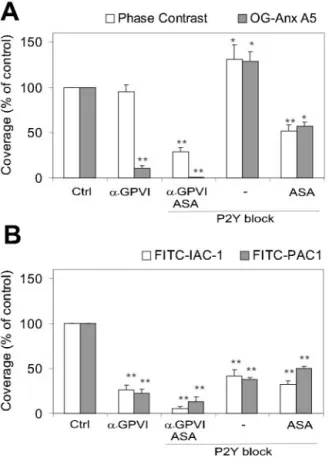
During perfusions under control condition, fluorescence staining of FITC-IAC-1 increased in time; it covered 9.5⫾2.1% (n⫽8 subjects) of the surface area after 4 minutes. Addition of 9O12 (50 g/mL) greatly reduced staining to 23⫾1.9% of the control condition, despite the presence of many adherent platelets (Figures 1C and 2B). Addition of P2Y blockers also reduced IAC-1 staining to 41.2⫾22.1% of control. Blockade of GPVI and P2Y and presence of ASA resulted in almost complete suppression of IAC-1 fluores-cence (Figure 2B).
Figure 2. Quantitative effect of GPVI and P2Y receptor blockade
on platelet aggregation, PS exposure, and integrin activation. Whole blood was perfused over collagen, and platelets were stained with fluorescent annexin-A5, IAC-1, or PAC-1 (Figure 1). Blood was untreated (control) or treated with anti-GPVI Fab 9O12 (50g/mL). Alternatively, blood was treated with P2Y blockers (40mol/L MRS2179 and 20 mol/L AR-C69931MX) in combination with anti-GPVI Fab and ASA; with P2Y blockers alone; or with P2Y blockers and ASA. A, Surface area coverage of all platelets and PS-exposing platelets. B, Surface area cov-erage of FITC-IAC-1 and PAC-1 staining detecting activated ␣21 and ␣IIb3, respectively. Per parameter, data were expressed as percentages of respective control condition set at 100% (mean⫾SEM; n⫽4 to 5; *P⬍0.05; **P⬍0.01 vs control).
To ensure that the reduced IAC-1 binding was not caused by a low detection level of the fluorescence camera, platelets were counterstained for actin with fluorescent phalloidin and examined by confocal microscopy. Under control conditions, individual platelets in aggregates were strongly labeled with FITC-IAC-1, as apparent from overlays of IAC-1 and phal-loidin images (Figure IV, available online at http://atvb. ahajournals.org). With 9O12 present, FITC-IAC-1 fluores-cence of collagen-adherent platelets was reduced greatly, in contrast to the still bright phalloidin staining.
Because 9O12 inhibited platelet aggregation, its effect was studied on signaling toward integrin ␣IIb3 using fluores-cent-labeled PAC1, which is a mAb against activated ␣IIb3.33Under control conditions, addition of FITC-PAC1
to blood or postperfusion with FITC-PAC1 gave fluorescent-labeled platelet aggregates on collagen (Figure 1D). After 4 minutes of perfusion, staining with FITC-PAC1 was 7.5⫾2.1% (n⫽8) of the surface. Blocking of GPVI or P2Y receptors decreased PAC1 staining (Figure 2B). Similarly, as observed with IAC-1, the combination of GPVI and P2Y blockers plus ASA almost completely suppressed PAC-1 staining.
To verify that ADP could activate integrins on collagen-adherent platelets in the absence of GPVI activity, 9O12-treated platelets were postperfused with 10 mol/L ADP. Perfusion with ADP but not with vehicle gave a substantial ⬇5-fold increase in IAC-1-labeling or PAC1-labeling of adherent platelets (Figure 3).
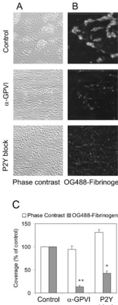
Addition of OG488-labeled human fibrinogen to the blood provided another means to measure ␣IIb3 activation on flow. TPLSM with low detection threshold showed that platelet aggregates on collagen were labeled diffusely with OG488-fibrinogen (Figure 4A and 4B). In the presence of 9O12, only some of the single, collagen-adherent platelets showed fluorescence. Total surface area coverage with
OG488-fibrinogen decreased greatly to 13.0⫾2.8% of control (Figure 4C). P2Y blockers were less effective in reducing OG488-fibrinogen binding (ie, to 42.4⫾5.0% of control); many individual platelets still bound fibrinogen (Figure 4B). With 9O12, ASA, and P2Y blockers present, fibrinogen binding was no longer detected (data not shown).
These results indicate that antagonism of GPVI or the P2Y receptors severely impaired exposure of activation-dependent epitopes on ␣21 and ␣IIb3. Only combined blockade of GPVI and secondary mediators resulted in full inhibition of integrins. This confirms that autocrine ADP and thromboxane play key roles in thrombus build-up24,25and reveals that these
mediators, along with GPVI, mediate ␣21 and ␣IIb3 activation.
GPIb Involved in Collagen Platelet Adhesion but not in Integrin Activation
GPIb-IX-V mediates platelet adhesion to collagen under shear. Its role in integrin activation was studied using the anti-GPIb␣ mAb 12G1, which specifically hinders shear-dependent adhesion to vWF.34At maximally effective con-Figure 3. Effect of ADP addition on integrin activation of
GPVI-inhibited platelets. Blood containing 50g/mL 9O12 was per-fused over collagen, and FITC-PAC1 or FITC-IAC-1 binding was measured. Thereafter, vehicle buffer or ADP (10mol/L) with same fluorescent antibody was superfused at 1000 s⫺1for 1 minute. Percentages of area coverage with fluorescence are given at respective control condition (mean⫾SEM; n⫽3; *P⬍0.05 vs 9O12 alone).
Figure 4. Effect of GPVI and P2Y receptor blockade on platelet–
fibrinogen binding. Blood with OG488-labeled fibrinogen (0.25 mg/mL) was perfused for 4 minutes over collagen in the pres-ence of vehicle (control), anti-GPVI 9O12, or P2Y blockers (Fig-ure 1). A, Phase-contrast images (120⫻120m) and B, merged stacks of TPLSM images (110⫻110m) after perfusion. C, Quantitative effect of receptor blockade on OG488-fibrinogen fluorescence. Data are percentages of control (mean⫾SEM; n⫽3; **P⬍0.01).
centration of 40 g/mL, 12G1 F(ab⬘)2 completely blocked
adhesion of platelets to immobilized vWF. However, the F(ab⬘)2only partially reduced adhesion to collagen fibers at
1000 s⫺1(Figure V, available online at http://atvb.ahajournal-s.org). Although less platelet-collagen contacts were formed, aggregate formation was not prevented; the area covered by platelets remained 72.2⫾10.6% (n⫽7) of control. GPIb blockade with 12G1 inhibited staining with OG488-annexin-A5 to 41.7⫾3.3% of control (Figure V). This inter-vention reduced staining with FITC-PAC1 and FITC-IAC-1 only moderately to 70.7⫾11.4% and 73.8⫾16.3% of control, respectively. However, with 9O12 present, 12G1 markedly inhibited platelet deposition to 12.0⫾5.3% of control.
The results with blocking antibody were corroborated by studies with blood from a patient with Bernard-Soulier syndrome, displaying GPIb-deficient platelets (Figure 5A). After 4 minutes of perfusion, surface area covered with patient platelets was 7.6⫾0.9% (ie, slightly lower than the averaged value for healthy subjects [16.4⫾1.3%; n⫽11]). With 9O12, adhesion of patient platelets was almost
abol-ished to 8.2⫾1.8% of control conditions (Figure 5A and 5B). Thus, at this shear rate, GPIb and GPVI together determine adhesion to collagen, but GPIb does not have a major role in integrin activation.
Functional Importance of␣21 Activation
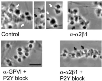
Under control conditions, most platelets on collagen dis-played pseudopods, lamellipods, or blebs. When 9O12 was combined with ASA and P2Y blockers, platelets adhered individually (coverage 28.9⫾4.8% of control) and did not show morphological signs of activation (Figure 6). The remaining adhesion was integrin-dependent because blocking anti-␣21 mAb 6F1 (10 g/mL) severely abrogated adhesion with 9O12 (18.0⫾3.5% versus control; n⫽4), and extra addition of ␣IIb3-blocking arg-gly-asp-ser (400 mol/L) eliminated all platelet deposition (⬍5%). Thus, integrins participated in platelet adhesion in the absence of detectable binding of IAC-1, PAC1, or fibrinogen.
Experiments in which 6F1 was added to the blood in-formed on the functional importance of (activated) ␣21. Blocking of ␣21 with 6F1 notably reduced pseudopod formation but still allowed bleb formation (Figure 6). The 6F1 effect was complete with P2Y blockers present as well (only blebs formed because of GPVI activation). Pseudopod and lamellipod formation was restored when 9O12-treated platelets were postperfused with ADP (data not shown). This suggested that␣21-dependent pseudopod formation, which correlated with IAC-1 binding, was responsible for increased platelet– collagen contact.
Discussion
In this study, we used newly developed tools to determine the role of human GPVI and ADP in integrin activation during collagen-induced thrombus formation under flow. The O12 mAb, directed against the collagen-binding site of human GPVI, was used to block GPVI activity. This inhibited
Figure 5. Abolished adhesion by blockade of GPVI of platelets
from Bernard-Soulier (BSS) patient. Blood from a healthy control subject or a patient displaying BSS was perfused over collagen with/without 9O12 (50g/mL). A, Representative phase-contrast images (120⫻120m) of deposited platelets; inserts are 3-fold magnifications showing giant size of BSS platelets. B, Quantita-tive effect on platelet deposition. Values are percentage of sur-face area coverage (mean⫾SEM; n⫽3; *P⬍0.05; **P⬍0.01 vs absence of 9O12).
Figure 6. Platelet shape and activation of integrin␣21. Blood
was perfused over collagen (Figure 2). Blood was treated with anti-␣21 6F1 (10 g/mL), anti-GPVI 9O12 (50 g/mL), or P2Y blockers (MRS2179⫹AR-C69931MX) plus ASA, as indicated. Shown are representative phase-contrast images (bar⫽10m). White arrows indicate pseudopods; black arrows, blebbing platelets.
formation of platelet aggregates and staining with annexin-A5 (detecting PS exposure) and IAC-1 (detecting activated ␣21), as well as fibrinogen and PAC1 (detecting activated␣IIb3). FITC-IAC-1 is the first described mAb to specifically recognize high activation states of ␣21.32
Blockade of the P2Y1and P2Y12receptors partially inhibited
binding of IAC-1, PAC1, and fibrinogen to platelets, but blockade of GPVI and P2Y receptors in combination with ASA treatment was needed to abolish all binding completely. Conversely, postperfusion with ADP resulted in increased IAC-1 and PAC1 binding to GPVI-inhibited platelets. To-gether, these results provide direct evidence for a role of GPVI and autocrine ADP in inside-out integrin signaling. The inhibitory effects of 9O12 are consistent with studies using isolated platelets showing that stimulation with GPVI ago-nists results in integrin activation6,13and exposure of
proco-agulant PS.27In addition, they extend recent evidence that
GPVI and␣21 contribute to human thrombus formation.17,23
Although anti-GPVI 9O12 efficiently suppressed aggrega-tion, procoagulant activity, and integrin activation under flow, it did not abolish platelet adhesion to collagen, even not at a high dose of 200g/mL. This is remarkably similar to the effects of anti-human GPVI scFv, 10B12, which is also directed against the collagen-binding domain of GPVI.17
Thus, 2 different antibodies against human GPVI appear to suppress platelet activation under shear but not adhesion to collagen. However, we find that combined blockade of human GPVI and ADP/thromboxane effects does lower the adhesion. For mouse blood, this is less clear because block-ade or absence of murine GPVI has been found to either abolish5,18,22 or still permit21platelet– collagen adhesion
un-der flow. This discrepancy is probably attributable to differ-ent experimdiffer-ental conditions.
At the moderately high shear rate of 1000 s⫺1 used, the anti-GPIb mAb 12G1 only partially reduced platelet adhesion to collagen/vWF. When given alone, 12G1 inhibited adhesion slightly and hardly influenced binding of IAC-1 and PAC1. However, in combination with GPVI blockade, GPIb antag-onism or absence of GPIb (in a Bernard-Soulier patient) completely abolished platelet adhesion to collagen. This indicates that GPIb-V-IX only partially contributes to integrin activation under conditions in which GPVI and P2Y receptors are also signaling.
Adhesion of platelets treated with GPVI and P2Y antago-nists was mostly blocked when anti-␣21 mAb 6F1 was added. Furthermore, staining of platelets with IAC-1 or PAC1 correlated with␣21-dependent pseudopod formation of the platelets on collagen. These observations suggest that platelet adhesion to collagen can occur under conditions in which the IAC-1/PAC1 epitopes are not or only partially exposed (ie, with no or local integrin activation), basically in agreement with earlier suggestions by Inoue et al.35In conclusion, our
results indicate that GPVI is responsible for integrin affinity regulation on platelet adhesion to collagen under high shear. Furthermore, autocrine released ADP and subsequent engage-ment of P2Y receptors play assisting roles. Thus GPVI- and P2Y-coupled signaling act synergistically to achieve full integrin activation and thereby stable thrombus formation.
Acknowledgments
C.L. was supported by a Marie-Curie Fellowship from the European Community. We acknowledge grant 902-16 to 276 from the Neth-erlands Organization for Scientific Research. We thank AgroBio for production of 9O12 ascites.
References
1. Ruggeri ZM. Platelets in atherothrombosis. Nat Med. 2002;8:1227–1234. 2. Moroi M, Jung SM. Platelet receptors for collagen. Thromb Haemost.
1997;78:439 – 444.
3. Siljander P, Farndale RW, Feijge MAH, Comfurius P, Kos S, Bevers EM, Heemskerk JWM. Platelet adhesion enhances glycoprotein VI-dependent procoagulant response. Arterioscler Thromb Vasc Biol. 2001;21: 618 – 627.
4. Savage B, Ginsberg MH, Ruggeri ZM. Influence of fibrillar collagen structure on the mechanisms of platelet thrombus formation under flow.
Blood. 1999;94:2704 –2715.
5. Nieswandt B, Brakebusch C, Bergmeier W, Schulte V, Bouvard D, Mohtari-Nejad R, Lindhout T, Heemskerk JWM, Zirngibl H, Fa¨ssler R. Glycoprotein VI but not␣21 integrin is essential for platelet interaction with collagen. EMBO J. 2001;20:2120 –2130.
6. Shattil SJ, Kashiwagi H, Pampori N. Integrin signaling, the platelet paradigm. Blood. 1998;91:2645–2657.
7. Savage B, Sixma JJ, Ruggeri ZM. Functional self-association of von Willebrand factor during platelet adhesion under flow. Proc Natl Acad Sci
U S A. 2002;99:425– 430.
8. Shattil SJ, Ginsberg MH. Integrin signaling in vascular biology. J Clin
Invest. 1997;100:1–5.
9. Kasirer-Friede A, Ware J, Leng L, Marchese P, Ruggeri ZM, Shattil SJ. Lateral clustering of platelet GP Ib-IX complexes leads to up-regulation of adhesive function of integrin ␣IIb3. J Biol Chem. 2002;277: 11949 –11956.
10. Daniel JL, Dangelmaier C, Jin JG, Kim YB, Kunapuli SP. Role of intracellular signaling events in ADP-induced platelet aggregation.
Thromb Haemost. 1999;82:1322–1326.
11. Eckly A, Gendrault JL, Hechler B, Ceazenave JP, Gachet C. Differential involvement of P2Y1and P2YTreceptors in morphological changes of
platelet aggregation. Thromb Haemost. 2001;85:694 –701.
12. Wang Z, Leisner TM, Parise LV. Platelet ␣21 integrin activation: contribution of ligand internalization and the␣2-cytoplasmic domain.
Blood. 2003;102:1307–1315.
13. Jung SM, Moroi M. Platelet collagen receptor integrin␣21 activation involves differential participation of ADP-receptor subtypes P2Y1and
P2Y12. Eur J Biochem. 2001;268:3513–3522.
14. Takagi J, Petre BM, Walz T, Springer TA. Global conformational re-arrangements in integrin extracellular domains in outside-in and inside-out signaling. Cell. 2002;110:599 –511.
15. Goto S, Tamura N, Handa S, Arai M, Kodama K, Takayama H. Involvement of glycoprotein VI in platelet thrombus formation on both collagen and von Willebrand factor surfaces under flow conditions.
Cir-culation. 2002;106:266 –272.
16. Nieswandt B, Watson SP. Platelet collagen interaction: is GPVI the central receptor? Blood. 2003;102:449 – 461.
17. Siljander PRM, Munnix ICA, Smethurst PA, Deckmyn H, Lindhout T, Ouwehand WH, Farndale RW, Heemskerk JWM. Platelet receptor interplay regulates collagen-induced thrombus formation in flowing human blood. Blood. 2004;103:1333–1341.
18. Massberg S, Gawaz M, Gru¨ner S, Schulte V, Konrad I, Zohlho¨fer D, Heinzmann U, Nieswandt B. A crucial role of glycoprotein VI for platelet recruitment to the injured arterial wall in vivo. J Exp Med. 2003;197: 41– 49.
19. Chen H, Locke D, Liu Y, Liu C, Kahn ML. The platelet receptor GPVI mediates both adhesion and signaling responses to collagen in receptor density-dependent fashion. J Biol Chem. 2002;277:3011–3019. 20. Best D, Senis YA, Jarvis GE, Eagleton HJ, Roberts DJ, Saito T, Jung SM,
Moroi M, Harrison P, Green FR, Watson SP. GPVI levels in platelets: relationship to platelet function at high shear. Blood. 2003;102: 2811–2818.
21. Kato K, Kanaji T, Russell S, Kunicki TJ, Furihata K, Kanaji S, Marchese P, Reininger A, Ruggeri ZM, Ware J. The contribution of glycoprotein VI to stable platelet adhesion and thrombus formation illustrated by targeted gene deletion. Blood. 2003;102:170 –1707.
22. Kuijpers MJE, Schulte V, Bergmeier W, Lindhout T, Brakebusch C, Offermanns S, Fa¨ssler R, Heemskerk JWM, Nieswandt B.
Comple-mentary roles of glycoprotein VI and␣21 integrin in collagen-induced thrombus formation in flowing whole blood ex vivo. FASEB J. 2003;17: 685– 687.
23. Chen H, Kahn ML. Reciprocal signaling by integrin and nonintegrin receptors during collagen activation of platelets. Mol Cell Biol. 2003;23: 4764 – 4777.
24. Suzuki-Inoue K, Inoue O, Frampton J, Watson SP. Murine GPVI stim-ulates weak integrin activation in PLC␥2 ⫺/⫺ platelets. Blood. 2003; 102:1367–1373.
25. Turner NA, Moake JL, McIntire LV. Blockade of ADP receptors P2Y12
and P2Y1is required to inhibit platelet aggregation in whole blood under
flow. Blood. 2001;98:3340 –3345.
26. Remijn JA, Wu YP, Heninga EH, IJsseldijk MJW, van Willigen G, de Groot PG, Sixma JJ, Nurden AT, Nurden P. Role of ADP receptor P2Y12
in platelet adhesion and thrombus formation in flowing blood.
Arte-rioscler Thromb Vasc Biol. 2002;22:686 – 691.
27. Heemskerk JWM, Bevers EM, Lindhout T. Platelet activation and blood coagulation. Thromb Haemost. 2002;88:186 –193.
28. Gu MY, Xi XD, Englund GD, Berndt MC, Du XP. Analysis of the roles of 14-3-3 in platelet glycoprotein Ib-IX-mediated activation of integrin
␣IIb3 using a reconstituted mammalian cell expression model. J Cell
Biol. 1999;147:1085–1096.
29. Yap CL, Hughan SC, Cranmer SL, Nesbitt WS, Rooney MM, Giuliano S, Kulkarni S, Dopheide SM, Yuan YP, Salem HH, Jackson SP. Synergistic adhesive interactions and signaling mechanisms operating between
plate-let glycoprotein Ib/IX and integrin ␣IIb3. J Biol Chem. 2000;275: 41377– 41388.
30. Lecut C, Feeney LA, Kingsbury G, Hopkins J, Lanza F, Gachet C, Villeval JL, Jandrot-Perrus M. Human platelet glycoprotein VI function is antagonised by monoclonal antibody-derived Fab fragments. J Thromb
Haemost. 2003;1:2653–2662.
31. Heemskerk JWM, Vuist WMJ, Feijge MAH, Reutelingsperger CPM, Lindhout T. Collagen but not fibrinogen surfaces induce bleb formation, exposure of phosphatidylserine and procoagulant activity of adherent platelets. Blood. 1997;90:2615–2625.
32. Schoolmeester A, Vanhoorelbeke K, Depraetere H, Feys HB, Heemskerk JWM, Hoylaerts M, Deckmyn H. Monoclonal antibody IAC-1 is specific for activated␣21 and binds to amino acid 199–201 of the integrin ␣2 I-domain. Blood 2004;in press.
33. Abrams C, Deng YJ, Steiner B, O’Toole T, Shattil SJ. Determinants of specificity of a baculovirus-expressed antibody Fab fragment that binds selectively to activated form of integrin␣IIb3. J Biol Chem. 1994;269: 18781–18788.
34. Cauwenberghs N, Vanhoorelbeke K, Vauterin S, Westra DF, Romo G, Huizinga EG, Lopez JA, Berndt MC, Harsfalvi J, Deckmyn H. Epitope mapping of inhibitory antibodies against platelet glycoprotein Ib␣ reveals interaction between the leucine-rich repeat N-terminal and C-terminal flanking domains of glycoprotein Ib␣. Blood. 2001;98:652–660. 35. Inoue O, Suzuki-Inoue K, Dean WL, Frampton J, Watson SP. Integrin
␣21 mediates outside-in regulation of platelet spreading on collagen