HAL Id: hal-01712186
https://hal-mines-albi.archives-ouvertes.fr/hal-01712186
Submitted on 31 Oct 2019
HAL is a multi-disciplinary open access
archive for the deposit and dissemination of
sci-entific research documents, whether they are
pub-lished or not. The documents may come from
teaching and research institutions in France or
abroad, or from public or private research centers.
L’archive ouverte pluridisciplinaire HAL, est
destinée au dépôt et à la diffusion de documents
scientifiques de niveau recherche, publiés ou non,
émanant des établissements d’enseignement et de
recherche français ou étrangers, des laboratoires
publics ou privés.
Camila Alves Bandeira Falcao, Maria-Inês Ré, Bartira Rossi-Bergmann
To cite this version:
Ariane de Jesus Sousa-Batista, Wallace Pacienza-Lima, Natalia Arruda-Costa, Camila Alves
Ban-deira Falcao, Maria-Inês Ré, et al.. Depot subcutaneous injection with chalcone CH8-loaded Poly
(Lactic-Co-Glycilic Acid) microspheres as a single-dose treatment of cutaneous leishmaniasis.
Antimi-crobial Agents and Chemotherapy, American Society for Microbiology, 2018, 62 (3), pp.art. e01822-17.
�10.1128/AAC.01822-17�. �hal-01712186�
Depot Subcutaneous Injection with Chalcone CH8-Loaded
Poly(Lactic-Co-Glycolic Acid) Microspheres as a Single-Dose
Treatment of Cutaneous Leishmaniasis
Ariane de Jesus Sousa-Batista,
aWallace Pacienza-Lima,
aNatalia Arruda-Costa,
aCamila Alves Bandeira Falcão,
aMaria Ines Ré,
bBartira Rossi-Bergmann
aaInstituto de Biofísica Carlos Chagas Filho, Universidade Federal do Rio de Janeiro, Rio de Janeiro, Brazil
bUniversité de Toulouse, Mines Albi, CNRS, Centre Rapsodee, Campus Jarlard, F-81013 Albi Cedex 09, France
ABSTRACT
Conventional chemotherapy of cutaneous leishmaniasis (CL) is based on
multiple parenteral or intralesional injections with systemically toxic drugs. Aiming at
a single-dose localized therapy, biodegradable poly(lactic-co-glycolic acid) (PLGA)
mi-croparticles loaded with 7.8% of an antileishmanial nitrochalcone named CH8 (CH8/
PLGA) were constructed to promote sustained subcutaneous release. In vitro, murine
macrophages avidly phagocytosed CH8/PLGA smaller than 6
m without triggering
oxidative mechanisms. Upon 48 h of incubation, both CH8 and CH8/PLGA were 40
times more toxic to intracellular Leishmania amazonensis than to macrophages. In
vivo, BALB/c were given one or three subcutaneous injections in the infected ear
with 1.2 mg/kg of CH8 in free or CH8/PLGA forms, whereas controls received three
CH8-equivalent doses of naked PLGA microparticles or meglumine antimoniate
(Glu-cantime; Sanofi-Aventis). Although a single injection with CH8/PLGA reduced the
parasite loads by 91%, triple injections with free CH8 or CH8/PLGA caused 80 and
97% reductions, respectively, in relation to saline controls. Meglumine antimoniate
treatment was the least effective (only 36% reduction) and the most toxic, as
indi-cated by elevated alanine aminotransferase serum levels. Together, these findings
show that CH8/PLGA microparticles can be effectively and safely used for
single-dose treatment of CL.
KEYWORDS
cutaneous leishmaniasis, microparticles, chemotherapy, depot, PLGA,
polymeric microparticle
L
eishmaniasis is a collective term describing vector-borne diseases caused by
differ-ent species of the protozoan parasite Leishmania where clinical manifestations vary
from cutaneous leishmaniasis (CL) to life-threatening visceral leishmaniasis (VL). CL, the
object of this study, is the most common form, affecting 1.2 million people annually in
98 countries where it constitutes a serious health problem (1). At the site of the sandfly
bite, the dermotropic parasites infect local macrophages, causing primary skin lesions
that typically evolve from papules to ulcers with a raised border and central depression
which may reach many centimeters in diameter. Spontaneous healing is common in the
Old World but rare in the New World (2, 3). Although in a few CL patients infected in
the New World with Leishmania amazonensis and L. braziliensis, the initial lesion may
spread to different parts of the skin (diffuse CL) or to the oronasal mucosa (mucosal
leishmaniasis), respectively, in the great majority (
⬎95%) of cases worldwide, the lesion
remains localized (localized CL [LCL]) (3). Despite that, the mainstay LCL treatment is
similar to other more serious CL and VL forms, advocating one or more series of
parenteral injections with antimonials, pentamidine, or amphotericin B. The efficacy of
oral drugs such as miltefosine, fluconazole, or ketoconazole is controversial (4). All
Received 30 August 2017 Returned for modification 30 October 2017 Accepted 5
December 2017
Accepted manuscript posted online 20
December 2017
Citation Sousa-Batista AJ, Pacienza-Lima W,
Arruda-Costa N, Falcão CAB, Ré MI, Rossi-Bergmann B. 2018. Depot subcutaneous injection with chalcone CH8-loaded poly(lactic-co-glycolic acid) microspheres as a single-dose treatment of cutaneous leishmaniasis. Antimicrob Agents Chemother 62:e01822-17.https://doi.org/10.1128/AAC .01822-17.
Copyright © 2018 American Society for
Microbiology.All Rights Reserved. Address correspondence to Bartira Rossi-Bergmann, bartira@biof.ufrj.br. A.J.S.-B. and W.P.-L. contributed equally to this article.
crossm
on October 16, 2019 by guest
http://aac.asm.org/
of these treatments produce systemic toxicity, ranging from arthralgia to kidney failure
(5), which is unacceptable for patients with uncomplicated LCL.
Local therapies have also presented restrictions. Although cryotherapy and
thermo-therapy require specific equipment and trained personnel to avoid burnings (6), topical
paromomycin (7) and amphotericin B (Drugs for Neglected Diseases Initiative [DNDi])
creams have shown partial or no efficacy. Failure to permeate skin and reach the deep
dermis where infected macrophages reside may be due not only to the thickened LCL
epidermis (8) but also to drug physical inappropriateness (e.g., molecular size and logP)
(9), and the demanding strong permeation enhancers that are frequently irritating to
the skin (10). Strategies aiming at bypassing skin permeation have included
intral-esional infiltrations with antimonials and amphotericin B (11–14). However, due to the
rapid clearance to the circulation, repeated injections are needed, leading to
discom-fort, occasional systemic toxicity, and reduced patient compliance (15). Besides,
re-peated hospital visits are impracticable for patients with limited mobility, and those
living in remote areas or in a conflict zone. Therefore, a local, effective, safe, and short
therapy remains urgently needed for the most common and uncomplicated LCL, such
as those with up to four lesions, each no more than 3 to 4 cm in diameter, afflicting
more than 1 million people worldwide (6).
Drug release depots comprised of biodegradable polymeric microparticles have
appeared as an effective alternative to repeated drug intake in chronic therapy against
cancer, diabetes, hormone, inflammatory diseases, and mental disorders (16–20). In this
context, poly(lactic acid) (PLA), poly(glycolic acid) (PGA), and their copolymer PLGA are
appropriate polymers to prepare slow-release systems due to their favorable
biocom-patibility and biodegradability (21). Chalcones are a new class of potential
antileish-manials (22), and their hydrophobic nature seems adequate for association with PLGA.
In murine LCL caused by L. amazonensis, Piñero et al. used transchalcone-loaded PLGA
film disks implanted at a site distant from the lesion for sustained systemic delivery (23).
Aiming at a local intracellular delivery, we showed previously that PLA nanoparticles
entrapped with a plant-derived chalcone (DMC) are effectively internalized by infected
macrophages and more active than the naked drug when given by intralesional route
(24). Due to the low plant extraction yield, DMC analogues were chemically synthesized,
among them the nitrosylated chalcone CH8 (Fig. 1) was selected as a model drug for
its high antileishmanial activity (50% inhibitory concentration [IC
50]
⫽ 0.7
M) and
selectivity index (SI; SI
⫽ 143). In addition, CH8 showed an intralesional efficacy that was
60-fold higher than the pentavalent antimonial pentostam in a murine model of LCL
caused by L. amazonensis (25).
In the present study, we proposed to prepare and use CH8-loaded PLGA
micropar-ticles for a safe and effective treatment for LCL that bypassed skin permeation and
promoted both the drug uptake by infected macrophages and local sustained release.
RESULTS
CH8/PLGA microparticles characteristics. Both naked PLGA and CH8/PLGA
parti-cles displayed unimodal size distribution (Fig. 2). The average mean sizes at day 4.3
were 5.3 and 6.2
m, respectively, with 2.4 and 2.3 (span) values (Table 1). PLGA and
CH8/PLGA zeta potentials were
⫺11 and ⫺12.5 mV, respectively, indicating a slight
increase in size and negative surface charge due to loading with CH8.
The percentage of drug loading (calculated as the weight of drug in grams loaded
in the microparticles/the weight of polymer and drug in grams initially added in the
FIG 1 Chalcone CH8 (3-nitro-2=-hydroxy-4=,6=-dimetoxychalcone).
on October 16, 2019 by guest
http://aac.asm.org/
formulation
⫻ 100) was 7.8%, corresponding to an entrapment efficiency of 78% (that
is, the percentage of drug that was successfully entrapped into microparticles). The
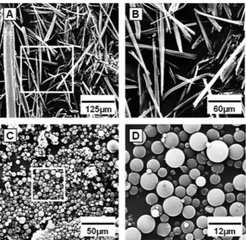
scanning electron microscopic images in Fig. 3 show that particles were spherical and
smooth, with no visible pores. Figure 3A and B (a higher magnification) show the CH8
crystals measuring, on average, 120
m prior to CH8-loaded microparticles preparation.
Figure 3C and D (higher magnification) show the obtained microparticles, which were
about 20 times smaller than the drug crystals. The scarce CH8 crystal outside CH8/PLGA
confirms the high drug entrapment efficiency.
Intracellular uptake and parasite killing. The capacity of macrophages to
inter-nalize CH8/PLGA microparticles was evaluated under optical microscopy (Fig. 4). After
the addition of 50 microparticles/macrophage for 3 h, an average of 10.5
⫾ 1.2 particles
smaller than 6
m was seen inside the cells. No further increase in the numbers of
internalized particles was seen after 6 h (not shown). Despite the efficient particle
uptake by macrophages, entrapment in CH8/PLGA did not increase CH8 activity against
intracellular amastigotes (IC
50⫽ 10.7 and 7.2
g/ml, respectively) as measured after 48
h of incubation (Table 2). However, entrapment in PLGA did reduce CH8 cytotoxicity to
macrophages (50% cytotoxic concentrations [CC
50]
⫽ 290.2 and 429.7
g/ml,
respec-tively), resulting in similar selectivity index values.
Activation of macrophage oxidative mechanisms. To verify whether PLGA uptake
interfered with oxidative mechanisms important for leishmanial parasite killing by
macrophages, the production of nitric oxide (NO) and reactive oxygen species (ROS)
was compared in infected and uninfected macrophages after the addition of CH8,
PLGA, and CH8/PLGA to cells. Figure 5A shows that Leishmania-infected macrophages
were less responsive to lipopolysaccharide (LPS) for NO production than uninfected
cells, in line with previous studies (26–28). CH8 inhibited LPS-stimulated NO response
in infected cells, which is compatible with chalcone’s antioxidant properties (29, 30).
Despite the normal capacity of infected macrophages to respond to zymosan with ROS
production, neither CH8 nor PLGA displayed ROS-activating properties (Fig. 5B). In all,
FIG 2 Microparticle size distributions. Naked PLGA (continuous lines) and drug-entrapped CH8/PLGA(dotted lines) microspheres were analyzed for the percentage of differential volume (Gaussian lines) and cumulative volume (increasing lines).
TABLE 1 Size, surface charge, and drug loadinga
Sample
Distribution of particle diam (m)
Zeta potential (mV) Drug loading (%) d(4.3) d10 d90 SD Spanb PLGA 5.3 1.3 10.4 2.3 2.4 ⫺11.0 ⫾ 2.6 NA CH8/PLGA 6.2 0.6 13.4 4.9 2.3 ⫺12.5 ⫾ 2.8 7.8⫾ 1.5
aZeta potential and drug loading are expressed as means⫾ the standard errors of the mean (n ⫽ 4 to 10).
NA, not applicable.
bSpan⫽ (d90 ⫺ d10)/d50.
on October 16, 2019 by guest
http://aac.asm.org/
these results show that PLGA does not revert the antioxidative mechanisms of
macro-phages by CH8, which would otherwise contribute with the CH8 killing of intracellular
parasites.
Efficacy of treatment of murine CL. The capacity of CH8/PLGA to reduce the
necessary number of drug injections to one was evaluated in the mouse ear model of
infection with L. amazonensis, where treatment started on day 9 postinfection to
maximize the opportunity of demonstrating differences between the formulations. For
that, the infected ears were injected subcutaneously with one or three doses of free
CH8, CH8/PLGA, or the same dose (27
g) of the reference drug meglumine
antimo-niate (Glucantime; Sanofi-Aventis). The curative parameters were lesion growth (Fig. 6A)
and parasite loads measured on day 90 of infection (Fig. 6B). Although local
inflam-mation caused by the subcutaneous injection may unrelatedly increase the lesion sizes,
that was not the case here since untreated and phosphate-buffered saline
(PBS)-injected ears showed similar sizes at most time points. With the three-dose schedule,
meglumine antimoniate was less effective than free CH8 in controlling lesion growth
and reducing the parasite loads (42% versus 78%) compared to animals given PBS
alone (0%). It is noteworthy that entrapment of CH8 in CH8/PLGA further increased its
efficacy to 97%. That was not due to an intrinsic PLGA effect, since naked microparticles
FIG 3 Scanning electron microscopy images of CH8 crystals (raw material) (A and B) and CH8-loadedPLGA microparticles (C and D). Inset squares are shown magnified on the right (B and D).
FIG 4 Microparticle uptake by macrophages. Untreated macrophages (A) and macrophages incubated
for 3 h with CH8/PLGA microparticles (B). Cells and microparticles alone (inset image in panel B) were stained with Giemsa prior to imaging by optical microscopy.
on October 16, 2019 by guest
http://aac.asm.org/
failed to control lesion and parasite growth. To verify the sustained release effect, mice
were given a single dose of CH8/PLGA on day 9. After 81 days, the parasite loads were
significantly lower than in animals given three weekly doses of PBS, CH8, or meglumine
antimoniate (91, 50, and 82%, respectively) (Fig. 6B).
Treatment toxicity. Although there were no visible signs of local toxicity such as
redness or edema, confirmed by the observation that ears were never thicker than
untreated controls even during treatment (days 9 to 23, Fig. 6), it was interesting to
evaluate the possible systemic effects of drugs leaking into the circulation. Apart from
TABLE 2 In vitro antiamastigote and antimacrophage cytotoxicitySample
Mean concn (g/ml) ⴞ SDa
SIb Amastigotes (IC50) Macrophages (CC50)
CH8 7.2⫾ 1.9 290.2⫾ 46.7 40.3
CH8/PLGA 10.7⫾ 2.2 429.7⫾ 60.2* 40.2
PLGA ⬎1,000† ⬎1,000† ND
Glucantime 45.5⫾ 1.6 175.5⫾ 2.6 3.9
aMean values⫾ SD (n ⫽ 3). The IC
50and CC50values were determined by logarithmic regression analysis.
bThe selectivity index (SI) was calculated as CC
50/IC50.*, P⬍0.01 in relation to free CH8. †, value
extrapolated from curve fitting. ND, not determined.
FIG 5 NO and ROS production by macrophages. L. amazonensis-infected and uninfected peritoneal
macrophages were treated with CH8/PLGA, PLGA, or free CH8 (15g of CH8/ml and equivalent amounts
of PLGA) or left untreated. (A) For NO production, LPS (1 g/ml) was added just prior to CH8 or
CH8/PLGA, as indicated. Cells were incubated for 48 h, when the NaNO2content was determined in the
culture supernatants. (B) For ROS production, Zymosan (250g/ml) was added as a control. H2DCFDA (10
M) was immediately added after the indicated compounds, and after 20 min of incubation, ROS
production was evaluated by fluorescence emission. FU, fluorescence units. Means ⫾ the standard
deviations are presented (n⫽ 3). *, P ⬍ 0.05 in relation to the same additive in uninfected cells.
on October 16, 2019 by guest
http://aac.asm.org/
the slight (P
⬍ 0.05) weight loss of meglumine antimoniate-treated mice detected on
days 16 to 23, no other group presented weight variations significantly different from
PBS controls (Fig. 7).
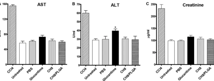
As more sensitive parameters, biochemical markers of hepatic (aspartate
transami-nase [AST] and alanine transamitransami-nase [ALT]), and kidneys (creatinine) toxicity were
measured in the sera after termination of the three-dose schedule with meglumine
antimoniate, CH8, and CH8/PLGA. As seen in Fig. 8, with the exception of meglumine
antimoniate, which increased (P
⬍ 0.05) ALT levels, no other treatment affected AST,
ALT, or creatinine levels. These findings indicate that, unlike treatment with meglumine
antimoniate, which led to a transient weight loss and hepatic toxicity, treatment with
CH8 and PLGA led to no detectable toxicity.
DISCUSSION
The great majority of LC cases are uncomplicated, presenting up to four localized
lesions. Still, due to difficult skin permeation after topical application or rapid clearance
to the circulation after intralesional injections, available drugs are often given by
systemic routes unacceptably exposing patients to systemic toxicity, besides
demand-FIG 6 Effect of CH8/PLGA microparticle depot in murine CL. Mice were infected in the ear with L. amazonensis and given
intralesional subcutaneous injections on days 9, 16, and 23 postinfection with meglumine antimoniate, CH8, or CH8/PLGA, with all drugs administered at 1.2 mg/kg/dose. The controls were vehicle (PBS) or PLGA given on the same days. CH8/PLGA was also given as a single injection on day 9. (A) Lesion sizes were measured on the indicated days. (B) Parasite loads in the ear,
measured on day 90 by a limiting dilution assay. Means⫾ the standard errors of the mean (SEM) are presented (n ⫽ 5 to 8).
FIG 7 Body weight gain. Mice were infected and treated on days 9, 16, and 23 (three times) or day 9 only
(once) as described in Fig. 6 and periodically weighed on the indicated days. Means⫾ the SEM are
presented (n⫽ 5 to 8).
on October 16, 2019 by guest
http://aac.asm.org/
ing frequent visits to a physician for medication. For this reason, formulations designed
to reduce dosing frequency and potential drug toxicity, useful for patients who adhere
poorly to frequent oral or injectable medication and have limited mobility, are urgently
needed for CL. In the United States in 2016, there were eleven U.S. Food and Drug
Administration-approved PLA/PLGA drug products available as microparticulate
de-vices for subcutaneous or intramuscular administration to treat alcohol dependence,
prostate cancer, hormonal disorders, schizophrenia, periodontitis, diabetes, and
acro-megaly (16). In CL, Piñero et al. surgically implanted 3-mm-diameter PLGA and PLA
disks loaded with transchalcone in mouse ears to treat L. amazonensis infection in the
footpads. These authors found that PLGA was superior to PLA (31 and 10% reductions
in lesion sizes, respectively) (23), probably because of the faster PLGA degradation rate
(31). However, all these devices are meant for sustained release in the circulation, not
avoiding the systemic side effects. Local injections would therefore appear more
appropriate for localized CL.
Although used for decades in Asia and Africa to treat CL (13), intralesional injections
with meglumine antimoniate (
ⱕ5 ml/lesion every 2 weeks) has only recently been
officially approved by the Ministry of Health in Brazil (15). Therefore, meglumine
antimoniate would appear a first-choice drug for sustained intralesional release in
microsphere depot. However, because it is a highly hydrophilic drug, a complex
double-wall process is necessary to counteract the strong initial burst, short release
period, and low encapsulation efficiency that normally accompanies encapsulation of
hydrophilic drugs in PLA/PLGA particles (32, 33). Local injections with 130-nm
lipo-somes loaded with meglumine antimoniate, miltefosine, and paromomycin have also
been tested in L. major-infected mice, but only multiple doses of miltefosine-loaded
formulations showed significant therapeutic effects (34). In this context, the
nitrochal-cone CH8 was used here as a model drug for its selective antileishmanial activity and
lipophilicity, suitable for encapsulation in PLGA microparticles for sustained release in
the infected tissue.
The CH8/PLGA microparticles were constructed with unimodal size distribution of
0.6 to 13
m (days 10 to 90) to allow both rapid macrophage uptake of smaller particles
(35) and extracellular depot with the larger ones (36). Indeed, the results showed rapid
internalization of particles
ⱕ6
m within 3 h of incubation, with no further increase
afterward. The negative surface charge (
⫺12.5 mV) was sufficient to avoid cytotoxicity
normally provoked by cationic particles (37), and yet it was mild enough not to produce
repulsion and prevent uptake, as seen with PLGA microparticles of similar sizes but with
FIG 8 Evaluation of systemic toxicity. Mice were infected and treated with the indicated compounds on days 9, 16, and 23 as described in Fig. 6. Positive control
was 1% CCl4given by intraperitoneal injection on day 23. Three days after the last dose, the sera were collected and individually assayed for AST (A), ALT (B),
and creatinine (C). Means⫾ the SEM are presented (n ⫽ 3). *, P ⬍ 0.05.
on October 16, 2019 by guest
http://aac.asm.org/
less cytotoxic and yet maintained its high selectivity index at approximately 40 (Table
2). This may be either (i) due to a diminished phagocytic capacity of
Leishmania-infected macrophages or (ii) an indication that more than 48 h is necessary for the
particles to release additional drug in the intracellular milieu. Likewise, CH8 and
CH8/PLGA did not activate macrophage microbicidal mechanisms (NO and ROS) (Fig. 5).
This is consistent with the antioxidant (39) and anti-inflammatory (40) properties of
chalcones and the lack of ROS activation by similar naked PLGA microparticles (41).
Together, these results indicate that, in vitro, the CH8/PLGA antileishmanial activity was
due to the direct action of the intracellularly released CH8 and not to macrophage
activation.
In vivo, a lesion suppression model of infection with L. amazonensis was used to
minimize the number of animals used and decrease their suffering. Although a triple
CH8/PLGA intralesional injection was required to reduce the parasite loads by 97%, it
is noteworthy that a single injection produced 91% reduction, whereas three injections
with meglumine antimoniate, the gold standard for CL intralesional treatment, yielded
only a 36% reduction. Despite the reported capacity of PLGA microparticles to activate
macrophages for tumor necrosis factor alpha and interleukin-1
 production in vitro
(38), CH8/PLGA efficacy was not due to intrinsic PLGA activity since triple injection with
naked microparticles had no effect on parasite growth (Fig. 6). None of the CH8
intralesional treatments affected body weight gain (Fig. 7) or induced detectable
hepatic or nephrotoxicity, as measured by AST, ALT, and creatinine levels in the serum
3 days after the last of three doses (Fig. 8). Surprisingly, during the three-dose
meglumine antimoniate treatment, a slight weight loss was verified, and significantly
elevated ALT was observed at the end of treatment, a finding indicative of
hepatotox-icity. This is in line with various studies showing that systemic absorption of meglumine
antimoniate occurs after intralesional administration, leading to systemic adverse
ef-fects during and after intralesional meglumine antimoniate that include nausea,
vom-iting, dyspnea, dizziness, and anaphylactic shock (13), as well as altered hepatic
function and white cell count changes (42).
Due to the low solubility of CH8 in aqueous physiological solutions, it was not
possible to measure the in vitro release rate from PLGA particles in sink conditions. In
higher concentrations, the drug precipitated together with the microparticles and
could not be independently measured. However, in the more relevant in vivo situation,
a single dose of CH8/PLGA was found to be superior in controlling parasite growth than
three doses of free drug, supporting the notion that PLGA effectively promoted in situ
sustained CH8 release for at least 81 days. Future process adjustments to increase drug
loading will likely improve its single-dose efficacy.
In conclusion, this study proposes a novel single-dose therapy for uncomplicated CL
consisting of a delivery system that is stable under relatively high temperatures,
suitable for field use in tropical countries. In addition to the greater patient compliance,
this approach may help reduce hospital and personnel costs compared to the current
treatments.
MATERIALS AND METHODS
Drugs. Chalcone CH8 was synthesized by aldol condensation as previously described (25).
Glucan-time (meglumine antimoniate, powder) was obtained from Sanofi-Aventis.
Animals and ethics statement. BALB/c mice (female, 8 weeks old, 23 g) used in the experiments
were maintained under controlled temperature, filtered air, filtered water, autoclaved bedding, and using commercial food at our facilities at Federal University of Rio de Janeiro.
The animal protocols for this study were approved by the Federal University of Rio de Janeiro institutional Animal Care and Use Committee (protocol CAUAP118). The research was conducted in compliance with the principles stated in the National Institutes of Health Guide for the Care and Use of
Laboratory Animals (43).
on October 16, 2019 by guest
http://aac.asm.org/
CH8/PLGA microparticles preparation. Microparticles were prepared by multiple emulsion-solvent
evaporation method (24). Briefly, 500 mg of PLGA (Purasorb PDLG 5004, 50:50, 0.83 dl/g; Corbion) was added to 10 ml of dichloromethane (DCM; 45% [wt/vol]; Vetec) containing prediluted chalcone CH8 (50 mg). The organic solution was agitated at 7,220 rpm for 30 s in an ice bath using a mechanic homogenizer (Heidolph DIAX 900). This first solution was gradually dropped into an aqueous solution of
3% (wt/vol) of polyvinyl alcohol (Mw⫽ 13,000 to 17,000; Sigma-Aldrich) in ultrapure water under 7,220
rpm for 1 min. The final emulsion was further stirred at 400 rpm for 4 h at 40°C under vacuum in a rotary evaporator (Logen) to totally evaporate the DCM solvent. The microparticles were pelleted by centrifu-gation at 9,000 rpm for 10 min at 4°C (Hitachi centrifuge), washed three times in ultrapure water, and then vacuum filtered. The formed particles (CH8/PLGA) were allowed to dry overnight in an oven (Panasonic) at 40°C. Blank particles (PLGA) were prepared in the same way without CH8.
CH8/PLGA microparticles characterization. The particle size distribution was determined by laser
scattering (Zetasizer 3000; Malvern Instruments). CH8/PLGA and PLGA were dispersed in 0.01% (wt/vol) Tween 80 solution in water under ultrasound for 20 s. To quantify entrapped drug, a dry powder sample of CH8/PLGA was dissolved in acetonitrile (Tedia) under bath sonication. The CH8 concentration was determined by UV/high-pressure liquid chromatography using a system consisting of a Shimadzu pump
(LC-20AT), a reversed-phase C18column (5m by 25 cm by 4.6 mm; Rexchrom ODS-S5-100), and a UV
detector (SPDM20A). The parameters used were a mobile phase containing acetonitrile-aqueous 0.01% phosphoric acid (Tedia; 80:20 [vol/vol]), flow at 1 ml/min, and detection at a wavelength of 337 nm.
For morphological imaging, the dry microparticles were coated by sputtering with gold (JFC-1300; JEOL) and analyzed by using a scanning electron microscope (JSM-5600LV; JEOL) at an acceleration of 20 kV under a nitrogen atmosphere.
Microparticle uptake by macrophages. Mouse peritoneal macrophages (5⫻ 105cells in 50l of
RPMI medium) were allowed to adhere for 1 h at 37°C onto glass coverslips placed in triplicates in 24-well
culture plates. The cells were washed twice with PBS and treated or not in triplicates with 2.5⫻ 107
PLGA-CH8 for 3 h at 37°C. At the end of treatment, the coverslips were dipped in prewarmed PBS to remove noninternalized microparticles. Dried cells were fixed in methanol and stained with Giemsa, and
the internalized microparticles inⱖ50 macrophages/coverslip were counted under an optical
micro-scope equipped with a micro ruler (Nikon Ellipse Ti).
Antiamastigote cytotoxicity. Mouse peritoneal macrophages (5 ⫻ 105cells in 50 l of RPMI
medium) were allowed to adhere for 1 h at 37°C onto glass coverslips placed in triplicates in 24-well culture plates. After the nonadherent cells were washed away using prewarmed PBS, the remaining cells
were infected with 5⫻ 106promastigotes of L. amazonensis (strain WHOM/BR/75/JOSEFA) for 4 h at 34°C.
After removal of noninternalized parasites, the cells were cultured for a further 24 h at 37°C in RPMI supplemented with 5% heat-inactivated fetal bovine serum (Cultilab) to allow for intracellular amastigote growth. The infected macrophages were then treated for 48 h in triplicates with either CH8 (0.7, 1.5, 3,
6.1, 12.5, 25, 50, or 100g/ml) or PLGA and CH8/PLGA (CH8 equivalents). Meglumine antimoniate (6.1,
12.5, 25, 50, 100, and 200g/ml) was added as a positive control. The negative controls were incubated
solely with the culture medium. At the end of the incubation period, the coverslips were washed in prewarmed PBS, and the cells were stained with Giemsa. The numbers of amastigotes (in total, 200
macrophages/coverslip) were counted under ⫻400 magnification microscopy, and the results were
expressed as the drug concentration that inhibited parasite growth by 50% (i.e., the IC50).
Anti-macrophage cytotoxicity. The leakage of cytoplasmic lactate dehydrogenase (LDH) into the
culture supernatant was used as a measure of macrophage lysis, considering that leishmanial parasites do not produce LDH (44). Macrophages were infected and treated for 48 h with CH8, PLGA, CH8/PLGA, or meglumine antimoniate as described above for antiamastigote cytotoxicity, using higher drug concentration ranges. At the end of the incubation time, the culture plates were gently centrifuged (500 rpm for 5 min), the supernatants were collected, and LDH was quantified using a colorimetric assay kit according to the manufacturer’s instructions (Doles, Brazil). The results were determined by using the
following equation: % specific release⫽ [(test release ⫺ spontaneous release)/(maximal release ⫺
spontaneous release)]⫻ 100, where the maximum release refers to supernatants from cells treated with
1% Triton X-100, and spontaneous release cells were cultured without additives.
NO and ROS production. For NO production, macrophages were left uninfected or were infected
with L. amazonensis, as described below, for antiamastigote activity. After 24 h of infection, the
macrophages were incubated for 48 h with CH8 (15g/ml), CH8/PLGA (15 g/ml of CH8/135 g/ml of
PLGA) or PLGA (135g/ml), and/or 1 g/ml of LPS (Escherichia coli [Sigma-Aldrich]). The stable NaNO2
product was measured in the culture supernatants using the Griess colorimetric method (45) and a
NaNO2standard curve. For ROS, infected and noninfected macrophages were treated with CH8 (15
g/ml), CH8/PLGA (15 g/ml of CH8 per 135 g/ml of PLGA), PLGA (135 g/ml), or 250 g/ml of Zymosan (Saccharomyces cerevisiae [Sigma-Aldrich]) for 24 h at 37°C. In the last 20 min of culture, a 10
M concentration of the oxidation-sensitive fluorescent dye H2DCFDA (Invitrogen) was added in the
dark, and fluorescence was quantitated at 485-nm excitation and 528-nm emission.
Mouse infection and treatment. Mice (n⫽ 5 to 8) were infected in the ear pinnae with 2 ⫻ 106
promastigotes of L. amazonensis. On days 9, 16, and 23 of infection (three doses) or on day 9 only (one
dose), the ears were injected subcutaneously with CH8 or CH8/PLGA at a dose of 1.17 mg/kg (27g of
CH8 equivalents in 10l of PBS). Controls received the same dose of naked PLGA, reference drug
meglumine antimoniate, or PBS.
Efficacy parameters. Lesion sizes and parasite burdens were evaluated as infection parameters. Ear
thicknesses were periodically measured during infection with a caliper gauge, and lesion sizes were expressed as the difference between infected and noninfected ears. On day 90 of infection, the parasite
on October 16, 2019 by guest
http://aac.asm.org/
relative to a single amastigote at the start of incubation.
Systemic toxicity. Mice were individually weighted during treatment with CH8, CH8/PLGA, PLGA,
and meglumine antimoniate on days 9, 16, and 23 of infection to assess weight loss. Animals were killed on day 26 of infection, the sera were collected and assayed for AST, ALT, and creatinine as parameters of liver and kidney toxicity, using a colorimetric commercial kit (Doles, Brazil) adapted for microvolumes
(47). The positive-control sera were taken from mice which received 200l of 1% carbon tetrachloride
(CCl4) in soybean oil by the intraperitoneal route 3 days before serum collection (48).
Statistical analysis. One-way analysis of variance and the Bonferroni posttest were performed to
demonstrate statistical differences (P ⬍ 0.05). To compare the difference between two groups, a
nonparametric t test was used. The IC50s were calculated from a curve of sigmoidal dose response. All
analyses were conducted using GraphPad Prism 6 software.
REFERENCES
1. Alvar J, Vélez ID, Bern C, Herrero M, Desjeux P, Cano J, Jannin J, de Boer M. 2012. Leishmaniasis worldwide and global estimates of its incidence.
PLoS One 7:e35671.https://doi.org/10.1371/journal.pone.0035671.
2. Masmoudi A, Hariz W, Marrekchi S, Amouri M, Turki H. 2013. Old World cutaneous leishmaniasis: diagnosis and treatment. J Dermatol Case Rep
7:31– 41.https://doi.org/10.1186/1752-1947-7-31.
3. Cota GF, De Sousa MR, Fereguetti TO, Saleme PS, Alvarisa TK, Rabello A. 2016. The cure rate after placebo or no therapy in American cutaneous leishmaniasis: a systematic review and meta-analysis. PLoS One 11:1–15.
https://doi.org/10.1371/journal.pone.0149697.
4. Kevric I, Cappel MA, Keeling JH. 2015. New World and Old World leishmania infections: a practical review. Dermatol Clin 33:579 –593.
https://doi.org/10.1016/j.det.2015.03.018.
5. Iqbal H, Ishfaq M, Wahab A, Abbas MN, Ahmad I, Rehman A, Zakir M. 2016. Therapeutic modalities to combat leishmaniasis, a review. Asian
Pacific J Trop Dis 6:1–5.https://doi.org/10.1016/S2222-1808(15)60975-6.
6. Sundar S, Chakravarty J. 2015. An update on pharmacotherapy for
leishmaniasis. Expert Opin Pharmacother 2:237–252.https://doi.org/10
.1517/14656566.2015.973850.
7. Kim DH, Chung HJ, Bleys J, Ghohestani RF. 2009. Is paromomycin an effective and safe treatment against cutaneous leishmaniasis? A meta-analysis of 14 randomized controlled trials. PLoS Negl Trop Dis 3:e381.
https://doi.org/10.1371/journal.pntd.0000381.
8. Rethi B, Eidsmo L. 2012. Fasl and TRAIL signaling in the skin during cutaneous leishmaniasis: implications for tissue immunopathology and
infectious control. Front Immunol 3:1– 8.https://doi.org/10.3389/fimmu
.2012.00163.
9. Bos JD, Meinardi MM. 2000. The 500 Dalton rule for the skin penetration
of chemical compounds and drugs. Exp Dermatol 9:165–169.https://doi
.org/10.1034/j.1600-0625.2000.009003165.x.
10. Williams AC, Barry BW. 2012. Penetration enhancers. Adv Drug Deliv Rev
64:128 –137.https://doi.org/10.1016/j.addr.2012.09.032.
11. Ferreira EVasconcellos ÉDC, Fernandes Pimentel MI, De Oliveira Schu-bach A, Carvalhaes De Oliveira RDV, Azeredo-Coutinho RB, Da Conceição Silva F, De Matos Salgueiro M, Soares Moreira J, De Fátima Madeira M, Baptista C, Valete-Rosalino CM. 2012. Short report: Intralesional meglu-mine antimoniate for treatment of cutaneous leishmaniasis patients with contraindication to systemic therapy from Rio de Janeiro (2000 to
2006). Am J Trop Med Hyg 87:257–260.https://doi.org/10.4269/ajtmh
.2012.11-0612.
12. Goyonlo VM, Vosoughi E, Kiafar B, Nahidi Y, Momenzadeh A, Taheri AR. 2014. Efficacy of intralesional amphotericin B for the treatment of
cuta-neous leishmaniasis. Indian J Dermatol 59:631– 636.https://doi.org/10
.4103/0019-5154.143571.
13. Esfandiarpour I, Farajzadeh S, Rahnama Z, Fathabadi EA, Heshmatkhah A. 2012. Adverse effects of intralesional meglumine antimoniate and its influence on clinical laboratory parameters in the treatment of
cutane-ous leishmaniasis. Int J Dermatol 51:1221–1225.https://doi.org/10.1111/
j.1365-4632.2012.05460.x.
14. Mushtaq S, Dogra D, Dogra N. 2016. Clinical response with intralesional amphotericin B in the treatment of Old World cutaneous leishmaniasis:
a preliminary report. Dermatol Ther 29:398 – 405. https://doi.org/10
.1111/dth.12377.
15. Ministério da Saúde, Secretaria de Vigilância em Saúde. 2017. Manual de vigilância da leishmaniose tegumentar recurso eletrônico. Ministério da Saúde, Secretaria de Vigilância em Saúde, Departamento de Vigilância das Doenças Transmissívei, Sao Paolo, Brazil.
16. Wang Y, Qu W, Choi SH. 2016. FDA’s regulatory science program for generic PLA/PLGA-based drug products. Am Pharm Rev 19:5–9. 17. Abadi SSH, Moin A, Veerabhadrappa GH. 2016. Review article: fabricated
microparticles: an innovative method to minimize the side effects of
NSAIDs in arthritis. Crit Rev Ther Drug Carrier Syst 33:433– 488.https://
doi.org/10.1615/CritRevTherDrugCarrierSyst.2016016624.
18. Westphal M, Ram Z, Riddle V, Hilt D, Bortey E. 2006. Gliadel® wafer in initial surgery for malignant glioma: long-term follow-up of a
multi-center controlled trial. Acta Neurochir 148:269 –275.https://doi.org/10
.1007/s00701-005-0707-z.
19. Hornstein MD, Surrey ES, Weisberg GW, Casino LA. 1998. Leuprolide acetate depot and hormonal add-back in endometriosis: a 12-month study. Lupron Add-Back Study Group. Obstet Gynecol 91:16 –24. 20. Bobo WV, Shelton RC. 2010. Risperidone long-acting injectable
(Risper-dal Consta®) for maintenance treatment in patients with bipolar
disor-der. Expert Rev Neurother 10:1637–1658.https://doi.org/10.1586/ern.10
.143.
21. Makadia HK, Steve S. 2011. Poly lactic-co-glycolic acid (PLGA) as biode-gradable controlled drug delivery carrier. Polymers (Basel) 3:1377–1397.
https://doi.org/10.3390/polym3031377.
22. Tajuddeen N, Isah MB, Suleiman MA, van Heerden FR, Ibrahim MA. 2017. The chemotherapeutic potential of chalcones against leishmaniases: a
review. Int J Antimicrob Agents 2017:1–28. https://doi.org/10.1016/j
.ijantimicag.2017.06.010.
23. Piñero J, Temporal RM, Silva-Gonçalves AJ, Jiménez IA, Bazzocchi IL, Oliva A, Perera A, Leon LL, Valladares B. 2006. New administration model of trans-chalcone biodegradable polymers for the treatment
of experimental leishmaniasis. Acta Trop 98:59 – 65.https://doi.org/
10.1016/j.actatropica.2006.02.001.
24. Torres-Santos EC, Rodrigues JMJ, Moreira DL, Kaplan MAC, Rossi-Bergmann B. 1999. Improvement of in vitro and in vivo antileishmanial activities of
2=,6=-dihydroxy-4=-methoxychalcone by entrapment in poly(D,L-lactide)
nanoparticles. Antimicrob Agents Chemother 43:1776 –1778.
25. Boeck P, Bandeira Falcão CA, Leal PC, Yunes RA, Filho VC, Torres-Santos EC, Rossi-Bergmann B. 2006. Synthesis of chalcone analogues with increased antileishmanial activity. Bioorganic Med Chem 14:1538 –1545.
https://doi.org/10.1016/j.bmc.2005.10.005.
26. Lapara NJ, Kelly BL. 2010. Suppression of LPS-induced inflammatory responses in macrophages infected with leishmania. J Inflamm 7:8.
https://doi.org/10.1186/1476-9255-7-8.
27. Olivier M, Gregory DJ, Forget G. 2005. Subversion mechanisms by which leishmania parasites can escape the host immune response: a signaling
point of view. Clin Microbiol Rev 18:293–305.https://doi.org/10.1128/
CMR.18.2.293-305.2005.
28. Perrella Balestieri FM, Pires Queiroz AR, Scavone C, Assis Costa VM, Barral-Netto M, De Abrahamsohn IA. 2002. Leishmania amazonensis-induced inhibition of nitric oxide synthesis in host macrophages.
Mi-crobes Infect 4:23–29.https://doi.org/10.1016/S1286-4579(01)01505-2.
29. Nowakowska Z. 2007. A review of anti-infective and anti-inflammatory
on October 16, 2019 by guest
http://aac.asm.org/
chalcones. Eur J Med Chem 42:125–137.https://doi.org/10.1016/j.ejmech .2006.09.019.
30. Torres-Santos EC, Moreira DL, Kaplan MAC, Meirelles MN, Rossi-Bergmann B. 1999. Selective effect of 2=,6=-dihydroxy-4=-methoxy-chalcone isolated from Piper aduncum on Leishmania amazonensis. An-timicrob Agents Chemother 43:1234 –1241.
31. Lee BK, Yun Y, Park K. 2016. PLA micro- and nanoparticles. Adv Drug
Deliv Rev 107:176 –191.https://doi.org/10.1016/j.addr.2016.05.020.
32. Pandey P, Jain D, Chakraborty S. 2016. Poly-lactic-co-glycolic acid (PLGA) copolymer and its pharmaceutical application, p 151–172. In Thakur VK, Thakur MK (ed), Handbook of polymers for pharmaceutical technologies. Wiley, New York, NY.
33. Navaei A, Rasoolian M, Momeni A, Emami S, Rafienia M. 2014. Double-walled microspheres loaded with meglumine antimoniate: preparation, characterization, and in vitro release study. Drug Dev Ind Pharm 40:
701–710.https://doi.org/10.3109/03639045.2013.777734.
34. Momeni AA, Rasoolian M, Navaei A, Emami S, Shaker Z, Mohebali M, Khoshdel A. 2013. Development of liposomes loaded with antileishma-nial drugs for the treatment of cutaneous leishmaniasis. J Liposome Res
23:134 –144.https://doi.org/10.3109/08982104.2012.762519.
35. Champion J, Walker A, Mitragotri S. 2008. Role of particle size in
phago-cytosis of polymeric microspheres. Pharm Res 25:1815–1821.https://doi
.org/10.1007/s11095-008-9562-y.
36. Gaumet M, Vargas A, Gurny R, Delie F. 2008. Nanoparticles for drug delivery: the need for precision in reporting particle size parameters. Eur
J Pharm Biopharm 69:1–9.https://doi.org/10.1016/j.ejpb.2007.08.001.
37. Fröhlich E. 2012. The role of surface charge in cellular uptake and cytotoxicity of medical nanoparticles. Int J Nanomed 7:5577–5591. 38. Nicolete R, Santos Dos DF, Faccioli LH. 2011. The uptake of PLGA
micro-or nanoparticles by macrophages provokes distinct in vitro
inflamma-tory response. Int Immunopharmacol 11:1557–1563.https://doi.org/10
.1016/j.intimp.2011.05.014.
39. Sivakumar PM, Prabhakar PK, Doble M. 2011. Synthesis, antioxidant evaluation, and quantitative structure-activity relationship studies of
chalcones. Med Chem Res 20:482– 492.https://doi.org/10.1007/s00044
-010-9342-1.
40. Herencia F, Ferrándiz ML, Ubeda A, Guillén I, Dominguez JN, Charris JE, Lobo GM, Alcaraz MJ. 1999. Novel anti-inflammatory chalcone deriva-tives inhibit the induction of nitric oxide synthase and cyclooxygenase-2
in mouse peritoneal macrophages. FEBS Lett 453:129 –134.https://doi
.org/10.1016/S0014-5793(99)00707-3.
41. Prior S, Gander B, Blarer N, Merkle HP, Subirá ML, Irache JM, Gamazo C. 2002. In vitro phagocytosis and monocyte-macrophage activation with poly(lactide) and poly(lactide-co-glycolide) microspheres. Eur J Pharm
Sci 15:197–207.https://doi.org/10.1016/S0928-0987(01)00218-4.
42. Da Neves DBJ, Caldas ED, Sampaio RNR. 2009. Antimony in plasma and skin of patients with cutaneous leishmaniasis: relationship with side effects after treatment with meglumine antimoniate. Trop Med Int Heal
14:1515–1522.https://doi.org/10.1111/j.1365-3156.2009.02408.x.
43. National Academy of Sciences. 2011. Guide for the care and use of laboratory animals. National Academy of Sciences, Bethesda, MD. 44. Martin E, Simon MW, Schaefer FW, Mukkada AJ. 1976. Enzymes of
carbohydrate metabolism in four human species of leishmania: a
com-parative survey. J Protozool 23:600 – 607.https://doi.org/10.1111/j.1550
-7408.1976.tb03850.x.
45. Bredt DS, Snyder SH. 1994. Nitric oxide: a physiologic messenger
mole-cule. Annu Rev Biochem 63:175–195.https://doi.org/10.1146/annurev.bi
.63.070194.001135.
46. Lima HC, Bleyenberg JA, Titus RG. 1997. A simple method for quantifying leishmania in tissues of infected animals. Parasitol Today 13:80 – 82.
https://doi.org/10.1016/S0169-4758(96)40010-2.
47. Da Cunha-Júnior EF, Pacienza-Lima W, Ribeiro GA, Netto CD, do Canto-Cavalheiro MM, Da Silva AJM, Costa PRR, Rossi-Bergmann B, Torres-Santos EC. 2011. Effectiveness of the local or oral delivery of the novel naph-thopterocarpanquinone LQB-118 against cutaneous leishmaniasis. J
Anti-microb Chemother 66:1555–1559.https://doi.org/10.1093/jac/dkr158.
48. Otsuka T, Takagi H, Horiguchi N, Toyoda M, Sato K, Takayama H, Mori
M. 2002. CCl4-induced acute liver injury in mice is inhibited by
hepato-cyte growth factor overexpression but stimulated by NK2
overexpres-sion. FEBS Lett 532:391–395. https://doi.org/10.1016/S0014-5793(02)
03714-6.