Suivi de la survie de Geotrichum candidum pendant la
digestion in vitro du fromage de type Camembert
Mémoire
Rihab Farhat
Maîtrise en sciences et technologie des aliments
Maître ès sciences (M. Sc.)
Québec, Canada
Suivi de la survie de Geotrichum candidum pendant la
digestion in vitro du fromage de type Camembert
Mémoire
Rihab Farhat
Sous la direction de :
Sylvie Turgeon, directrice de recherche
Steve Labrie, codirecteur de recherche
iii
Résumé
Au contraire du Camembert traditionnel, la pré-acidification du Camembert stabilisé est limitée par l’inoculation des bactéries lactiques thermophiles à une température inférieure à celle de leur croissance optimale (35-39 °C). Geotrichum candidum est une levure essentielle pour l’affinage du fromage Camembert grâce à ses activités biochimiques. En outre, quelques études ont rapporté que cette levure a été détectée dans les fèces humaines suite à la digestion du Camembert. Cette présence pourrait être due soit à la résistance intrinsèque des souches de G. candidum ou en lien avec les propriétés protectrices de la matrice fromagère. L’objectif de notre étude était d’examiner l’effet protecteur procuré à la souche G. candidum LMA-1028, par les propriétés de la matrice du fromage Camembert pendant la digestion statique in vitro. Afin d’y parvenir, deux matrices liquides (i.e. lait 3,25 % matières grasses et un milieu de culture) ainsi que deux matrices fromagères (i.e. Camembert traditionnel et Camembert stabilisé) ont été analysées. La survie de G. candidum et la désintégration de matrices étudiées ont été évaluées à différents temps de digestion aux étapes buccale, gastrique et duodénale. La désintégration du lait et du milieu de culture était plus élevée que celle des matrices fromagères en raison de leur structure liquide. La désintégration du Camembert stabilisé est plus importante que celle du Camembert traditionnel, ceci pourrait être attribué entre autres à une composition en lipides plus élevée. Globalement, la teneur en matière grasse des matrices laitières contrôle la progression de la désintégration. Lors de la digestion in vitro, la survie de G. candidum a été évaluée. Les résultats sur la viabilité de G. candidum LMA-1028 ont montré que cette souche est hautement résistante. La composition, la structure et les propriétés physicochimiques des matrices laitières n’ont pas amélioré la viabilité de G. candidum LMA-1028 pendant le transit gastro-intestinal.
iv
Abstract
Compared to traditional Camembert-type cheese, stabilized Camembert’s pre-acidification is limited using thermophilic lactic acid bacteria that are inoculated and used under their optimal growth temperature (35-39 °C). Geotrichum candidum is an essential ripening yeast of Camembert cheese due to its biochemical activities. Incidentally, it has been detected in human feces after Camembert consumption. However, this observation could be due either to the intrinsic G. candidum resistance to the gastrointestinal condition or to the protective properties of the Camembert cheese matrix. This study examines the putative protective effect of the cheese matrix on G. candidum LMA-1028 viability during static in vitro digestion. For this purpose, two liquid matrices (i.e. culture medium and pasteurized whole milk (3.25 %fat)) and two Camembert-type cheese variety (i.e. traditional and stabilized) were analyzed. G. candidum LMA-1028 survival under digestive stress was investigated at five digestion times (oral: 2 min, gastric: 60 and 120 min and duodenal: 60 and 120 min), while matrix disintegration was evaluated at three times (oral: 2 min, gastric: 120 min and duodenal: 120 min). Milk and culture medium matrices displayed higher disintegration than cheese matrices due to their liquid nature. The lowest measured disintegration of traditional Camembert compared to stabilized cheese matrix could be attributed to the higher fat content. Overall, dairy matrices disintegration was significantly modulated by the matrix fat content. The structure of the casein networks of milk and Camembert cheeses appears to modulate the accessibility of digestive juice into these matrices during gastric digestion. The difference in the original structure of both Camembert cheese matrices led to different rates of gastric disintegration and resulted in different rates of fat release. When comparing viability counts, G. candidum LMA-1028 showed a high intrinsic resistance to simulated gastrointestinal stresses. Camembert cheese matrices as well as milk didn’t bring additional protection to the studied strain LMA-1028.
v
Table of content
Résumé ... iii Abstract ... iv Table of content ... v List of tables ... ix List of figures ... x Abbreviation list ... xi Remerciements ... xii Avant-propos ... xiii Introduction ... 1Chapter 1 : Literature review ... 4
I. Surface mold-ripened cheese: Camembert ... 4
1. Camembert-type cheese making ... 4
1.1. Coagulation ... 4
1.2. Draining of the curd ... 5
1.3. Salting ... 6
1.4. Ripening ... 6
2. Traditional vs. Stabilized Camembert cheese ... 7
3. Camembert cheese ecosystem ... 10
3.1. Lactic acid bacteria and surface microflora ... 10
vi 4. Geotrichum candidum: Biochemical and physicochemical changes during Camembert cheese
ripening ... 12
4.1. Taxonomy and strains diversity ... 12
4.2. Morphology ... 13
4.3. pH change ... 13
4.4. Flavor-formation ... 15
II. Survival of cheese microbiota during in vitro and in vivo digestion ... 18
1. Gastrointestinal stress ... 18
1.1. Gastric acid stress ... 18
1.2. Bile salts toxicity ... 19
2. Protective effect of cheese matrix ... 20
3. Survival of microbial microflora of soft surface-ripened cheese ... 20
III. In vitro gastrointestinal digestion ... 21
1. In vitro digestion ... 21
2. Models: study of microorganisms viability during in vitro digestion of dairy products ... 22
2.1. Static models ... 22
2.2. Dynamic digestion model ... 23
3. Disintegration kinetics of solid food during in vitro digestion ... 26
3.1. The process of oral digestion ... 27
3.2. The process of gastric digestion ... 28
3.3. The process of intestinal digestion ... 29
4. Nutrient bioaccessibility: dairy matrices ... 31
vii
4.2. Impact of some manufacturing steps on food bioaccessibility ... 33
Hypothèse et objectifs... 36
Chapter 2 : Survival of Geotrichum candidum in Camembert-type cheese during simulated gastro-intestinal transit ... 38
Abstract ... 38
Introduction ... 39
Materials and methods... 42
1. Material ... 42
2. Sample preparation ... 42
3. Matrices characterization ... 43
3.1. Compositional and biochemical analysis ... 43
3.2. Cheeses textural properties ... 44
3.3. G. candidum LMA-1028 enumeration ... 44
4. Survival of G. candidum during in vitro simulated gastrointestinal disintegration of dairy matrices ... 44
4.1. Static in vitro digestion model ... 44
4.2. Matrices mass balance during in vitro digestion ... 45
4.3. Milk and soft-ripened cheese disintegration ... 46
4.4. Viability of G. candidum LMA-1028 during in vitro digestion ... 46
5. Statistical analysis ... 46
Results and discussion ... 47
1. Physicochemical and biochemical properties of dairy matrices ... 47
2. Camembert Cheese texture ... 50
viii
3.1. Liquid matrices ... 54
3.2. Solid matrices ... 55
3.3. Liquid vs solid matrices ... 56
4. Survival of Geotrichum candidum to simulated gastro-intestinal stress ... 57
Conclusion ... 59
General conclusion ... 61
Annex ... 63
ix
List of tables
Chapter 1
Table 1-1: Manufacturing differences between traditional and stabilized Camembert-type cheese:
physicochemical, textural and microbial consequences (Tamime and Law, 2001; Walstra et al., 2005) ... 7
Table 1-2 : Characterization of G. candidum yeast-like morphotype (Guéguen and Jacquet, 1982). ... 13
1-3: Preparation of stock solutions of simulated digestion fluids a final volume of 500 mL). ... 30
Chapter 2 Table 2-1: Chemical composition (%) of CM, Milk, CTRAD and CSTAB ... 48
Table 2-2: Proteolysis degree (%) and pH values1 of Camembert-type cheese (CTRAD and CSTAB) ... 49
Table: 2-3 Texture profile Analysis of Camembert cheese (CTRAD; CSTAB) at room temperature ... 51
x
List of figures
Chapter 1
Figure 1-1: General diagram of mold surface-ripened cheese process (e. g Camembert-type) ... 5
Figure 1-2: Cascade of physical and chemical reactions during ripening of Camembert cheese ... 6
Figure 1-3: Description of (a) Stabilized curd versus (b) Traditional curd manufacture ... 8
Figure 1-4: Stabilized (A) and traditional (B) Camembert-type cheese ... 9
Figure 1-5: Evolution of pH and fungal growth during Camembert cheese ripening. ... 14
Figure 1-6: Protein degradation during surface-ripened cheese maturation: Expression data observed for genes encoding proteases (A) and peptidases (B). ... 15
Figure 1-7: An overview of static digestion model... 22
Figure 1-8: Schematic presentation of TIM-1 equipped with membranes to study nutrients bioaccessibility .... 25
Figure 1-9: Scheme of the transit of food structures during digestion ... 26
Figure 1-10: The structure of produced dairy products during processing by non-specific association at the sub-micron level of three major components of milk matrix ... 34
Chapter 2 Figure 2-1: Disintegration rate of CM ( ), Milk ( ), CTRAD ( ) and CSTAB ( ) during static in vitro digestion. ab denotes significant differences (p < 0.05) between disintegration of each matrix at the same period of in vitro digestion. ... 53
Figure 2-2: Mass balance of pellet ( ), fat ( ) and liquid ( ) layer through in vitro gastrointestinal digestion of CM, Milk, CTRAD and CSTAB. ... 55
xi
Abbreviation list
CFU Colony Forming Unit CM Culture medium FAA Free Amino Acids
FFA Free Fatty Acids
FRQNT Fonds de recherche du Québec - Nature et les Technologies
FRAP Fluorescence Recovery After Photobleaching HDM Human Duodenal Model
LAB Lactic Acid Bacteria
NPN Non Protein Nitrogen
PDO Protected Designation of Origin
TN Total Nitrogen
TPA Texture Profile Analysis
xii
Remerciements
Je remercie tout particulièrement ma directrice de maitrise, Dre Sylvie Turgeon, pour son assistance et ses conseils si précieux, et surtout de m’avoir aidé à développer mon esprit critique.
Par ailleurs, j’adresse mes remerciements les plus sincères à mon codirecteur, Dr Steve Labrie, pour sa bienveillance et son excellent mentorat de toute l’équipe.
Je remercie principalement Dr Yves Pouliot pour son soutien, sa confiance et surtout de m’avoir recommandé pour intégrer deux équipes de recherche au sein desquelles j’ai pu beaucoup apprendre.
J’adresse aussi mes remerciements, aux membres de deux équipes de recherche pour leur collaboration et au personnel de l’Université Laval: Catherine Viel, Diane Gagnon, Dre Laurie-Eve Rioux et Dre Marie-Hélène Lessard pour l’aide et les conseils qu’elles m’ont offerts au laboratoire.
Le bouquet de mes remerciements, je le présente affectueusement à mes parents, mes frères, mon oncle et Fourat pour leur soutien si efficace tout le long de mon cheminement.
Je souhaite également remercier tous mes amis et plus particulièrement Adriana Paredés pour son écoute, ses conseils et soutien moral tout le long de ma maitrise, ainsi Awa, Olfa, Zeineb, Dorra, Nesrine Fatma et Mariem. Merci pour votre temps, votre aide, votre bonne humeur et pour les agréables moments passés ensemble.
xiii
Avant-propos
Les travaux de ce mémoire font partie d’un projet de recherche financé par le Fonds de recherche du Québec – Nature et technologies (FRQNT). L’objectif général de ce mémoire était l’évaluation de l’effet protecteur des matrices fromagères sur la survie de Geotrichum candidum pendant la digestion in vitro du fromage Camembert.
Le mémoire comprend deux chapitres. Le premier chapitre correspond à une Revue de la littérature abordant les propriétés physico-chimiques du fromage Camembert, une description de la levure G. candidum et ses activités enzymatiques (lipolytique et protéolytique), la caractérisation du stress gastro-intestinal (i.e. stress acide et biliaire), la description des modèles de digestion in vitro utilisés en recherche et la cinétique de la désintégration des matrices laitières suite à la digestion orale, gastrique et duodénale.
Le deuxième chapitre, intitulé « Survival of Geotrichum candidum from Camembert-type cheese during simulated gastro-intestinal transit », est un article qui sera soumis à une revue scientifique à déterminer. J‘ai contribué à ce travail en réalisant toutes les expérimentations au laboratoire et en rédigeant entièrement l‘article. Sylvie Turgeon, Steve Labrie, Marie-Hélène Lessard et Laurie-Eve Rioux ont apporté leur soutien scientifique dans les décisions expérimentales et lors de la rédaction et la correction de l‘article. Sylvie Turgeon et Steve Labrie ont également obtenu le financement pour la réalisation de la recherche.
1
Introduction
The worldwide production of cheese increased with an average annual growth rate of 4.0% over the last 30 years (Fox et al., 2017). This increase is due to their nutritive quality, their appealing flavors, textural properties and the large possibility to use them in a meal (Fox et al., 2017). In Canada, cheese consumption reached 13.38 kg per capita per annum in 2016 (Canadian Dairy Commission, 2016). Among all cheese categories, the highest increase of consumption belongs to Cheddar and specialty cheese types (Canadian Dairy Commission, 2016). For instance, soft cheese is the third largest cheese production with 77,194 Kg in 2016 (Canadian Dairy Commission, 2017).
From a structural point of view, cheese matrix is a protein-based gel consisting in a cross-linked casein-calcium phosphate network; physically entrapping fat globules. The final structure of the gel is a function of pH, calcium concentration and milk processing history. The variation of cheese-making steps (e.g., coagulation, maturation, whey draining, salting, pressing, and ripening) provides a large variety of cheese products. For instance, a mixed coagulation by microbial acidification and rennet action on casein gel gives a Camembert-type cheese curd. Depending on the nature of the selected starter, two categories of Camembert cheese are produced; namely traditional- and stabilized-Camembert cheeses. For the traditional Camembert cheese, acidification requires the use of mesophilic bacteria (25 °C), whereas for the stabilized curd, thermophilic species are used below their optimal growth temperature (34-39 °C) to limit acidification (Lawrence et al., 1987). Camembert cheese stabilization results in different physicochemical properties, such as pH and water activity, which in turn influences the composition of Camembert cheese microflora (Arteau et al., 2010; Lessard et al., 2012; Spinnler and Gripon, 2004). Specifically, Penicillium camemberti predominates on the rind of stabilized cheeses whereas Geotrichum candidum seems more abundant on traditional curd (Arteau et al., 2010).
G. candidum is an aerobic acid-tolerant and salt-sensitive yeast. It is a key microorganism in the development of the organoleptic properties of Camembert-type cheese. It governs the lipolytic and proteolytic activities during the ripening of Camembert cheese matrix (Boutrou et al., 2006b; Dugat-Bony et al., 2015; Leclercq-Perlat et al., 2004a; Lessard et al., 2014). G. candidum simultaneously assimilates the lactate produced by lactic acid bacteria and produces ammonia which contributes to the alkalinization of the curd. This subsequently promotes the proteolytic activity of the surface microflora during the first 21 days of the cheese ripening (Boutrou et al., 2006b). As with proteolysis, lipolysis occurs extensively within mould-ripened cheeses matrices; about 5-20% of the triglycerides are hydrolyzed depending on the ripening period (Fox et al., 2004).
The metabolic activities (i.e. proteolysis and lipolysis) of the cheese microflora, in addition to technological processing, modulate the particular microstructural and physicochemical properties of the cheese matrix. These properties have an important influence on nutrient bioaccessibility during the gastrointestinal transit (Turgeon and
2 Rioux, 2011). Few studies focused on the disintegration and the nutrients release of different cheese matrices such as aged, young, mild and light Cheddar, Mozzarella and stabilized Camembert-type cheeses (Ayala-Bribiesca et al., 2016; Fang et al., 2016; Lamothe et al., 2012). Overall, high-fat commercial cheese varieties (Camembert and Cheddar) showed higher disintegration rates compared to low-fat Mozzarella (Fang et al., 2016). A high calcium content limited the lipolysis extent, decreased the Cheddar disintegration, and also limited and delayed the fatty acids bioavailability (Ayala-Bribiesca et al., 2016). Specifically, the gastric disintegration of cheeses is modulated by the composition and the textural profile of the matrix (i.e., elasticity, hardness, and cohesiveness) (Lamothe et al., 2012). These studies showed that the bioaccessibility of the cheese nutrients was proportional to the disintegration kinetics of the cheese matrix during the gastrointestinal transit (Lamothe et al., 2012; Rinaldi et al., 2014). Noteworthy, liquid and semi-liquid dairy matrices such as milk and yogurt reached full ʺdisintegrationʺ more rapidly, i.e. after about 120 min of gastric digestion (Rinaldi et al., 2014), while the majority of the analyzed cheese matrices showed an almost complete disintegration after 300 min of gastrointestinal digestion.
The slow disintegration of cheese matrix made this product a favorable delivery system of probiotic microorganisms (Ouwehand et al., 2010; Stanton et al., 1998). Mainly, the dense matrix (i.e., high protein and fat contents), the high pH in some varieties and the buffering capacity have been reported to bring protection against gastrointestinal stresses (Champagne et al., 2011; Plessas et al., 2012). These properties also promoted the development of a diversified microflora within cheese during ripening which provides an average of 108–109 CFU microorganisms per g
of ready-to-eat cheese (Beresford et al., 2001). The diversity of cheese ecosystems raises the question whether this microflora has a beneficial effect on human microbiota when ingested (Montel et al., 2014). Lay et al. (2004), showed that the microflora of traditional Camembert cheese enhanced the metabolic activity of the human-associated rat’s microbiota during consumption. Subsequently, the same team confirmed the beneficial effect of Camembert-type cheese consumption on the microbiota of a human subject group. G. candidum showed a significant resistance to gastrointestinal stresses (Firmesse et al., 2008). Adouard et al. (2015b) , compared the viability of microbial mixture, including G. candidum ATCC 204307 when inoculated into liquid culture medium, rennet gel, and smear-ripened cheese, using a dynamic in vitro digestion model. They observed that during the digestion of smear-ripened cheese, the strains of the yeast species D. hansenii, K. lactis and G. candidum displayed high resistance to digestive stresses.
A high viability of G. candidum strains is observed through digestive stress, after the consumption of Camembert type cheese (Firmesse et al., 2008). Given this, it may be hypothesized that the resistance of G. candidum to these harsh conditions is due to the protective effect of the structural and physicochemical properties of cheese matrix. Our study aims to investigate the viability of G. candidum LMA-1028, during the gastrointestinal transit using an in vitro static
3 model. The protective effect of pasteurized whole milk (3.25 % fat content) and of two Camembert-type cheese matrices with different protein and fat content was studied.
4
Chapter 1 : Literature review
I.
Surface mold-ripened cheese: Camembert
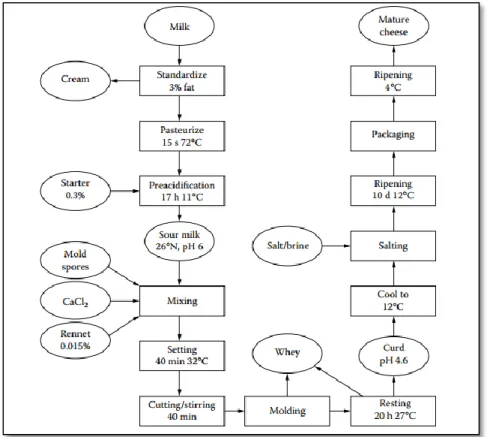
Numerous soft-cheese varieties are produced such as Pont-l’Évêque, Munster, and Italian Crescentia. Camembert and Brie-type cheeses are the best known and their popularity is increasing especially in Australia, USA, and Canada (Tamime and Law, 2001). Those varieties are made from either raw or pasteurized milk and do not have the Protected Designation of Origin (PDO) status (Leclercq-Perlat, 2011). The manufacturing scheme of Camembert-type cheese production is discussed in the following sections with a simplified flow chart (Figure 1-1).
1. Camembert-type cheese making
1.1. Coagulation
The Camembert cheese is produced using a mixed coagulation method combining milk acidification and the activity of the rennet that result in a mixed coagulated curd. Coagulation time depends on the cheese type and varies from 30 to 90 min (Leclercq-Perlat, 2011). During coagulation, the temperature is maintained between 32-35 °C to promote both the rennet and the lactic acid bacteria activities. In general, in Camembert cheese, coagulation is performed using an inoculation rate around 5.70 log CFU/mL and the addition of 0.018- 0.022% (v/v) concentrated rennet (Leclercq-Perlat, 2011).
According to Tamime and Law (2001) as reported from Mietton (1986), during mold-ripened cheese manufacture, the pH of the curd at molding and unmolding is influenced by the renneting pH. The acidification controls the extent of curd demineralization and syneresis which induces several physicochemical and structural changes in cheese. When the acidification approaches the isoelectric point of casein (pH 4.6), the colloidal calcium phosphate (CCP) is extensively solubilized. This weakens the bonds between individual caseins which favors their dissociation from the micelle and reduces the water binding capacity of caseins (Fox et al., 2004). Hydrophobic interactions between caseins are favored and as the pH approaches the isoelectric point of caseins, the para-casein network displays a more compact conformation (Fox et al., 2004). These chemical changes result in the decrease of the protein matrix porosity and the increase of the serum expulsion. When acidification is limited (pH > 5.2), casein network porosity is higher and serum release is lower than in a more acidic curd (Fox, 2000; Lucey et al., 1996). The final physicochemical and structural properties of cheese matrix also depend on other factors as temperature, and the equilibrium pH of the gel before drainage.
5 1.2. Draining of the curd
Draining takes place spontaneously at 26-28 °C as driven by the acidification and the rennet action on casein gel structure. Both acts simultaneously with antagonist effects on the final properties of the curd (Leclercq-Perlat, 2011). The syneresis of cheese curd depends on milk gel firmness at cutting and the surface area of the curd after being cut into cubes (Walstra et al., 2005). The strong bonds between casein particles as a result of low renneting pH, limits the syneresis. While cutting the curd into smaller cubes increases the whey-curd interface and so the extent of syneresis. In the case of Camembert-type cheese, the brittle structure of the curd requires a moderate mechanical handling (i.e., cutting and stirring) to get a smooth final curd (Tamime and Law, 2001; Walstra et al., 2005). This practice explains the final high moisture and moisture in non-fat basis of this cheese variety (Bylund, 2003). For traditional Camembert cheese, acidification continues during draining until a pH around 4.6 is reached within one day. To sum up, syneresis depends on the acidity, the temperature, and mechanical working in the vat of cheese curd during manufacturing. These factors modulate the final moisture content and textural properties of the cheese matrix.
Figure 1-1: General diagram of mold surface-ripened cheese process (e. g Camembert-type)
Reproduced from Walstra et al. (2005)
6 1.3. Salting
In general, salting is done either using rubbing or brining techniques (Guinee, 2004). Brining is the most common practice in Camembert-type cheese making. Depending on the moisture content, shape and size of cheese curd, brining time might vary from 30 min, some hours to one day (Spinnler and Gripon, 2004; Walstra et al., 2005). These factors also modulate the time required for salt-in-moisture (SM) equilibrium after salting. Due to the high moisture and the specific surface area of Camembert-type cheese salt absorption is fast. The brine solution does not only contain NaCl but also other solutes, notably lactic acid and salts that are leached out from the cheese (Walstra et al., 2005). The targeted concentration of salt within soft surface-ripened cheese matrix is 1-2 % (g NaCl/100g of wet cheese) (Leclercq-Perlat, 2011; Walstra et al., 2005) . For Camembert-type cheese, salt content influences the proteolysis and pH change. It also has a selective effect on the fungal microflora with salt sensitivity (Guinee and Fox, 1993). Overall brining allows further draining all with limiting pathogenic or spoilage microorganisms growth (Guinee, 2004).
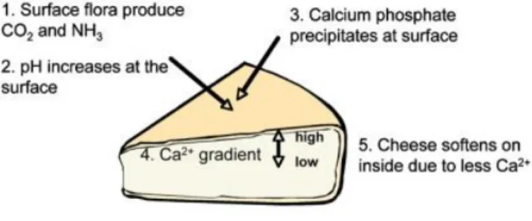
1.4. Ripening
Cheese ripening starts before curd making is finished, and continues until the desired organoleptic and textural properties are reached (Figure 1-4). It includes the biochemical, physical and microbiological changes driven by enzymatic activities of the cheese microflora (Spinnler and Gripon, 2004). Camembert-type cheese ripening occurs in two steps (1) in a ripening chamber at a temperature between 10-14 °C, relative humidity 90 to 95%, during 9 to 14 days, and (2) ripening after packaging at a temperature between 4-6 °C using a specific wrapping (Leclercq-Perlat, 2011). The total ripening time varies between 12 and 35 days depending on the wanted organoleptic qualities (Spinnler and Gripon, 2004). The impact of the microbial activities on the physicochemical and structural properties of Camembert-type cheese are summarized in Figure 1-2 and will be detailed in sections I.4.3 & .
Figure 1-2: Cascade of physical and chemical reactions during ripening of Camembert cheese
Reproduced from Everett and Auty (2008)
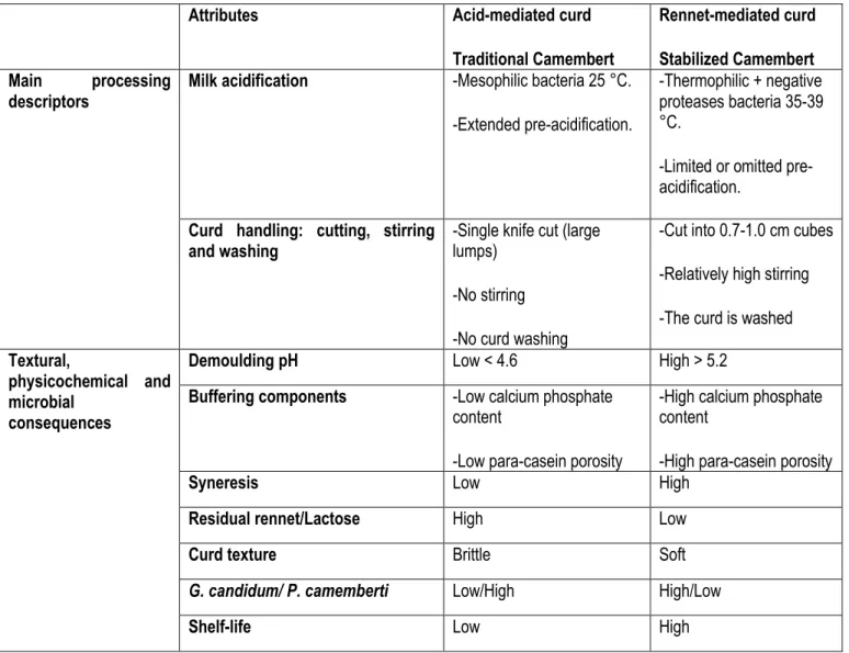
7 2. Traditional vs. Stabilized Camembert cheese
Camembert-type cheese is manufactured through mixed coagulation by acidification and rennet action on casein gel structure. Rennet-mediated coagulation and acid-mediated coagulation provide two Camembert cheese categories (Tamime and Law, 2001). Processing dissimilarities result in physico-chemical, textural and microbiological differences. These are briefly listed in the following Table 1-1.
Table 1-1: Manufacturing differences between traditional and stabilized Camembert-type cheese: physicochemical, textural and microbial consequences (Tamime and Law, 2001; Walstra et al., 2005)
Attributes Acid-mediated curd
Traditional Camembert
Rennet-mediated curd Stabilized Camembert Main processing
descriptors Milk acidification -Mesophilic bacteria 25 °C.
-Extended pre-acidification.
-Thermophilic + negative proteases bacteria 35-39 °C.
-Limited or omitted pre-acidification.
Curd handling: cutting, stirring
and washing -Single knife cut (large lumps) -No stirring
-No curd washing
-Cut into 0.7-1.0 cm cubes -Relatively high stirring -The curd is washed
Textural,
physicochemical and microbial
consequences
Demoulding pH Low < 4.6 High > 5.2
Buffering components -Low calcium phosphate content
-Low para-casein porosity
-High calcium phosphate content
-High para-casein porosity
Syneresis Low High
Residual rennet/Lactose High Low
Curd texture Brittle Soft
G. candidum/ P. camemberti Low/High High/Low
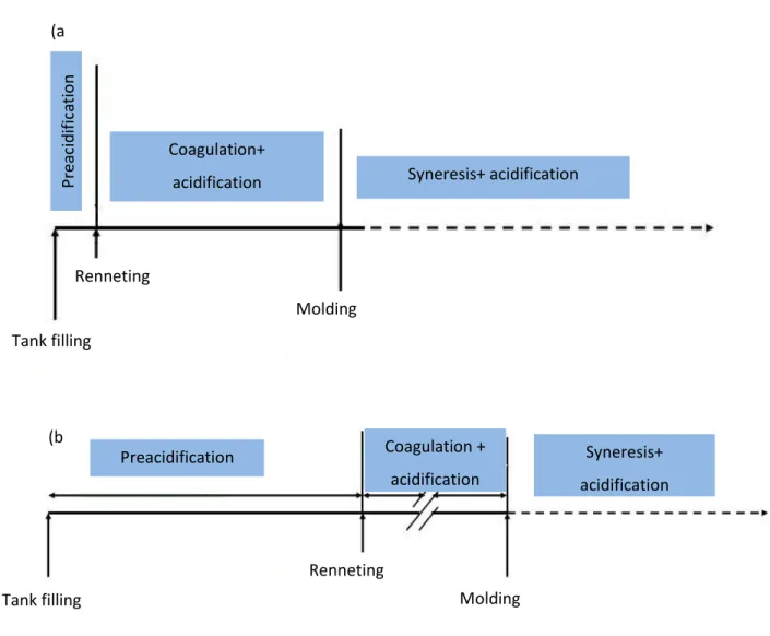
8 For acid-mediated or traditional Camembert, coagulation includes lactic acidification without vat working. Contrarily, stabilized Camembert is produced through a limited or omitted lactic acidification with a moderate vat working (Figure 1-4).
Figure 1-3: Description of (a) Stabilized curd versus (b) Traditional curd manufacture
For the acidification of the stabilized curd, proteinase negative strains such as thermophilic streptococci or the mixture of streptococci and lactococci are used below their optimal growth temperature 34-39 °C (Spinnler and Gripon, 2004). This procedure decelerates the lactic acid production to maintain the pH ≥ 5.2 which in turn reduces the demineralization of cheese matrix (Lawrence et al., 1987). The curd of stabilized cheese is cut into 0.7 to 1.0 cm cubes, and then the whey-mixture is stirred leading to faster and higher syneresis and so lower moisture in the final curd (Spinnler and Gripon, 2004; Walstra et al., 2005).
Coagulation + acidification Preacidification Syneresis+ acidification Tank filling Renneting Molding (b ) Syneresis+ acidification Coagulation+ acidification P reacid ifi ca tio n Tank filling Renneting Molding (a )
9 The resulting cheese has a firm and smooth texture (Figure 1-4) (Lortal et al., 2004). Conventionally, during stabilized cheese making the initial level of rennet is reduced decreasing the residual rennet and increase its shelf life (Lawrence et al., 1987).
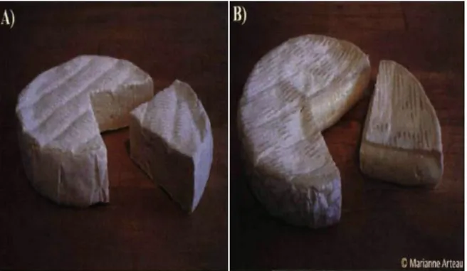
Figure 1-4: Stabilized (A) and traditional (B) Camembert-type cheese
Reproduced from Arteau (2009) The traditional Camembert cheese is manufactured industrially with a mechanized system but could also be produced using a traditional ‘moulage à la louche’ (Tamime and Law, 2001). Inversely to stabilized curd, the extended acidification of traditional cheese requires the use of a mesophilic starter at 25-28 °C that decreases the curd pH to 4.6. These leads to a higher residual rennet activity and extensive colloidal calcium-phosphate solubilization (Lawrence et al., 1987; Lucey et al., 2000; Walstra et al., 2005). The acid-mediated curd is merely cut without being stirred, which limits syneresis rate and provides a smooth, free ‘pea’ macrostructure (Tamime and Law, 2001; Walstra et al., 2005). The cutting is followed by hooping or molding the curd to allow further whey drainage. The curd is not washed which induces a high residual lactose content within cheese matrix compared to stabilized Camembert (Walstra et al., 2005).
10 3. Camembert cheese ecosystem
The diversity of Camembert microflora is reduced when cheese is made from pasteurized milk (Beuvier and Buchin, 2004; Skeie and Ardö, 2000). To compensate for the impact of heat treatment on the natural milk microflora, the manufacturer uses specific lactic acid bacteria (e.g., mesophilic and thermophilic species) and ripening microorganisms (e.g., Brevibacterium aurantiacum, Penicillium camemberti, Geotrichum candidum, Debaryomyces hansenii and Kluyveromyces lactis) (Spinnler and Gripon, 2004). The physicochemical and biochemical changes during Camembert cheese ripening promote the development of a complex microflora (Monnet et al., 2015). Lessard et al. (2012), used an accurate real-time quantitative PCR to investigate the growth kinetics and the interactions between the fungal species in a Camembert model curd ecosystem during a 31-day ripening period. In a previous study, the same team showed that the diversity and distribution of the fungal microflora within Commercial Camembert cheese curd depend on cheese size, surface area, and cheese matrix properties as modulated during manufacturing (Arteau et al., 2010).
3.1. Lactic acid bacteria and surface microflora
Depending on the Camembert cheese technology used, the lactic acid bacteria might include either mesophilic bacteria such as Lactococcus lactis, Leuconostoc mesenteroides (Walstra et al., 2005), or protease negative thermophilic strains (Lawrence et al., 1987). These lactic acid bacteria allow the acidification of cheese except for the Leuconostoc mesenteroides that reduces this phenomenon in the first six days of the cheese ripening (Leclercq-Perlat et al., 2004a). The growth of lactic acid bacteria begins with the addition of the starter up to the salting stage to reach around 3-5x10⁹ CFU/g of wet cheese. Afterward, the concentration of the microflora remains stable until the
10th day of ripening, then decreases progressively to around 5x10⁸ CFU/g of wet cheese on the 41st day. The surface
bacterial microflora is either sprayed on the cheese curd after ripening or inoculated in the cheese milk before renneting, simultaneously with the starters. The growth of these bacteria (e.g., Brevibacterium linens) starts when the pH of cheese surface is above 6.0 due to the cheese alkalinization by the fungal microflora (Leclercq-Perlat, 2011). This higher pH promotes the proteolytic activity of lactic acid bacteria to produce small peptides and free amino acids (Leclercq-Perlat, 2011). Overall, lactic acid bacteria represents a minor fraction of the surface community due to the competition with other microorganisms that consume a large proportion of the lactic acid, amino acids and lipids energy sources such as the yeast G. candidum (Leclercq-Perlat et al., 2004a; Monnet et al., 2015).
3.2. Yeasts and molds
Arteau et al. (2010) investigated the microbiota of Canadian Camembert cheeses made of pasteurized milk and identified nine fungal genera including Cladosporium, Debaryomyces, Geotrichum, Kluyveromyces, Mucor,
11 Penicillium, Pichia, Saccharomyces and Yarrowia. According to this study, the source of such fungal microflora might be either the ripening starter, the native ecosystem of milk or processing conditions (reviewed in Irlinger et al. (2014) ). In the core of Camembert cheese, yeast count is two-to-three log lower than that at the surface (Leclercq-Perlat et al., 2006). Another study reported that the concentration of yeasts in the center of cheese is around 1% of that on the surface (Beresford et al., 2001). The study of the microflora of Camembert cheese showed that Geotrichum sp., Penicillium sp. and Debaryomyces sp. are the main genus of the surface while, Kluyveromyces sp. and Saccharomyces sp. were typically found in the core (Arteau et al., 2010; Beresford et al., 2001; Lessard et al., 2012). These studies also showed that during cheese-making, the growth and the enzymatic activities of the fungi depend on the pH, the water activity, and salt content of the cheese curd (Addis et al., 2001; Leclercq-Perlat et al., 2004a; Leclercq-Perlat, 2011; Roostita and Fleet, 1996). The fungal microflora is not affected at a salt concentration < 2 % (Tabla et al., 2015), however, at a decreased salt content, G. candidum growth predominates over P. camemberti which might cause the ‘’toad skin’’ defect (Fox et al., 2004). Inversely, the very high salt concentration causes the ‘’ bitterness defect ’’ due to the excessive proteolytic activity of P. camemberti (Spinnler and Gripon, 2004). When comparing traditional and stabilized Camembert cheese microflora, Arteau et al. (2010) showed that cheese stabilization modifies the composition of the fungal community of the core. Specifically, G. candidum seems to predominate traditional curd while P. camemberti predominates in stabilized cheese.
The growth rate and the distribution of the fungus are significantly affected by the interaction with other members of the cheese microbiota (Mounier et al., 2008; Roostita and Fleet, 1996). Using classic microbial count (Addis et al., 2001; Arteau et al., 2010; Leclercq-Perlat et al., 2004a; Mounier et al., 2008; Roostita and Fleet, 1996), terminal restriction-fragment length polymorphisms (Arteau et al., 2010) and metagenomics and meta-transcriptomic analysis (Dugat-Bony et al., 2015; Lessard et al., 2014), authors showed that K. lactis and K. marxianus are the first yeasts to grow in cheese curd at the beginning of the ripening period. They are subsequently followed by G. candidum, P. camemberti and D. hansenii. The enzymatic activities of these species promote further cheese alkalinization and induces the growth of acid-sensitive species (Leclercq-Perlat et al., 2004a). Simultaneously, this microflora displays a key ripening role. D. hansenii uses lactose and lactate, G. candidum does not metabolize lactose but uses lactate instead, and governs the proteolytic and lipolytic activities that occur during Camembert-type cheese ripening. These metabolic activities contribute in the development of typical flavors and textural properties (Leclercq-Perlat et al., 2004a; Lessard et al., 2014; Roostita and Fleet, 1996; Schlesser et al., 1992). Because of this D. hansenii, Y. lipolytica and G. candidum are often inoculated as a starter culture (Boutrou and Guéguen, 2005; Ferreira and Viljoen, 2003; Van den Tempel and Jakobsen, 2000). However, proteases and peptidases activities are detected only after two to three weeks of ripening (Boutrou and Guéguen, 2005; Boutrou et al., 2006a; Engel et al., 2001; Leclercq-Perlat et al., 2004a; Lessard et al., 2014).
12 Camembert cheese fungal microflora produces several aromatic compounds during their growth, such as dimethyl disulfide (DMDS) by G. candidum (Demarigny et al., 2000; Jollivet et al., 1994; Leclercq-Perlat et al., 2004b) and an ester of fruity flavor by D. hansenii (Gori et al., 2012). To sum up, the microbial interaction in soft cheese ecosystems and related biochemical activities contribute to the development of desired organoleptic quality, shelf life and safety of ready-to-eat cheese (Addis et al., 2001; Lessard et al., 2014).
4. Geotrichum candidum: Biochemical and physicochemical changes during Camembert cheese ripening
Originally isolated from milk by Fresenius in 1850 (reported by (Wouters et al., 2002)). Geotrichum candidum is an aerobic acid-tolerant and salt-sensitive yeast. Some authors cite it as a mold because the fungi arbor sometime a fluffy phenotype, but G. candidum is now correctly assigned as a yeast (Boutrou and Guéguen, 2005). The name G. candidum was attributed to anamorphic yeast species used specifically in dairy products such as surface-ripened cheese (Prillinger et al., 1999). The selected strains of G. candidum provide different desirable sensorial properties to cheese (i.e., flavor and texture) through their lipolytic and proteolytic activities (Boutrou and Guéguen, 2005). Moreover, certain strains have shown a significant inhibition of undesirable microbes such as Listeria monocytogenes (Dieuleveux et al., 1998). Currently, G. candidum is used as a starter and colonizes a large number of surface-ripened cheese varieties during the first stages of ripening (Berger et al., 1999).
4.1. Taxonomy and strains diversity
Several synonyms have been attributed to this microorganism. The name Geotrichum candidum, known as the anamorph of Galactomyces Geotrichum, was associated to the specie Link (1809) then Link: Fries (1832) with CBS 772.71 the type strain of Ga. geotrichum and neotype strain of G. candidum (De Hoog et al., 1998). The taxonomic position was revised by de Hoog et al. (2004). The anamorphic state has been characterized as follows; Candidacae (family) and Geotrichum (genus). Prillinger et al. (1999) assigned all isolated Geotrichum from dairy products to Ga. geotrichum and gave the name of Geotrichum candidum to anamorphic yeast species. A type strain of this yeast was defined in Brie cheese (France), CBS 615.84 (De Hoog et al., 1998). In the last decade, molecular methods, allowed the identification of G. candidum at the species and strain levels (Gente et al., 2006) and strain diversity (Alper et al., 2013; Lessard et al., 2014; Lessard et al., 2012) which improved the characterization of dairy strains specifically in cheese industry (Alper et al., 2011; Alper et al., 2013; Leclercq-Perlat et al., 2004a; Sacristán et al., 2012; Sacristán et al., 2013).
13 4.2. Morphology
G. candidum is a dimorphic yeast because it involves strains that can display three distinct colonial morphotypes (Table 1-2) (Gente et al., 2002; Guéguen and Jacquet, 1982; Guéguen and Schmidt, 1992; Wyder and Puhan, 1999). According to Guéguen and Jacquet (1982), the three types of Geotrichum candidum morphotypes were well correlated to their physicochemical properties (i.e., lipolysis, proteolysis, and alkalinization). More details about yeast-like colony type characteristics are summarized in table 1-2.
Table 1-2 : Characterization of G. candidum yeast-like morphotype (Guéguen and Jacquet, 1982).
Characteristics Yeast-like Colonies color Cream-colored
Pattern Cloudy bottom and lowly developed mycelium
Arthrospore Arthrospore predominance
Growth Media Surface and core
Optimum Temperature 22-25 °C
Proteolytic/ lipolytic activities High/ moderate
Alkalinization High
4.3. pH change
During Camembert-type cheese ripening, the pH of the rind remains unchanged until about the 6th day. Hence, it
increases from about 4.6 to 7.8 within six days and remains stable until the end of ripening (Leclercq-Perlat et al., 2004a; Lessard et al., 2012). This change was attributed to the growth of surface yeasts and their biochemical activities and mainly the presence of G. candidum (Aldarf et al., 2004; Boutrou et al., 2006b). G. candidum assimilates lactate produced by lactic acid bacteria and releases ammonia which reduces rind acidity. Given this, Leclercq et al. (2004) showed that the pH of the surface of Camembert cheese was positively correlated to NPN and ASN concentrations that are metabolized by G. candidum proteases. These proteolytic enzymes are produced on the rind and did not diffuse to cheese core (Noomen, 1978). Subsequently, ammonia is produced at the cheese surface, and a pH gradient occurs from the rind to the core due to the diffusion of the metabolites (Aldarf et al., 2004; Leclercq-Perlat et al., 2004a). Similarly, lactate was reported to diffuse from the core to the surface allowing a further rise of core pH. These phenomena (i.e., ammonia and lactate diffusion) contribute to the pH gradient between the rind and the core. Recently, two key studies confirmed these previous statements using metatranscriptome analysis
14 (Dugat-Bony et al., 2015; Lessard et al., 2014). Authors detected G. candidum‘s transcripts confirming that G. candidum catabolises peptides, amino acids and lactate principally, and produces NH3 to alkalinize cheese curd.
These pH changes also occur during the simultaneous growth of G. candidum and P. camemberti (Figure 1-5).
Figure 1-5: Evolution of pH and fungal growth during Camembert cheese ripening.
The ripening culture was a mixture of (□) G. candidum LMA-1028 and (▲) P. camemberti LMA- 1029.
Each strain was quantified individually using a TaqMan real-time qPCR method pH (×) measures were
taken weekly until day 50.
15 4.4. Flavor-formation
Proteolysis
From the 8th day of ripening, the increase of pH promotes the proteolytic activity within Camembert cheese matrix.
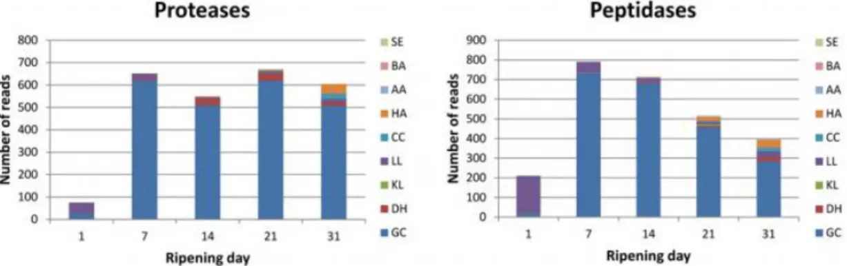
The proteolysis is mainly governed by the growth of G. candidum and P. camemberti on the surface of the Camembert cheese. For this reason, it is known to be more intensive near the rind than in the core. Particularly, G. candidum is considered to be the main proteolytic yeast, responsible for 85 % of peptidasic and proteolytic activity (Figure 1-6) (Baroiller et al., 1990; Dugat-Bony et al., 2015; Guéguen and Schmidt, 1992). To discriminate the technological features of G. candidum strains, Sacristan et al. (2012) focused on the enzymatic profile, including the proteolytic and amino-peptidase activity of several G. candidum strains. Among the 41 strains characterized only eight strains showed an extracellular proteolytic activity. These strains have been divided into two strains sub-groups, respectively with weak or strong proteolytic activity. Besides, among these strains, intracellular proteolytic activity was suggested to be higher than extracellular activity. Dugat-Bony et al. (2015) observed the same behavior and showed that during milk coagulation, lactic acid bacteria and rennet activities allow the release of casein fragments, which are catabolized by extracellular proteases or by vacuolar proteases and peptidases of G. candidum after being adsorbed by endocytosis.
Figure 1-6: Protein degradation during surface-ripened cheese maturation: Expression data observed for
genes encoding proteases (A) and peptidases (B).
SE: Staphylococcus equorum. BA: Brevibacterium aurantiacum. AA: Arthrobacter arilaitensis. HA: Hafnia
alvei. CC: Corynebacterium casei. LL: Lactococcus lactis. KL: Kluyveromyces lactis. DH: Debaryomyces
hansenii. GC: G. candidum
16 In another study, Boutrou et al. (2006) characterized the proteolytic activity of G. candidum in Camembert-type cheese. They used reverse-phase high-performance liquid chromatography to determine the contribution of G. candidum to the primary and secondary proteolysis at the surface of this soft mold-ripened cheese. Electrophoretic profiles showed a low intensity of the casein bands after the first week of ripening. The transcriptomic analysis indicated that the metabolic pathways responsible for ammonia production and amino acid metabolism are active. This is referred as the primary proteolytic activity of G. candidum, which is allowed due to the increase of cheese surface pH up to the G. candidum proteinase’s optimal pH of 5.5-6.2 (Boutrou et al., 2006b). Primary proteolysis results in the production of large- or medium-sized casein fragments and peptides. The generation rate of these products decreases by the third week making authors suggest a lower level of hydrolysis and/or the degradation of a part of primary proteolysis’ products (Boutrou et al., 2006b). After that, the secondary proteolysis takes place after the 15th day of ripening showing peptides decrease coupled with a FAA and small peptides increase on the cheese rind. Secondary proteolysis is referred to peptides degradation by G. candidum peptidases which are active at current cheese surface pH, but lactococcal peptidases were also functional when cheese surface pH increased to their optimal pH (6.5-9.5). Furthermore, casein fractions were hydrolyzed by G. candidum, particularly β-A2 and αs1-casein
with preferred hydrolysis of β-casein (Dugat-Bony et al., 2015).
Using meta-omics analyses Lessard et al. (2014) and Dugat-Bony et al. (2015) proved that G. candidum governs aroma compounds production through amino acids catabolism especially sulfured amino acids (i.e., methionine) and glutamate (Jollivet et al., 1994) to produce further compounds that are important in the development of the aroma (Latrasse et al., 1987; Gripon, 1997). Among the volatile compounds generated during Camembert cheese-type ripening, the 3-methyl-butanol, DMS and isoamyl acetate were mainly associated to the G. candidum proteolytic activity. Likewise, thanks to its enzymatic properties, specifically carboxypeptidase and amino-peptidase activities, G. candidum reduces the bitterness provided by some hydrophobic peptides of low metabolic weight produced through the degradation of β-casein. This effect has been observed in Camembert cheese by several authors (Wyder and Puhan, 1999; Thammavongs et al., 2000; Marcellino et al., 2001, Boutrou et al., 2006).
Lipolysis
During soft mould-ripened cheese ripening, lipid hydrolysis may reach 5 to 20% (Fox et al., 2004). As for proteolysis, lipolysis contributes to the development of cheese flavor. Lipolysis occurs intensively from the first to the 7th day of
ripening then decreases slightly or remains stable until the 4th week (Dugat-Bony et al., 2015). It has been correlated
to the surface-fungal microflora including, essentially, P. camemberti, K. lactis and G. candidum (Molimard et al., 1997). Also, the concentration of lipases has been reported to be twice higher under the rind when compared to the core due to the relatively high pH (<6.0) (Hassouna and Guizani, 1995). G. candidum‘s lipases are responsible for
17 soft mould-ripened and Armada cheeses lipolysis during curd ripening when rind pH is between 5.5 and 7.5 (Fresno et al., 1997; Tornadijo et al., 1998). However, the intensity of the lipolytic activity, as well as lipase forms, are strain dependent (Baillargeon et al., 1989; Boutrou and Guéguen, 2005). Lipases from G. candidum have been purified and characterized on relatively ‘’simple’’ medium cultures (Baillargeon et al., 1989; Jacobsen and Poulsen, 1992; Sugihara et al., 1990). Authors identified six lipases forms (Lipase A, B, I, II, III and IV) with different substrate specificities. Nevertheless, G. candidum lipolysis was mainly due to the extracellular synthesized Lipases I and II (reviewed in Boutrou et al. (2006b)). These forms have unique specificity to cis-9 unsaturated fatty acids (i.e., oleic acid) and unsaturated C18 fatty acids at the Sn2 position of the triglycerides, respectively (Bertolini et al., 1995; Veeraragavan et al., 1990). Fresno et al. (1997) correlated the low ratio of palmitic (16:0) to oleic acid (18:1) to the activity of G. candidum Lipase I whose affinity is to unsaturated fatty acids with a double bond cis-9 and cis-cis 9,12 position, with preference to 18:1. The screening of free fatty acids (FFAs) profile during the ripening of a traditional (Leclercq-Perlat et al., 2007) and a freeze-dried Tibetan kefir co-cultured (Jun et al., 2015) Camembert cheeses showed that the main liberated FFAs are long chain ones including palmitic (16:0), myristic (14:0) and stearic (18:0).
The specific lipolytic activity of G. candidum was not/barely characterized until the last ten years (Boutrou and Guéguen, 2005). Recently, Sacristàn et al. (2012) examined the extracellular and cell-bound lipases activities in Armada cheese and revealed significant differences between strains. About 40% of G. candidum strains showed high extracellular activity, and 10% presented cell bound lipase activity. Lessard et al. (2014), established the enzymatic activity (i.e., proteolysis, lipolysis) profile of P. camemberti and G. candidum during 77 ripening days of Commercial Canadian Camembert-type cheese. This study explored the lipolytic activity of both P. camemberti and G. candidum. The metabolic activities of pilot-scale made surface-ripened cheese ecosystem inoculated with K. lactis, D. hansenii and G. candidum ATCC 204307 showed that cheese lipolysis accounted, majorly, for G. candidum lipases activity (Dugat-Bony et al., 2015). Most of the produced FFAs were detected in the rind and the core of Camembert-type cheese. Hence their diffusion phenomenon from the rind to the core has been highlighted (Leclercq-Perlat, 2011). Known as a major flavor precursor in Camembert-type cheese, G. candidum develops volatile compounds by metabolizing lipid and free fatty acids degradation. Indeed, it contributes to the production of several methyl-ketones through fat catabolism, which constitutes the main flavor component of Camembert-type cheese (Leclercq-Perlat et al., 2004b; Leclercq-Perlat et al., 2004c; Molimard and Spinnler, 1996).
18
II.
Survival of cheese microbiota during in vitro and in vivo digestion
In addition to their nutrient values, fermented dairy products (e.g., yogurt, kefir and cheese) provide a diverse microflora that is composed of lactic starter culture and non-starter culture (Walther et al., 2008). As detailed above, ripened cheese categories such as surface mould-ripened cheese comprise a wide variety of acidifying strains such as Lactococcus lactis and Streptococcus thermophilus and several flavoring yeasts and molds, like Debaromyces hansenii, Geotrichum candidum, Kluyveromyces spp., Pichia spp., Rhodotorula spp., Saccharomyces spp., Trichosporon spp., Torulospora spp., Yarrowia spp. and Zygosaccharomyces spp (Arteau et al., 2010; Leclercq-Perlat, 2011; Lessard et al., 2012). Cheese ecosystem provides about 108 – 109 CFU per gram of ready-to-eat
cheese (Beresford et al., 2001). Along with their technological role during cheese ripening, such abundant microbial diversity raises the question whether this microflora has a beneficial effect, such as an antimicrobial activity or a probiotic potential, on human microbiota when ingested (Hatoum et al., 2012). Beforehand, the behavior of these microorganisms through the stressful gastrointestinal conditions should be investigated. To exert their beneficial properties the ingested microorganisms should resist the harsh physiological stress during digestion, such as the temperature, acidic pH of the stomach, intestinal bile salts, organic acids, gastrointestinal enzymes and secondary metabolism metabolites (e.g. H2S, bacteriocins) (Fioramonti et al., 2003). Since the tolerance of dairy yeasts to these
stresses are barely investigated, some examples using lactic acid bacteria as models will be illustrated in the next sections.
1. Gastrointestinal stress
1.1. Gastric acid stress
Depending on the kinetics of food matrix disintegration, ingested microorganisms are exposed to a gastric pH ranging from 1 to 3 units during an average exposure time of 90 min (Kong and Singh, 2008b). The low acidity of the stomach induces a drop of the intracellular pH of the microorganisms which affects their cellular metabolism (i.e., inhibits cell growth and product formation) (Lohmeier-Vogel et al., 1998; Matsushika and Sawayama, 2012). Eukaryotic cells control their intracellular pH and nutrients uptake by maintaining a proton gradient force over the plasma membrane (Madshus, 1988). Several yeasts can grow at pH 3.0 (Deak and Beuchat, 1994; Miller, 1979; Praphailong and Fleet, 1997; Walker, 1977) and resist to an acidic pH as low as 1.5 units (Czerucka et al., 2007; Praphailong and Fleet, 1997). However, acidic pH tolerance and the metabolic response is strain-dependent (Fietto et al., 2004). The tolerance of low pH depends on the activity of the plasma membrane H+-ATPase that controls the intracellular pH
through proton exchange (Eraso and Gancedo, 1987; Praphailong and Fleet, 1997). For instance, some Saccharomyces strains use a cell buffering capacity when exposed to acid stress or produce a particular protein
19 profile (Marešová et al., 2010). Fietto et al. (2004). Given this, in the presence of lactic acid stress, recent studies performed metabolomics analysis using capillary electrophoresis time-of-flight mass spectrometry (CE-TOFMS) to investigate the metabolic tolerance mechanism of S. cerevisae (Nugroho et al., 2015). They highlighted three metabolic responses interfered by low pH, namely the energy metabolism, redox balance, and amino acid composition. Authors reported the accumulation of ATP suggesting the reduction of ATPase activity under intensive acid stress. Furthermore, they observed the increase of the concentration of cysteine, glutamine and γ-amino-butyric-acid (GABA) and the decrease of glycine. Both induced the accumulation of glutathione and this has been suggested to improve the tolerance of cells to oxidative stress. Similarly, the accumulation of proline in cells was shown to protect yeast cells from an intracellular pH drop under acid stress.
1.2. Bile salts toxicity
Bile toxicity in the small intestine is a serious barrier to withstand by ingested microorganisms. Bile salts are known to be ‘biological detergent’ because they ‘’have a detergent action on particles of dietary fat which causes fat globules to break down or be emulsified into minute’’ (Hofmann and Small, 1967). They are the conjugated form of bile acids secreted by the liver from cholesterol (Russell and Setchell, 1992). Due to microbial activities, these acids go through different chemical reactions such as de-conjugation and dehydrogenation (reviewed in (Prabha and Ohri, 2006)). Both de-conjugated and conjugated acids attack the lipid bilayer structure and the protein integrity of cellular membrane as a result of the cellular homeostasis disruption (Cabral et al., 1987). In the neutral pH environment of the duodenum, the resistance to bile salts stress depends on the concentration of salts and the exposure time (Hill, 1993). The average concentration of bile salts in the intestine is around 0.3%. However, peaks of 1.5 to 2.0% might be found within the first hour of the intestinal digestion bile tolerance is often evaluated after microbial exposition to a range of 0.1 to 2.0% for probiotic bacteria selection (Noriega et al., 2004). A microorganism is considered to have a good bile tolerance if it could withstand a concentration of 0.3% v/v of bile (Gilliland et al., 1984; Gotcheva et al., 2002; Lankaputhra and Shah, 1995). This concentration was selected to simulate the physiological conditions and is used to screen pure cultures or a mixture of bacterial strains for their bile tolerance (Gotcheva et al., 2002). Many yeasts, more specifically dairy genera such as Debaromyces hansenii (Psani and Kotzekidou (2006)), Kluyveromyces lactis, Yarrowia lipolytica (Chen et al., 2010), and Kluyveromyces marxianus ((Kumura et al., 2004), have shown resistance to high bile salts concentration. Likewise, genetically unrelated Saccharomyces strains, isolated from dairy products, survived the simulated gastrointestinal stresses, specifically bile salts toxicity (Fietto et al., 2004). The resistance to such ‘biological detergents’ includes mainly bile salts hydrolase activity(Smet et al., 1995) that results from a ‘’detergent shock’’ protein response. This response is displayed through the immunomodulation of the gastrointestinal tract (Buts et al., 1990).
20 A similar immunomodulatory response of G. candidum strains had been reported by Plé et al. (2015) when investigating the immune effect of dairy products. Also, Adouard et al. (2015a) determined the viability of smear-ripened cheese microflora and their digestive stress response. G. candidum strains displayed a high resistance to the simulated gastrointestinal juices with a moderate immunomodulatory activity microflora (Adouard et al., 2015a). The high viability of the species of cheese microflora through gastrointestinal stress might be attributed to their intrinsic tolerance or the properties of their ecosystem.
2. Protective effect of cheese matrix
Since the ‘90s, a full range of probiotic dairy products has been developed, e.g., pasteurized milk, ice cream, and fermented milk products (Boylston et al., 2004; Fondén et al., 2003; Ross et al., 2002). Mainly, fermented dairy products are considered an excellent delivery system due to their structural features, physicochemical properties, and their extended shelf life (Buriti et al., 2012; Tamime, 2008). Notably, the firm consistency (i.e., dense casein network), the high pH and the buffering capacity of cheese matrix of the soft-ripened cheese promote the development and the protection of its microflora (Ross et al., 2002; Vinderola et al., 2002). These advantages also allow the protection of probiotic microorganisms against the digestive stresses which made of cheese matrix a possible alternative probiotic carrier to yogurts and other fresh fermented dairy products (Dinakar and Mistry, 1994; Gardiner et al., 1999; Karimi et al., 2011; Plessas et al., 2012; Possemiers et al., 2010; Sharp et al., 2008).
Particularly, fat protects microbes properly through the digestive tract by reducing their exposure to acid and bile acids stress (Karimi et al., 2011; Possemiers et al., 2010; Ranadheera et al., 2012). The fat content of food matrix provides better protection than protein content. Given this, the viability of probiotic bacteria when ingested in milk chocolate vs. half-skimmed milk and skim milk vs. whole milk, with the same protein content, increased in high-fat dairies (Possemiers et al., 2010; Tompkins et al., 2011; Varcoe et al., 2002). Furthermore, the addition of protein hydrolysates during the manufacture of Gouda-type cheese didn’t appear to improve the viability nor did protect probiotic bacteria during cheese ripening (Champagne et al., 2011; Gomes et al., 1995; Ong et al., 2006; Stanton et al., 1998). Even-though cheese has been widely optimized as a probiotic carrier; few studies investigated the viability and the beneficial impact of its microflora on human metabolism and health.
3. Survival of microbial microflora of soft surface-ripened cheese
Diet is one among the important factors affecting the composition of human gut microbiota (Walker et al., 2011; Wu et al., 2011). Marteau et al. (1994) found Bifidobacterium (yogurt) and Lactobacillus (cheese) in human subject feces after dairy products intake and reported a possible beneficial effect on intestinal metabolism. Given this, Lay et al.
21 (2004) assessed the beneficial effect of the traditional Camembert-type cheese consumption on rat Human-associated microbiota using specific medium and PCR–temporal temperature gradient gel electrophoresis. Cheese microflora showed significant tolerance to harsh gastrointestinal conditions, particularly, Streptococcus thermophilus, Lactobacillus sp. and G. candidum. These resistant genera have shown potential enhancement of the intestinal metabolism. Subsequently, the same team investigated the survival of traditional Camembert-type cheese microorganisms in a small clinical trial. The study was conducted with twelve healthy volunteers who have consumed cheese during four weeks after two exclusion weeks, followed by a wash out period. Overall, the final analyzed fecal samples contain Lactococcus lactic, Leuconostoc mesenteroides and G. candidum. However, no intestinal metabolic changes have been shown after Camembert cheese consumption (Firmesse et al., 2008). David et al. (2014), used 16S rRNA and ITS gene sequencing to study the effect of animal diet on human microbiome composition. The main bacterial (thermophilic strains) and fungal (Penicillium and Candida sp.) species of Camembert-type cheese survived the digestive stresses and were predominantly detected in the human distal gut. Adouard et al. (2015b), evaluated the survival of a mixture of nine microbial strains grown on a smear-ripened cheese during gastrointestinal transit. Interestingly, G. candidum, K. lactis, and D. hansenii showed high resistance to simulated digestive stress with less than 1.0 Log (CFU.mL-1) of viability loss with a moderate immunomodulatory and anti-inflammatory effects (Adouard et al., 2015a; Plé et al., 2015). G. candidum displayed significantly high resistance during gastrointestinal transit specifically when tested as part of the cheese matrix microflora.
III.
In vitro gastrointestinal digestion
In general, the digestive system is divided into four main sections, starting by the mouth, the stomach, the small intestine (duodenum, jejunum, and ileum) and finally the colon. Each of these digestive compartments is characterized by different physicochemical properties and a complex microflora. Since the human and the animal digestive systems aretime-consuming, ethically complicated, and expensive, in vitro digestion systems were developed to facilitate the study of the digestion process.
1. In vitro digestion
In vitro digestion models were developed as an alternative to animal and human subjects or to be complementary to them. Hur et al. (2011), reported about 80 studies using in vitro models of which seven were dedicated to dairy product digestibility and several for probiotic microorganisms’ survival before human studies. In vitro digestion models are being used in different fields such as pharmacology, biotechnology or nutrition. In the latter case, it allows a rapid screening of the bioaccessibility and bioavailability of food nutrients’ as a function of their composition, structure and functional properties. Likewise, simulated digestion models are used to investigate food microbiology
22 particularly, the optimization of delivery systems for probiotic microorganisms, and the viability of ingested strains through gastrointestinal transit as detailed in (section II). Digestive juices are prepared to simulate physiologic human conditions.
2. Models: study of microorganisms viability during in vitro digestion of dairy products
In vitro digestion models have been classified into ‘batch’ and ‘dynamic’ systems depending on whether the temporal profile of in vivo digestion are controlled\simulated or not (e.g., mechanical force, liquid flow, shear stress, dilutions by gastric secretions over time, gastric emptying and the removal of resulting digestion products) (Guerra et al., 2012; Thomas et al., 2007; Vieira et al., 2014). A large number of in vitro digestion models has been reviewed by (Guerra et al., 2012) and (Verhoeckx et al., 2015). The physiological, chemical and enzymatic properties of simulated oral, gastric and duodenal steps will be detailed in the section III.3.3.
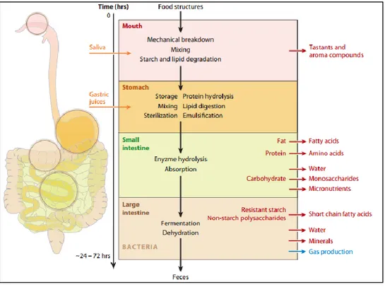
2.1. Static models
The static digestion system, also called ‘batch’ model, comprises a series of vessels (Figure 1-7), each simulating a digestive compartment (mouth, stomach, and small intestine: duodenum) as described by (Versantvoort et al., 2005).