Volume 2013, Article ID 827621,16pages http://dx.doi.org/10.1155/2013/827621
Review Article
Class A
𝛽-Lactamases as Versatile Scaffolds to Create Hybrid
Enzymes: Applications from Basic Research to Medicine
Céline Huynen,
1Patrice Filée,
2André Matagne,
1Moreno Galleni,
3and Mireille Dumoulin
11Laboratory of Enzymology and Protein Folding, Centre for Protein Engineering, Institute of Chemistry, University of Liege,
(Sart-Tilman) 4000 Liege, Belgium
2ProGenosis, Boulevard du Rectorat, 27b-B22, (Sart-Tilman) 4000 Liege, Belgium
3Laboratory of Biological Macromolecules, Centre for Protein Engineering, Institute of Chemistry, University of Liege,
(Sart-Tilman) 4000 Liege, Belgium
Correspondence should be addressed to Mireille Dumoulin; [email protected] Received 31 May 2013; Accepted 4 July 2013
Academic Editor: Bidur Prasad Chaulagain
Copyright © 2013 C´eline Huynen et al. This is an open access article distributed under the Creative Commons Attribution License, which permits unrestricted use, distribution, and reproduction in any medium, provided the original work is properly cited. Designing hybrid proteins is a major aspect of protein engineering and covers a very wide range of applications from basic research
to medical applications. This review focuses on the use of class A𝛽-lactamases as versatile scaffolds to design hybrid enzymes
(referred to as𝛽-lactamase hybrid proteins, BHPs) in which an exogenous peptide, protein or fragment thereof is inserted at
various permissive positions. We discuss how BHPs can be specifically designed to create bifunctional proteins, to produce and to characterize proteins that are otherwise difficult to express, to determine the epitope of specific antibodies, to generate antibodies against nonimmunogenic epitopes, and to better understand the structure/function relationship of proteins.
1. Introduction
Engineering proteins with improved properties or new functions is an important goal in biotechnology. In this context, numerous bi- or multifunctional proteins have been created for applications in a broad range of fields including biochemical analysis, protein purification, immunodetection, protein therapies, vaccine development, functional genomics, analysis of protein trafficking and analysis of protein
interac-tion network [1]. The most common strategy to create such
proteins involves fusing polypeptides into an end-to-end con-figuration. For example, the fusion of proteins with specific tagging domains is nowadays a standard tool for protein engineering. Indeed, numerous commercial kits allow the fusion of a protein of interest to an affinity polypeptide such as 6-Histidine tag, maltose-binding protein, and glutathione
transferase [1]. Moreover, the fusion of a protein of interest
to the green fluorescent protein (GFP) has become one of the most important tools used in contemporary bioscience, and O. Shimomura, M. Chalfie, and R. Y. Tsien were rewarded
the Chemistry Nobel prize in 2008 for the initial discovery of GFP and development of important GFP-based applications. Another strategy to create hybrid proteins, although much less commonly used, is to insert (or graft) a pep-tide/protein/fragment thereof at a permissive site of a carrier
protein. For example, Collinet et al. [2] have successfully
created bi- or trifunctional hybrid enzymes by inserting
the dihydrofolate reductase (159 aa) and/or the TEM-1
𝛽-lactamase (263 aa) into four different positions of
phospho-glycerate kinase (415 aa). Betton et al. [3] have created hybrid
proteins by inserting the TEM-1𝛽-lactamase at various sites
into the MalE maltodextrin-binding protein, that is, positions 120, 133, and 303, or by fusing it to the carboxy terminus of MalE. Insertion at positions 133 and 303 or fusion at the C-terminus allows the production in the periplasm of hybrid proteins exhibiting both parental activities; indeed, the maltose binding and the penicillinase activity of these hybrid proteins is indistinguishable from that of, respectively,
MalE and TEM-1 [3]. Interestingly, the two proteins with
their C-terminal fusion counterpart: (i) they were more resistant to degradation by endogenous proteases and, (ii) even more remarkably, the TEM-1 moiety was stabilized against urea denaturation through the binding to MalE of maltose, that is, its natural ligand. This latter observation clearly demonstrates that TEM-1 is structurally dependent on MalE or in other words that an allosteric interaction
occurs between the two proteins [1, 3]. Thus, insertion of
a polypeptide inside a scaffold protein can present some advantages compared to the more common end-to-end fusion. This strategy actually mimics that used by nature to generate protein diversity. Indeed, genetic rearrangements, such as the introduction of a sequence into an unrelated
coding sequence, naturally occur in the genome [2]. The
resulting proteins are composed of two or more domains, and the linear sequence of one domain is interrupted by the insertion, forming discontinuous domains. This process has been shown for large natural proteins such as disulfide bond
isomerase A (dsbA) [4], DNA polymerase [5], and pyruvate
kinase [6]. A systematic survey of structural domains showed
that ∼28% of them are actually not continuous, clearly
indicating that the sequence continuity of a domain is not
required for correct folding and function [2,7].
In this review, we report the use of two class A
𝛽-lactam-ases to create hybrid proteins (𝛽-lactamase hybrid proteins, BHPs) in which exogenous peptides/proteins/fragments
thereof are inserted within 𝛽-lactamase sequences. Such
hybrid proteins can be designed for a range of applications
such as (i) the creation of bifunctional hybrid proteins [8–
11], (ii) the production, purification, and characterization of
proteins otherwise difficult to express [10, 12, 13], (iii) the
determination of the epitope of a specific antibody at the
surface of an antigen [8,11], (iv) the generation of antibodies
against specific antigens or fragments thereof [9,13–15], and
(v) the investigation of fundamental aspects of structure,
stability, and function of proteins [10,12,16].
2. Class A
𝛽-Lactamases as Scaffolds to
Create Hybrid Enzymes
2.1. Why the Use of Class A 𝛽-Lactamases? The two class
A 𝛽-lactamases that have been extensively used as model
proteins to insert exogenous polypeptides are BlaP from
Bacillus licheniformis 749/C and TEM-1, the latter being the
most encountered 𝛽-lactamase in gram-negative bacteria
(Figure 1). These two enzymes are, respectively, secreted
by bacteria in the external environment and expressed in
the periplasmic space to inactivate 𝛽-lactam antibiotics.
About 40% and 20% of the amino acids are, respectively, identical and homologous between these two serine-active
𝛽-lactamases (Figure 1). Note that the residue numbering
used in this review is that specific to class A𝛽-lactamases
[17]. These enzymes, especially TEM-1, have been extensively
used as reporters to study gene expression [21–23] and for
immunoassays [24]. They have been selected as scaffolds
to create hybrid proteins based on a series of observations. Firstly, they are fairly small (30 kDa) and stable proteins that are easily overexpressed in E. coli and subsequently
purified [9–11]. Their three-dimensional structure has been
determined by X-ray crystallography (Figure1) [18, 19, 25,
26]. They exhibit the typical class A𝛽-lactamase 3D structure
which is composed of two domains: an𝛼/𝛽 domain and an
all-𝛼 domain, with the catalytic site at the interface between
the two domains [19, 25, 26]. Finally, their catalytic
prop-erties have been thoroughly characterized and can be easily
monitored using fluorescent [22, 23, 27] or chromogenic
substrates [28]. This latter point is crucial as exemplified in
the following sections. The enzymatic activity of the BHPs
serves as a reporter to select clones resistant to 𝛽-lactam
antibiotics, such as ampicillin, expressing well-folded and active BHP. It is assumed that the folding of the exogenous polypeptide and the carrier enzyme are interdependent to
some degree. Indeed, the correct folding of the carrier
𝛽-lactamase requires the exogenous polypeptide to be exposed
to the solvent; otherwise, the 𝛽-lactamase cannot adopt
its native structure. Moreover, the correct folding of the carrier protein imposes constraints at the N- and C-terminal extremities of the inserted polypeptide, maintaining its two extremities in close proximity of the insertion site. These constraints limit therefore the conformational flexibility of the insert and could assist the insert in adopting its native structure. When a greater flexibility is required for the correct folding of the insert, small peptides, that is, linkers, can be added at both of its extremities. Note that, in some applications, these linkers have an additional functionality allowing the excision of the insert thereafter, as discussed in detail below.
2.2. A Brief History of Class A𝛽-Lactamase Hybrid Proteins.
The design of class A hybrid𝛽-lactamases was initiated with
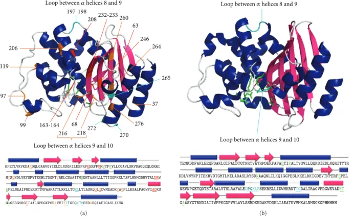
the work of Hallet et al. in 1997 [20]. A random pentapeptide
cassette was inserted at 23 different positions of TEM-1 using
pentapeptide-scanning mutagenesis (Figure1(a)) [20]. This
method consists of transposing the transposon Tn4430 into the TEM-1 gene which duplicates 5 base pairs (bp) of host sequence at the insertion point. This transposon contains
KpnI restriction enzyme sites 5 bp from the outer ends of its
terminal inverted repeats. Digestion with KpnI followed by ligation results therefore in the insertion of 10 pb derived from Tn4430 and 5 bp from the 5-nucleotide duplication of the target gene. Each transposition event leads to a 15 bp insertion into the TEM-1 gene encoding for a random pentapeptide cassette. The 23 insertion sites have been classified into 3 groups depending on the level of resistance that the hybrid
protein confers to E. coli cells against ampicillin, a𝛽-lactam
antibiotic (Figure1(a)): (i) 5 permissive sites (197, 198, 265,
270, and 272) with high level of resistance. These sites are located into two loops distant from the catalytic site, one loop
between𝛼 helices 8 and 9 and the other between 𝛽-strand 5
and the C-terminal helix. (ii) Eight non-permissive sites (63, 68, 163, 164, 208, 232, 233 and 246) associated with the loss
of the𝛽-lactamase activity and clustered around the catalytic
site. (iii) Ten semi-permissive sites (37, 97, 99, 119, 206, 216, 218, 260, 264, and 276) are conferring an intermediate level of ampicillin resistance.
197-198 265 270 272 63 68 163-164 37 99 97 246 208232-233 260 218 216 206 119 276 264
HPETLVKVKDAEDQLGARVGYIELDLNSGKILESFRPARVGYIELDL ILESFR ERFPEERFPMMMSTFKVLLCGAVLSRVDAGQEQLGRRI HYSQNDLVEYSPVTEKHLTDGMTVRELCSAAITMSDNTAANLLLTTIGGPKELTAFLHNMGDHVTRLDRW EPELNEAIPNDERDTTMPAAMATTLRKLLTGDTTMP ELLTLASRQQLIDWMEADKVAGPLLRSALPAGWFIADKS GAGERGSRGIIAALGPDGKPSRIVVIYTTGSQATMDERNRQIAEIGASLIKHW
GAG SRGIIAALG SRIVVIYYTT
Loop between𝛼 helices 8 and 9
Loop between𝛼 helices 9 and 10
(a) TEMKDDFAKLEEQFDAKLGIFALDTGTNRTVAYRPDERFAFASTIKALTVGVLLQQKSIEDLNQRITYTR DDLVNYNPITEKHVDTGMTLKELADASLRYSDNAAQNLILKQIGGPESLKKELRKIGDEVTNPERFEPEL NEVNPGETQDTSTARALVTSLRAFALEDPGKLPSEKRELLIDWMKRNTTGDALIRAGVPDGWEVADKT GAASYGTRNDIAIIWPPKGDPVVLAVLSSRDKKDAKYDDKLIAEATKVVMKALNMNGKGPHHHHH QDTSTARTTT AKLGIFALDTGTNRTVAYRPDERFAR GTRNDIAIIWPPK Q GDPVVLAVLSSRDK GAASYGYG EVTNPERNNN Q
Loop between𝛼 helices 8 and 9
Loop between𝛼 helices 9 and 10
(b)
Figure 1: X-ray crystal structure and sequence of TEM-1 (a) and BlaP (b). The residue numbering is based on homology to class A
𝛽-lactamases [17]. The structures were produced using PyMOL (DeLano Scientific LLC, South San Francisco, CA, USA) and the PDB ID is
4BLM for BlaP [18] and 1XPB for TEM-1 [19]. (a) TEM-1. The residues of the active site are represented in green on the structure and are
coloured and underlined in the sequence. The different insertion sites of pentapeptides reported in Hallet et al. [20] are coloured on the
structure and are coloured and underlined in the sequence. Light blue, orange and red are associated with permissive, semi-permissive, and non-permissive insertion sites, respectively. (b) BlaP from Bacillus licheniformis 749/C. The residues of the active site are represented in green on the structure, and are coloured and underlined in the sequence. The two insertion sites most commonly used to design BHPs, located in the
loop between𝛼 helices 8 and 9 (197-198) and in the loop between 𝛼 helices 9 and 10 (216-217), are indicated in light blue on the structure and
are coloured and underlined in the sequence. The PG dipeptide between residues 197 and 198 indicated in bold in the sequence corresponds to the SmaI restriction site inserted into the gene of BlaP for the cloning of exogenous polypeptides at this position.
Later, Ruth et al. [9] have inserted longer and more
structured polypeptides in eight of the TEM-1 positions
investigated by pentapeptide-scanning mutagenesis (Table1).
The thermostable 18-residues STa enterotoxin from entero-toxic Escherichia coli has been introduced, for example, into one of the two permissive loops (at position 195 and 198,
between 𝛼 helices 8 and 9), in five semi-permissive sites
(37, 206, 216, 218 and 260) and into one non-permissive site
(232). Insertions between𝛼 helices 8 and 9 (positions 195–
200) and between 𝛼 helices 9 and 10 (positions 213–220)
allow the production of a soluble hybrid protein which retains both activities of parental proteins (i.e., ampicillin resistance and enterotoxicity). The level of activities and the amount of soluble hybrid enzymes produced depend however on the position of insertion.
TEM-1 is a𝛽-lactamase sensitive to many proteases [11,
29]. Some BHPs that do not confer resistance to ampicillin
to E. coli (i.e., BHPs containing insertions at sites identified as non-permissive) could however be successfully expressed in protease-deficient E. coli strains. This observation suggests that insertions at these positions into TEM-1 actually increase
its sensitivity to proteolysis [11]. This increased susceptibility
to proteolysis could be due to local conformational changes or to a destabilisation of the enzyme upon the insertion of the exogenous polypeptide. In contrast, BlaP presents an advantage over TEM-1 in being much less sensitive to proteases. Indeed, this enzyme evolved to withstand high levels of various and numerous proteases, which are secreted
by the gram-positive Bacillus licheniformis bacterium [11,30].
Based on this observation, it was anticipated that BlaP could constitute an alternative scaffold to TEM-1 to generate hybrid proteins. Since, as mentioned above, BlaP and TEM-1 share a high sequence identity and a similar three-dimensional structure, the permissive sites described for TEM-1 served as a basis to engineer hybrid BlaP proteins with the insertion of
heterologous polypeptides (Figure1(b)) [11].
2.3. How Were BHPs Designed? TEM-1 hybrid proteins were
produced in the periplasm of E. coli via pFHX plasmids derived from the pBR332 plasmid, the X corresponding to the position of insertion into TEM-1. These plasmids carry a KpnI restriction site at the position of insertion
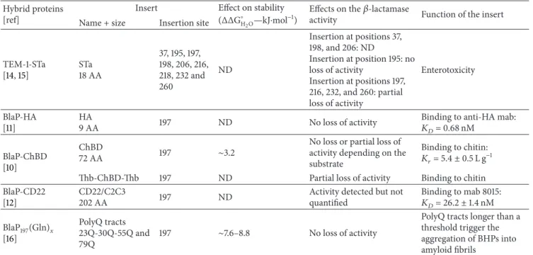
Table 1: Properties of the different BHPs discussed in this review. Hybrid proteins
[ref]
Insert Effect on stability
(ΔΔG∘
H2O—kJ⋅mol
−1) Effects on theactivity 𝛽-lactamase Function of the insert
Name + size Insertion site
TEM-1-STa [14,15] STa 18 AA 37, 195, 197, 198, 206, 216, 218, 232 and 260 ND Insertion at positions 37, 198, and 206: ND Insertion at position 195: no loss of activity Insertion at positions 197, 216, 232, and 260: partial loss of activity Enterotoxicity BlaP-HA [11] HA 9 AA 197 ND No loss of activity
Binding to anti-HA mab:
𝐾𝐷= 0.68 nM
BlaP-ChBD
[10]
ChBD
72 AA 197 ∼3.2
No loss or partial loss of activity depending on the substrate
Binding to chitin:
𝐾𝑟= 5.4± 0.5 L g−1
Thb-ChBD-Thb 197 ND Partial loss of activity Binding to chitin
BlaP-CD22
[12]
CD22/C2C3
202 AA 197 ND
Activity detected but not quantified Binding to mab 8015: 𝐾𝐷= 26.2± 1.4 nM BlaP197(Gln)𝑥 [16] PolyQ tracts 23Q-30Q-55Q and 79Q 197 ∼7.6–8.8 No loss of activity
PolyQ tracts longer than a threshold trigger the aggregation of BHPs into amyloid fibrils
The name of the BHPs, the name of the insert, its size, and function are indicated. The effects of the insertion on the stability and on the enzymatic activity of the𝛽-lactamase are also reported. ND: not determined.
allowing the insertion of the exogenous sequence. They allow a constitutive expression of the BHPs which are purified by
ion-exchange chromatography [9].
Most BlaP hybrid proteins, on the other hand, have so far been created by inserting the exogenous polypeptide at position 197 within the permissive loop located between the
𝛼 helices 8 and 9 (Figure1(b)) [10]. A constitutive expression
vector in E. coli (pNYBlaP), which allows the insertion of heterologous polypeptide sequences into the SmaI restriction
site at position 197 (Figure1(b)), has been developed [10,11].
This vector allows the expression, in the periplasm of E. coli, of the BHP in fusion with a C-terminal His-tag to facilitate their purification using affinity chromatography.
3. BHPs as Tools to Produce and Study
Difficult to Express Peptides/Proteins
Some proteins are difficult to produce recombinantly because of their intrinsic properties such as relative insolubility, instability and/or large size. One strategy to study their
properties (i.e., structure, function,. . .) consists of inserting
these proteins or their structural subdomains into a carrier protein.
Vandevenne et al. [10] have, for example, designed,
produced, and characterized a hybrid protein in which the chitin binding domain (ChBD) of the human macrophage chitotriosidase has been inserted in BlaP at position 197; this
hybrid protein is referred to as BlaP-ChBD (Table 1). The
study of the chitin binding domain of this chitinase is of interest since (i) the physiological function of mammalian
chitinases remains unknown [31] and, more importantly, (ii)
the concentration of chitotriosidase, a chitinase expressed in
lipid-laden macrophages, is highly elevated in Gaucher
dis-ease [31–33]. Some evidence suggests that mammalian
chiti-nases confer a defensive function against chitin-containing pathogens, such as fungals, which have cell walls consisting mainly of chitin; indeed, high levels of chitinases are present in the serum and tissues of guinea pigs after infection by
Aspergillus fumigates [31,34,35]. It is also proposed that the enzyme could modulate the extracellular matrix in the vessel wall affecting the downstream tissue-remodelling processes, associated with atherogenesis. This hypothesis is based on the fact that macrophages expressing chitinases are abundant in
atherosclerotic plaques [31,33].
ChBD is a small domain of 8 kDa composed of 72 amino acids which binds insoluble chitin. It contains 3 disulfide bridges that are essential for both its structural stability and its
binding to chitin [10,13,31]. This protein domain is extremely
difficult to produce, and its three-dimensional structure has
not yet been solved [10,13,31].
The BlaP-ChBD hybrid protein has been successfully produced in the periplasm of E. coli and purified in a two-step procedure, that is, affinity chromatography via a His-tag designed at the C-terminus of BlaP, followed by anion-exchange chromatography. Remarkably, the hybrid protein
exhibits both parental activities (Table 1). The 𝛽-lactamase
moiety hydrolyzes its 𝛽-lactam substrates, in the presence
and/or absence of chitin, and the ChBD domain binds to
insoluble chitin with similar𝐾𝑟 value (relative equilibrium
association constant),5.4 ± 0.5 L g−1, of other
carbohydrate-binding domains [10, 13]. It is important to note that the
bifunctionality of the hybrid protein allowed the 𝐾𝑟 value
between BlaP-ChBD and insoluble chitin to be easily
the hybrid protein allowed the detection of chitin in fungal
cell walls [24]. The fact that ChBD conserves its binding
properties within BlaP suggests that its N- and C-terminal extremities are proximal in the three-dimensional structure. In the presence of reducing agents, BlaP-ChBD still exhibited 𝛽-lactamase activity but failed to bind to chitin, confirming that the 3 disulfide bridges of ChDB are essential to maintain its functionality.
The effects of the insertion of ChBD on the thermo-dynamic stability of BlaP were extensively characterized. Despite the multidomain character of BlaP-ChBD, urea unfolding occurs according to a simple two-state mechanism
and is fully reversible [10]. The insertion of ChBD in
posi-tion 197, however, slightly destabilizes BlaP by 3.2 kJ⋅mol−1
(Table 1). Moreover, the thermal unfolding of BlaP-ChBD
was found to be less reversible and less cooperative than that of BlaP. Indeed, the thermal transition observed for BlaP-ChBD by differential scanning calorimetry (DSC) and far UV circular dichroism is characteristic of a three-state transition while the one observed for BlaP is characteristic of a two-state transition. This suggests that, within the hybrid protein, ChBD and BlaP unfold individually upon
temperature increase [10].
The ChBD domain has also been inserted in position 197 of BlaP between two thrombin (Thb) cleavage sites; this
hybrid protein is referred to as BlaP-Thb/CHBD/Thb (Table1)
[13]. This construction allowed easy separation of the ChBD
domain from the BlaP carrier protein, following the action of the thrombin and subsequent purification. Upon proteolytic cleavage, the N- and C-terminal parts of BlaP remain strongly linked together and are separated from the ChBD domain by depletion using affinity chromatography via the C-terminal
His-tag (Figure2). Remarkably, the thrombin cleavage did
not significantly alter either the activity of BlaP or that of
of ChBD (Table1), and∼18 mg of highly soluble and active
ChBD was obtained per litre of culture [13,24].
These studies clearly demonstrate that BlaP is an effi-cient scaffold to create bifunctional enzyme and to produce, purify, and characterize protein fragments that are otherwise difficult to express. This approach has been further used to produce various polypeptides of up to 40 kDa in size (unpublished results). Moreover, these results show that the BHPs can be used (i) to easily investigate the biological function of the polypeptide/protein inserted such as its
interaction with a series of ligands using the 𝛽-lactamase
activity as a reporter, (ii) to screen therapeutic molecules, that is, molecules neutralizing the biological properties of the polypeptide inserted, as further exemplified in the section of this review describing the use of BlaP for vaccine devel-opment, (iii) to screen molecules stabilizing and preventing protein aggregation (see section “BHPs as model proteins to investigate structure/function relationships”), and (iv) to facilitate the determination of the three-dimensional struc-ture of polypeptides that are otherwise unnameable to X-ray crystallography due to their insolubility, since BHPs allow an
increase in the solubility of the insert [10,12,13].
4. BHPs: an Original Approach to Map and
Functionalize Epitopes
The determination of the epitope (i.e., the region of the antigen that is recognized by an antibody or an antibody fragment, which can be linear or conformational) is of crucial importance to understand, at the molecular level, the rela-tionship between the structure and function at the
antigen-antibody interface [8]. For example, the determination of the
epitope recognized by two heavy-chain antibody fragments specific to human lysozyme has allowed a better understand-ing of how they prevent in vitro amyloid fibril formation
by the amyloidogenic variants of human lysozyme [36,37].
Moreover, the identification of the epitope enables the design and isolation of new antibody analogues, having higher affinity for their antigen or exhibiting different biological activities, or smaller antibody mimetics (i.e., peptides and
small molecules interacting with the same epitope region) [8].
Epitope mapping is also essential in diagnostic, immunother-apy, and vaccine development.
Different techniques have been developed to identify epitopes. The gold standard for epitope identification is the determination of the three-dimensional structure of the antigen-antibody complex by X-ray crystallography. This technique is, however, not always easy to perform due to the difficulty of obtaining high concentrations of a good quality complex and finding the proper conditions of
crys-tallisation [8,38]. Alternative biophysical techniques include
(i) H/D exchange experiments on the antigen-antibody complex coupled to proteolysis with pepsin and subsequent analysis of the digested fragments by mass spectrometry
(deuterium exchange mass spectrometry) [39] and (ii) NMR
spectroscopy by comparing, for example, heteronuclear single
quantum coherence (HSQC) spectra, of small target proteins
in the presence or absence of the antibodies [40]. For all
these structural approaches, high quantities of pure and stable antigens are however required, and some of them are limited
by the size of the complex [8]. Another set of approaches is
based on the exposition of numerous small peptides (overlap-ping sequences from the natural antigen or peptide mixtures derived from a combinatorial library if the antigen sequence
is unknown) on a chip (peptide arrays) [41], or at the surface
of phages (phage display) [42–46]. Phage display is based
on the expression of a peptide/protein/protein fragment of interest at the surface of phage particles, fused to one of
their coat proteins [42,47,48]. The power of this technique
is the physical link between the phenotype (the expression of the peptide/protein/protein fragment of interest that can interact with its ligand) and the genotype (the nucleotide sequence of the peptide/protein/protein fragment cloned
into the phagemid genome) [42, 48]. Phage display allows
high-throughput screening of libraries of variant nucleotide
sequences with diversity up to 106to 1010[11,42,49].
Innovative phage display strategies are continuously
implemented [50]. One of them is based on the use of the
𝛽-lactamase hybrid protein to expose random peptides/protein fragments at the surface of the phages and therefore to
B laP -C te r His-tag ChBD Thrombin Digestion Purification His B laP -N te r B laP -N te r His His IMAC column B laP -C te r B laP -C te r B laP -N te r + Ni2+
Figure 2: Schematic representation of the protocol used to cleave the ChBD domain from BlaP and to purify it. Purified ChBD is collected in the flow through after loading the hybrid protein digested by thrombin on an immobilized metal ion affinity chromatography (IMAC).
Figure adapted from Vandevenne et al., 2008 [13]. BlaP-Cter: C-terminal sequence of BlaP, starting at the insertion site. BlaP-Nter: N-terminal
sequence of BlaP ending at the insertion site.
Gene of the targeted antigen (Ag)
Estimation of the library enrichment
with a chromogenic substrate
Positive selection in the presence of ampicillin of the phages
Determination of the minimal sequence encompassing the epitope
Nucleotidic sequencing of phages specific to anti-Ag mAb
Cloning into pNYBlaP/SmaI
Determination of the kinetic parameters of hybrid BlaP and of the
affinity constant to anti-Ag MAb
Positive selection in the presence of ampicillin of clones producing
functional hybrid BlaP
SmaI fdTet Blap/SmaI Antigen fragment BlaP-Cter BlaP-Nter pIII pIII BlaP Antigen fragment Phag e a m p lifica tio n P o si ti ve s elec tio n in t h e p res ence o f a m p icil lin o f t h e p h ag es Nitrocefin BlaP
Gene fragments of antigen
Peptide array
Construction of a phage library
exp re ssin g ac ti ve 𝛽 -lac ta mas e expressing active𝛽-lactamase
specific to anti-Ag mAb Selection of phages
measuring the𝛽-lactamase activity
Nitrocefin-A482
Anti-Ag mAb Anti-Ag mAb
Elution of specific phages
Production of hybrid BlaP in
ampicillin+ purification
Alanine scanning mutagenesis Multiple alignment
Random DNase1 digestion
Cloning into fdTet Blap/SmaI
10056 bp
Phage amplification (3x)
E.coli-positive selection with
Figure 3: BHPs as unique tools for epitope mapping. Schematic representation of the epitope mapping procedure developed by Bannister
et al. and Chevign´e et al. [8,11]. The steps for which the enzymatic activity of BlaP, as a reporter or as factor for selection, is critical are
highlighted in red. BlaP-Cter: C-terminal sequence of BlaP, starting at the insertion site. BlaP-Nter: N-terminal sequence of BlaP ending at the insertion site. Anti-Ag mab: monoclonal antibody specific to the targeted antigen. Following the elution of phages after the last round of panning, a peptide array can be carried out to verify that the entire antigen sequence is well represented in the phage library. Alanine scanning mutagenesis can be carried out to determine which residues of the minimal sequence encompassing the epitope is in direct contact with the
antigen. Figure adapted from Chevign´e et al., 2007 [11].
51]. The advantages of this strategy are numerous [11, 51].
First, the polypeptide exposed within the𝛽-lactamase could
have a restricted conformational freedom and be somehow
protected from proteolytic cleavage [3]. As illustrated in
Figure3, the main implementation is, however, associated
with the use of 𝛽-lactamase activity which can be easily
detected with chromogenic substrates such as nitrocefin [11].
The𝛽-lactamase activity is used (i) as a reporter to directly
assess the interactions between the inserted peptides and the target antibodies, and to easily monitor the enrichment of
the phage library during the successive rounds of selection, and (ii) as an agent of selection of phages expressing active BHPs. Indeed, in order to amplify phages, phage-infected cells are grown in a liquid medium in the presence of ampicillin. Under these conditions, the bacterial growth is directly associated with antibiotic resistance, and thus only cells expressing active BlaP at the surface of phages are able
to grow [11].
This approach was first introduced by Legendre et al., who inserted random peptides within TEM-1, which was
displayed on phage fd in fusion with the coat protein pIII [51].
The peptides were displayed on different loops surrounding the active site of TEM-1: in one library, peptides replaced residues A103 to 106; in a second library, they replaced residues Thr271 and/or Met272, and these two libraries
were combined in a third one [51]. By selection from the
different libraries using three unrelated conformational mon-oclonal antibodies (mabs) recognizing distinct epitopes on the prostate-specific antigen (PSA), several hybrid proteins expressing small peptides with an affinity for these mabs in the micro- and nanomolar range were isolated. The sequence of these small peptides presents no similarity with that of PSA, suggesting that mimotopes (i.e., peptides which bind the antibody without having any identity to the antigen sequence) were selected. These results indicate that BHPs could effectively be used to display peptides at the surface of phages.
An original epitope mapping strategy, inspired by the work of Legendre et al., was developed by Bannister et al. and
Chevign´e et al. [8,11]. It involves constructing a phage display
library in which random fragments of different lengths of the gene coding for the antigen of interest are exposed within
the 𝛽-lactamase BlaP (at position 197) in fusion with the
pIII protein at the surface of phages (Figure 3). A given
region of the target protein is displayed within peptides of different lengths and could therefore adopt different conformations. Sufficiently long peptides are likely to adopt
a fold similar to what they adopt in the full target protein [11].
As exemplified below, this technology allows the minimal region of an antigen interacting with an antibody to be
identified with high accuracy (Figure3) [11]. As the peptides
exposed within the 𝛽-lactamase are directly related to the
nucleotide sequence of the antigen, they do not correspond to mimotopes. Moreover, the key amino acids within this minimal region that are directly in contact with the antibody can be further determined by point mutations as discussed
below (Figure3) [8].
4.1. Mapping of Linear Epitopes. The BHP-implemented
phage display approach, developed by Chevign´e et al. [11],
has allowed the determination of the linear epitope (i.e., an epitope composed of amino acids that are in close proximity in the sequence) of a high-affinity monoclonal antibody
(anti-HA mab) specific to the virus influenza hemagglutinin ((anti-HA1).
Overlapping peptides of different sizes (from gene fragments of 50 to 300 bp) of HA1 were displayed on the solvent exposed
loop between𝛼 helices 8 and 9 of BlaP, which was fused to
pIII coat protein of phages (Figure3). The phages expressing
peptides containing the epitope region of HA1 were selected by panning against anti-HA mab. After three rounds of selection and amplification in the presence of ampicillin, a series of phages expressing BHPs specific to anti-HA mab was selected. The sequencing of these phages shows a consensus
9-residues linear epitope of anti-HA mab (Figure 4). This
epitope is present in fragments of various sizes (from 13 to 70 residues) and can thus in principle adopt different folds and solvent accessibilities. The gene coding for the BHP displaying the consensus 9-residues epitope sequence was cloned into the constitutive expression vector pNY. The hybrid protein, referred to as BlaP-HA, was subsequently expressed in the periplasm of E. coli and purified as a
bifunctional hybrid protein. The kinetic parameters (i.e.,𝐾𝑚,
𝑘cat, and 𝑘cat/𝐾𝑚) of BlaP-HA are similar to that of
wild-type BlaP, and it binds anti-HA mab with an affinity(𝐾𝐷) of
∼0.68 nM (Table1) [11].
4.2. Mapping of Conformational Epitopes. A similar approach
was used by Bannister et al. to identify the conformational epitope (i.e., epitope composed of amino acids located far apart in the sequence but brought together by protein folding)
of the anti-CD22 immunotoxin CAT-8015 (mab 8015) [8].
CD22 (cluster of differentiation-22) is a cell surface glyco-protein composed of a N-terminal Ig-like V-type domain
and various Ig-like C-type domains (Figure5) [52–55]. This
protein is of particular therapeutic interest because it is a specific marker present on the surface of malignant B-cells and it is rapidly internalized upon binding; it constitutes therefore a relevant target for an antibody drug conjugate or immunotoxin approach. Despite the therapeutic interest of CD22, very little is known concerning its structure or that of the other related family members due to their high
level of glycosylation [8]. CAT-8015 is an immunotoxin which
combines a CD22-specific antibody variable fragment (Fv, derived from the antibody mab 8015) with a Pseudomonas exotoxin A (PE38); it exhibits a noteworthy clinical activity in three leukaemia (i.e., chronic lymphocyte leukaemia, hairy
cell leukaemia, and acute lymphoblastic leukaemia) [8,56,
57]. Antibody competition-binding studies have revealed that
the epitope of CAT-8015 is localized in the C-like domain 2 of CD22, but there is no information on its precise location
[8,58].
In order to identify which residues of CD22 bind to CAT-8015, Bannister et al. introduced random CD22 extracellular domain gene fragments of 50 to 1000 bp into the permissive exposed loop of BlaP (position 197) fused to the phage
pIII coat protein (Figure 3) [8]. A library containing 6 ×
105 transformants was obtained and screened against
CAT-8015 using the same strategy as that explained above. A
peptide-array analysis (Figure3) confirmed that the entire
CD22 gene was well represented in the𝛽-lactamase-positive
infectious phages, except two small regions of 7 residues each. The minimal region of CD22 which binds to mab 8015 was identified. This region, referred to as CD22/C2C3, is composed of 202 residues, from V234 to G435, located in the C-terminal of the C-type domain 1, in the C-type domains
2 and 3, and in the N-terminal of the C-type domain 4
(Figure5).
The gene coding for the BHP displaying the minimal region of CD22 was cloned into the constitutive expression vector pNY. The hybrid BlaP-CD22/C2C3, produced as a
bifunctional hybrid protein in E. coli, conserves the
𝛽-lactamase activity and binds to mab 8015 with a high affinity
(𝐾𝐷 ∼26.2 nM) (Table1). To identify the key amino acids
involved in the antibody binding, an alanine-scanning
muta-genesis was performed (Figure 3) [11]. Thirty-four
single-point mutations to alanine were performed in three clusters (referred to as clusters 1, 2, and 3) of CD22 encompassing the epitope, and the resulting hybrid proteins were produced and purified. Cluster 1 is located in the junction of C-type domains 1 and 2, cluster 2 is located in C-type domain 3 and cluster 3 encompasses the junction between C-type domain 3 and 4 and the N-terminal of C-type domain 4
(Figure5). The binding of each mutant to mab 8015 was then
measured by enzyme-linked immunosorbent assay (ELISA) and compared to that of the parent hybrid protein BlaP-CD22/C2C3. This technique has allowed the identification of the amino acids of high or intermediate importance in the CD22-mab 8015 complex. The most important residues encompass three discontinuous regions and they are mainly located in C-type domains 2 and 3 (clusters 1 and 2), and few are located in cluster 3. Modelling of different orientations of these domains strongly suggests that they adopt a U-shaped arrangement which allows the three clusters to be in close
enough proximity to form the epitope (Figure5) [8].
These two examples show that alternating successive affinity selections of phages and growth of phage-infected cells in the presence of antibiotic permits the selection of BHPs exhibiting both a high affinity for the target antigen and an efficient enzymatic activity. They clearly demonstrate that BHP technology using BlaP is a unique tool that allows the identification of the antigen region involved in the antigen-antibody interactions. It allows (i) the identification of linear epitopes (i.e., the linear epitope of HA for anti-HA mab) and (ii) the characterisation of complex epitopes (i.e., the U-shaped discontinuous CD22 epitope of mab 8015). Moreover, BlaP has enough conformational flexibility to allow inserted antigen fragments of various lengths to fold and interact with the antibody. BHPs allow therefore the identification of non-linear epitopes (i.e., conformational epitopes) that are directly linked to the nucleotide sequence of the antigen (i.e., not
a mimotope) [8,11]. This approach can be further used to
identify the protein region involved in any protein-ligand interactions.
4.3. BHPs as a Unique Tool for Immunoassay Develop-ment. The 𝛽-lactamase implemented phage-display
tech-nique described above allows the selection of a particular epitope as a bifunctional hybrid protein which associates the epitope recognizing its specific target with an efficient enzy-matic activity. This functionalization of the epitope allows the rapid characterisation of the antigen-antibody interaction. For example, the BlaP-HA hybrid protein was successfully coated on solid surface to perform an ELISA, suggesting that
the solvent accessibility of the inserted polypeptide (i.e., the epitope) was not altered when the hybrid protein is coated
[11]. Moreover, the enzymatic activity of BlaP can be used to
check the coating efficiency and to quantify the amount of coated protein.
5. BHPs as Immunogenic Carriers for
Vaccine Development
Antibodies are specialized fighter proteins that are effective in preventing infectious diseases. This property is based on the recognition of specific epitopes on the surface of the antigen that promotes the neutralization of the biological activity of the antigen or the opsonisation of the pathological agent. It is assumed that immunization with a precise epitope, corresponding to an effective neutralizing antibody, would elicit the generation of similarly potent antibodies in the
vaccine [38]. Thus, the insertion of these particular epitopes
into a carrier protein is often the starting point for the development of a new generation of safe vaccines. Within the carrier protein, the properties of the polypeptides, including their antigenicity and immunogenicity, can be further
modi-fied [59]. In this context, the BHP technology, allowing the
insertion of a domain, subdomain, or a short polypeptide
from the native targeted antigen into a carrier𝛽-lactamase,
is of particular interest to design such de novo antigens
(Figure6). This approach is particularly relevant to antibody
raised against cryptic epitopes and non-immunodominant antigens.
5.1. Generation of Vaccines against Poorly Immunogenic Polypeptides/Proteins. TEM-1 hybrid proteins were created
in an attempt to develop a vaccine against the poorly antigenic and non-immunogenic STa enterotoxin from enterotoxic E.
coli strains (ETEC). This toxin, which is associated with
enteric colibacillosis (characterized by severe diarrhoea) in calves and piglets, leads to significant losses in agriculture
due to the death of newborns [60,61]. STa is composed of 18
amino acids including 6 cysteins involved in three disulfide
bridges which are necessary for the toxic activity [62,63]. STa
is poorly antigenic and is not immunogenic due to its small size. No vaccine is yet available against this thermostable toxin despite many attempts to design a safe vaccine (i.e., a non-toxic form of STa). The latter includes the chemical
coupling of the toxin to bovine serum albumin [64, 65],
to the 𝛽-subunit of cholera toxin [66], and to the
heat-labile enterotoxin (LT) [65,67], and its fusion to the major
protein subunit ClpG of E. coli CS31A surface antigen [68],
to several subunits of cholera toxin [66,69], to LT [70,71], to
OmpC [70], and to flagellin [72]. These constructions either
failed to induce the production of neutralizing antibodies against STa or retained a certain degree of toxicity. However, Wu and Chung have managed to elicit the production of protective antibodies against the antigen STLT (i.e., a fusion of the thermostable ST and thermolabile LT enterotoxins) in mice using a protein fusion between GFP and STLT. These immunized mice presented a subsequent full protection
Cl 1 FVERSKAFSNCYPYDVPDYASLRSLVASSGTLEFITEGFTWTEV Cl 2 VERSKAFSNCYPYDVPDYASLRSLVASSGTLEFITEGFTWTEVTQNGGS Cl 3 VERSKAFSNCYPYDVPDYASLRSLVASSGTLEFITEGFTWTEVTQNGGS Cl 4 VERSKAFSNCYPYDVPDYASLRSLVASSGTLEFITEGFTWTEV Cl 5 VERSKAFSNCYPYDVPDYASLRSLVASSGTLEFITEGFTWTEV Cl 6 VERSKAFSNCYPYDVPDYASLRSLVASSGTLEFITEGFTWTEVTQNGGSNACKRGPGSGFFS Cl 7 RSKAFSNCYPYDVPDYASL Cl 8 RSKAFSNCYPYDVPDYASLRSLVASSGTLEFITEGFTWTEVTQNGGSNACKRGPGSGFFSRLNWLTKSEV Cl 9 NCYPYDVPDYASLRSLVASSGTLEFITEGFTWTEVTQN Cl 10 CYPYDVPDYASLRSLVASSGTLEFITEGFTWTEVTQNGGSNACKRGPGSGFFS Cl 11 NCYPYDVPDYASLRSL Consensus CYPYDVPDYASL
Figure 4: Consensus sequence recognized by anti-HA mab. Multiple sequence alignment from eleven individual clones selected after three rounds of selection of phages on anti-HA mab. The residues corresponding to the epitope recognized by the anti-HA mab are underlined.
Figure from Chevign´e et al. [11].
C-terminal C-type domain 6 C-type domain 5 C-type domain 4 C-type domain 3 C-type domain 2 C-type domain 1 V-like domain Cluster 1 Cluster 2
Cluster 3 U-shaped epitope
of mAb 8015 C-type domain 6 C-type domain 5 C-type domain 4 C-type domain 3 C-type domain 2 C-type domain 1 V-like domain N-terminal
Figure 5: Schematic representations of the glycoprotein CD22 in a linear and U-shaped arrangement. In the linear arrangement, the three
clusters forming the epitope are far away from each other whereas they are close in a U-shaped arrangement [8]. The residues involved in
the complex with mab 8015 are clustered in cluster 1, 2, and 3 and are shown in light blue, red, and green, respectively. Figure adapted from
Bannister et al. [8].
Different approaches based on the use of TEM-1 hybrid proteins have been used in order to design a vaccine against
STa [9,14, 15]. First, Ruth et al. have designed DNA
vacci-nation to induce the production of neutralizing antibodies
against STa [14]. STa and three variant forms, in which
one (STaC6A, STaC17A) or two (STaC6A-C17A) of the six cysteines have been mutated (in order to disrupt one or two disulphide bridges and cause a complete loss of the toxicity of the toxin), have been inserted either in position 195 (loop A) or 216 (loop B) of TEM-1. Plasmid DNA encoding these hybrid proteins has been used to immunize
mice. Following immunizations, different levels of anti TEM-1 antibodies were generated. The level of antibody production against TEM-1 was higher for mice immunized with plasmid DNA coding for BHPs displaying STa and its variants in position 216 (loop B) than in position 195 (loop A). Since insertions in loop B interfere less with anti TEM-1 antibody production than insertion in loop A, this suggests that the immunodominant epitope of TEM-1 is located in the region of loop A. In contrast, no antibody specific to STa could be detected, even after 3 injections of the different plasmids. Two subsequent boosts with a STa toxin obtained by peptide
synthesis, which is not immunogenic, did however induce the production of STa-specific antibodies in mice initially primed with plasmids encoding hybrid proteins, but not when primed with plasmid encoding TEM-1. This clearly indicates that hybrid proteins were expressed in mice and have primed the production of antibodies specific to STa. The ability of sera containing STa specific antibodies to neutralize the toxin was determined by suckling mouse assays. Only the sera from mice immunized with the plasmid coding for the double disulfide bridge mutated variant (STaC6A-C17A) and boosted with STa injections show neutralizing activity. These results indicate that the use of the toxic form of STa is therefore not needed to induce the production of neutralizing antibodies. These results also indicate that TEM-1 is an appropriate carrier to present nonimmunogenic peptide to the immune system. Indeed, the carrier is required for the induction of helper T cells allowing the production of antibodies against the carrier, TEM-1, and the hapten, STa. Moreover, the best location for the insertion seems to be located at position 195, the immunodominant epitope of TEM-1. Indeed, exposing the insert at this position reduces the production of anti TEM-1 antibodies and should therefore decrease the competition between the hapten and the carrier
for the B-cell immune response [14].
Subsequently, recombinant hybrid TEM-1 proteins in which STa has been inserted at different positions, including positions 195 and 216, were designed, produced, and used for
mice immunization (Figure6) [9]. This study revealed that
the hybrid proteins elicit the production of antibodies mainly against TEM-1, and in much lower quantity against STa. TEM-1 with the insertion at position 195 induces the highest production of anti-STa antibody and most importantly was the only hybrid protein that leads to the production of antibodies neutralizing the toxicity of STa. These results are in good agreement with results obtained with DNA vaccination and confirm that B-cell epitopes of the carrier are immunodominant. Moreover, the carrier, TEM-1, con-tains the functional helper T cell epitopes necessary for the
immune response against the hapten, STa [9].
Following these encouraging results, Zervosen et al. immunized cattle with a TEM-1 hybrid protein containing the STa in position 197 in the presence of 3 different adjuvants
(Table1) (Montanide ISA70 - ISA206 and IMS1313) [15]. High
levels of different IgG types specific to TEM-1 were detected,
and in vitro neutralization of the𝛽-lactamase activity was
observed by mixing purified TEM-1 with sera. In contrast, specific anti-STa IgG and IgG1 antibodies were only detected at non-significant levels, and no IgG2 were detected in the sera of the immunized cattle. These results suggest that the response of the bovine immune system is different from that
observed in mice [9,15]. The use of a series of other adjuvants
to immunize cattle needs to be investigated.
5.2. Production of Neutralizing Antibodies against Difficult-to-Express Antigens. As discussed above, BlaP can be used as a
scaffold to produce and to purify the chitin binding domain
from chitotriosidase (ChBD) [13]. The purified ChBD has
been used to immunize a rabbit (Figure 6). The resulting
serum contains antibodies that are able to bind to the free ChBD, the hybrid BlaP-Thb/ChBD/Thb, and the native human macrophage chitotriosidase. Moreover, these anti-ChBD antibodies are able to prevent the interaction of anti-ChBD
with chitin [13].
Taken all together, the results of these studies indicate that both BlaP and TEM-1 are appropriate scaffolds for producing and presenting antigens and inducing the production of antibodies neutralizing biological properties of the inserted
fragment (Figure6).
6. BHPs as Model Proteins to Investigate
Structure/Function Relationships
6.1. BlaP: A Scaffold to Create Protein Models to Study the Mechanism of Amyloid Fibril Formation by Polyglutamine Pro-teins. Ten progressive neurodegenerative disorders, referred
to as polyglutamine (polyQ) diseases, and, including Hunt-ington’s disease and several spinocerebellar ataxias, are asso-ciated with ten unrelated proteins containing an expanded polyglutamine (polyQ) tract (i.e., a tract that is longer than a pathological polyglutamine threshold). PolyQ tracts are encoded by a repetition of an unstable CAG trinucleotide
repeat in the corresponding genes [74–77]. The ten
disease-associated proteins show no sequence or structural similarity apart from the expanded polyQ tract, which is located at a different positions in each protein. The polyQ tract appears therefore to be a critical determinant in polyQ diseases, and several lines of evidence suggest that it confers a gain of toxic function to the mutant proteins by triggering the aggregation
of the proteins into amyloid fibrils [74,75,78–80]. Indeed, all
polyQ diseases share a number of features which suggest a
common physiopathological mechanism [75,79–81]: (i) the
existence of a polyQ threshold for the aggregation of polyQ proteins and the disease development, generally comprised
between 35 and 45Q [82], (ii) the so-called anticipation
phenomenon, which indicates that the longer the polyQ,
the earlier and more severe the disease [76, 83–85], and
(iii) the presence of intranuclear inclusion bodies, containing amyloid fibrils made of polyQ proteins, in neuronal cells
[75,76,85,86].
So far, there is no preventive or curative treatment for these pathologies and existing therapies only treat the symp-toms (i.e., they alleviate the sympsymp-toms without modifying the course of the disease). In order to design curative and/or preventive treatments, it is crucial to better understand how polyQ tracts trigger protein aggregation and by which mechanism some of the aggregates formed are cytotoxic.
Although the presence of the expanded polyQ tract is the critical trigger factor of the aggregation phenomenon, a growing number of studies suggest that the non-polyQ regions can, however, modulate both the kinetics and the
aggregation pathway of polyQ proteins [74,78,87–90]. The
non-polyQ regions can protect from aggregation by (i)
ster-ically hindering polyQ intermolecular interactions [91], (ii)
restricting polyQ conformational changes which are required
for fibril formation [89] and (iii) increasing the protein
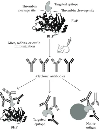
Mice, rabbits, or cattle immunization
Serum containing antibodies specific to the targeted epitope and recognizing it within the BHP used for the immunization, in its free form,
and within the native antigen Targeted epitope
BlaP
BHP
Targeted epitope Ligand
Antigen-ligand complex
Neutralizing antibodies
Antibody binding to the targeted epitope preventing its interaction with its ligand Thrombin
cleavage site Thrombin cleavage site
Native antigen Targeted epitope BHP Polyclonal antibodies +
Figure 6: Schematic representation of the BHP technology used to design de novo antigens in order to induce the production of neutralizing antibodies. A specific region of a large antigen, that is known to promote the neutralization of the biological activity of the antigen or promote its opsonization, is inserted into BlaP. This approach can also be used to elicit the production of specific, neutralizing antibodies against
polypeptides that are insoluble, difficult to express (i.e., ChBD, [13]), and poorly immunogenic (such as STa, [9]).
could assist aggregation by providing additional
aggregation-prone domains [74, 92]. There is, therefore, a complex
interplay between the tendency of the polyQ tract to trigger aggregation and the modulating effect of non-polyQ regions
[16]. To better understand the general principles governing
this complex phenomenon, it is crucial to investigate in detail which properties of the host protein (sequence, size, structure, and stability) influence the ability of polyQ tracts to mediate aggregation. The ten disease-associated proteins
are difficult to produce recombinantly due to their large
size and/or relative insolubility [74,92–94]. Scarafone et al.
tackled this problem by creating and characterizing model
polyQ proteins consisting of the𝛽-lactamase BlaP and polyQ
tracts inserted at position 197 [16]. Significant amounts (i.e.,
10–20 mg per liter of culture) of hybrid enzymes containing 23, 30, 55, and 79Q were successfully produced and purified. It is important to note that the longest polyQ tract that has been
Time (h) 0 100 200 300 400 500 600 700 800 A ggr eg at ed fracti o n 0 0.2 0.4 0.6 0.8 1 1.2 A ggr ega ted f rac ti o n 0 0.2 0.4 0.6 0.8 1 1.2 Fibrils B laP t o lera tes in se rt io n o f u p t o 79Q: The lo n gest p o lyQ t rac t r ep o rt ed in mo del p ro tein s Time (h) 0 50 100 150 200 250 300 350 Fibrils Modulating effects imposed by BlaP depend
on the polyQ tract length N U F N U F F aggregates Amorphous BlaP197(Gln)55 BlaP197(Gln)79 BlaP197(Gln)x 100% N 100% N 100% U 100% U 1.85 M urea - 25∘C 1.85 M urea - 25∘C 50% N - 50% U 50% N - 50% U 0 M urea - 37∘C 0 M urea - 37∘C 3.5 M urea - 25∘C 3.5 M urea - 25∘C
Figure 7: Model polyQ proteins using BlaP to investigate the amyloid fibril formation. The advantage of using BlaP as a scaffold to create polyQ proteins are highlighted in red. Left side panel: schematic representation of the polyQ-BlaP hybrid protein. Middle panel: kinetics of
aggregation of BlaP197(Gln)55and BlaP197(Gln)79at 110𝜇M under the following conditions of incubation: (i) PBS, pH 7.5 and 0 M urea at 37∘C
(pink), (ii) PBS, pH 7.5, and 1.85 M urea at 25∘C (blue) and (iii) PBS, pH 7.5, and 3.5 M urea at 25∘C (green). BlaP197(Gln)55does not form
amyloid fibrils under native conditions, in contrast to BlaP197(Gln)79. Under native conditions, the threshold value is therefore comprised
between 55 and 79Q. Under denaturing conditions, it is comprised between 30 and 55Q. Moreover, the kinetics of aggregation are faster with
longer polyQ tracts. Right side panel: schematic pathway of aggregation for BlaP197(Gln)55and BlaP197(Gln)79. N, native state; U, unfolded
state; F, amyloid fibril. The native conformation of BlaP imposes constraints to the 55Q tract that prevent it to trigger the formation of amyloid
fibrils. Figure adapted from Scarafone et al. [16].
BlaP is therefore a unique scaffold to investigate the effects of
long inserted polyQ sequences (Figure7).
The effects of the polyQ insertions on the activity, struc-ture, and aggregation of BlaP were investigated using a range of biophysical techniques. The activity and the secondary and tertiary structures of BlaP are essentially not affected by the insertion of the polyQ tract as long as 79Q. The polyQ tract adopts a disordered structure at the surface of the protein irrespective of the number of glutamines. BlaP is significantly destabilized, however, by the insertion of the polyQ tract. Remarkably, the extent of destabilization is
largely independent of the polyQ length (7.6–8.8 kJ⋅mol−1;
Table 1). This behaviour therefore makes it possible to
investigate independently the role of (i) the length of the
polyQ sequence and (ii) the conformational state of the
𝛽-lactamase moiety on the aggregating properties of the hybrid proteins. Accordingly, the aggregating properties of BlaP and of the different hybrid proteins were investigated under both native and denaturing conditions. The aggregation behaviour of BlaP-polyQ hybrid proteins recapitulates that of disease-associated polyQ proteins. Only hybrid proteins with 55Q and 79Q readily form amyloid fibrils; therefore, analogous to the proteins associated with diseases, there is a polyglutamine threshold required for the formation of amyloid fibrils.
Moreover, above this threshold, the longer the polyQ, the
faster the aggregation rate (Figure7). Most importantly, the
threshold-value critically depends on the structural integrity of BlaP. BlaP with 55Q forms fibrils under denaturing but not
under native conditions (Figure7). This means that the native
conformation of BlaP imposes some conformational and/or steric constraints to the 55Q tract that inhibit fibril formation. On the other hand, the hybrid protein containing 79Q forms amyloid fibrils at similar rates whether BlaP is folded or not
(Figure7). These results therefore suggest that the influence
of the protein context on the aggregation properties of polyQ disease-associated proteins could be negligible when the
latter contain particularly long polyQ tracts [16].
Taken all together, these results indicate that BlaP is an appropriate host to study the aggregation of polyQ proteins. Its utility could be extended to the study of how other amyloidogenic peptides trigger protein aggregation.
6.2. Effect of the Structure of the Insert on BHP Stabilities.
Vandevenne et al. [10] extensively investigated the effects of
the insertion of the ChBD domain on the thermodynamic stability of BlaP, as indicated above. They observed that the insertion of this 72-residue folded domain at position
This destabilization is significantly lower than that induced by the insertion of unstructured polyQ tracts of 23 to 79
residues, inserted into the same position (7.6–8.8 kJ⋅mol−1,
Table 1) [16]. This observation suggests that the insertion
of unstructured polypeptides is more destabilizing than the insertion of folded polypeptides.
6.3. BHPs as Models to Screen Molecules Stabilizing Polypep-tides. A growing number of peptide and protein drugs
are utilized in therapy [96]. Unfortunately, because of
unfavourable solubility, stability, and aggregation, their
appli-cations are sometimes difficult [96]. To circumvent this
prob-lem, low molecular weight additives, called cosolutes, have
been developed, including cyclodextrins [96,97].
Cyclodex-trins are circular oligosaccharides containing a central cavity forming the resting site for hydrophobic molecules of an
appropriate dimension [98]. They are generally used to
prevent protein aggregation during the renaturation process
[12,96]. BCD07056 is a modified𝛽-cyclodextrin, one of the
most abundant classes of cyclodextrin [96].
Vandevenne et al. used the hybrid protein BlaP-ChBD, incubated under drastic conditions, to investigate the effects
of BCD07056 on the stability of proteins [12]. They observed
that BCD07056 does not affect the chitin binding of
BlaP-ChBD, yet increases its 𝛽-lactamase activity. More
inter-estingly, this𝛽-cyclodextrin minimizes the inactivation of
BlaP-ChBD upon storage at room temperature; its addition
cannot, however, reverse the inactivation process. The
𝛽-cyclodextrin has a moderate effect on the thermal stability of BlaP-ChBD. However, its presence restores a cooperative reversible thermal unfolding, with a simple two-state tran-sition, characteristic of BlaP without insert. This suggests that BCD07056 prevents the aggregation of BlaP-ChBD by interacting with the protein during the unfolding process. BCD07056 is therefore an effective additive to stabilize proteins during their storage and prevent their aggregation,
without interfering with their activity [12]. It could be used to
facilitate the application of a growing number of peptide and
protein drugs in therapy [96].
7. Conclusion
Class A 𝛽-lactamase hybrid proteins, in which
exoge-nous polypeptides of various sizes are inserted (up to 40 kDa), can be readily designed and used for multi-ple purposes, notably to produce difficult-to-express pep-tides/proteins/protein fragments, to map epitopes, to dis-play antigens, and to study protein structure/function rela-tionships. The wide ranging impact of the BHP approach essentially originates from (i) the efficient enzymatic activity that can be easily measured and that can serve as a reporter or factor for selection and (ii) the facility with which they can be recombinantly produced and purified. In addition to the numerous applications summarized in this review, many other applications can be envisaged: BHPs could be used as biosensors and in affinity chromatography, drug screening, and drug targeting. They are also of special interest to better
understand more fundamental aspects of protein evolution and structure/function relationships.
Abbreviations
Ag: Antigen
BHPs: 𝛽-lactamase hybrid proteins
BlaP: 𝛽-lactamase from Bacillus licheniformis 749/C
CD22: Cluster of differentiation-22 ChBD: Chitin binding domain dsbA: Disulfide bond isomerase A
ELISA: Enzyme-linked immunosorbent assay ETEC: Enterotoxic E. coli
GFP: Green fluorescent protein
HA1: Virus influenza hemagglutinin
H/D: Hydrogen/deuterium
LT: Thermolabile enterotoxin
mab: Monoclonal antibody
MalE: Maltodextrin-binding protein NMR: Nuclear magnetic resonance polyQ: Polyglutamine
PSA: Prostate specific antigen
Thb: thrombin.
Acknowledgments
The authors would like to acknowledge the Fonds de la Recherche Scientifique (1.C039.09, FRFC 24.58.112.F and MIS-F.4505.11), the Belgian program of Interuniversity Att-raction Poles administered by the Federal Office for Scientific Technical and Cultural Affairs (PAI nos. P6/19 and P7/44), the University of Liege (FSRC-11/108), the Walloon region and the funds from DGO6 for their support of those parts of their own research that are described in this review. We would like to thank Dr. David Thorn and Coralie Pain for their critical reading of the paper.
References
[1] P. Beguin, “Hybrid enzymes,” Current Opinion in Biotechnology, vol. 10, pp. 336–340, 1999.
[2] B. Collinet, M. Herv´e, F. Pecorari, P. Minard, O. Eder, and M. Desmadril, “Functionally accepted insertions of proteins within protein domains,” Journal of Biological Chemistry, vol. 275, no. 23, pp. 17428–17433, 2000.
[3] J.-M. Betton, J. P. Jacob, M. Hofnung, and J. K. Broome-Smith,
“Creating a bifunctional protein by insertion of𝛽-lactamase
into the maltodextrin-binding protein,” Nature Biotechnology, vol. 15, no. 12, pp. 1276–1279, 1997.
[4] J. L. Martin, J. C. A. Bardwell, and J. Kuriyan, “Crystal structure of the DsbA protein required for disulphide bond formation in vivo,” Nature, vol. 365, no. 6445, pp. 464–468, 1993.
[5] M. Delarue, O. Poch, N. Tordo, D. Moras, and P. Argos, “An attempt to unify the structure of polymerases,” Protein
Engineering, vol. 3, no. 6, pp. 461–467, 1990.
[6] M. Levine, H. Muirhead, D. K. Stammers, and D. I. Stu-art, “Structure of pyruvate kinase and similarities with other enzymes: possible implications for protein taxonomy and evo-lution,” Nature, vol. 271, no. 5646, pp. 626–630, 1978.
[7] S. Jones, M. Stewart, A. Michie, M. B. Swindells, C. Orengo, and J. M. Thornton, “Domain assignment for protein structures using a consensus approach: characterization and analysis,”
Protein Science, vol. 7, no. 2, pp. 233–242, 1998.
[8] D. Bannister, B. Popovic, S. Sridharan et al., “Epitope mapping and key amino acid identification of anti-CD22 immunotoxin CAT-8015 using hybrid-lactamase display,” Protein Engineering,
Design and Selection, vol. 24, no. 4, pp. 351–360, 2011.
[9] N. Ruth, B. Quinting, J. Mainil et al., “Creating hybrid proteins by insertion of exogenous peptides into permissive sites of a
class A𝛽-lactamase,” FEBS Journal, vol. 275, no. 20, pp. 5150–
5160, 2008.
[10] M. Vandevenne, P. Filee, N. Scarafone et al., “The Bacillus
licheniformis BlaP𝛽-lactamase as a model protein scaffold to
study the insertion of protein fragments,” Protein Science, vol. 16, no. 10, pp. 2260–2271, 2007.
[11] A. Chevign´e, N. Yilmaz, G. Gaspard et al., “Use of bifunctional
hybrid𝛽-lactamases for epitope mapping and immunoassay
development,” Journal of Immunological Methods, vol. 320, no. 1-2, pp. 81–93, 2007.
[12] M. Vandevenne, G. Gaspard, E. M. Belgsir et al., “Effects of monopropanediamino-𝛽-cyclodextrin on the denaturation process of the hybrid protein BlaPChBD,” Biochimica et
Bio-physica Acta, vol. 1814, no. 9, pp. 1146–1153, 2011.
[13] M. Vandevenne, G. Gaspard, N. Yilmaz et al., “Rapid and easy development of versatile tools to study protein/ligand interactions,” Protein Engineering, Design and Selection, vol. 21, no. 7, pp. 443–451, 2008.
[14] N. Ruth, J. Mainil, V. Roupie, J.-M. Fr`ere, M. Galleni, and K. Huygen, “DNA vaccination for the priming of neutralizing antibodies against non-immunogenic STa enterotoxin from enterotoxigenic Escherichia coli,” Vaccine, vol. 23, no. 27, pp. 3618–3627, 2005.
[15] A. Zervosen, C. Saegerman, I. Antoniotti et al., “Character-ization of the cattle serum antibody responses against TEM 𝛽-lactamase and the nonimmunogenic Escherichia coli heat-stable enterotoxin (STaI),” FEMS Immunology and Medical
Microbiology, vol. 54, no. 3, pp. 319–329, 2008.
[16] N. Scarafone, C. Pain, A. Fratamico et al., “Amyloid-like fibril formation by polyq proteins: a critical balance between the polyq length and the constraints imposed by the host protein,”
PLoS One, vol. 7, no. 3, Article ID e31253, 2012.
[17] R. P. Ambler, A. F. W. Coulson, J.-M. Frere et al., “A standard
numbering scheme for the class A𝛽-lactamases,” Biochemical
Journal, vol. 276, no. 1, pp. 269–270, 1991.
[18] J. R. Knox and P. C. Moews, “-Lactamase of Bacillus
licheni-formis 749/C. Refinement at 2 ˚A resolution and analysis of
hydration,” Journal of Molecular Biology, vol. 220, no. 2, pp. 435– 455, 1991.
[19] E. Fonze, P. Charlier, Y. To’th et al., “TEM1 beta-lactamase struc-ture solved by molecular replacement and refined strucstruc-ture of the S235A mutant,” Acta Crystallographica D, vol. 51, no. 5, pp. 682–694, 1995.
[20] B. Hallet, D. J. Sherratt, and F. Hayes, “Pentapeptide scanning mutagenesis: random insertion of a variable five amino acid cassette in a target protein,” Nucleic Acids Research, vol. 25, no. 9, pp. 1866–1867, 1997.
[21] J. T. Moore, S. T. Davis, and I. K. Dev, “The development of
𝛽-lactamase as a highly versatile genetic reporter for eukaryotic cells,” Analytical Biochemistry, vol. 247, no. 2, pp. 203–209, 1997. [22] G. Zlokarnik, P. A. Negulescu, T. E. Knapp et al., “Quantitation of transcription and clonal selection of single living cells with
𝛽-lactamase as reporter,” Science, vol. 279, no. 5347, pp. 84–88, 1998.
[23] J. Oosterom, E. J. P. van Doornmalen, S. Lobregt, M. Blomenr¨ohr, and G. J. R. Zaman, “High-throughput
screen-ing usscreen-ing 𝛽-lactamase reporter-gene technology for
identi-fication of low-molecular-weight antagonists of the human gonadotropin releasing hormone receptor,” Assay and Drug
Development Technologies, vol. 3, no. 2, pp. 143–154, 2005.
[24] M. Vandevenne, M. Galleni, and P. Filee, “How to make a good use of a “Bad Enzyme”: utilisation of efficient beta-lactamase for the benefits of biochemical research,” in Beta-Lactamases, Nova Science, New York, NY, USA, 2011.
[25] C. Jelsch, L. Mourey, J.-M. Masson, and J.-P. Samama, “Crystal
structure of Escherichia coli TEM1𝛽-lactamase at 1.8 ˚A
resolu-tion,” Proteins, vol. 16, no. 4, pp. 364–383, 1993.
[26] P. C. Moews, J. R. Knox, O. Dideberg, P. Charlier, and J.-M. Frere, “𝛽-Lactamase of Bacillus licheniformis 749/C at 2 ˚A resolution,” Proteins, vol. 7, no. 2, pp. 156–171, 1990.
[27] X. Charpentier and E. Oswald, “Identification of the secre-tion and translocasecre-tion domain of the enteropathogenic and enterohemorrhagic Escherichia coli effector Cif, using TEM-1 𝛽-lactamase as a new fluorescence-based reporter,” Journal of
Bacteriology, vol. 186, no. 16, pp. 5486–5495, 2004.
[28] A. Matagne, A.-M. Misselyn-Bauduin, B. Joris, T. Erpicum, B. Granier, and J.-M. Frere, “The diversity of the catalytic
properties of class A𝛽-lactamases,” Biochemical Journal, vol.
265, no. 1, pp. 131–146, 1990.
[29] X.-C. Wu, W. Lee, L. Tran, and S.-L. Wong, “Engineering a Bacillus subtilis expression-secretion system with a strain deficient in six extracellular proteases,” Journal of Bacteriology, vol. 173, no. 16, pp. 4952–4958, 1991.
[30] B. Veith, C. Herzberg, S. Steckel et al., “The complete genome sequence of Bacillus licheniformis DSM13, an organism with great industrial potential,” Journal of Molecular Microbiology
and Biotechnology, vol. 7, no. 4, pp. 204–211, 2004.
[31] L. W. Tjoelker, L. Gosting, S. Frey et al., “Structural and functional definition of the human chitinase chitin- binding domain,” Journal of Biological Chemistry, vol. 275, no. 1, pp. 514– 520, 2000.
[32] C. E. M. Hollak, S. Van Weely, M. H. J. Van Oers, and J. M. F. G. Aerts, “Marked elevation of plasma chitotriosidase activity. A novel hallmark of Gaucher disease,” Journal of Clinical
Investigation, vol. 93, no. 3, pp. 1288–1292, 1994.
[33] R. G. Boot, T. A. E. Van Achterberg, B. E. Van Aken et al., “Strong induction of members of the chitinase family of proteins in atherosclerosis: chitotriosidase and human carti-lage gp-39 expressed in lesion macrophages,” Arteriosclerosis,
Thrombosis, and Vascular Biology, vol. 19, no. 3, pp. 687–694,
1999.
[34] B. Overdijk and G. J. Van Steijn, “Human serum contains a chitinase: identification of an enzyme, formerly described as 4-metbylumbelliferyl-tetra-N-acetylchitotetraoside hydro-lase (MU-TACT hydrohydro-lase),” Glycobiology, vol. 4, no. 6, pp. 797– 803, 1994.
[35] B. Overdijk, G. J. Van Steijn, and F. C. Odds, “Chitinase levels in guinea pig blood are increased after systemic infection with
Aspergillus fumigatus,” Glycobiology, vol. 6, no. 6, pp. 627–634,
1996.
[36] M. Dumoulin, A. M. Last, A. Desmyter et al., “A camelid antibody fragment inhibits the formation of amyloid fibrils by human lysozyme,” Nature, vol. 424, no. 6950, pp. 783–788, 2003.
![Figure adapted from Vandevenne et al., 2008 [13]. BlaP-Cter: C-terminal sequence of BlaP, starting at the insertion site](https://thumb-eu.123doks.com/thumbv2/123doknet/6186964.159331/6.900.90.805.446.865/figure-adapted-vandevenne-blap-terminal-sequence-starting-insertion.webp)
![Figure from Chevign´e et al. [11].](https://thumb-eu.123doks.com/thumbv2/123doknet/6186964.159331/9.900.237.677.402.807/figure-from-chevign-e-et-al.webp)