HAL Id: dumas-01956399
https://dumas.ccsd.cnrs.fr/dumas-01956399
Submitted on 15 Dec 2018
HAL is a multi-disciplinary open access
archive for the deposit and dissemination of sci-entific research documents, whether they are pub-lished or not. The documents may come from teaching and research institutions in France or abroad, or from public or private research centers.
L’archive ouverte pluridisciplinaire HAL, est destinée au dépôt et à la diffusion de documents scientifiques de niveau recherche, publiés ou non, émanant des établissements d’enseignement et de recherche français ou étrangers, des laboratoires publics ou privés.
transfusion sanguine en chirurgie prothétique de hanche
ou genou ? Une méta-analyse
Chloé Alliana
To cite this version:
Chloé Alliana. L’acide tranexamique diminue-t-il le risque de transfusion sanguine en chirurgie pro-thétique de hanche ou genou ? Une méta-analyse. Médecine humaine et pathologie. 2018. �dumas-01956399�
Faculté de Médecine de Nice - CHU Nice
THESE D’EXERCICE DE MEDECINE
En vue de l’obtention du
Diplôme d’état de docteur en médecine
Présentée par Chloé ALLIANA
L’ACIDE TRANEXAMIQUE DIMINUE T’IL LE RISQUE DE
TRANSFUSION SANGUINE EN CHIRURGIE PROTHETIQUE DE
HANCHE ET GENOU ? UNE META-ANALYSE
Soutenue publiquement le 06 juillet 2018 à 19h00
Amphithéâtre 1 René BOURGEON - Faculté de Médecine - Nice
Membres du jury :
Pr Marc RAUCOULES-AIMÉ
Président
Pr Patrick BAQUÉ
Assesseur
Université de Nice-Sophia Antipolis
Faculté de Médecine de Nice - CHU Nice
THESE D’EXERCICE DE MEDECINE
En vue de l’obtention du
Diplôme d’état de docteur en médecine
Présentée par Chloé ALLIANA
L’ACIDE TRANEXAMIQUE DIMINUE T’IL LE RISQUE DE
TRANSFUSION SANGUINE EN CHIRURGIE PROTHETIQUE DE
HANCHE ET GENOU ? UNE META-ANALYSE
Soutenue publiquement le 06 juillet 2018 à 19h00
Amphithéâtre 1 René BOURGEON - Faculté de Médecine - Nice
Membres du jury :
Pr Marc RAUCOULES-AIMÉ
Président
Pr Patrick BAQUÉ
Assesseur
UNIVERSITÉ NICE-SOPHIA ANTIPOLIS FACULTÉ DE MÉDECINE
Doyen M. BAQUÉ Patrick
Vice-Doyen M. BOILEAU Pascal
Assesseurs M. ESNAULT Vincent
M DELLAMONICA Jean Mme BREUIL Véronique M. MARTY Pierre
Conservateur de la bibliothèque Mme AMSELLE Danièle
Directrice administrative des services Mme CALLEA Isabelle
Doyens Honoraires M. AYRAUD Noël
M. RAMPAL Patrick M. BENCHIMOL Daniel Professeurs Honoraires
M ALBERTINI Marc M. GRELLIER Patrick
M. BALAS Daniel M. GRIMAUD Dominique
M. BATT Michel M. HARTER Michel
M. BLAIVE Bruno M. INGLESAKIS Jean-André
M. BOQUET Patrice M. JOURDAN Jacques
M. BOURGEON André M. LALANNE Claude-Michel
M. BOUTTÉ Patrick M. LAMBERT Jean-Claude
M. BRUNETON Jean-Noël M. LAZDUNSKI Michel
Mme BUSSIERE Françoise M. LEFEBVRE Jean-Claude
M. CAMOUS Jean-Pierre M. LE FICHOUX Yves
M. CANIVET Bertrand Mme LEBRETON Elisabeth
M. CASSUTO Jill-patrice M. LOUBIERE Robert
M. CHATEL Marcel M. MARIANI Roger
M. COUSSEMENT Alain M. MASSEYEFF René
Mme CRENESSE Dominique M. MATTEI Mathieu
M. DARCOURT Guy M. MOUIEL Jean
M. DELLAMONICA Pierre Mme MYQUEL Martine
M. DELMONT Jean M. ORTONNE Jean-Paul
M. DEMARD François M. PRINGUEY Dominique
M. DESNUELLE Claude M. SAUTRON Jean Baptiste
M. DOLISI Claude M. SCHNEIDER Maurice
M . FRANCO Alain M. TOUBOL Jacques
M. FREYCHET Pierre M. TRAN Dinh Khiem
M. GÉRARD Jean-Pierre M VAN OBBERGHEN Emmanuel
M. GILLET Jean-Yves M. ZIEGLER Gérard
Liste des professeurs au 1er septembre 2017 à la Faculté de Médecine de Nice
UNIVERSITÉ NICE-SOPHIA ANTIPOLIS FACULTÉ DE MÉDECINE
Liste des professeurs au 1er septembre 2017 à la Faculté de Médecine de Nice
M.C.A. Honoraire Mlle ALLINE Madeleine
M.C.U. Honoraires
M. ARNOLD Jacques M. BASTERIS Bernard
Mlle CHICHMANIAN Rose-Marie Mme DONZEAU Michèle M. EMILIOZZI Roméo M. FRANKEN Philippe M. GASTAUD Marcel M. GIUDICELLI Jean M. MAGNÉ Jacques Mme MEMRAN Nadine M. MENGUAL Raymond M. PHILIP Patrick M. POIRÉE Jean-Claude Mme ROURE Marie-Claire
UNIVERSITÉ NICE-SOPHIA ANTIPOLIS FACULTÉ DE MÉDECINE
Liste des professeurs au 1er septembre 2017 à la Faculté de Médecine de Nice
PROFESSEURS CLASSE EXCEPTIONNELLE
M. AMIEL Jean Urologie (52.04)
M. BERNARDIN Gilles Réanimation Médicale (48.02)
M. BOILEAU Pascal Chirurgie Orthopédique et Traumatologique (50.02)
M. DARCOURT Jacques Biophysique et Médecine Nucléaire (43.01)
M. ESNAULT Vincent Néphrologie (52-03)
Mme EULLER-ZIEGLER Liana Rhumatologie (50.01)
M. FENICHEL Patrick Biologie du Développement et de la Reproduction (54.05)
M. FUZIBET Jean-Gabriel Médecine Interne (53.01)
M. GASTAUD Pierre Ophtalmologie (55.02)
M. GILSON Éric Biologie Cellulaire (44.03)
M. HASSEN KHODJA Reda Chirurgie Vasculaire (51.04)
M. HÉBUTERNE Xavier Nutrition (44.04)
M. HOFMAN Paul Anatomie et Cytologie Pathologiques (42.03)
Mme ICHAI Carole Anesthésiologie et Réanimation Chirurgicale (48.01)
M. LACOUR Jean-Philippe Dermato-Vénéréologie (50.03)
M. LEFTHERIOTIS Geogres Physiologie- médecine vasculaire
M. MARQUETTE Charles-Hugo Pneumologie (51.01)
M. MARTY Pierre Parasitologie et Mycologie (45.02)
M. MICHIELS Jean-François Anatomie et Cytologie Pathologiques (42.03)
M. MOUROUX Jérôme Chirurgie Thoracique et Cardiovasculaire (51.03)
Mme PAQUIS Véronique Génétique (47.04)
M. PAQUIS Philippe Neurochirurgie (49.02)
M. QUATREHOMME Gérald Médecine Légale et Droit de la Santé (46.03)
M. RAUCOULES-AIMÉ Marc Anesthésie et Réanimation Chirurgicale (48.01)
M. ROBERT Philippe Psychiatrie d’Adultes (49.03)
M. SANTINI Joseph O.R.L. (55.01)
M. THYSS Antoine Cancérologie, Radiothérapie (47.02)
M. TRAN Albert Hépato Gastro-entérologie (52.01)
UNIVERSITÉ NICE-SOPHIA ANTIPOLIS FACULTÉ DE MÉDECINE
Liste des professeurs au 1er septembre 2017 à la Faculté de Médecine de Nice
PROFESSEURS PREMIERE CLASSE
Mme ASKENAZY-GITTARD Florence Pédopsychiatrie (49.04)
M. BAQUÉ Patrick Anatomie - Chirurgie Générale (42.01)
M. BARRANGER Emmanuel Gynécologie Obstétrique (54.03)
M. BÉRARD Étienne Pédiatrie (54.01)
Mme BLANC-PEDEUTOUR Florence Cancérologie – Génétique (47.02)
M. BONGAIN André Gynécologie-Obstétrique (54.03)
Mme BREUIL Véronique Rhumatologie (50.01)
M. CASTILLO Laurent O.R.L. (55.01)
M. DE PERETTI Fernand Anatomie-Chirurgie Orthopédique (42.01)
M. DRICI Milou-Daniel Pharmacologie Clinique (48.03)
M. FERRARI Émile Cardiologie (51.02)
M. FERRERO Jean-Marc Cancérologie ; Radiothérapie (47.02)
M. GIBELIN Pierre Cardiologie (51.02)
M. GUGENHEIM Jean Chirurgie Digestive (52.02)
M. HANNOUN-LEVI Jean-Michel Cancérologie ; Radiothérapie (47.02)
M. LONJON Michel Neurochirurgie (49.02)
M. MOUNIER Nicolas Cancérologie, Radiothérapie (47.02)
M. PADOVANI Bernard Radiologie et Imagerie Médicale (43.02)
M. PICHE Thierry Gastro-entérologie (52.01)
M. PRADIER Christian Épidémiologie, Économie de la Santé et Prévention (46.01)
Mme RAYNAUD Dominique Hématologie (47.01)
M. ROSENTHAL Éric Médecine Interne (53.01)
M. SCHNEIDER Stéphane Nutrition (44.04)
M. STACCINI Pascal Biostatistiques et Informatique Médicale (46.04)
UNIVERSITÉ NICE-SOPHIA ANTIPOLIS FACULTÉ DE MÉDECINE
Liste des professeurs au 1er septembre 2017 à la Faculté de Médecine de Nice
PROFESSEURS DEUXIEME CLASSE
Mme ALUNNI Véronique Médecine Légale et Droit de la Santé (46.03)
M. ANTY Rodolphe Gastro-entérologie (52.01)
M. BAHADORAN Philippe Cytologie et Histologie (42.02)
Mme BAILLIF Stéphanie Ophtalmologie (55.02)
M. BENIZRI Emmanuel Chirurgie Générale (53.02)
M. BENOIT Michel Psychiatrie (49.03)
M. BREAUD Jean Chirurgie Infantile (54-02)
M. CARLES Michel Anesthésiologie Réanimation (48.01)
M. CHEVALIER Nicolas Endocrinologie, Diabète et Maladies Métaboliques (54.04)
M. CHEVALLIER Patrick Radiologie et Imagerie Médicale (43.02)
Mme CHINETTI Giulia Biochimie-Biologie Moléculaire (44.01)
M. CLUZEAU Thomas Hématologie (47.01)
M. DELLAMONICA Jean réanimation médicale (48.02)
M. DELOTTE Jérôme Gynécologie-obstétrique (54.03)
M. FONTAINE Denys Neurochirurgie (49.02)
M. FOURNIER Jean-Paul Thérapeutique (48-04)
Mlle GIORDANENGO Valérie Bactériologie-Virologie (45.01)
M. GUÉRIN Olivier Gériatrie (48.04)
M. IANNELLI Antonio Chirurgie Digestive (52.02)
M JEAN BAPTISTE Elixène Chirurgie vasculaire (51.04)
M. LEVRAUT Jacques Anesthésiologie et Réanimation Chirurgicale (48.01)
M. PASSERON Thierry Dermato-Vénéréologie (50-03)
M. ROGER Pierre-Marie Maladies Infectieuses ; Maladies Tropicales (45.03)
M. ROHRLICH Pierre Pédiatrie (54.01)
M. ROUX Christian rhumatologie (50.01)
M. RUIMY Raymond Bactériologie-virologie (45.01)
Mme SACCONI Sabrina Neurologie (49.01)
M. SADOUL Jean-Louis Endocrinologie, Diabète et Maladies Métaboliques (54.04)
M. TROJANI Christophe Chirurgie Orthopédique et Traumatologique (50.02)
M. VENISSAC Nicolas Chirurgie Thoracique et Cardiovasculaire (51.03)
UNIVERSITÉ NICE-SOPHIA ANTIPOLIS FACULTÉ DE MÉDECINE
Liste des professeurs au 1er septembre 2017 à la Faculté de Médecine de Nice
PROFESSEUR DES UNIVERSITÉS
M. HOFLIGER Philippe Médecine Générale (53.03) MAITRE DE CONFÉRENCES DES UNIVERSITÉS
M. DARMON David Médecine Générale (53.03) PROFESSEURS AGRÉGÉS
Mme LANDI Rebecca Anglais
MAITRES DE CONFÉRENCES DES UNIVERSITÉS - PRATICIENS HOSPITALIERS
M. AMBROSETTI Damien Cytologie et Histologie (42.02)
Mme BANNWARTH Sylvie Génétique (47.04)
M. BENOLIEL José Biophysique et Médecine Nucléaire (43.01)
Mme BERNARD-POMIER Ghislaine Immunologie (47.03)
M. BRONSARD Nicolas Anatomie Chirurgie Orthopédique et Traumatologique (42.01)
Mme BUREL-VANDENBOS Fanny Anatomie et Cytologie pathologiques (42.03)
M. DOGLIO Alain Bactériologie-Virologie (45.01)
M DOYEN Jérôme Radiothérapie (47.02)
M FAVRE Guillaume Néphrologie (52.03)
M. FOSSE Thierry Bactériologie-Virologie-Hygiène (45.01)
M. GARRAFFO Rodolphe Pharmacologie Fondamentale (48.03)
Mme GIOVANNINI-CHAMI Lisa Pédiatrie (54.01)
Mme HINAULT Charlotte Biochimie et biologie moléculaire (44.01)
M. HUMBERT Olivier Biophysique et Médecine Nucléaire (43.01)
Mme LAMY Brigitte Bactérilogie-virologie ( 45.01)
Mme LEGROS Laurence Hématologie et Transfusion (47.01)
Mme LONG-MIRA Elodie Cytologie et Histologie (42.02)
Mme MAGNIÉ Marie-Noëlle Physiologie (44.02)
Mme MOCERI Pamela Cardiologie (51.02)
Mme MUSSO-LASSALLE Sandra Anatomie et Cytologie pathologiques (42.03)
M. NAÏMI Mourad Biochimie et Biologie moléculaire (44.01)
Mme POMARES Christelle Parasitologie et mycologie (45.02)
Mme SEITZ-POLSKI barbara Immunologie (47.03)
M. TESTA Jean Épidémiologie Économie de la Santé et Prévention (46.01)
UNIVERSITÉ NICE-SOPHIA ANTIPOLIS FACULTÉ DE MÉDECINE
Liste des professeurs au 1er septembre 2017 à la Faculté de Médecine de Nice
PRATICIEN HOSPITALIER UNIVERSITAIRE
M. DURAND Matthieu Urologie (52.04)
M. ILIE Marius Anatomie et Cytologie pathologiques (42.03)
PROFESSEURS ASSOCIÉS
M. GARDON Gilles Médecine Générale (53.03)
Mme HURST Samia Thérapeutique (48.04)
M. PAPA Michel Médecine Générale (53.03)
MAITRES DE CONFÉRENCES ASSOCIÉS
M BALDIN Jean-Luc Médecine Générale (53.03)
Mme CASTA Céline Médecine Générale (53.03)
M. HOGU Nicolas Médecine Générale (53.03)
Mme MONNIER Brigitte Médecine Générale (53.03)
PROFESSEURS CONVENTIONNÉS DE L’UNIVERSITÉ
M. BERTRAND François Médecine Interne
M. BROCKER Patrice Médecine Interne Option Gériatrie
M. CHEVALLIER Daniel Urologie
Mme FOURNIER-MEHOUAS Manuella Médecine Physique et Réadaptation
M. JAMBOU Patrick Coordination prélèvements d’organes
M. ODIN Guillaume Chirurgie maxilo-faciale
M. PEYRADE Frédéric Onco-Hématologie
M. PICCARD Bertrand Psychiatrie
M. QUARANTA Jean-François Santé Publique
REMERCIEMENTS:
A Monsieur le Professeur Marc RAUCOULES, qui m’a fait l’honneur de présider ce jury. Veuillez trouver ici l’expression de ma profonde et respectueuse reconnaissance pour m’avoir permis de réaliser ce travail.
A monsieur le Professeur Christophe TROJANI, vous me faites l’honneur d’apporter votre expérience à la critique de ce travail en siégeant dans ce jury, veuillez trouver ici l’expression de mes sincères remerciements et de mon profond respect.
A Monsieur le Doyen Patrick BAQUE, vous me faites l’honneur de participer à ce jury, je vous remercie de l’intérêt que vous avez porté à mon travail. Ma demande s'est faite un peu au pied levé, mais vous avez su répondre présent. Votre pédagogie à l’égard des internes et étudiants en médecine est remarquable.
A Madame le Docteur Laurie TRAN, pour avoir accepter de diriger ce travail. Je te remercie pour ton soutien, ta disponibilité et tes nombreuses relectures. J’espère un jour être aussi forte que toi, notamment quand il s’agit de s’imposer dans cette « jungle », parfois hostile qu’est le bloc opératoire.
A mes parents ; A ma mère, médium (depuis le bac) qui m’a assuré que « Oui, tout allait bien se passer » chaque million de fois ou je lui ai posé la question, qui a toujours trouvé les mots pour me réconforter, qui a cru en moi à ma place. A mon père, qui, je l’espère, mesure aujourd’hui combien les heures passées à me faire réciter des pages et des pages n’ont pas été vaines ; Merci pour votre investissement, pour votre soutient infaillible et pour votre renoncement à toute vie sociale pendant ces longues années ou vous avez abrité mes hauts et mes bas. Grâce à vous je suis arrivée au bout du chemin.
A mon frère, merci pour ta patience, pour tes conseils plein de sagesse (En réalité l’aîné c’est toi…), ces pool party à la maison que tu as annulées avec le sourire pour que je travaille… heureusement ta popularité naturelle ne s’est jamais jouée la dessus !
A ma famille du 243, à ma Tante et son grain de folie qui me fait me sentir un peu plus normale, à mon Oncle et sa capacité à dédramatiser, je me rappelle de ta présence à un certain anniversaire veille de P1 !!!, à ma Cousine Chérie à qui j’ai voulu ressembler presque toute ma vie, à l’exemple que tu m’as donné en tout point, 1 an en avance.
Aux Karimdjee, mes chirurgiens préférés, j’ai fait mes premiers pas dans un bloc avec vous…avant de basculer de l’autre côté de la force !
A mes fillottes, Angélique, Charlotte, Diane, Faustine, Lauren et Virginie, mes amies « conçues pour faire la différence », mes partenaires de galère, mes acolytes de soirée… Cette décennie d’étude a été longue mais tellement plus pétillante à vos côtés.
A Lulu, mon allié, qui a rendu cet internat beaucoup plus drôle. Merci de partager ma vision de la spécialité, de dire tout fort et avec panache, ce que je pense tout bas. J’espère continuer à refaire le monde avec toi pendant encore de longues années.
A Marine, à nos soirées à l’étoile, à nos diners presque parfaits avec Charles et Alice, à nos traits d’union, à notre retour en force au semestre dernier. J’espère que nos chemins continueront à se croiser.
A Philou : Tic&Tac forever Je me rappellerai de nos débuts périlleux en viscéral, des retours sur Nice en partant de Grasse à 14h parce que après « il y a trop de bouchons »… le hasard fait bien les choses on se retrouve en ortho pour clôturer cet internat plein de péripéties ;
A mes super co-internes : Ben et ses combines ; Quentin, Matty, Vincent, Juliette, Marion, Julie… Aux plus vieux qui sont des chefs (certains même des Rufs) plutôt cool : Romain, Nicolas, Fabien, Abdi, Max…
A la Team du PO, Marine, Thibault, Lucky et Rémi ces mois ont été difficiles mais qu’est ce qu’on a ri depuis notre cave. A toutes les IDE de l’ombre, à Steph et son talent d’artiste.
A Noémie, mon infirmière de réveil préférée partie pour de nouvelles aventures.
A la team Réa Poly, Audrey, Seb, Claire, Micka et Eve Marie… Merci pour tout ce que vous m’avez appris.
A PED, merci sincèrement pour tout ce que tu m’as enseigné sur nos amis les cirrhotiques et sur la nage à contre courant, au sens propre comme au sens figuré.
A andreea, ma chef préférée et aussi le plus gros chat noir que je n’ai jamais rencontré ;)
A mes Iades chouchou, celles et ceux qui m’ont beaucoup appris et dans la bonne humeur, qui m’ont donné leurs astuces, qui m’ont tenu la main pendant ces longues nuits de garde : Sigrid, Betty, Marta, Kristen, Rosiane, Oliv (même si tu crois toujours que je suis en premier semestre), Yoanna, Agnès, Florence, Caro, Franck, Yann, Seb, Sandrine, Joelle, Béné et tous les autres qui font du bloc un endroit sympa où on a envie de rester !!
Au club des buveuses de thé, Steph, Isa, Elodie, Anna lisa, merci de m’avoir transmis le virus de la mater. J’espère vous rejoindre très bientôt.
A Seb, ma plus belle rencontre en anesthésie avant de devenir ma plus belle rencontre tout court. L’année de la thèse est la plus difficile à ce qu’on dit, merci d’avoir supporté mon caractère encore pire qu’a son état basal ; Ce travail sans toi n’aurait pas existé, merci d’y avoir apporté ton éclairage brillant. Et pour le reste merci pour ton amour inconditionnel.
Table des matières
ABREVIATIONS ... 15
PREMIERE PARTIE : PRE-REQUIS ... 16
GENERALITES ... 16
MODE D’ACTION DE L’ACIDE TRANEXAMIQUE, INTERET EN CHIRURGIE: ... 16
PHARMACOCINETIQUE ... 17
SECURITE D’EMPLOI ET POPULATIONS PARTICULIERES ... 18
ACIDE TRANEXAMIQUE ET CHIRURGIE ORTHOPEDIQUE ... 19
REFERENCES ... 19 DEUXIEME PARTIE: ... 21 BACKGROUND ... 21 MATERIALS ET METHODS ... 21 CRITERIA FOR CONSIDERING STUDIES FOR THIS REVIEW ... 21
TYPES OF OUTCOME MEASURES ... 22
SEARCH METHODS FOR IDENTIFICATION OF STUDIES ... 22
RESULTS ... 24 DESCRIPTION OF STUDIES ... 24
STUDY DESIGN ... 25
TOTAL HIP ARTHROPLASTY (THA) ... 29
TOTAL KNEE ARTHROPLASTY (TKA) ... 31
ADVERSE EVENTS ... 34
DISCUSSION ... 35 AUTHOR’S CONCLUSION ... 37 IMPLICATION FOR THE PRACTICE ... 37
IMPLICATION FOR THE RESEARCH ... 37
REFERENCES ... 38 SERMENT D’HIPPOCRATE ... 43 ABSTRACT ... 44
ABREVIATIONS
ASA : Société Américaine d’Anesthésie (ASA) AVC : Accident Vasculaire Cérébral
CI: Confidence Intervals Df : Degree of Freedom
ERAS: Enhanced Recovery After Surgery HAS: Haute Autorité de Santé
MD : Mean Difference Mls: Milliliters
N/A: data Not Available
NVPO: Nausées Vomissements Post-Opératoires RBC : Red Blood Cell
RCP: Résumé des Caractéristiques du Produit RR : Relative Risk
RRR : Relative Risk Reduction SD: Standard Deviations
SFAR : Société Française d’Anesthésie et de Réanimation TBL : Total Blood Loss
THA : Total Hip Arthroplasty TKA : Total Knee Artrhoplasty TXA : Tranexamic Acid
PREMIERE PARTIE : PRE-REQUIS
GENERALITES
L’acide tranexamique est l’antifibrinolytique de référence en raison d’un bon rapport bénéfice/risque et d’une efficacité prouvée dans toutes les indications associées à une activation
de la fibrinolyse. Il réduit la mortalité en traumatologie1, en chirurgie2 et réduirait la mortalité par
saignement dans l’hémorragie du post-partum3,4.
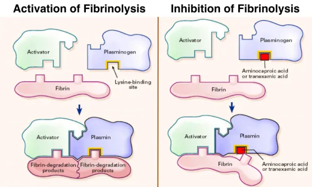
MODE D’ACTION DE L’ACIDE TRANEXAMIQUE, INTERET EN CHIRURGIE:
Une lésion tissulaire (comme la chirurgie) entraîne, en même temps que le saignement, l’activation du facteur tissulaire contenu dans les cellules endothéliales. L’activation du facteur tissulaire induit le déclenchement de la cascade de la coagulation à l’origine de la formation du caillot sanguin (enchevêtrement de plaquettes et de fibrine) qui permet de combler la brèche vasculaire.
Simultanément, afin de limiter cette réaction de coagulation, toute lésion tissulaire induit l’activation d’un processus de fibrinolyse qui vise à contenir l’extension du thrombus et à le dégrader pour maintenir la perméabilité des vaisseaux sanguins.
Cette réaction de fibrinolyse est déclenchée par la plasmine, enzyme protéolytique, qui va dégrader la trame de fibrine. L’ischémie vasculaire (induite par un garrot ou une plicature des vaisseaux), entraîne elle aussi une activation de la fibrinolyse, plus ou moins importante en fonction de la durée de cette ischémie.
Ceci permet de comprendre l’intérêt de l’acide tranexamique en chirurgie et les variations de son efficacité à réduire non seulement les pertes sanguines mais aussi la transfusion.
L’acide tranexamique est une molécule de synthèse dérivée des analogues de la lysine, il agit en bloquant de manière réversible les récepteurs à la lysine du plasminogène permettant, dans les conditions physiologiques normales, la fixation de ce dernier à la fibrine. (Figure 1) Ainsi il inhibe la formation d’un complexe ternaire plasminogène-fibrine-activateur (t-PA), retarde la fibrinolyse naturelle et donc la dégradation du caillot. Ceci est son principal mode d’action sur la diminution du saignement. Il a été également décrit que l’acide tranexamique pourrait avoir un effet plaquettaire et un effet anti-inflammatoire pouvant participer à la réduction du risque
Figure 1 : mécanisme d’action de l’acide tranexamique.5
PHARMACOCINETIQUE
L’acide tranexamique possède une demi vie d’élimination courte d’environ 3h. Après administration orale la biodisponibilité est de 30 à 50%. Le volume de distribution plasmatique est de 9 à 12L.
L’acide tranexamique n’est que très légèrement métabolisé par le foie. Il est éliminé à 90% par voie urinaire (excrétion glomérulaire sans réabsorption tubulaire). D’après la RCP, l’acide tranexamique ne doit pas être utilisé en cas d’insuffisance rénale « grave » et la posologie doit être adaptée à la créatininémie chez les patients présentant une insuffisance rénale légère à modérée. Selon le Vidal pour une créatininémie entre 120 et 249 µmol/l, la posologie recommandée est de 10 mg/kg deux fois par jour dans l’indication de l’accident hémorragique au lieu de 2 à 4 prises par jour dans les conditions de créatininémie < 120 µmol/l; pour des créatininémies entre 250 et 500 µmol/l, la posologie recommandée est de 10 mg/kg par jour, enfin au delà de 500 µmol/l la posologie recommandée est de 5 mg/kg par jour.
L’acide tranexamique se présente sous forme de comprimé ou de solution injectable. Il se présente sous différentes appellations dont la plus connue est Exacyl®.
explique l’absence de consensus sur les doses employées en orthopédie qui varient de 10 à 100 mg/kg selon les études.
SECURITE D’EMPLOI ET POPULATIONS PARTICULIERES
L’effet indésirable le plus fréquent lié à l’acide tranexamique est la survenue de nausées/vomissements lors d’une administration intraveineuse chez un patient éveillé. En obstétrique, l’acide tranexamique donné lors d’une hémorragie du post-partum a augmenté le
seuil des NVPO de 2 à 15% et des phosphènes de 2 à 9%.3
Les antifibrinolytiques ont longtemps été contre-indiqués chez des patients aux antécédents thrombotiques artériels ou veineux en raison d’un risque théorique d’aggravation des thromboses dû à l’effet antifibrinolytique. Cependant, dans les études CRASH 2 et WOMAN, incluant respectivement 20211 et 20060 patients, ainsi que dans les 129 essais randomisés en chirurgie incluant un total de 10488 patients, aucune augmentation des infarctus du myocarde, des AVC ou
des thromboses veineuses n’a été observée avec l’acide tranexamique.1,2,4 D’après le RCP, l’acide
tranexamique est contre-indiqué en cas de thrombose veineuse ou artérielle aigüe, il n’est plus contre indiqué en cas d’antécédent de thrombose.
L’aprotinine augmente le risque de dysfonction rénale de 33% par rapport aux patients qui ne
reçoivent pas d’antifibrinolytique.6 Cet effet n’est pas retrouvé avec l’acide tranexamique dans les
études citées précédemment.
L’effet indésirable principal est le risque neurologique qui se manifeste principalement par des
crises convulsives généralisées. Elles sont essentiellement rapportées en chirurgie cardiaque7 et le
risque convulsif est proportionnel à la dose d’acide tranexamique utilisée.8 Les doses élevées
(>2g/prise) doivent être évitées en chirurgie cardiaque ce d’autant qu’elles ne sont pas plus
efficaces que des doses inférieures.9 En cas de crises convulsives, il convient de prolonger la
sédation par du propofol ou d’administrer une benzodiazépine chez un patient ne pouvant être anesthésié. L’augmentation du risque convulsif par l’acide tranexamique chez un patient aux antécédents convulsifs n’est pas connue. Cependant l’acide tranexamique est contre-indiqué chez ces patients dans le résumé des caractéristiques du produit.
Une neurotoxicité a également été décrite chez des patients en insuffisance rénale terminale ayant
eu des doses répétées d’acide tranexamique.10 Comme il s’agit d’un produit à élimination rénale,
RCP, l’acide tranexamique ne doit pas être utilisé en cas d’insuffisance rénale « grave » (sans précision d’une valeur seuil). Il n’y a pas d’étude pharmacocinétique réalisée avec l’acide tranexamique chez l’insuffisant rénal sévère. Les suggestions qui peuvent être avancées sont d’utiliser pour la première administration la dose minimale efficace, de réinjecter seulement si cela est nécessaire et de réduire la dose des réinjections proportionnellement à la diminution de la
filtration glomérulaire.11
ACIDE TRANEXAMIQUE ET CHIRURGIE ORTHOPEDIQUE
Depuis les années 50, de nombreux essais cliniques rapportés dans la littérature et menés sur de petits effectifs de prothèses de hanche ou de genou, ont montré l’efficacité de l’acide tranexamique sur la réduction du risque de transfusion de culot globulaire et sur la réduction des pertes sanguines.
Par la suite, la méta analyse de la Cochrane publiée en 2011 a confirmé l’efficacité des antifibrinolytiques pour la réduction de la transfusion érythrocytaire dans de nombreuses
indications chirurgicales (chirurgie orthopédique, cardiaque, hépatique, urologique et vasculaire).9
La Société Américaine d’Anesthésie (ASA)12 ainsi que la Société Française d’Anesthésie et de
Réanimation (SFAR)13 et la Haute Autorité de Santé (HAS) l’ont intégré dans leurs
recommandations de bonne pratique pour les chirurgies à risque hémorragique.14
A l’heure actuelle il persiste des interrogations quant à la dose et la durée d’administration pour obtenir une efficacité maximale.
REFERENCES
1. CRASH-2 trial collaborators, Shakur H, Roberts I, Bautista R, Caballero J, Coats T, Dewan Y,
El-Sayed H, Gogichaishvili T, Gupta S, Herrera J, Hunt B, Iribhogbe P, Izurieta M, Khamis H, Komolafe E, Marrero M-A, Mejía-Mantilla J, Miranda J, Morales C, Olaomi O, Olldashi F, Perel P, Peto R, Ramana PV, Ravi RR, Yutthakasemsunt S: Effects of tranexamic acid on death, vascular occlusive events, and blood transfusion in trauma patients with significant haemorrhage (CRASH-2): a randomised, placebo-controlled trial. Lancet Lond Engl 2010; 376:23–32
2. Ker K, Edwards P, Perel P, Shakur H, Roberts I: Effect of tranexamic acid on surgical
bleeding: systematic review and cumulative meta-analysis. BMJ 2012; 344:e3054
3. Ducloy-Bouthors A-S, Jude B, Duhamel A, Broisin F, Huissoud C, Keita-Meyer H, Mandelbrot
4. WOMAN Trial Collaborators: Effect of early tranexamic acid administration on mortality, hysterectomy, and other morbidities in women with post-partum haemorrhage (WOMAN): an international, randomised, double-blind, placebo-controlled trial. Lancet Lond Engl 2017; 389:2105–16
5. Mannucci PM: Hemostatic drugs. N Engl J Med 1998; 339:245–53
6. Hutton B, Joseph L, Fergusson D, Mazer CD, Shapiro S, Tinmouth A: Risks of harms using
antifibrinolytics in cardiac surgery: systematic review and network meta-analysis of randomised and observational studies. BMJ 2012; 345:e5798
7. Lecker I, Wang D-S, Whissell PD, Avramescu S, Mazer CD, Orser BA: Tranexamic
acid-associated seizures: Causes and treatment. Ann Neurol 2016; 79:18–26
8. Lin Z, Xiaoyi Z: Tranexamic acid-associated seizures: A meta-analysis. Seizure 2016; 36:70–3
9. Henry DA, Carless PA, Moxey AJ, O’Connell D, Stokes BJ, Fergusson DA, Ker K:
Anti-fibrinolytic use for minimising perioperative allogeneic blood transfusion. Cochrane Database Syst Rev 2011:CD001886 doi:10.1002/14651858.CD001886.pub4
10. Ma TK-W, Chow KM, Kwan BC-H, Leung CB, Szeto CC, Li PK-T: Manifestation of tranexamic
acid toxicity in chronic kidney disease and kidney transplant patients: A report of four cases and review of literature. Nephrol Carlton Vic 2017; 22:316–21
11. Yang QJ, Jerath A, Bies RR, Wąsowicz M, Pang KS: Pharmacokinetic modeling of tranexamic
acid for patients undergoing cardiac surgery with normal renal function and model simulations for patients with renal impairment. Biopharm Drug Dispos 2015; 36:294–307
12. American Society of Anesthesiologists Task Force on Perioperative Blood Transfusion and
Adjuvant Therapies: Practice guidelines for perioperative blood transfusion and adjuvant therapies: an updated report by the American Society of Anesthesiologists Task Force on Perioperative Blood Transfusion and Adjuvant Therapies. Anesthesiology 2006; 105:198– 208
13. Duranteau J, Asehnoune K, Pierre S, Ozier Y, Leone M, Lefrant J-Y: Guidelines on the
treatment of hemorrhagic shock 2015:62–74
14. Haute Autorité de Santé: Transfusions de globules rouges homologues : produits,
DEUXIEME PARTIE:
BACKGROUND
Perioperative blood management in major orthopedic surgery is relevant in front of higher
morbidity and mortality 1 linked to postoperative anemia and allogeneic blood transfusion.2,3
Among interventions developed to reduce allogeneic blood transfusion including preoperative
autologous blood donation,4 recombinant human erythropoietin protocol,5 perioperative cell
salvage blood device 6 and surgical measures with use of tourniquet, electro cautery or drain
clamping,7 pharmacological agents such as tranexamic acid (TXA) have been investigated to reduce
perioperative blood loss 8. By interfering with fibrinolysis, TXA is a synthetic lysine analogue,
blocking the lysine binding sites on plasminogen molecules, inhibiting the formation of plasmin and
therefore inhibiting fibrinolysis 8.
Two previous studies 1,9 analyzed TXA compared to placebo for blood transfusion and blood loss
reduction in perioperative time. Although positive results about TXA efficacy, these two studies
present an extent of heterogeneity (due to inclusion of all surgeries 9 or general orthopedic
surgery) 1 fail to draw overall conclusions in lower limb arthroplasty.
Moreover, numerous small recent studies evaluate use of TXA in total hip arthroplasty (THA) or total knee arthroplasty (TKA) without determine optimal protocol of TXA administration.
In order to clarify the optimal TXA dosing regimen, we conducted a meta-analysis, including studies from 1950 to 2017, examining efficacy, administration regimen and safety of TXA in patients undergoing total hip and total knee arthroplasty.
MATERIALS ET METHODS
Criteria for considering studies for this review
Types of studies
Randomized controlled trials (RCTs) with a concurrent control group, or randomized head-to-head comparative trials.
Types of participants
The study participants were adults (over 18 years) and undergoing total hip arthroplasty or total knee arthroplasty.
Types of outcome measures
Primary outcomes
• The proportion of patients who was transfused with allogeneic blood, autologous blood, or with both;
Secondary outcomes
• Peri-operative blood loss
• Post-operative complications (myocardial infarction, stroke, deep vein thrombosis, pulmonary embolism, any thrombosis, renal failure),
Search methods for identification of studies
Literature Search
An exhaustive literature search computer-assisted in MEDLINE was conducted to identify all randomized studies comparing intravenous tranexamic acid with placebo in total hip arthroplasty and total knee arthroplasty at the end of July 2017. Only key words “tranexamic acid”, “total hip arthroplasty” and “total knee arthroplasty” were used. Only English or French articles were included. All identified articles (from title and abstract) about tranexamic acid in total hip arthroplasty or total knee arthroplasty were analyzed. Furthermore, reference lists in reviews, studies and previous meta-analyses were checked.
Study Selection
Among identified articles about TXA in scheduled surgeries, only controlled studies with a double-blind randomization and a presence of a transfusion protocol were considered. Studies not controlled (no placebo given) and/or unclear randomization or in pediatric patients were excluded. Efficacy and safety of the tranexamic acid needed to be evaluated in screened studies.
For efficacy of TXA, proportion of patients receiving at least one unit of allogeneic/autologous erythrocyte transfusion according to a transfusion protocol, and total volume of blood loss in milliliters (intra-operative and postoperative blood loss) are considered.
To limit evaluation bias on transfusion criteria, screened studies had to have a transfusion protocol including a transfusion threshold of allogeneic/autologous erythrocytes with an adjusted statistical analysis of preoperative hemoglobin between tranexamic acid group and control group. Trials with transfusion protocol missing or not mentioned were excluded. Finally, transfusion protocol could differ between studies.
For safety of TXA, venous thromboembolism event (confirmed by ultrasound or venography for deep venous thrombosis, ventilation-perfusion lung scanning or pulmonary angiography or spiral computed tomography for pulmonary embolism), or arterial thrombosis (myocardial infarction, stroke or limb ischemia) are considered.
Data Extraction
At least two authors (SP, CA) independently extracted study characteristics and outcomes using an article extraction form. The extraction form was used to record information regarding randomization criteria, methodology descriptions, presence of a transfusion protocol, type of surgery involved, treatment outcomes and adverse events.
Data on the following outcomes were recorded on the data extraction form: number of patients receiving any transfusion (allogeneic blood, autologous blood, or both), number of patients experiencing post-operative complications (thrombosis, myocardial infarction, renal failure), and mortality. Data were also recorded on total, intraoperative and postoperative blood loss. Information regarding type of surgery, tranexamic acid dose range, tranexamic acid use protocol, preoperative hemoglobin, transfusion thresholds, preoperative autologous blood donation, cell salvage blood were also recorded.
If more than one group was treated, treatment group with smaller dose of TXA was chosen.
Statistical Analysis
Publication bias was checked using funnel plot technique.
For dichotomous outcomes such as the proportion of patients transfused or having postoperative venous thromboembolism, meta-analyses were performed using a logarithm of relative risk (RR). The RR in intervention group was compared to control group and corresponding 95% confidence intervals (CI) were calculated for each trial using the random effects model. Relative risk reduction (RRR) is calculated from formula “1-RR” and means how much TXA reduces the risk of allogeneic blood transfusion relative to the control group who did not receive TXA. RRR is expressed in percentage. Association and heterogeneity test were performed for each meta-analysis. A value of p ≤ 0.05 in an association test was considered to be statistically significant proving an effect of tranexamic acid. The presence of heterogeneity of treatment effect was assessed using the Q statistic, which has an approximate chi-square distribution with degrees of freedom equal to the number of studies minus one. A P-value less than or equal to 0.1 was used to define statistically
significant heterogeneity. Statistical heterogeneity was also assessed using the I2 test. The I2 test
describes the percentage of total variation across studies due to heterogeneity rather than chance. A value of 0% indicates no observed heterogeneity and larger values show increasing
heterogeneity. Substantial heterogeneity is considered to exist when I2 > 50%.
were collected for each trial. When confidence intervals were reported rather SD, the CI was transformed to SD using formula:
SD = !"
!! !
× n.
Like dichotomous outcomes, the standardized MD and the corresponding 95% CI, were calculated for each trial using the random effects model. Association and heterogeneity test were performed for each meta-analysis. A value of p ≤ 0.05 in an association test were considered to be statistically significant and to occur an effect of tranexamic acid. The presence of heterogeneity of treatment
effect was defined using the Q statistic with p-value less than or equal to 0.1 and using the I2 test
with substantial heterogeneity is considered to exist when I2 > 50%.
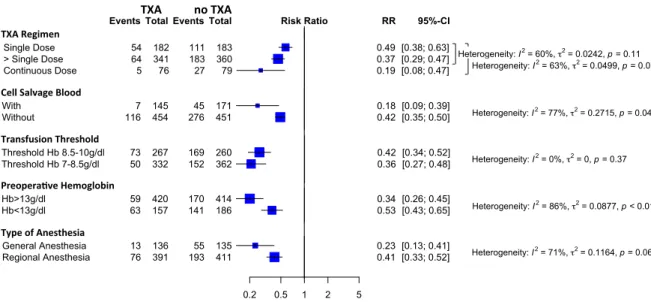
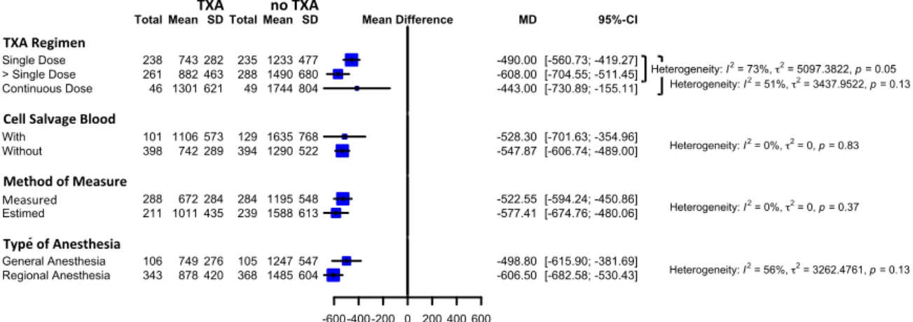
Analysis of subgroups was performed to determine whether effect sizes varied according factors, to explain heterogeneity, such as:
• Tranexamic acid dose regimens • Blood cell salvage use
• Transfusion thresholds • Preoperative hemoglobin
• The way to measure or estimate the blood loss • Type of anesthesia.
All analyses were performed using R-project software, package meta/metafor/rmeta.
RESULTS
Description of studies
For THA, the literature search, “tranexamic acid” AND “total hip arthroplasty”, identified 113 studies. Only 17 trials remained potentially relevant after reading through title and abstract. Articles were excluded for several reasons: uncertainly randomization, lack of control group, tranexamic acid not administrated intravenously. Among these 17 trials, 6 studies were withdrawn
because trial was not randomized,11 transfusion protocol was not applied,12,13,14 hemoglobin
preoperative was not analyzed between TXA group and control group,15 article was not available.16
For blood loss analysis, 1 study was withdrawn because data were presented as median with lower
and upper quartiles.17
For TKA, the literature search, “tranexamic acid” AND “total knee arthroplasty”, identified 262 studies. Only 23 trials remained potentially relevant after reading through title and abstract. Articles were excluded for several reasons: uncertainly randomization, lack of control group, tranexamic acid not administrated intravenously. Among these 23 trials, 3 studies were withdrawn
because trial was not clearly randomized,18 hemoglobin preoperative was not analyzed between
TXA group and control group,19 data were not available.20 For blood loss analysis, 5 studies were
withdrawn because data were presented as median with lower and upper quartiles,21,22,23,24,25 and
1 study was withdrawn because data were not available.26 Figure 1 presents the PRISMA flow
chart10 of our meta-analysis.
Study design
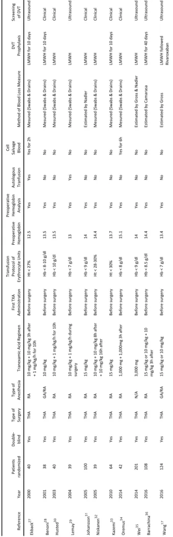
The tables 1 and 2 show the design of the included studies. Total Hip Arthroplasty (table 1).
Eleven studies using tranexamic acid in total hip arthroplasty were selected comprising a total of
830 randomized patients. 17,27,28,29,30,31,32,33,34,35,36 Trials were conducted in many countries including:
Sweden (n = 3), Finland (n = 1), Denmark (n = 1), Spain (n = 1), Croatia (n = 1), Iran (n = 1), Canada (n = 1), China (n = 2). Trials were small (38 to 62 patients) and only 4 trials randomized more than
100 patients. 17,31,35,36 In each study, total dose of tranexamic acid was variable : first TXA dose was
ranged from 10 to 15 mg/kg intravenous associated or not with second or third TXA dose three hours after or associated with continuous administration ranged from 1 to 10 mg/kg/h during surgery or for 10 h. In all cases, first TXA dose was delivered just before the surgery beginning (or before skin incision). The transfusion trigger threshold differed between trials. Only 2 studies used
partially autologous blood transfusion 27,29 and only 2 studies used currently a cell salvage blood
device.27,34 Prophylaxis of deep venous thrombosis used Low Molecular Weight Heparin (LMWH)
17,27,29,35,36 Ar#cles iden#fied through Pubmed using the keywords: “Total hip arthroplasty” AND “tranexamic acid” n = 113 Titles and abstracts screened n = 113 Full-text of the arEcles assessed for the eligibility n = 17 Ar#cles included in the meta-analysis n = 11 Ar#cles excluded n = 96 Uncertainly randomizaEon Lack of control group Other route for TXA than IV Ar#cles excluded n = 6 1 not randomized 3 no transfusion protocol 1 no preoperaEve hemoglobin 1 not available Ar#cles iden#fied through Pubmed using the keywords: “Total knee arthroplasty” AND “tranexamic acid” n = 262 Titles and abstracts screened n = 262 Full-text of the arEcles assessed for the eligibility n = 23 Ar#cles included in the meta-analysis n = 20 Ar#cles excluded n = 239 Uncertainly randomizaEon Lack of control group Other route for TXA than IV Ar#cles excluded n = 3 1 not randomized 1 no preoperaEve hemoglobin 1 not available Fig. 1: PRISMA flow chart of meta-analysis Id en % fic a% on Sc re ening Eligibility In clu d ed
Re fe re nc e Ye ar Pati en ts ran do m ize d Double -blind Ty pe of Sur ge ry Ty pe of An es th es ia Tr an ex am ic A ci d Re gi m en Fi rs t T XA Ad m in is tr ati on Tr an sf us io n Pr oto co l o f Er yth ro cy te U ni ts Pr eo pe rati ve He m og lo bi n Pr eo pe rati ve He m og lo bi n An al ys is Au to lo go us Tr an fu si on Ce ll Sal vag e Bl ood Me th od o f B lo od L os s Me as ur e DVT Pr op hy lax is Sc re ening of DVT Ek bac k 27 2000 40 Ye s THA RA 10 m g/ kg + 10 m g/ kg 3 h af te r + 1 m g/ kg /h fo r 1 0h Before su rgery Ht < 27 % 12. 5 Ye s Ye s Ye s for 2 h Me su re d (S w ab s & D rai ns ) LMW H fo r 1 0 day s U ltr as ou nd Ben on i 28 2001 38 Ye s THA G A/ RA 10 mg/kg Before su rgery Hb < 8-10 g /d l 13. 5 Ye s No No Me su re d (S w ab s & D rai ns ) LMW H fo r 1 0 day s Cl in ic al Hu ste d 30 2003 40 Ye s THA RA 10 m g/ kg + 1 m g/ kg /h fo r 1 0h Before su rgery Hb < 10 g /d l 13. 5 Ye s No No Me su re d (S w ab s & D rai ns ) LMW H Cl in ic al Le m ay 29 2004 39 Ye s THA RA 10 m g/ kg + 1 m g/ kg /h d ur in g sur ge ry Before su rgery Hb < 7 g/ dl 13 Ye s Ye s No Me su re d (S w ab s & D rai ns ) LMW H U ltr as ou nd Jo han ss on 31 2005 100 Ye s THA RA 15 mg/kg Before su rgery Hb < 9 g/ dl 14 Ye s No No Es tim ate d by N ad le r LMW H Cl in ic al N is kan en 32 2005 39 Ye s THA RA 10 m g/ kg + 10 m g/ kg 8 h af te r + 10 m g/ kg 1 6h af te r Before su rgery Ht < 28 -3 0% 14. 4 Ye s No No Me su re d (S w ab s & D rai ns ) LMW H Cl in ic al Kaze m i 33 2010 64 Ye s THA RA 15 mg/kg Before su rgery Ht < 30 % 13. 7 Ye s No No Me su re d (S w ab s & D rai ns ) LMW H fo r 1 0 day s Cl in ic al Or emus 34 2014 42 Ye s THA RA 1, 00 0 m g + 1, 00 0m g 3h af te r Before su rgery Hb < 8 g/ dl 15. 1 Ye s No Ye s for 6 h Me su re d (S w ab s & D rai ns ) LMW H Cl in ic al Wei 35 2014 201 Ye s THA N/ A 3,000 mg Before su rgery Hb < 9 g/ dl 14 Ye s No No Es tim ate d by G ro ss & N ad le r LMW H U ltr as ou nd Bar rac hi na 36 2016 108 Ye s THA RA 15 m g/ kg o r 1 0 m g/ kg + 10 m g/ kg 3 h af te r Before su rgery Hb < 8. 5 g/ dl 14. 4 Ye s No No Es tim ate d by C am ar as a LMW H fo r 4 0 day s U ltr as ou nd W an g 17 2016 124 Ye s THA G A/ RA 15 mg/kg o r 10 mg/kg Before su rgery Hb < 7 g/ dl 13. 4 Ye s No No Es tim ate d by G ro ss LMW H fo llo w ed Ri var ox ab an U ltr as ou nd
Ta
bl
e
1.
C
ha
ra
ct
er
is
0c
s
an
d
O
ut
co
m
es
in
S
el
ec
te
d
St
ud
ie
s
in
T
ot
al
H
ip
Ar
th
ro
pl
as
ty
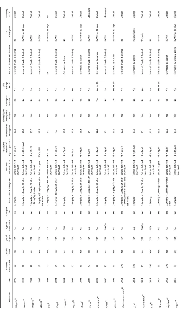
Total Knee Arthroplasty (table 2).
Twenty studies using tranexamic acid in total knee arthroplasty were selected comprising a total of
1221 randomized patients.21,22,23,24,25,26,34,37,38,39,40,41,42,43,44,45,46,47,48,49 Trials were conducted in many
countries including: Sweden (n = 2), Finland (n = 2), Denmark (n = 1), Belgium (n = 1), Germany (n = 1), Spain (n = 3), United Kingdom (n = 1), Croatia (n = 1), Greece (n = 1), Israel (n = 2), Dubai (n = 1), Japan (n = 1), Taiwan (n = 1), Thailand (n = 1), South Korea (n = 1). Trials were small and only 6 trials
randomized more than 95 patients.45,46,47,48 All studies used pneumatic tourniquet during surgery.
In each study, total dose of tranexamic acid was variable: first TXA dose was ranged from 10 to 20 mg/kg intravenous associated with second or third TXA dose three hours after or associated with continuous administration ranged from 1 to 10 mg/kg/h for 6 h or 12 h. Only 2 studies
administered tranexamic acid during several days.40,46 In all case, first TXA dose was delivered just
before the deflation of tourniquet, excluding 2 studies in which tranexamic acid was administered
before the surgery. 34,40 The transfusion trigger threshold differed between trials. Only 2 studies
used partially autologous blood transfusion 42,43 and only 5 studies used currently a blood cell
salvage device.25,34,42,43,44 Prophylaxis of deep venous thrombosis (when described) used Low
Molecular Weight Heparin (LMWH) excluding 2 studies using Warfarin44 or Indomethacin45. Venous
thrombosis screening was done by ultrasound in only 2 studies.26,41 Type of anesthesia varied
Re fe re nc e Ye ar Pati en ts ran do m ize d Double -blind Ty pe of Sur ge ry Ty pe of An es th es ia To ur ni qu et Us e Tr an ex am ic A ci d Re gi m en Fi rs t T XA Ad m in is tr ati on Tr an sf us io n Pr oto co l o f Er yth ro cy te U ni ts Pr eo pe rati ve He m og lo bi n Pr eo pe rati ve He m og lo bi n An al ys is Au to lo go us Tr an fu si on Ce ll Sal vag e Bl ood Me th od o f B lo od L os s Me as ur e DVT Pr op hy lax is Sc re ening of DVT Hi ip pal a 37 1995 28 Ye s TK A RA Ye s 15 mg/kg Be fo re d ef late d to ur ni qu et Hb < 10 g /d l 13. 2 Ye s No No Me su re d (S w ab s & D rai ns ) No Cl in ic al Ben on i 38 1996 86 Ye s TK A RA Ye s 10 m g/ kg + 10 m g/ kg 3 h af te r Be fo re d ef late d to ur ni qu et Hb < 8. 5-10 g /d l 13. 6 Ye s No No Me su re d (S w ab s & D rai ns ) LMW H fo r 1 0 day s Cl in ic al Hi ip pal a 39 1997 77 Ye s TK A RA Ye s 15 m g/ kg + 10 m g/ kg 3 h af te r + 10 m g/ kg 6 h af te r Be fo re d ef late d to ur ni qu et Hb < 10 g /d l 13. 6 Ye s No No Me su re d (S w ab s & D rai ns ) LMW H Cl in ic al Jan se n 40 1999 42 Ye s TK A GA Ye s 15 m g/ kg +1 5 m g/ kg e ve ry 8 h fo r 3 d ay s Before su rgery PC V < 26 % 13. 2 Ye s No No Me su re d (S w ab s & D rai ns ) LMW H Cl in ic al Ellis 24 2001 20 Ye s TK A GA Ye s 15 m g/ kg + 10 m g/ kg /h fo r 1 2h Be fo re d ef late d to ur ni qu et Ht < 27 % NA Ye s No No NA LMW H fo r 3 5 day s No Eng el 22 2001 24 Ye s TK A RA Ye s 15 m g/ kg + 10 m g/ kg 3 h af te r Be fo re d ef late d to ur ni qu et Hb < 10 g /d l NA Ye s No No Me su re d (S w ab s & D rai ns ) LMW H Cl in ic al Tan ak a 23 2001 48 Ye s TK A N/ A Ye s 20 mg/kg Be fo re d ef late d to ur ni qu et Hb < 7 g/ dl 11. 7 Ye s No No Es tim ate d by G ro ss NA Cl in ic al Ve ie n 25 2002 30 Ye s TK A RA Ye s 10 m g/ kg + 10 m g/ kg 3 h af te r Be fo re d ef late d to ur ni qu et Ht < 28 % 13. 4 Ye s No Ye s Me su re d (S w ab s & D rai ns ) LMW H Cl in ic al Good 21 2003 51 Ye s TK A RA Ye s 10 m g/ kg + 10 m g/ kg 3 h af te r Be fo re d ef late d to ur ni qu et Hb < 9 g/ dl 13. 8 Ye s No No Es tim ate d by N ad le r LMW H fo r 1 0 day s Cl in ic al Zo har 26 2004 40 Ye s TK A GA Ye s 15 m g/ kg + 10 m g/ kg /h fo r 1 2h Be fo re d ef late d to ur ni qu et Ht < 28 % 14 Ye s No No Me su re d (S w ab s & D rai ns ) LMW H U ltr as ou nd Cam ar as a 42 2006 95 Ye s TK A RA Ye s 10 m g/ kg + 10 m g/ kg 3 h af te r Be fo re d ef late d to ur ni qu et Hb < 8 g/ dl 12. 6 Ye s Ye s Ye s for 5 h Es tim ate d by C am ar as a LMW H fo r 4 0 day s Cl in ic al Or pe n 41 2006 29 Ye s TK A G A+RA Ye s 15 mg/kg Be fo re d ef late d to ur ni qu et Hb < 9 g/ dl 13 Ye s No No Me su re d (S w ab s & D rai ns ) LMW H U ltr as ou nd Al var ez 43 2008 95 Ye s TK A RA Ye s 10 m g/ kg + 1 m g/ kg /h fo r 6 h Be fo re d ef late d to ur ni qu et Hb < 8 g/ dl 13. 5 Ye s Ye s Ye s for 6 h Es tim ate d by B re ch er LMW H fo r 3 0 day s Cl in ic al Ch ar oe nc ho lv an ic h 46 2011 100 Ye s TK A RA Ye s 10 m g/ kg + 10 m g/ kg 3 h af te r + 50 0m g or al th re e tim es d ai ly fo r 5 d ay s Be fo re d ef late d to ur ni qu et Hb < 10 g /d l 12. 5 Ye s No No Me su re d (S w ab s & D rai ns ) No Cl in ic al Lin 45 2011 100 Ye s TK A GA Ye s 10 mg/kg Be fo re d ef late d to ur ni qu et Hb < 8. 5 g/ dl 13. 3 Ye s No No Es tim ate d by N ad le r In do m eth ac in Cl in ic al Mac G ill iv ray 44 2011 40 Ye s TK A G A+RA Ye s 10 m g/ kg + 10 m g/ kg 3 h af te r Be fo re d ef late d to ur ni qu et Hb < 8 g/ dl 13 Ye s No Ye s Me su re d (S w ab s & D rai ns ) W ar far in Cl in ic al Se o 47 2013 100 Ye s TK A N/ A Ye s 1,500 mg Be fo re d ef late d to ur ni qu et Hb < 8 g/ dl 11. 4 Ye s No No Me su re d (S w ab s & D rai ns ) NA Cl in ic al Or emus 34 2014 56 Ye s TK A RA Ye s 1, 00 0 m g + 1, 00 0m g 3h af te r Before su rgery Hb < 8 g/ dl 15. 1 Ye s No Ye s for 6 h Me su re d (S w ab s & D rai ns ) LMW H Cl in ic al Agu ilera 48 2015 100 Ye s TK A RA Ye s 1, 00 0 m g + 1, 00 0m g 60 -9 0m in af te r Be fo re d ef late d to ur ni qu et Hb < 8 g/ dl 13. 5 Ye s No No Es tim ate d by N ad le r LMW H Cl in ic al D ig as 49 2015 60 Ye s TK A RA Ye s 15 mg/kg Be fo re d ef late d to ur ni qu et Hb < 8. 5 g/ dl 13. 2 Ye s No No Es tim ate d by G ro ss & N ad le r LMW H fo r 3 0 day s Cl in ic al
Ta
bl
e
2.
C
ha
ra
ct
er
is
0c
s
an
d
O
ut
co
m
es
in
S
el
ec
te
d
St
ud
ie
s
in
T
ot
al
K
ne
e
Ar
th
ro
pl
as
ty
Total Hip Arthroplasty (THA)
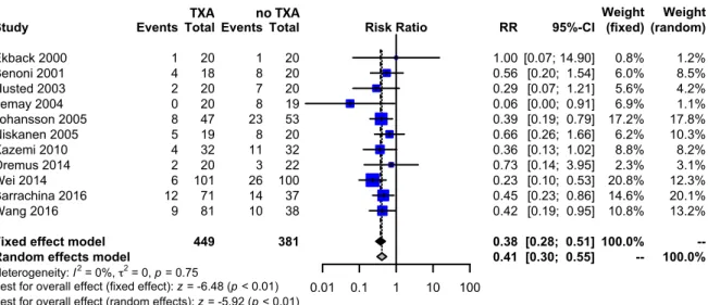
For exposure to allogeneic/autologous blood transfusion according to a transfusion protocol, 11 trials of TXA versus control involved total hip arthroplasty. These trials included a total of 830 patients, of whom 449 were randomized to TXA, and 381 were randomized to a control group who did not receive TXA. There was a significant 59% relative reduction in the rate of exposure to allogeneic blood transfusion in those patients with TXA (RR 0.41, 95% CI 0.30 to 0.55).
Heterogeneity between these trials was not statistically significant (Chi2 = 6.70, df = 10, p = 0.7530;
I2 = 0%). (Figure 2)
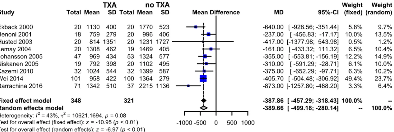
Nine trials of TXA versus control involving total hip arthroplasty reported data on the total volume of blood loss. These trials included a total of 669 patients, of whom 348 were randomized to TXA, and 321 were randomized to a control group who did not receive TXA. The use of TXA in THA reduced total volume of blood loss by around 390 mls per patient (MD -389.66 mls, 95% CI -499.18
to -280.14 mls). Heterogeneity between these trials was statistically significant (Chi2 = 14.05, df = 8,
2
Study
Fixed effect model Random effects model
Heterogeneity: I2 = 0%, τ2 = 0, p = 0.75
Test for overall effect (fixed effect): z = -6.48 (p < 0.01) Test for overall effect (random effects): z = -5.92 (p < 0.01)
Ekback 2000 Benoni 2001 Husted 2003 Lemay 2004 Johansson 2005 Niskanen 2005 Kazemi 2010 Oremus 2014 Wei 2014 Barrachina 2016 Wang 2016 Events 1 4 2 0 8 5 4 2 6 12 9 Total 449 20 18 20 20 47 19 32 20 101 71 81 Experimental Events 1 8 7 8 23 8 11 3 26 14 10 Total 381 20 20 20 19 53 20 32 22 100 37 38 Control 0.01 0.1 1 10 100 Risk Ratio RR 0.38 0.41 1.00 0.56 0.29 0.06 0.39 0.66 0.36 0.73 0.23 0.45 0.42 95%-CI [0.28; 0.51] [0.30; 0.55] [0.07; 14.90] [0.20; 1.54] [0.07; 1.21] [0.00; 0.91] [0.19; 0.79] [0.26; 1.66] [0.13; 1.02] [0.14; 3.95] [0.10; 0.53] [0.23; 0.86] [0.19; 0.95] (fixed) 100.0% --0.8% 6.0% 5.6% 6.9% 17.2% 6.2% 8.8% 2.3% 20.8% 14.6% 10.8% Weight (random) --100.0% 1.2% 8.5% 4.2% 1.1% 17.8% 10.3% 8.2% 3.1% 12.3% 20.1% 13.2% Weight Fig. 2. Meta-analysis of use of tranexamic acid compared with placebo in total hip arthroplasty (THA) on the proporAon of paAent requiring allogenic/autologous transfusion according to a transfusion protocol. The relaAve raAo (RR) for individual studies is represented as a square within a bar represenAng the 95% confidence interval (CI). Symbol size is proporAonal to the study weight. The RR for summaries is represented as a diamond. The length of the diamond corresponds to the 95% CI. An RR < 1 indicated that the tranexamic acid is more effecAve than placebo. If the value of 1 is included within 95% CI, the result is not significant.
TXA
Funnel plots did not suggest that low-quality studies and/or publication bias (absence of studies showing no significant beneficial effect) exaggerated the magnitude of estimated treatment effect, confirmed by trim and fill method (no added studies). (Figure 4)
Fig. 3. Meta-analysis of use of tranexamic acid compared with placebo in total hip arthroplasty (THA) on the total volume of blood loss in milliliters of paBent. The standardized mean difference (MD) for individual studies is represented as a square within a bar represenBng the 95% confidence interval (CI). Symbol size is proporBonal to the study weight. The MD for summaries is represented as a diamond. The length of the diamond corresponds to the 95% CI. An MD < 0 indicated that the tranexamic acid is more effecBve than placebo. If the value of 0 is included within 95% CI, the result is not significant. Study
Fixed effect model Random effects model
Heterogeneity: I2 = 43%, τ2 = 10621.1694, p = 0.08
Test for overall effect (fixed effect): z = -10.95 (p < 0.01) Test for overall effect (random effects): z = -6.97 (p < 0.01)
Ekback 2000 Benoni 2001 Husted 2003 Lemay 2004 Johansson 2005 Niskanen 2005 Kazemi 2010 Wei 2014 Barrachina 2016 Total 348 20 18 20 20 47 19 32 101 71 Mean 1130 759 814 1308 969 792 1024 958 1342 SD 400 279 1351 462 434 398 544 422 510 Experimental Total 321 20 20 20 19 53 20 32 100 37 Mean 1770 996 1231 1469 1324 1102 1399 1364 2215 SD 523 406 1727 405 577 495 587 279 1136 Control -1000 -500 0 500 1000 Mean Difference MD -387.86 -389.66 -640.00 -237.00 -417.00 -161.00 -355.00 -310.00 -375.00 -405.70 -873.00 95%-CI [ -457.29; -318.43] [ -499.18; -280.14] [ -928.56; -351.44] [ -456.83; -17.17] [-1377.98; 543.98] [ -433.32; 111.32] [ -553.81; -156.19] [ -591.29; -28.71] [ -652.29; -97.71] [ -504.48; -306.92] [-1257.80; -488.20] (fixed) 100.0% --5.8% 10.0% 0.5% 6.5% 12.2% 6.1% 6.3% 49.4% 3.3% Weight (random) --100.0% 9.7% 13.5% 1.2% 10.4% 14.9% 10.0% 10.2% 23.7% 6.4% Weight TXA no TXA 0.05 0.20 0.50 2.00 5.00 1.4 1.2 1.0 0.8 0.6 0.4 0.2 0.0 Risk Ratio St an da rd Erro r 0.05 0.20 0.50 2.00 5.00 1.4 1.2 1.0 0.8 0.6 0.4 0.2 0.0 Risk Ratio St an da rd Erro r Fig. 4. Funnel plot of use of tranexamic acid compared with placebo in THA, upper on the propor=on of pa=ent requiring allogenic/ autologous transfusion according to a transfusion protocol, and bo@om on the total volume of blood loss in milliliters of pa=ent. The leB funnel plots are a sca@er plot of the treatment effect es=mated from individual studies (logarithm of RR or MD, horizontal axis) against a measure of study size (SE of logarithm of RR or MD, ver=cal axis). Dashed line is pooled RR or MD. In the absence of publica=on bias, studies are symmetrically distributed around the pooled RR or MD, and the plot resembles a symmetric inverted funnel. Asymmetric funnel plots may indicate publica=on bias or be due to exaggera=on of treatment effects in small studies of low quality. The right funnel plots represented funnel plot by the trim and fill method. This method es=mate the number of studies missing from a meta-analysis due to the suppression of the most extreme results on one side of the funnel plot. The method then augments the observed data so that the funnel plot is more symmetric. -1000 -500 0 500 500 400 300 200 100 0 Mean Difference St an da rd Erro r -1000 -500 0 500 500 400 300 200 100 0 Mean Difference St an da rd Erro r
Total Knee Arthroplasty (TKA)
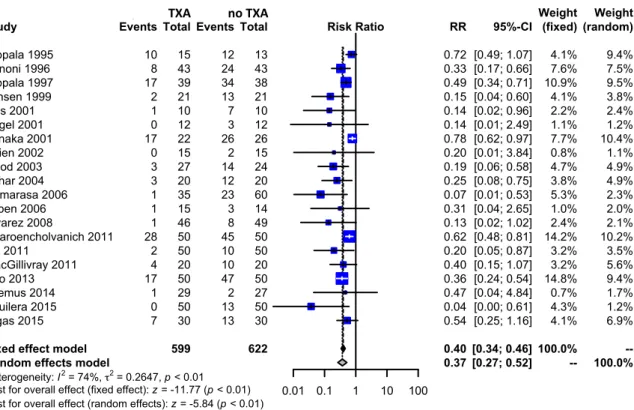
For exposure to allogeneic/autologous blood transfusion according to a transfusion protocol, twenty trials of TXA versus control involved total knee arthroplasty. These trials included a total of 1221 patients, of whom 599 were randomized to TXA, and 622 were randomized to a control group who did not receive TXA. There was a significant 63% relative reduction in the rate of exposure to allogeneic blood transfusion in those patients with TXA (RR 0.37, 95% CI 0.27 to 0.52).
Heterogeneity between these trials was statistically significant (Chi2 = 72.08, df = 19, p < 0.0001; I2
= 74%). (Figure 5)
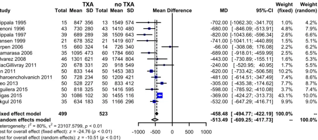
Fourteen trials of TXA versus control involving total knee arthroplasty reported data on the total volume of blood loss. These trials included a total of 1022 patients, of whom 499 were randomized to TXA, and 523 were randomized to a control group who did not receive TXA. The use of TXA in THA reduced total volume of blood loss by around 513 mls per patient (MD 513.49 mls, 95% CI
-609.25 to -417.73 mls). Heterogeneity between these trials was statistically significant (Chi2 =
2
Study
Fixed effect model Random effects model
Heterogeneity: I2 = 74%, τ2 = 0.2647, p < 0.01
Test for overall effect (fixed effect): z = -11.77 (p < 0.01) Test for overall effect (random effects): z = -5.84 (p < 0.01)
Hippala 1995 Benoni 1996 Hippala 1997 Jansen 1999 Ellis 2001 Engel 2001 Tanaka 2001 Veien 2002 Good 2003 Zohar 2004 Camarasa 2006 Orpen 2006 Alvarez 2008 Charoencholvanich 2011 Lin 2011 MacGillivray 2011 Seo 2013 Oremus 2014 Aguilera 2015 Digas 2015 Events 10 8 17 2 1 0 17 0 3 3 1 1 1 28 2 4 17 1 0 7 Total 599 15 43 39 21 10 12 22 15 27 20 35 15 46 50 50 20 50 29 50 30 Experimental Events 12 24 34 13 7 3 26 2 14 12 23 3 8 45 10 10 47 2 13 13 Total 622 13 43 38 21 10 12 26 15 24 20 60 14 49 50 50 20 50 27 50 30 Control 0.01 0.1 1 10 100 Risk Ratio RR 0.40 0.37 0.72 0.33 0.49 0.15 0.14 0.14 0.78 0.20 0.19 0.25 0.07 0.31 0.13 0.62 0.20 0.40 0.36 0.47 0.04 0.54 95%-CI [0.34; 0.46] [0.27; 0.52] [0.49; 1.07] [0.17; 0.66] [0.34; 0.71] [0.04; 0.60] [0.02; 0.96] [0.01; 2.49] [0.62; 0.97] [0.01; 3.84] [0.06; 0.58] [0.08; 0.75] [0.01; 0.53] [0.04; 2.65] [0.02; 1.02] [0.48; 0.81] [0.05; 0.87] [0.15; 1.07] [0.24; 0.54] [0.04; 4.84] [0.00; 0.61] [0.25; 1.16] (fixed) 100.0% --4.1% 7.6% 10.9% 4.1% 2.2% 1.1% 7.7% 0.8% 4.7% 3.8% 5.3% 1.0% 2.4% 14.2% 3.2% 3.2% 14.8% 0.7% 4.3% 4.1% Weight (random) --100.0% 9.4% 7.5% 9.5% 3.8% 2.4% 1.2% 10.4% 1.1% 4.9% 4.9% 2.3% 2.0% 2.1% 10.2% 3.5% 5.6% 9.4% 1.7% 1.2% 6.9% Weight Fig. 5. Meta-analysis of use of tranexamic acid compared with placebo in total knee arthroplasty (TKA) on the proporBon of paBent requiring allogenic/autologous transfusion according to a transfusion protocol. The relaBve raBo (RR) for individual studies is represented as a square within a bar represenBng the 95% confidence interval (CI). Symbol size is proporBonal to the study weight. The RR for summaries is represented as a diamond. The length of the diamond corresponds to the 95% CI. An RR < 1 indicated that the tranexamic acid is more effecBve than placebo. If the value of 1 is included within 95% CI, the result is not significant.
Funnel plots did suggest that low-quality studies and/or publication bias (absence of studies showing no significant beneficial effect) exaggerated the magnitude of estimated treatment effect, confirmed by trim and fill method (10 added studies for exposure to allogeneic/autologous blood transfusion, and 3 added studies for total blood loss). (Figure 7)
Study
Fixed effect model Random effects model
Heterogeneity: I2 = 80%, τ2 = 23107.5799, p < 0.01
Test for overall effect (fixed effect): z = -24.76 (p < 0.01) Test for overall effect (random effects): z = -10.51 (p < 0.01)
Hippala 1995 Benoni 1996 Hippala 1997 Jansen 1999 Orpen 2006 Camarasa 2006 Alvarez 2008 MacGillivray 2011 Lin 2011 Charoencholvanich 2011 Seo 2013 Aguilera 2015 Digas 2015 Akgul 2016 Total 499 15 43 39 21 15 35 46 20 50 50 50 50 30 35 Mean 847 730 689 678 660 1095 1301 678 833 728 528 818 1086 634 SD 356 280 289 352 324 473 621 331 144 234 227 325 102 183 Experimental Total 523 13 43 38 21 14 60 49 20 50 50 50 50 30 35 Mean 1549 1410 1509 1419 726 1784 1744 918 1453 1209 833 1416 1455 1166 SD 574 480 643 607 340 660 804 549 383 421 412 595 116 296 Control -1000 -500 0 500 1000 Mean Difference MD -458.48 -513.49 -702.00 -680.00 -820.00 -741.00 -66.00 -689.00 -443.00 -240.00 -620.00 -481.00 -305.00 -598.00 -369.00 -532.00 95%-CI [ -494.77; -422.19] [ -609.25; -417.73] [-1062.30; -341.70] [ -846.09; -513.91] [-1043.66; -596.34] [-1041.11; -440.89] [ -308.08; 176.08] [ -918.01; -459.99] [ -730.89; -155.11] [ -520.95; 40.95] [ -733.42; -506.58] [ -614.51; -347.49] [ -435.38; -174.62] [ -785.92; -410.08] [ -424.27; -313.73] [ -647.29; -416.71] (fixed) 100.0% --1.0% 4.8% 2.6% 1.5% 2.2% 2.5% 1.6% 1.7% 10.2% 7.4% 7.7% 3.7% 43.1% 9.9% Weight (random) --100.0% 4.2% 7.9% 6.6% 5.1% 6.2% 6.5% 5.3% 5.5% 9.0% 8.6% 8.7% 7.4% 10.0% 9.0% Weight Fig. 6. Meta-analysis of use of tranexamic acid compared with placebo in total knee arthroplasty (TKA) on the total volume of blood loss in milliliters of paCent. The standardized mean difference (MD) for individual studies is represented as a square within a bar represenCng the 95% confidence interval (CI). Symbol size is proporConal to the study weight. The MD for summaries is represented as a diamond. The length of the diamond corresponds to the 95% CI. An MD < 0 indicated that the tranexamic acid is more effecCve than placebo. If the value of 0 is included within 95% CI, the result is not significant. TXA no TXA 0.05 0.20 0.50 2.00 5.00 1.5 1.0 0.5 0.0 Risk Ratio St a n d a rd Erro r 0.02 0.05 0.20 0.50 2.00 5.00 1.5 1.0 0.5 0.0 Risk Ratio St an da rd Erro r -800 -600 -400 -200 150 100 50 0 Mean Difference St an da rd Erro r -800 -600 -400 -200 150 100 50 0 Mean Difference St an da rd Erro r Fig. 7. Funnel plot of use of tranexamic acid compared with placebo in TKA, upper on the propor=on of pa=ent requiring allogenic/ autologous transfusion according to a transfusion protocol, and bo@om on the total volume of blood loss in milliliters of pa=ent. The leB funnel plots are a sca@er plot of the treatment effect es=mated from individual studies (logarithm of RR or MD, horizontal axis) against a measure of study size (SE of logarithm of RR or MD, ver=cal axis). Dashed line is pooled RR or MD. In the absence of publica=on bias, studies are symmetrically distributed around the pooled RR or MD, and the plot resembles a symmetric inverted funnel. Asymmetric funnel plots may indicate publica=on bias or be due to exaggera=on of treatment effects in small studies of low quality. The right funnel plots represented funnel plot by the trim and fill method. This method es=mate the number of studies missing from a meta-analysis due to the suppression of the most extreme results on one side of the funnel plot. The method then augments the observed data so that the funnel plot is more symmetric.