HAL Id: hal-01213889
https://hal.sorbonne-universite.fr/hal-01213889
Submitted on 9 Oct 2015
HAL is a multi-disciplinary open access
archive for the deposit and dissemination of
sci-entific research documents, whether they are
pub-lished or not. The documents may come from
teaching and research institutions in France or
abroad, or from public or private research centers.
L’archive ouverte pluridisciplinaire HAL, est
destinée au dépôt et à la diffusion de documents
scientifiques de niveau recherche, publiés ou non,
émanant des établissements d’enseignement et de
recherche français ou étrangers, des laboratoires
publics ou privés.
Distributed under a Creative Commons Attribution - NonCommercial - NoDerivatives| 4.0
International License
The SENP7 SUMO-Protease Presents a Module of Two
HP1 Interaction Motifs that Locks HP1 Protein at
Pericentric Heterochromatin
Kelly Romeo, Yann Louault, Sylvain Cantaloube, Isabelle Loiodice, Geneviève
Almouzni, Jean-Pierre Quivy
To cite this version:
Kelly Romeo, Yann Louault, Sylvain Cantaloube, Isabelle Loiodice, Geneviève Almouzni, et al..
The SENP7 SUMO-Protease Presents a Module of Two HP1 Interaction Motifs that Locks HP1
Protein at Pericentric Heterochromatin.
Cell Reports, Elsevier Inc, 2015, 10 (5), pp.771-782.
Article
The SENP7 SUMO-Protease Presents a Module of
Two HP1 Interaction Motifs that Locks HP1 Protein at
Pericentric Heterochromatin
Graphical Abstract
Highlights
d
Loss of the SENP7 SUMO-protease impacts mitosis
d
SENP7 interacts with HP1
a via a module of two PxVxL HP1
interaction motifs
d
In mouse 3T3 cells, HP1
a enrichment at pericentric domains
requires this module
d
HP1
a mobility at pericentric heterochromatin is restricted by
this module
Authors
Kelly Romeo, Yann Louault, ...,
Genevie`ve Almouzni, Jean-Pierre Quivy
Correspondence
genevieve.almouzni@curie.fr (G.A.),
jpquivy@curie.fr (J.-P.Q.)
In Brief
HP1 enrichment at pericentric domains is
essential for mitosis. Romeo et al. show in
mouse cells that SENP7 contains a
module, comprising two HP1 interaction
motifs, that restricts HP1 mobility at
pericentric domains. They propose that
this module locks HP1 molecules docked
on H3K9me3-modified nucleosomes to
promote stable HP1 accumulation.
Romeo et al., 2015, Cell Reports10, 771–782 February 10, 2015ª2015 The Authors
Cell Reports
Article
The SENP7 SUMO-Protease Presents a Module
of Two HP1 Interaction Motifs that Locks
HP1 Protein at Pericentric Heterochromatin
Kelly Romeo,1,2,3,4,5Yann Louault,1,2,3,4,5Sylvain Cantaloube,1,2,3,4,5Isabelle Loiodice,1,2,3,4,5 Genevie`ve Almouzni,1,2,3,4,5,*and Jean-Pierre Quivy1,2,3,4,5,*
1Institut Curie, Centre de Recherche, Paris 75248, France
2Centre National de la Recherche Scientifique (CNRS), UMR3664, Paris 75248, France 3Equipe Labellise´e Ligue contre le Cancer, UMR3664, Paris 75248, France
4Universite´ Pierre et Marie Curie (UPMC), UMR3664, Paris 75248, France 5Sorbonne University, PSL, Paris 75005, France
*Correspondence:genevieve.almouzni@curie.fr(G.A.),jpquivy@curie.fr(J.-P.Q.)
http://dx.doi.org/10.1016/j.celrep.2015.01.004
This is an open access article under the CC BY-NC-ND license (http://creativecommons.org/licenses/by-nc-nd/3.0/).
SUMMARY
HP1 enrichment at pericentric heterochromatin is
essential for proper chromosome segregation. While
H3K9me3 is thought to be a major contributor to HP1
enrichment at pericentric domains, in mouse cells,
the SUMO-protease SENP7 is required in addition
to H3K9me3. How this is achieved remains elusive.
Here, we find that loss of SENP7 leads to an increased
time spent in mitosis. Furthermore, we reveal that a
short module comprising two consecutive HP1
inter-action motifs on SENP7 is the determinant for HP1
enrichment and acts by restricting HP1 mobility at
pericentric domains. We propose a mechanism for
maintenance of HP1 enrichment in which this module
functions on top of H3K9me3 to lock contiguous HP1
molecules already docked on H3K9me3-modified
nucleosomes. H3K9me3 would thus promote HP1
enrichment only if a locking system is in place. This
mechanism may apply to other nuclear domains to
contribute to the control of genome plasticity and
integrity.
INTRODUCTION
A central question in the field of nuclear organization is how a nuclear domain is established at specific chromatin loci during development and then maintained following cellular perturba-tion, during the cell cycle, or in response to environmental stress (Cavalli and Misteli, 2013). A major role for histone posttransla-tional modifications, which enable the docking of reader pro-teins, has been underscored as a means to promote a local stable enrichment-enabling formation and maintenance of spe-cific nuclear domains with dedicated functions. This is exempli-fied by centromeres, which ensure the delivery of one copy of each chromosome to each daughter cell at every cell division and are, therefore, crucial for genetic stability (Grewal and Jia,
2007; Weaver and Cleveland, 2007). The centromeric organiza-tion is conserved in various species and comprises a centric re-gion enriched in centromeric H3 variant (CenH3) and flanking pericentric regions where heterochromatin protein 1 (HP1) pro-teins accumulate (Maison et al., 2010). Epigenetic marks of the underlying chromatin are believed to contribute to centromere identity and include nuclear RNA, higher-order organization, histone modifications, and histone-binding proteins (Ekwall, 2007; Grewal and Jia, 2007; Karpen and Allshire, 1997; Maison et al., 2010).
Enrichment in HP1 proteins and among mammals of the HP1a isoform at pericentric domains is a hallmark of these regions (Billur et al., 2010; Gilbert et al., 2003; Maison and Almouzni, 2004; Nielsen et al., 2001). Further, enrichment of HP1 at these regions is critical for centromere function since the loss or delo-calization of HP1 has been reported to lead to mitotic defects in mammals (De Koning et al., 2009; Obuse et al., 2004; Peters et al., 2001) and in S. pombe (Allshire et al., 1995; Ekwall et al., 1995). How maintenance of HP1 enrichment is achieved still remains to be characterized. HP1 features an N-terminal chromodomain, followed by a hinge domain and a C-terminal chromoshadow domain (Maison and Almouzni, 2004). The HP1 chromodomain specifically recognizes methylated H3K9 ( Ban-nister et al., 2001; Jacobs and Khorasanizadeh, 2002) and is critical for the recruitment to heterochromatin regions of the genome (Lachner et al., 2001; Peters et al., 2001). The hinge domain is reported to mediate association with RNA (Maison et al., 2002; Muchardt et al., 2002). The HP1 chromoshadow domain is able to dimerize (Brasher et al., 2000; Cowieson et al., 2000), creating an interface allowing interactions with pro-teins that contain a PxVxL motif (Murzina et al., 1999; Smothers and Henikoff, 2000; Thiru et al., 2004).
The general view has been that HP1 enrichment at mouse pericentric heterochromatin (PCH) domains is achieved by the recognition of the H3K9me3 modification imposed by Suv39h by the HP1 chromodomain (Bannister et al., 2001; Lachner et al., 2001). However, several results suggest that the H3K9me3 modification on its own is not the only critical param-eter for HP1 enrichment. The affinity of HP1 for a histone tail
peptide containing the H3K9me3 modification is weak (on the order of micromolar) (Jacobs and Khorasanizadeh, 2002; Niel-sen et al., 2002) compared to reconstituted nucleosomal arrays that do not present this mark (on the order of nanomolar) (Fan et al., 2004). Previous work suggested an important role for HP1 interactors in combination with a specific spatial arrange-ment of nucleosomes (Maison et al., 2002). Recent work in S. pombe led to the proposal that association of the
S. pombe HP1 ortholog, Swi6, with H3K9me-modified
nucleo-some arrays is stabilized by oligomerization of distinct Swi6 molecules bound on neighboring nucleosomes, which could lead to heterochromatin spreading or maintenance (Canzio et al., 2011, 2013). Binding assays of HP1 to chromatin assem-bled in vitro with H3K9me-modified histones indicated that, in addition to the histone modification, auxiliary factors are neces-sary for HP1 accumulation on chromatin (Eskeland et al., 2007). Moreover, in vivo depletion of HP1 interactors, such as ORC proteins (Prasanth et al., 2004, 2010) or the SENP7 small ubiq-uitin-like modifier (SUMO)-protease (Maison et al., 2012), leads to HP1 delocalization from heterochromatin sites without affecting the H3K9me3 mark. Importantly, HP1 accumulation at PCH does not mean that HP1 is necessarily immobile when bound to heterochromatin. Indeed, fluorescence recovery after photobleaching (FRAP) experiments showed that HP1 is very mobile at these sites (Cheutin et al., 2003; Festenstein et al., 2003; Schmiedeberg et al., 2004), indicating that the mainte-nance of HP1 enrichment involves the regulation of dynamic interactions. Taken together, the emerging picture is that while the H3K9 modification is necessary for HP1 recruitment, it is not sufficient for stable HP1 accumulation. Thus, addi-tional mechanisms distinct from H3K9 methylation may be involved to stabilize HP1 enrichment and they should be characterized.
Here, we explored further the mechanism and molecular de-terminants that maintain HP1a enrichment at pericentric do-mains in NIH 3T3 cells. By dissecting the protein dodo-mains of the SENP7 SUMO-protease, we identified a module of two HP1 interaction motifs that can individually interact with HP1a, but both prove to be required for HP1a enrichment at pericentric domains in cells. We discuss how the maintenance of HP1 enrichment on H3K9me3-modified nucleosomes requires such module to lock HP1 at heterochromatin loci and reduce its mobility.
RESULTS
Loss of SENP7 Leads to an Increase Time Spent in Mitosis
We used mouse cells (NIH 3T3), in which the clustering of peri-centric regions from individual chromosomes forms distinct entities, easily detected by DAPI staining (chromocenters), and enables direct access by microscopy of the pericentric HP1-en-riched domains (Guenatri et al., 2004). Given the importance of HP1 for chromosome segregation during mitosis and the role of SENP7 in promoting stable enrichment of HP1 at pericentric domains (Maison et al., 2012), we verified whether the SENP7-mediated maintenance of HP1a enrichment at pericentric re-gions has a functional role during mitosis by monitoring with time-lapse microscopy the time spent in mitosis in the absence of SENP7.
We used a knockdown strategy to deplete SENP7 from NIH 3T3 cells by transfecting a plasmid encoding both a microRNA (miRNA) to downregulate SENP7 and a GFP mRNA to enable the identification of miRNA-expressing cells (Figure S1A). We verified that in GFP-positive cells expressing miSENP7, SENP7 was depleted (Figure S1B) and that HP1a accumulation at peri-centric domains was lost (Figure S1C). We cotransfected 3T3 cells with a plasmid expressing GFP/miRNA and a plasmid ex-pressing H2B-mCherry fusion protein as a marker of chromatin and followed GFP- and mCherry-positive cells by time-lapse microscopy for 48 hr (Figure S1D). We determined the time required for cells to progress from late prophase, indicated by a large nucleus with clearly detectable condensed chromo-somes, to late telophase, indicated by two well-separated daughter nuclei (Figures S1E and S1F). We found an average time of 30 min for control cells, whereas for SENP7-depleted cells, the average time significantly increased up to 50 min ( Fig-ures 1A andS1F). This prolonged time does not lead to major change in cell cycle (Maison et al., 2012). The late prophase-early telophase cells might represent a small fraction of G2-M cells that might not impact on the proportion of G2-M cells when the cell cycle is analyzed by DNA content. We did not detect defects in chromosome condensation during prophase nor abnormal mitotic figures in SENP7-depleted cells as compared to control cells. These data show that, under our experimental conditions, depletion of SENP7, which abrogates HP1a enrich-ment from pericentric domains (Maison et al., 2012), almost
Figure 1. SENP7 Enrichment at Pericentric Domains Requires Two PxVxL HP1 Interaction Motifs
(A) Quantitative analysis of the time required to progress from late prophase to early telophase in control (micont) and SENP7-depleted (miSENP7) GFP-positive cells. The bars represent the mean and error bars the SD. ***p < 0.001. See alsoFigure S1.
(B) Schematic representation of mouse SENP7 domains. The cysteine 979 critical for the SENP7 SUMO-protease activity is indicated. The two PxVxL motifs on SENP7 (P1 and P2) are indicated and aligned with HP1 interacting peptides (Smothers and Henikoff, 2000). The residues important for the interaction with HP1 are highlighted in red within a gray box (Thiru et al., 2004).
(C) Schematic representation of the GFP-SENP7 constructs. The two PXVXL motifs and the SUMO-protease activity domain are indicated respectively by P1, P2, and Act. The red X indicates mutations in the PxVxL motifs and/or de-SUMOylation activity.
(D) Immunoprecipitation (IP) of GFP (control), GFP-SENP7 WT, and GFP-SENP7 mutants. Input (Inp) is 5% of total cell extract. Proteins were detected by western blot with anti-GFP and anti-HP1a antibodies. Molecular weights are indicated on the left.
(E) Immunofluorescence analysis of GFP and GFP-SENP7 constructs after transfection in NIH 3T3 cells. Corresponding DAPI images are shown. GFP-SENP7 localization patterns are indicated below.
(F) Quantitative analysis of the percentage of GFP-SENP7 constructs localization patterns in GFP-positive cells. The value for P1*, P2*, P1*Act*, and P2*Act* were identical and shown under the same bar. Bar represents the mean, and error bars indicate the SD from four experiments.
doubles the time required to progress from late prophase to early telophase. Thus, this suggests a critical role of the SENP7-medi-ated HP1 enrichment at pericentric domains during mitosis.
A Module of Two PxVxL HP1 Interaction Motifs on SENP7 Mediates Association with HP1
To investigate the mechanism by which SENP7 ensures HP1 enrichment, we first aimed to characterize functional domains of SENP7. As a SUMO-protease, SENP7 contains a split cata-lytic domain required for its de-SUMOylation activity at the C ter-minus with a critical cysteine residue (C979) (Hay, 2007; Shen et al., 2009; Figure 1B). As a partner of HP1 (Maison et al., 2012; Nozawa et al., 2010), SENP7 should contain an HP1 inter-action motif. In human cells, the interinter-action between SENP7 and HP1 requires a dimerized chromoshadow domain (Nozawa et al., 2010), which can mediate interactions with proteins containing a PxVxL motif (Murzina et al., 1999; Smothers and Henikoff, 2000; Thiru et al., 2004). We could identify in the N-terminal part of SENP7 an unusual and not yet identified domain comprising two putative PxVxL motifs, spaced by 61 amino acids at posi-tions 91–95 and 157–161, which we called P1 and P2, respec-tively (Figures 1B andS2A). These PxVxL motifs are similar to peptides identified previously by phage display experiments (Smothers and Henikoff, 2000; Figure 1B), and the P2 motif corresponds to the PxVxL domain identified in human SENP7 (Bawa-Khalfe et al., 2012; Garvin et al., 2013). These two PxVxL motifs are conserved in amniotes, suggesting a conserved func-tion (Figure S2A). To verify that these motifs indeed mediate the association between SENP7 and HP1, we used combinations of point mutations in P1 and P2 (named P1* and P2*) to abolish/ alter the interaction with HP1 (Murzina et al., 1999; Thiru et al., 2004) and in the catalytic domain (named Act*, catalytic-dead SENP7 mutant) (Maison et al., 2012). These constructs were fused to GFP to follow cellular localization and provide a tag for immunoprecipitation in transfected cells (Figure 1C).
We first tested whether these mutants could associate with HP1a in vivo. We transfected GFP-SENP7 constructs or control GFP into NIH 3T3 cells to prepare total cell extracts, and carried out immunoprecipitations with anti-GFP beads. Western blot analysis using anti-HP1a revealed the presence of endogenous HP1a in the wild-type (WT) and Act* GFP-SENP7 immunoprecip-itates, but not in the control GFP immunoprecipitate (Figure 1D). When a single PxVxL motif was mutated, HP1a was still de-tected, with amounts similar to WT when P1 was mutated ( Fig-ure 1D, P1* and P1*Act*) and slightly decreased when P2 was mutated (Figure 1D, P2* and P2*Act*). Only when both PxVxL motifs were mutated (P1*P2*) did we lose the association with HP1a. We conclude from these results that SENP7 contains two functional PxVxL motifs, which in cells can individually mediate association between SENP7 and HP1a. Importantly, the catalytic-dead mutation did not alter the association be-tween HP1a and the different PxVxL mutations (Figure 1D).
We next investigated HP1a association with SENP7 in vitro using recombinant proteins as inMaison et al., (2012). We found that, in contrast to the coimmunoprecipitations performed from cell extracts (Figure 1D), the double PxVxL mutant (P1*P2*) and the P2 mutant (P2*) interacted similarly with HP1 as with the WT (Figure S2C). Two contiguous PxVxL motifs could
increase the affinity for HP1 and compensate for the loss of action in the context of a single mutated PxVxL motif. Most inter-estingly, it suggests that posttranslational modifications and/or additional factors present in cells may attenuate the strength of the association between HP1 and a protein containing two contiguous PxVxL motifs, and thus be key for regulated interac-tion. We then examined whether SENP7 mutations in the HP1 interaction motifs can affect the capacity of SENP7 to de-SUMOylate HP1a. We found that, in our cell extract conditions, mouse SENP7 can deconjugate SUMO-1-modified HP1a when unable to interact with HP1a (Figures S2D–S2F).
Taken together, these results provide the molecular basis for the interaction between SENP7 and HP1, and indicate that, in our conditions, the capacity for SENP7 to interact with HP1 in cells and its capacity to de-SUMOylate SUMO-1-HP1 are independent.
SENP7 Localization at Pericentric Domains Depends on HP1 Interaction Motifs
Given that our previous results indicated that the localization of SENP7 at pericentric domains is lost when the localization of HP1 isoforms at these regions is impaired (Maison et al., 2012), we investigated whether the pericentric localization of SENP7 could be mediated via the two PxVxL HP1 interaction motifs. We transfected the GFP-SENP7 constructs into NIH 3T3 cells and analyzed their nuclear localization by monitoring GFP fluo-rescence. We identified and characterized three types of locali-zation patterns as follows: (1) localilocali-zation at pericentric domains similar to the endogenous SENP7 (Figure S3A;Maison et al., 2012), (2) faint localization at pericentric domain and diffuse throughout the nucleus, and (3) only diffuse throughout the nu-cleus identical to the GFP control (Figure 1E). We found that the WT and the Act* localize at the domains. Localization of con-structs with a single functional PxVxL motif is faint at the domains and diffuse throughout the nucleus. Localization of constructs with both PxVxL motifs mutated was only diffuse throughout the nucleus, mimicking that of GFP control (Figures 1E and 1F). To further document the localization of GFP-SENP7 con-structs at pericentric domains, we quantified the enrichment of the GFP fluorescence at the domains for the different constructs using the 3D fluorescence intensity enrichment determination (3D-FIED) method (Cantaloube et al., 2012). This method deter-mines the enrichment (E) of fluorescent proteins at pericentric domains as the ratio of the mean fluorescence intensity at the pericentric domains and the mean fluorescence intensity outside of the domains. A value below 1.1 indicates no enrichment, whereas values above 1.1 indicate enrichment at the domains. We found that the GFP-SENP7 constructs can be clustered into three values of enrichment at pericentric domains (Figure 1G) that parallel localization patterns (Figure 1F). Enrichment of GFP-SENP7 was highest (E = 1.5) with the two WT PxVxL motifs (WT and Act*), and decreased (E = 1.2) for all constructs with one mutated PxVxL motif (P1* or P2* constructs). When both PxVxL motifs were mutated, enrichment of SENP7 at pericentric domains was lost (P1*P2* constructs). Analysis of the GFP-SENP7 constructs is in agreement with the association between HP1 and GFP-SENP7 analyzed by coimmunoprecipitation from cell extracts (Figure 1D). Mutation in the catalytic domain does
not impact on SENP7 enrichment, suggesting that, in these con-ditions, the de-SUMOylation activity is not required for SENP7 localization at PCH domains.
Taken together, our data indicate that localization of SENP7 at pericentric domains depends on the PxVxL interaction motifs and that both PxVxL motifs are required to achieve the highest enrichment.
The Module of Two PxVxL Interaction Motifs on SENP7 Maintains HP1 Enrichment at Pericentric Domains
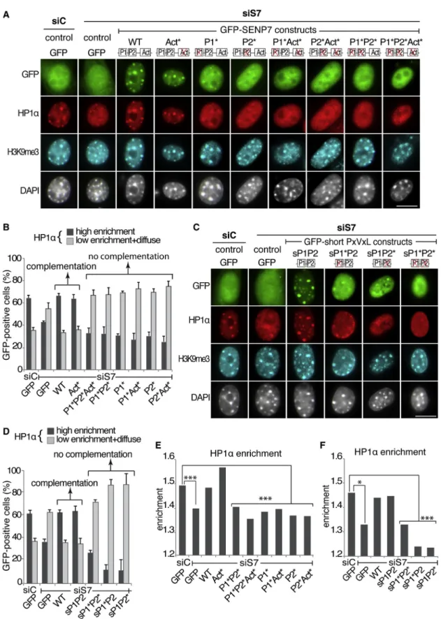
Having identified a complex mode of association between SENP7 and HP1a, we next aimed to characterize which function of SENP7 is involved in the maintenance of HP1a. We thus inves-tigated whether impairing (1) the interaction between SENP7 and HP1a, (2) SENP7 localization at pericentric domains, or (3) SENP7 deconjugation activity leads to a loss of endogenous HP1a accumulation at PCH domains. We designed a combina-tion of small interfering RNA (siRNA) targeting endogenous SENP7 and GFP-SENP7 constructs resistant to this siRNA to investigate whether mutated GFP-SENP7 can complement the depletion of endogenous SENP7. We verified that cotransfection of SENP7 siRNA with GFP-SENP7 siRNA-resistant constructs leads to efficient depletion of endogenous SENP7 (WT) (Figures S3A and S3B) and expression of the siRNA-resistant GFP-SENP7 construct (WTr) (Figure S3C). Importantly, the levels of HP1a in the cells remained unaffected by these cotransfections (Figures S3B and S3C). We performed immunofluorescence staining to visualize endogenous HP1a and H3K9me3 in cells cotransfected with siRNA and the GFP-SENP7 constructs ( Fig-ure 2A). Whereas we did not notice changes for the H3K9me3 enrichment at pericentric domains, HP1a localization was affected with some conditions showing high enrichment at peri-centric domains and others showing low enrichment diffuse throughout the nucleus (Figure 2A). When compared to control siRNA, we found that SENP7 depletion led to a decrease of cells showing high HP1a enrichment (Figures 2A and 2B). This is in agreement with our previous observation showing that depletion of SENP7 leads to a loss of HP1 enrichment at pericentric domains without affecting the cell cycle when analyzed by fluo-rescence-activated cell sorting (Maison et al., 2012).
In contrast, if WT GFP-SENP7 was expressed, the proportion of cells showing high HP1a enrichment remained identical to control siRNA (Figures 2A and 2B). This thus validates our approach using GFP-SENP7 constructs to complement endog-enous SENP7. We obtained an identical result when using a construct in which the SENP7 catalytic domain (amino acids 720–1037) was replaced by the catalytic domain of hSENP2 (data not shown), suggesting that the C-terminal part of SENP7 does not contain specific features necessary to maintain high HP1a enrichment. We next tested the GFP-SENP7 mutant con-structs. We found that when the single or double PxVxL motif mutants were expressed, the proportion of cells showing high HP1a enrichment decreased, indicating that these mutants cannot complement the loss of endogenous SENP7 (Figures 2A and 2B). Interestingly, when cells expressed the catalytic-dead mutant with two functional PxVxL motifs, the proportion of cells showing high HP1a enrichment remained identical to control siRNA, suggesting that the deconjugation activity is not
critical to complement for the loss of endogenous SENP7 ( Fig-ure 2B). Given that H3K9me3 is thought to contribute to HP1 accumulation (Bannister et al., 2001; Lachner et al., 2001), we verified whether H3K9me3 accumulation at pericentric domains would parallel that of HP1a under our conditions. The enrichment value of H3K9me3 was identical for each condition (Figure S3D), in line with the fact that we did not detect qualitative changes in H3K9me3 localization from the immunofluorescence stainings (Figure 2A).
We then investigated whether a simple module of two PxVxL motifs could be sufficient to maintain HP1 enrichment at pericen-tric domains following endogenous SENP7 depletion. We thus fused 125 amino acids of SENP7 (59–183) to GFP to generate a short construct containing only the two PxVxL motifs of SENP7. We generated constructs with two WT PxVxL motifs (sP1P2), a single PxVxL mutant (sP1*P2, sP1P2*), and the double PxVxL mutant (sP1*P2*) (Figure S3E). We verified that these GFP-short PXVXL constructs were expressed at similar levels as the full-length GFP-SENP7 (Figure S3F). Coimmunoprecipita-tion and immunofluorescence analyses showed that their asso-ciation with HP1a and their nuclear localization was identical to the full-length GFP-SENP7 constructs (Figures S3G and S3H). Since these GFP-short-PxVxL constructs are not targeted by SENP7 siRNA, we verified as above whether they could comple-ment the loss of endogenous SENP7 to maintain HP1a accumu-lation. We found that, following endogenous SENP7 depletion, the single PxVxL motif mutants (sP1*P2, sP1P2*) and the double PxVxL motif mutant (sP1*P2*) could not complement the loss of endogenous SENP7 (Figures 2C and 2D).
In contrast, when the WT double PxVxL motif (sP1P2) construct was expressed, high HP1a enrichment at pericentric domains was maintained in the same percentage of cells as con-trol or SENP7-depleted cells complemented by WT full-length SENP7 (Figures 2C and 2D). As above, we did not detect changes in H3K9me3 accumulation at pericentric domains ( Fig-ure S3I). We next analyzed quantitatively HP1a enrichment and found that, following endogenous SENP7 depletion, it remained identical to siRNA control transfection when WT or catalytic-dead full-length GFP-SENP7 (Figure 2E) and the GFP-short-PxVxL construct containing the two intact GFP-short-PxVxL motifs (sP1P2) (Figure 2F) were expressed. In contrast, the HP1a enrichment value significantly decreased when any of the other full-length GFP-SENP7 constructs and the short GFP-short-PxVxL constructs containing one or both mutant GFP-short-PxVxL motifs were expressed (Figures 2E and 2F). Taken together, these re-sults indicate that, in addition to the H3K9me3 modification, maintenance of HP1a enrichment at pericentric domains re-quires the two PxVxL HP1 interaction motifs on SENP7. In these conditions, we could not detect an effect of the desumoylation activity of SENP7. Importantly, the sole association of SENP7 with HP1a was not sufficient since a single PxVxL motif could not complement the loss of endogenous SENP7, whereas it associated with HP1a (Figures 1D andS3G). Furthermore, the behavior of the GFP-short-PxVxL construct containing the two intact PxVxL motifs was identical to that of the WT full-length GFP-SENP7.
We thus conclude from these results that a simple module of two PxVxL motifs displaying a bivalent HP1 interaction domain
Figure 2. A Simple Module of Two PxVxL Motifs Is Sufficient for HP1 Enrichment at Pericentric Domains
(A) Immunofluorescence analysis of full-length GFP-SENP7 constructs (green), endogenous HP1a (red), and H3K9me3 (blue) localization in GFP-positive NIH 3T3 cells after transfection of siRNA control (siC) or against SENP7 (siS7) and GFP control or GFP-SENP7 siRNA-resistant constructs. Corresponding DAPI images are shown. Scale bar, 10mm.
is required for the maintenance of HP1 enrichment at pericentric domains, in addition to the H3K9me3 mark.
Single PxVxL Mutants Behave as Dominant Negatives
Based on the above results, we predicted that single PxVxL mutants should act as monovalent HP1 interactor and could per-turb the association between endogenous HP1 and SENP7. We transfected full-length SENP7 constructs and the GFP-short-PxVxL constructs in NIH 3T3 and analyzed HP1a and H3K9me3 localization at pericentric domains, in comparison to GFP controls. Immunofluorescence analysis revealed that HP1a enrichment at pericentric domains resembled GFP con-trols, either when both PxVxL motifs are WT or both mutated for the full-length GFP-SENP7 constructs (Figure 3A) and the GFP-short-PxVxL constructs (Figure 3B). In contrast, when the constructs contained a mutation in a single PxVxL motif, HP1a enrichment decreased (Figures 3A and 3B). Thus, the single PxVxL motif mutants behave as dominant negatives for HP1a localization. We did not detect modifications of such interfering behavior of the single PxVxL mutants when using catalytic mu-tants (Figures 3A and 3B). Quantification of the proportion of cells displaying HP1 enrichment (Figures 3C and 3D) and quan-titative analysis of HP1 enrichment fully supported these obser-vations (Figures 3E and 3F). We next investigated the status of the H3K9me3 mark and found that it remained enriched at peri-centric domains in each condition, and, most importantly, when HP1a localization was perturbed (Figures 3A, 3B,S4A, and S4B). We did not detect differences in cell cycle profiles between cells expressing the GFP-short-PxVxL constructs (Figure S4C), indi-cating that the interference on HP1a localization mediated by expression of the single PxVxL motif mutants is not a conse-quence of a change in cell cycle. These results indicate that the presence of a monovalent module with only one motif able to interact with HP1 interferes with HP1a enrichment at pericen-tric domains. Furthermore, no interference was detected with the deconjugation mutant, suggesting that, in these conditions, the de-SUMOylation activity of SENP7 is not detected to be critical for HP1a enrichment.
Taken together, these findings strengthen our previous observations showing that the critical role of SENP7 for the maintenance of HP1 at pericentric domains lies in a bivalent HP1 interaction module.
The Two PxVxL Motifs of SENP7 Impact HP1 Dynamics at Pericentric Domains
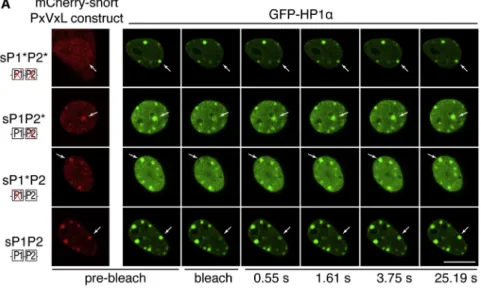
We next investigated by which mechanism a bivalent module of two PxVxL motifs affects HP1 enrichment at pericentric do-mains. Given that HP1 enrichment at heterochromatin is thought to result from a decreased mobility at these heterochromatic re-gions compared to euchromatin (Cheutin et al., 2003; Festen-stein et al., 2003; Schmiedeberg et al., 2004), we tested whether
the module of two PxVxL motifs could impact HP1 mobility at pericentric domains. We used FRAP to monitor HP1a mobility at pericentric domains in conditions where maintenance of HP1a enrichment is perturbed by expression of the interfering module. We cotransfected NIH 3T3 cells with GFP-HP1a and the short 125 amino acids of SENP7 (59–183) fused to mCherry to generate mCherry-short-PxVxL constructs that were WT (mCherry-sP1P2), single mutant (mCherry-sP1*P2 and mCherry-sP1P2*), or double mutant (mCherry-sP1*P2*). In the conditions used, we found that (1) GFP-HP1a was enriched at pericentric domains (Figure 4A), and (2) localization of the mCherry-short-PxVxL constructs was identical to the GFP-short-PxVxL and full-length GFP-SENP7 constructs (Figure 4A, prebleach). Further in agreement with our previous findings on endogenous HP1a, we found that localization of GFP-HP1a at pericentric domains was perturbed when interfering mCherry-short-PxVxL constructs were expressed (mCherry-sP1*P2 and mCherry-sP1P2*), when compared to expression of nonin-terfering mCherry-short-PxVxL constructs (mCherry-sP1P2 or mCherry-sP1*P2*) (Figure 4A, prebleach).
We selected cells expressing both GFP-HP1a and mCherry-short-PxVxL constructs, to ensure that HP1a mobility was analyzed in the presence of the mCherry-short-PxVxL proteins, and performed FRAP analysis at individual pericentric domains by bleaching and measuring fluorescence recovery of a single pericentric domain (Figure 4A). Analysis indicated that half re-covery at heterochromatin was reached within 1–5 s, in agree-ment with previous reported observations (Cheutin et al., 2003; Festenstein et al., 2003; Schmiedeberg et al., 2004;Figures 4B andS4E). When compared to the double-mutant sP1*P2*, which does not associate with HP1a or interfere with HP1a localization, single PxVxL mutants (sP1P2* and sP1*P2) conferred a faster recovery to HP1a, while WT PxVxL (sP1P2) conferred a slower recovery (Figures 4B andS4E). We performed kinetic modeling of the FRAP data and found, for every mCherry-short-PxVxL construct, the presence of a mobile and a very mobile population of HP1a molecules characterized by a distinct association time of HP1a to pericentric domains (t1 and t2 respectively, Fig-ure 4B), in agreement with previous results (Schmiedeberg et al., 2004). When the single PxVxL mutants (sP1P2* and sP1*P2) were expressed, both t1 and t2 decreased relative to the double PxVxL mutant (sP1*P2*), indicating a higher HP1a mobility. This finding correlates with the loss of HP1a enrichment at pericentric domains detected when the single PxVxL mutants are expressed (Figures 3B, 3D, and 3F). Conversely, when the WT PxVxL construct was expressed (sP1P2), both t1 and t2 increased relative to sP1*P2* (Figure 4B), indicating a decreased HP1a mobility.
Based on these results, we conclude that the interference observed on HP1a enrichment in the presence of a single HP1 interaction motif reflects an increase in HP1a mobility at
(B) Quantitative analysis of HP1a localization patterns in GFP-positive cells from immunofluorescence stainings above. Bar represents the mean, and error bars indicate the SD from four different experiments.
(C) As in (A) but with the GFP-short-PxVxL constructs. (D) As in (B) but with the GFP-short-PxVxL constructs.
(E) Quantitative analysis of endogenous HP1a enrichment at PCH domains when full-length GFP-SENP7 constructs are transfected. ***p < 0.001. (F) As in (E) but with the GFP-short-PxVxL constructs. *p < 0.05; ***p < 0.001.
Figure 3. Single PxVxL Mutants Behave as Dominant Negatives
(A) Immunofluorescence staining of full-length GFP-SENP7 constructs (green), endogenous HP1a (red), and H3K9me3 (blue) in GFP-positive cells after trans-fection of GFP control or the full-length GFP-SENP7 constructs in NIH 3T3 cells. Corresponding DAPI images are shown. Scale bar, 10mm.
pericentric domains. This thus indicates that the double PxVxL motif module of SENP7 participates in the maintenance of HP1a enrichment at pericentric regions by restricting HP1a mobility.
DISCUSSION
The results reported here indicate that the ability of SENP7 to interact with HP1 via a module comprising two PxVxL HP1 inter-action motifs is most critical to maintain HP1 enrichment at pericentric domains. We further provide evidence that, for this function in HP1 maintenance, SENP7 can be substituted for a simple bivalent module containing two PxVxL HP1 interaction motifs. Interfering with the two PxVxL motifs module leads to an increased HP1 mobility, suggesting that a function for this module is to restrict HP1 mobility at pericentric domains. Impor-tantly, we found that the loss of HP1 enrichment when SENP7 is either depleted or subjected to dominant-negative interference occurs without changes of H3K9me3 enrichment. This finding in-dicates that the H3K9me3 modification, although necessary for HP1 recruitment, is not sufficient to maintain stable HP1 accu-mulation at pericentric domains, and suggests that the module of two PxVxL HP1 interaction motifs of SENP7 might act on top of the H3K9me3 modification.
Based on these data, we propose a mechanism for the main-tenance of HP1 enrichment at H3K9me3-enriched regions involving a module of two PxVxL HP1 interaction motifs ( Fig-ure 4C). The docking of HP1 on contiguous H3K9me3-modified nucleosomes would provide exposure of several consecutive HP1-chromoshadow domains that could be recognized and connected by a module of two PxVxL motifs, resulting in a phys-ical link between several HP1 molecules. This would decrease global HP1 mobility, since dissociation from chromatin of con-nected HP1 molecules would necessitate a loss of interaction between every individual HP1 chromodomain and H3K9me3 residue to occur simultaneously. This ultimately would lock the HP1 molecules and, hence, favor their stable maintenance at H3K9me3-enriched regions. Importantly, such locking could occur between HP1 molecules docked on adjacent nucleo-somes from the same nucleosomal array, but also from distinct arrays, favoring a stabilization of higher-order chromatin organi-zation (Figure 4C). One could envisage that additional factors and/or posttranslational modifications regulating the physical association between HP1 and the module of two PxVxL HP1 interaction motifs could control this locking mechanism. At a functional cellular level, we could reveal that such an HP1-lock-ing function of SENP7 might be critical because of the loss of HP1 enrichment at pericentric domains as a result of prolonged time required to progress from late prophase to early telophase during mitosis (Figure 1A).
In our experimental conditions using 3T3 cells, in which HP1 enrichment at pericentric domains was established, we did not detect the de-SUMOylation activity of SENP7 to be critical for HP1 maintenance. However, important functions of SENP7 related to HP1, involving the catalytic de-SUMOylation domain, should not be dismissed. A recent report revealed a role of SENP7 in promoting a permissive environment for homolo-gous recombination repair role by regulating the levels of the SUMO-2/3 modification of the HP1 interactor KAP1 (Garvin et al., 2013). SENP7 de-SUMOylation activity with specificity for SUMO-2/3 also was reported to be involved in the regulation of HP1a recruitment to specific promoters and tumorigenesis (Bawa-Khalfe et al., 2012). Further, since we found that de novo targeting of HP1a involves HP1 SUMOylation by SUMO-1 and pericentric transcripts (Maison et al., 2011), SENP7 acting in combination with transcription-related mechanisms ( Bulut-Karslioglu et al., 2012) may be considered as a potential regu-lator of de novo HP1 targeting. It is thus tempting to envisage SENP7 with the combination of de-SUMOylation activity and a double PxVxL module as a factor that could regulate HP1 enrich-ment via several nonexclusive mechanisms that could adapt to the cellular context.
We identified here how HP1 accumulation depends on addi-tional mechanisms beyond the presence of the H3K9me3 modi-fication, and characterized a critical role for a module of two PxVxL HP1-binding motifs to ensure HP1 maintenance at het-erochromatic sites. Interacting with HP1 via two distinct interac-tion domains does not seem to be unique to SENP7, and could have a broad importance since it also has been reported for the EMSY protein in human (Huang et al., 2006), the DNA meth-yltransferase DIM-2 in Neurospora crassa (Honda and Selker, 2008), the Su(var)3-7 protein in Drosophila (Delattre et al., 2000), and the Orc3 protein (Prasanth et al., 2010). Although the in vivo relevance for a module of interaction with HP1 made of two distinct domains had remained unclear for these proteins besides SENP7, it is interesting to note that, similar to our data with SENP7, Orc3 depletion resulted in delocalization of HP1 from heterochromatin loci, though without impacting H3K9me3 (Prasanth et al., 2010). Further, although the interac-tion between Orc3 and HP1 may be related to replicainterac-tion of het-erochromatin domains, this does indicate that enrichment of HP1 at heterochromatin loci may depend on several factors able to bridge or connect several HP1 molecules, but which loss cannot systematically be compensated by another.
The mode of action of the two PxVxL modules on HP1 stability necessitates at least two critical requirements. First, the possibil-ity for HP1 molecules recruited on H3K9me3-modified nucleo-somes to oligomerize in order to expose contiguous chromosh-adow dimer interfaces that could be recognized and connected by the two PxVxL-containing module. Second, the connection of
(B) As in (A) but with transfection of the GFP-short-PxVxL constructs.
(C) Quantitative analysis of HP1a localization pattern at pericentric domains in GFP-positive cells from immunofluorescence stainings in (A). Bar represents the mean, and error bars indicate the SD from four experiments.
(D) Quantitative analysis as in (C) but with the GFP-short-PxVxL constructs from immunofluorescence stainings in (B).
(E) Quantitative analysis of HP1a enrichment at pericentric domains in GFP-positive cells transfected with full-length GFP-SENP7 constructs from immuno-fluorescence stainings in (A). ***p < 0.05.
two HP1 dimers by their respective chromoshadow dimer inter-face should be structurally possible. For the oligomerization aspect, human HP1 was reported to oligomerize as tetramers (Yamada et al., 1999), and Swi6 bound on H3K9me3-modified nucleosomes was shown to tetramerize (Canzio et al., 2011),
Figure 4. The Module of Two PxVxL Motifs Restricts Mobility of HP1a
(A) Images of confocal sections showing localiza-tion of mCherry-short-PxVxL constructs (red) and HP1a (green) before bleaching and GFP-HP1a fluorescence recovery at selected time points. The arrow indicates the bleached peri-centric domain. Scale bar, 10mm.
(B) FRAP curves of GFP-HP1a in the presence of the various mCherry-short-PxVxL constructs. The table shows the time of association of HP1a to pericentric domains for both HP1a population in the presence of the various mCherry-short-PxVxL constructs calculated from modeling. Number of pericentric domains analyzed (n) is indicated. See alsoFigure S4.
(C) Hypothetical model. The two PxVxL motifs of SENP7 connect contiguous HP1 molecules docked on H3K9me3-modified nucleosomes enabling locking of HP1. For simplification, only the minimal region of SENP7 containing the mod-ule of two PxVxL motifs and no additional factors are represented.
thus generating contiguous PxVxL-bind-ing sites on nucleosomal arrays. Further, dimers of Swi6 were shown to self-asso-ciate to promote elongation of Swi6 dimers chain (Canzio et al., 2013), sug-gesting that several binding sites for PxVxL motifs could be exposed on HP1-containing chromatin. For the structural aspect, the fact that two chromoshadow dimers are found in the crystal structure of the EMSY-HP1 complex (Huang et al., 2006) supports the absence of structural constraints that could prevent the binding of two chromoshadow dimers at the same time. Thus, taken together, current data support the idea of a partic-ular architecture at PCH involving factors with two HP1 interaction motifs, such as SENP7 to lock the chromoshadow domains of HP1 molecules docked on H3K9me3-modified nucleosomes.
The necessity of having closely spaced HP1 chromoshadow dimers to be able to be locked by SENP7 could help modu-late the strength of HP1 maintenance. A genomic loci made of homogenous ar-rays of H3K9me3-modified nucleosomes, such as blocks of heterochromatin, might be a better substrate to expose many regularly spaced chromoshadow do-mains that would be efficiently locked by the double PxVxL mod-ule of SENP7 and kept in a closed state. In contrast, genomic loci that would be made of H3K9me3 nucleosomes interspersed with non-H3K9me3 nucleosomes would not provide an efficient sub-strate to expose contiguous chromoshadow domains that could
be locked by double PxVxL module, and would not result in a stable accumulation of HP1. This would allow dynamic and reversible binding of factors to chromatin and DNA, such as pro-moter regions regulated by H3K9me3 and HP1 (Kwon and Workman, 2011). It is interesting then to envisage the double PxVxL module of SENP7 as a means to ensure HP1 stability and enrichment at heterochromatin loci as a consequence of the density of H3K9me3 modification.
Future work should aim at characterizing whether locking mechanisms acting on top of histone modifications to promote maintenance of protein concentration at specific genomic loci in the cell nucleus could apply to other nuclear domains and how they would impact on the plasticity of nuclear organization and genome stability.
EXPERIMENTAL PROCEDURES Cells, Transfections, and Extracts
We cultured NIH 3T3 cells (Maison et al., 2012) and used Lipofectamin 2000 for transfections (Invitrogen). For the complementation experiment, we cotrans-fected siRNA and DNA plasmids and performed analysis 33 hr after transfec-tion. We prepared total cell extracts by resuspending cells in RIPA buffer and performed immunoprecipitations (Maison et al., 2012).
Plasmids
GFP-SENP7 WT and Act* (C979S) expressing plasmid were fromMaison et al. (2012). Mutations of the GFP-SENP7 mutants were as follows: P1* (V93DL95N), P2* (V159DL161N), P1*P2* (V93DL95N V159DL161N), P1*Act* (V93DL95N C979S), and P1*P2*Act* (V93DL95N V159DL161N C979S).
Microscopy Analyses
We immunostained cells (Martini et al., 1998) and quantified the enrichment at PCH domains of SENP7, H3K9me3, and HP1a with the 3D-FIED method (Cantaloube et al., 2012). We performed time-lapse microscopy using the BioStation system (Nikon). We carried out FRAP experiments on a Zeiss LSM 780 confocal and used NIH Image J software for analysis.
Statistical Tests
We used the Mann-Whitney test to compare HP1a and H3K9me3 enrichments and the Wilcoxon test for time-lapse microscopy. Differences were considered significant when p < 0.05.
SUPPLEMENTAL INFORMATION
Supplemental Information includes Supplemental Experimental Procedures and four figures and can be found with this article online athttp://dx.doi.org/ 10.1016/j.celrep.2015.01.004.
AUTHOR CONTRIBUTIONS
K.R., G.A., and J.-P.Q. conceived and designed the experiments. K.R., Y.L., S.C., I.L., and J.-P.Q. performed the experiments. K.R., S.C., J.-P.Q., and G.A. analyzed the data. K.R., G.A., and J.-P.Q. wrote the paper.
ACKNOWLEDGMENTS
We thank Z. Gurard-Levin and Laura Attardi for critical reading of the manu-script, A. Corpet for help with statistical analysis, C. Billaudeau for modeling FRAP experiments, and thePICT-IBISA@PasteurImaging facility of the Institut Curie and the Nikon ImagingCentre@InstitutCurie-CNRS for the BioStation. We also thank C. Maison for helpful discussions and the gift of purified gluta-thione S-transferase (GST) and GST-HP1a proteins. K.R. received support from Ministe`re de l’Enseignement Supe´rieur et de la Recherche/Universite´ Pierre et Marie Curie and la Ligue Nationale contre le Cancer. This work was
supported by la Ligue Nationale contre le Cancer (Equipe labellise´e Ligue), PIC Programs, the European Commission Network of Excellence EpiGeneSys (HEALTH-F4-2010-257082), ERC Advanced Grant 2009-AdG_20090506 ‘‘Eccentric,’’ the European Commission large-scale integrating project FP7_HEALTH-2010-259743 ‘‘MODHEP,’’ ANR ‘‘ChromaTin’’ ANR-10-BLAN-1326-03, ANR Labex DEEP 11-LBX-0044, ANR ‘‘CHAPINHIB’’ ANR-12-BSV5-0022-02, and Aviesan-ITMO cancer project ‘‘Epigenomics of breast cancer.’’ Received: July 31, 2014 Revised: December 8, 2014 Accepted: December 24, 2014 Published: February 5, 2015 REFERENCES
Allshire, R.C., Nimmo, E.R., Ekwall, K., Javerzat, J.P., and Cranston, G. (1995). Mutations derepressing silent centromeric domains in fission yeast disrupt chromosome segregation. Genes Dev. 9, 218–233.
Bannister, A.J., Zegerman, P., Partridge, J.F., Miska, E.A., Thomas, J.O., All-shire, R.C., and Kouzarides, T. (2001). Selective recognition of methylated lysine 9 on histone H3 by the HP1 chromo domain. Nature 410, 120–124. Bawa-Khalfe, T., Lu, L.S., Zuo, Y., Huang, C., Dere, R., Lin, F.M., and Yeh, E.T. (2012). Differential expression of SUMO-specific protease 7 variants regulates epithelial-mesenchymal transition. Proc. Natl. Acad. Sci. USA 109, 17466– 17471.
Billur, M., Bartunik, H.D., and Singh, P.B. (2010). The essential function of HP1 beta: a case of the tail wagging the dog? Trends Biochem. Sci. 35, 115–123. Brasher, S.V., Smith, B.O., Fogh, R.H., Nietlispach, D., Thiru, A., Nielsen, P.R., Broadhurst, R.W., Ball, L.J., Murzina, N.V., and Laue, E.D. (2000). The struc-ture of mouse HP1 suggests a unique mode of single peptide recognition by the shadow chromo domain dimer. EMBO J. 19, 1587–1597.
Bulut-Karslioglu, A., Perrera, V., Scaranaro, M., de la Rosa-Velazquez, I.A., van de Nobelen, S., Shukeir, N., Popow, J., Gerle, B., Opravil, S., Pagani, M., et al. (2012). A transcription factor-based mechanism for mouse heterochromatin formation. Nat. Struct. Mol. Biol. 19, 1023–1030.
Cantaloube, S., Romeo, K., Le Baccon, P., Almouzni, G., and Quivy, J.P. (2012). Characterization of chromatin domains by 3D fluorescence micro-scopy: An automated methodology for quantitative analysis and nuclei screening. BioEssays 34, 509–517.
Canzio, D., Chang, E.Y., Shankar, S., Kuchenbecker, K.M., Simon, M.D., Mad-hani, H.D., Narlikar, G.J., and Al-Sady, B. (2011). Chromodomain-mediated oligomerization of HP1 suggests a nucleosome-bridging mechanism for het-erochromatin assembly. Mol. Cell 41, 67–81.
Canzio, D., Liao, M., Naber, N., Pate, E., Larson, A., Wu, S., Marina, D.B., Gar-cia, J.F., Madhani, H.D., Cooke, R., et al. (2013). A conformational switch in HP1 releases auto-inhibition to drive heterochromatin assembly. Nature 496, 377–381.
Cavalli, G., and Misteli, T. (2013). Functional implications of genome topology. Nat. Struct. Mol. Biol. 20, 290–299.
Cheutin, T., McNairn, A.J., Jenuwein, T., Gilbert, D.M., Singh, P.B., and Misteli, T. (2003). Maintenance of stable heterochromatin domains by dynamic HP1 binding. Science 299, 721–725.
Cowieson, N.P., Partridge, J.F., Allshire, R.C., and McLaughlin, P.J. (2000). Dimerisation of a chromo shadow domain and distinctions from the chromo-domain as revealed by structural analysis. Curr. Biol. 10, 517–525. De Koning, L., Savignoni, A., Boumendil, C., Rehman, H., Asselain, B., Sastre-Garau, X., and Almouzni, G. (2009). Heterochromatin protein 1alpha: a hall-mark of cell proliferation relevant to clinical oncology. EMBO Mol. Med. 1, 178–191.
Delattre, M., Spierer, A., Tonka, C.H., and Spierer, P. (2000). The genomic silencing of position-effect variegation in Drosophila melanogaster: interaction between the heterochromatin-associated proteins Su(var)3-7 and HP1. J. Cell Sci. 113, 4253–4261.
Ekwall, K. (2007). Epigenetic control of centromere behavior. Annu. Rev. Genet. 41, 63–81.
Ekwall, K., Javerzat, J.P., Lorentz, A., Schmidt, H., Cranston, G., and Allshire, R. (1995). The chromodomain protein Swi6: a key component at fission yeast centromeres. Science 269, 1429–1431.
Eskeland, R., Eberharter, A., and Imhof, A. (2007). HP1 binding to chromatin methylated at H3K9 is enhanced by auxiliary factors. Mol. Cell. Biol. 27, 453–465.
Fan, J.Y., Rangasamy, D., Luger, K., and Tremethick, D.J. (2004). H2A.Z alters the nucleosome surface to promote HP1alpha-mediated chromatin fiber folding. Mol. Cell 16, 655–661.
Festenstein, R., Pagakis, S.N., Hiragami, K., Lyon, D., Verreault, A., Sekkali, B., and Kioussis, D. (2003). Modulation of heterochromatin protein 1 dynamics in primary Mammalian cells. Science 299, 719–721.
Garvin, A.J., Densham, R.M., Blair-Reid, S.A., Pratt, K.M., Stone, H.R., Weekes, D., Lawrence, K.J., and Morris, J.R. (2013). The deSUMOylase SENP7 promotes chromatin relaxation for homologous recombination DNA repair. EMBO Rep. 14, 975–983.
Gilbert, N., Boyle, S., Sutherland, H., de Las Heras, J., Allan, J., Jenuwein, T., and Bickmore, W.A. (2003). Formation of facultative heterochromatin in the absence of HP1. EMBO J. 22, 5540–5550.
Grewal, S.I., and Jia, S. (2007). Heterochromatin revisited. Nat. Rev. Genet. 8, 35–46.
Guenatri, M., Bailly, D., Maison, C., and Almouzni, G. (2004). Mouse centric and pericentric satellite repeats form distinct functional heterochromatin. J. Cell Biol. 166, 493–505.
Hay, R.T. (2007). SUMO-specific proteases: a twist in the tail. Trends Cell Biol.
17, 370–376.
Honda, S., and Selker, E.U. (2008). Direct interaction between DNA methyl-transferase DIM-2 and HP1 is required for DNA methylation in Neurospora crassa. Mol. Cell. Biol. 28, 6044–6055.
Huang, Y., Myers, M.P., and Xu, R.M. (2006). Crystal structure of the HP1-EMSY complex reveals an unusual mode of HP1 binding. Structure 14, 703–712.
Jacobs, S.A., and Khorasanizadeh, S. (2002). Structure of HP1 chromodomain bound to a lysine 9-methylated histone H3 tail. Science 295, 2080–2083. Karpen, G.H., and Allshire, R.C. (1997). The case for epigenetic effects on centromere identity and function. Trends Genet. 13, 489–496.
Kwon, S.H., and Workman, J.L. (2011). The changing faces of HP1: From het-erochromatin formation and gene silencing to euchromatic gene expression: HP1 acts as a positive regulator of transcription. BioEssays 33, 280–289. Lachner, M., O’Carroll, D., Rea, S., Mechtler, K., and Jenuwein, T. (2001). Methylation of histone H3 lysine 9 creates a binding site for HP1 proteins. Nature 410, 116–120.
Maison, C., and Almouzni, G. (2004). HP1 and the dynamics of heterochro-matin maintenance. Nat. Rev. Mol. Cell Biol. 5, 296–304.
Maison, C., Bailly, D., Peters, A.H., Quivy, J.P., Roche, D., Taddei, A., Lachner, M., Jenuwein, T., and Almouzni, G. (2002). Higher-order structure in pericentric heterochromatin involves a distinct pattern of histone modification and an RNA component. Nat. Genet. 30, 329–334.
Maison, C., Quivy, J.P., Probst, A.V., and Almouzni, G. (2010). Heterochro-matin at mouse pericentromeres: a model for de novo heterochroHeterochro-matin forma-tion and duplicaforma-tion during replicaforma-tion. Cold Spring Harb. Symp. Quant. Biol.
75, 155–165.
Maison, C., Bailly, D., Roche, D., Montes de Oca, R., Probst, A.V., Vassias, I., Dingli, F., Lombard, B., Loew, D., Quivy, J.P., and Almouzni, G. (2011).
SUMOylation promotes de novo targeting of HP1a to pericentric heterochro-matin. Nat. Genet. 43, 220–227.
Maison, C., Romeo, K., Bailly, D., Dubarry, M., Quivy, J.P., and Almouzni, G. (2012). The SUMO protease SENP7 is a critical component to ensure HP1 enrichment at pericentric heterochromatin. Nat. Struct. Mol. Biol. 19, 458–460. Martini, E., Roche, D.M., Marheineke, K., Verreault, A., and Almouzni, G. (1998). Recruitment of phosphorylated chromatin assembly factor 1 to chro-matin after UV irradiation of human cells. J. Cell Biol. 143, 563–575. Muchardt, C., Guilleme, M., Seeler, J.S., Trouche, D., Dejean, A., and Yaniv, M. (2002). Coordinated methyl and RNA binding is required for heterochromatin localization of mammalian HP1alpha. EMBO Rep. 3, 975–981.
Murzina, N., Verreault, A., Laue, E., and Stillman, B. (1999). Heterochromatin dynamics in mouse cells: interaction between chromatin assembly factor 1 and HP1 proteins. Mol. Cell 4, 529–540.
Nielsen, A.L., Oulad-Abdelghani, M., Ortiz, J.A., Remboutsika, E., Chambon, P., and Losson, R. (2001). Heterochromatin formation in mammalian cells: interaction between histones and HP1 proteins. Mol. Cell 7, 729–739. Nielsen, P.R., Nietlispach, D., Mott, H.R., Callaghan, J., Bannister, A., Kouzar-ides, T., Murzin, A.G., Murzina, N.V., and Laue, E.D. (2002). Structure of the HP1 chromodomain bound to histone H3 methylated at lysine 9. Nature 416, 103–107.
Nozawa, R.S., Nagao, K., Masuda, H.T., Iwasaki, O., Hirota, T., Nozaki, N., Kimura, H., and Obuse, C. (2010). Human POGZ modulates dissociation of HP1alpha from mitotic chromosome arms through Aurora B activation. Nat. Cell Biol. 12, 719–727.
Obuse, C., Iwasaki, O., Kiyomitsu, T., Goshima, G., Toyoda, Y., and Yanagida, M. (2004). A conserved Mis12 centromere complex is linked to heterochromat-ic HP1 and outer kinetochore protein Zwint-1. Nat. Cell Biol. 6, 1135–1141. Peters, A.H., O’Carroll, D., Scherthan, H., Mechtler, K., Sauer, S., Scho¨fer, C., Weipoltshammer, K., Pagani, M., Lachner, M., Kohlmaier, A., et al. (2001). Loss of the Suv39h histone methyltransferases impairs mammalian heterochro-matin and genome stability. Cell 107, 323–337.
Prasanth, S.G., Prasanth, K.V., Siddiqui, K., Spector, D.L., and Stillman, B. (2004). Human Orc2 localizes to centrosomes, centromeres and heterochro-matin during chromosome inheritance. EMBO J. 23, 2651–2663.
Prasanth, S.G., Shen, Z., Prasanth, K.V., and Stillman, B. (2010). Human origin recognition complex is essential for HP1 binding to chromatin and heterochro-matin organization. Proc. Natl. Acad. Sci. USA 107, 15093–15098. Schmiedeberg, L., Weisshart, K., Diekmann, S., Meyer Zu Hoerste, G., and Hemmerich, P. (2004). High- and low-mobility populations of HP1 in hetero-chromatin of mammalian cells. Mol. Biol. Cell 15, 2819–2833.
Shen, L.N., Geoffroy, M.C., Jaffray, E.G., and Hay, R.T. (2009). Characteriza-tion of SENP7, a SUMO-2/3-specific isopeptidase. Biochem. J. 421, 223–230. Smothers, J.F., and Henikoff, S. (2000). The HP1 chromo shadow domain binds a consensus peptide pentamer. Curr. Biol. 10, 27–30.
Thiru, A., Nietlispach, D., Mott, H.R., Okuwaki, M., Lyon, D., Nielsen, P.R., Hirshberg, M., Verreault, A., Murzina, N.V., and Laue, E.D. (2004). Structural basis of HP1/PXVXL motif peptide interactions and HP1 localisation to hetero-chromatin. EMBO J. 23, 489–499.
Weaver, B.A., and Cleveland, D.W. (2007). Aneuploidy: instigator and inhibitor of tumorigenesis. Cancer Res. 67, 10103–10105.
Yamada, T., Fukuda, R., Himeno, M., and Sugimoto, K. (1999). Functional domain structure of human heterochromatin protein HP1(Hsalpha): involve-ment of internal DNA-binding and C-terminal self-association domains in the formation of discrete dots in interphase nuclei. J. Biochem. 125, 832–837.