Altered autonomic nervous system reactivity to pain in trigeminal neuralgia
Guillaume Léonard, Ph.D.1, Philippe Chalaye, M.Sc.2, Philippe Goffaux, Ph.D.2, David Mathieu, M.D.2, Isabelle Gaumond, Ph.D. 2, Serge Marchand, Ph.D.2
1Centre de recherche sur le vieillissement, 1036 Belvédère Sud, Sherbrooke (Québec), Canada, J1H 4C4; 2Centre de recherche du Centre hospitalier universitaire
de Sherbrooke, 3001 12e avenue Nord, Sherbrooke (Québec), Canada, J1H 5N4
Keywords: pain, trigeminal neuralgia, autonomic nervous system, heart rate variability, cold pressor test
Corresponding author: Serge Marchand, Ph.D. Université de Sherbrooke
Faculté de médecine, neurochirurgie 3001, 12e avenue nord
Abstract
BACKGROUND: In the last two decades, there has been increasing evidence to suggest that trigeminal neuralgia (TN) may be linked to a dysfunction of the
autonomic nervous system (ANS). The aim of the present study was to formally test this hypothesis by comparing the reactivity of the ANS to experimental pain in a population of TN patients and healthy controls. METHODS: Twelve patients diagnosed with classical TN and 12 healthy controls participated in the study. Cardiac activity was assessed while participants were instructed to rest and again during a cold pressor test (CPT). Heart rate variability analyses were performed off-line to obtain parasympathetic (high-frequency) and sympathetic (low-frequency) indices. RESULTS: At baseline, ANS measures did not differ between healthy controls and TN patients, and both groups showed a similar increase in heart rate during the CPT (all p-values > .05). However, TN patients showed a greater increase in cardiac sympathetic activity and a greater decrease in cardiac parasympathetic activity during CPT compared to healthy controls (all p-values < .05). Importantly, changes in sympathetic reactivity, from baseline to CPT, were negatively associated with the number of pain paroxysms experienced each day by TN patients in the preceding week (r = -.58, p < .05). CONCLUSION: These results suggest that TN, like many other short-lasting, unilateral facial pain conditions, is linked to ANS alterations. Future studies are required to determine if the altered ANS response observed in TN patients is a cause or a consequence of TN pain.
1. Introduction
Trigeminal neuralgia (TN) is a rare neuropathic pain disorder affecting the fifth cranial nerve and characterized by sharp paroxystic pain episodes lasting from a few seconds to two minutes1, 2. Although TN was described several decades ago3, 4, its physiopathology remains poorly understood5-7. According to Dandy8 and Janetta9, 10, TN is caused by a microvascular-induced compression of the dorsal root of the trigeminal nerve, most commonly from the superior cerebellar artery. Despite its popularity, the microvascular compression hypothesis has been challenged by several studies showing that: (a) many patients suffering from TN do not show evidence of microvascular compression11, 12, and (b) microvascular compression is present in a large proportion of individuals who do not suffer from TN13-16.
In the last two decades, several reports have suggested that TN could be linked to impaired autonomic nervous system (ANS) activity17-22. Until recently, however, evidence of ANS dysregulation in TN remained mostly speculative or anecdotal. For example, Noguchi et al.17 reported the case of a 29 year-old woman suffering from symptomatic TN who was significantly relieved following a stellate ganglion block (a sympathetic ganglion). Similar results were reported by Manahan et al.21 following a sphenopalatine ganglion block (a parasympathetic ganglion) on a 56 year-old woman.
More recently, Simms and colleagues23 sent questionnaires and reviewed the medical records of 92 TN patients. They noted that a majority of these patients (i.e., 67% of the patients sampled) had at least one autonomic symptom associated with their pain. Although some patients reported regional sweating (a symptom which can be linked to sympathetic activation), the vast majority reported parasympathetic-like symptoms (e.g., excessive salivation, nasal congestion, excessive tearing). Taken
together, these observations support the idea that TN could be linked to ANS
dysfunctions, and prod researchers to pursue future work to better understand the role of the ANS in TN pain.
A next logical step to the study of Simms and colleagues23 would be to evaluate the activity and reactivity of the ANS in a population of patients suffering from TN using objective sympathetic and parasympathetic measures. In this regard, heart rate variability (HRV) techniques, allowing the recording of both sympathetic and parasympathetic indices24, seem particularly appropriate. To our knowledge, no previous study used HRV techniques to evaluate the reactivity of cardiac ANS
responses in patients with TN. Given this knowledge gap, the aim of this study was to assess and compare the reactivity of the ANS to experimental pain in a population of TN patients and healthy controls using HRV analyses. Based on the results of Simms et al., we postulated that TN patients would show increased sympathetic and/ or parasympathetic reactivity when compared to healthy controls.
2. Methods
2.1 Participants
Twelve patients diagnosed with classical TN and 12 healthy controls participated in this study (mean age ± SD = 62 ± 11 yrs) (see Table 1). TN patients were recruited from the list of patients scheduled for Gamma-knife surgery (GKS) at the GKS clinic of the Sherbrooke University Hospital. Diagnosis of classical TN was confirmed by a neurosurgeon using the International Classification of Headache Disorders criteria25. Patients with atypical or symptomatic TN (e.g. TN secondary to multiple sclerosis) or with symptoms suggesting post-herpetic or deafferentation pain syndromes were excluded. None of our retained participants showed evidence of
tactile, thermal or pricking hypoesthesia. None showed signs of dysesthesia, hyperesthesia or allodynia. In addition to undergoing a conventional and sensory-specific neurological examination (conducted by both the appointed neurosurgeon and GL), every participant underwent magnetic resonance imaging (MRI) to rule out possible neuronal damage.
Healthy controls were recruited through local ads and were all community-dwelling individuals. They all had good general health and none suffered from a painful condition, with the exception of three participants who reported minor osteoarthritic pain. Every participant was asked to refrain from using short-term analgesics two hours before testing and from drinking coffee and smoking cigarettes six hours before testing. TN patients were also asked to stop all pain medications for a period of 24h before their appointment to minimize the effect that medication could have on pain perception and ANS measures. Group and patient characteristics are listed in Tables 1 and 2.
The experiment took place at the Centre de recherche du Centre hospitalier
universitaire de Sherbrooke (Sherbrooke, Quebec, Canada). The local institutional
ethics committee approved the study's procedures and each participant provided informed written consent before participating.
2.2 Experimental pain: cold pressor test
Participants were asked to immerse their right arm for 5 min in a bath of circulating cold water maintained at 10C. This procedure, commonly known as the cold pressor test (CPT), was used to trigger ANS responses to pain. Every 30 s, patients rated their immersion-induced pain intensity using a 0-100 numerical rating scale (NRS; 0 = no pain; 100 = intolerable pain).
2.3 Cardiac activity
Five-minute electrocardiogram (ECG) recordings were obtained during baseline testing (rest) and during experimental pain testing. Heart rate (HR) was monitored with a three lead (ECG) montage and sampled at 1000 Hz using a PowerLab recording system with Chart software (AD Instruments, Colorado). Instantaneous RR intervals were obtained from the ECG waveform with a peak detection algorithm to detect successive R-waves. All data were manually checked to ensure that only normal to normal (NN) intervals were analyzed. We analyzed HRV in the frequency domain. Specifically, fast Fourier transforms were used to calculate the power spectral density of HR oscillations. Two components can be distinguished from short-term spectrum analysis of HRV: a low frequency (LF) (0.04 - 0.15Hz) and a high frequency (HF) (0.15 - 0.4Hz) component. These two components are used as indices of cardiac autonomic nervous system activity24. HR fluctuations in the LF range reflect baroreflex-mediated sympathetic activity associated with Mayer waves of blood pressure26, 27. On the other hand, HRV in the HF range is generated by respiratory sinus arrhythmia (RSA) and constitutes a sensitive measure of cardiac vagal parasympathetic activity28, 29. Normalized HF and LF values were computed and analysed. Sympathovagal LF/HF ratios were not analysed because the
sympathovagal balance indexed by the LF/HF ratio is already accounted for when normalizing HF and LF values30.
2.4 Data analysis
Autonomic reactivity to pain was evaluated by calculating the delta score between baseline and CPT induced responses [delta score = (CPT – baseline)]. Heart rate and HRV delta scores were compared between the two groups using independent
t-tests. In order to examine the relationships between ANS reactivity to experimental pain and the characteristics of TN patients, we performed Pearson correlational analyses between the change in LF and HF observed from baseline to CPT and a series of demographic and clinical variables obtained at the moment of the
experiment. These demographic and clinical variables included the age and sex of the patient, disease duration, average intensity of pain paroxysms experienced in the preceding week (0-100 NRS), and the average number of daily pain paroxysms experienced in the preceding week. Statistical significance was set at p < 0.05. All tests were performed using SPSS (version 18.0 for Windows®, Chicago, IL, USA).
Because of the relatively small number of subjects included in the study and in spite of histograms suggesting the presence of normally distributed data,
non-parametric tests were additionally used to compare HR and HRV measures between the two groups (Mann-Whitney tests) and to evaluate the association between the HRV measures and the clinico-demographic variables (Spearman tests). Results confirmed no difference between parametric and non-parametric approaches. Therefore, only parametric statistics are reported.
3. Results
3.1 Pain perception
Every participant experienced the CPT as a painful procedure. The mean pain intensity score during the CPT was comparable between the two groups (mean ± SD = 48 ± 27 for healthy controls and 63 ± 26 for TN patients; t = -1.42; p = .17). None of the TN participants experienced facial pain during the testing session.
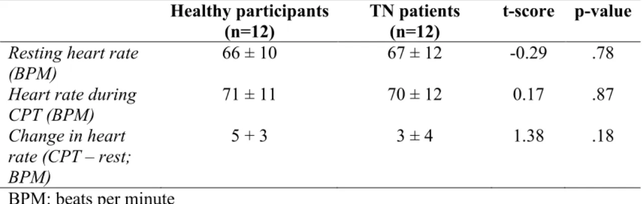
No difference in HR (both at rest and during CPT) was observed between the two groups. Healthy controls and TN patients showed a similar increase in HR during the CPT (see Table 3).
3.3 Autonomic activity and reactivity to pain
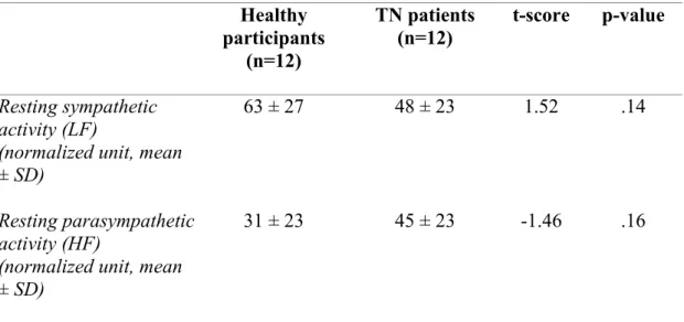
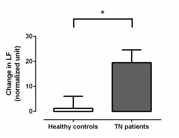
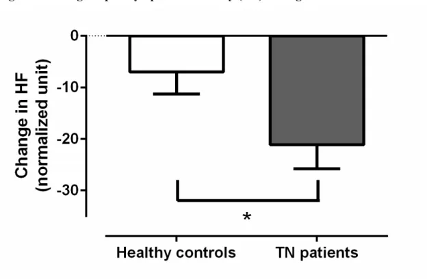
There was no difference in resting sympathetic (LF) or parasympathetic activity (HF) between healthy controls and TN patients (see Table 4). However, during the CPT, TN patients showed a greater increase in LF (t = -2.60, p < .05) and a greater decrease in HF (t = 2.23; p < .05) when compared to healthy controls,
indicating greater increment in sympathetic activity and greater decrement in
parasympathetic activity during experimental pain, respectively (see Figures 1 and 2). To extend the results obtained with the LF and HF measures, and better understand the role of the ANS in TN, we further explored HRV analysis in the time domain. More specifically, we calculated the root mean-square of successive
differences between adjacent normal-to-normal (NN) intervals (RMSSD), the number of pairs of adjacent NN intervals differing by more than 50 ms (pNN50) and the standard deviation of NN intervals (SDNN) 31, 32 and compared results between the two groups of participants. Contrary to LF and HF analyses, no significant group differences were noted for RMSSD, pNN50 and SDNN (all p-values > .05).
3.4 Associations between HRV measures and clinico-demographic variables
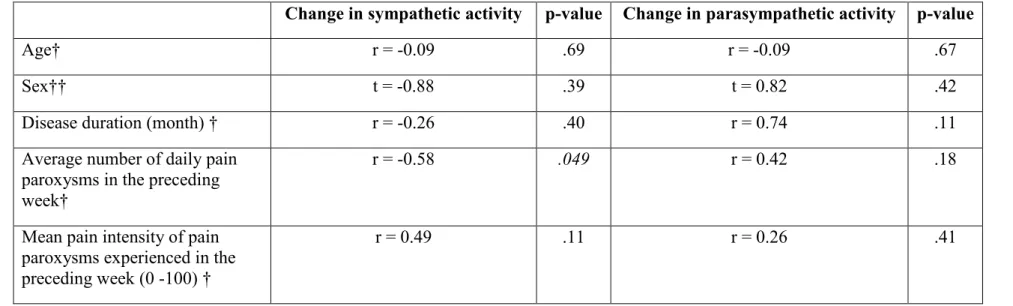
The correlation coefficients obtained between the HRV measures and the demographic and clinical variables obtained at the moment of the experiment are presented in Table 5. As can be seen from this table, there was a negative association between the CPT-induced change in LF activity and the average number of daily pain
paroxysms experienced by the patients in the preceding week. No other significant results were observed (all other p-values > .05).
4. Discussion
The purpose of this study was to test for differences in pain-evoked ANS activity between TN patients and healthy controls. Results indicate that TN patients and healthy controls have comparable autonomic cardiac responses at rest, but that in response to a tonic experimental pain challenge, TN patients show greater
sympathetic arousal and parasympathetic withdrawal than healthy controls. The results also revealed the presence of an association between sympathetic reactivity to pain and the number of pain paroxysms experienced each day by TN patients in the preceding week. Surprisingly, the direction of the association suggests that the ANS of patients with few pain paroxysms is more reactive to experimental pain than the ANS of patients with frequent pain paroxysms. The exact reason for the negative association between sympathetic reactivity and the number of pain paroxysms experienced by TN patients remains unclear and surely merits future attention.
4.1 Cause or consequence
At this point, it is difficult to determine if the altered autonomic reactivity to pain observed in TN patients is a cause or a consequence of TN pain. Some may for instance suggest that the increased sympathetic reactivity observed in TN patients represents a pathophysiological feature of TN, responsible (alone or combined with other factors) for the occurrence of pain episodes. This hypothesis can be supported by the observations of Noguchi et al.17, who reported clinical improvement (i.e., decreased pain) in a TN patient following a sympathetic ganglion block.
be attributable to an adaptation of the nervous system in response to the frequent pain paroxysms experienced by these individuals. Somewhat supportive of this view are the results of Chalaye et al.33 who noted the presence a positive relationship between the sympathetic response and the efficacy of pain inhibitory mechanisms (both triggered by the same CPT). In this regard, the increased sympathetic response noted in TN patients could be seen as an effective strategy to alleviate pain. Such an interpretation could help to explain why the number of pain paroxysms noted in this study in TN patients was lower in those with high sympathetic arousal. Future research is essential to better understand the link that exists between TN pain and ANS dysfunction.
4.2 Trigeminal neuralgia and trigeminal autonomic cephalalgias
In 1997, Goadsby and Lipton suggested that many headache disorders could be grouped together based on the presence of prominent autonomic features34. Goadsby and Lipton used the appellation “trigeminal autonomic cephalalgia” (TAC) to describe this group of headaches, characterized by short-lasting, unilateral and extremely severe pain associated with blatant cranial autonomic symptoms (e.g., tearing, ptosis, rhinorrhea). Today, the appellation TAC is a widely accepted
throughout the medical community, and includes disorders such as cluster headache (CH), chronic paroxysmal hemicrania (CPH), and short-lasting unilateral
neuralgiform headache attacks with conjunctival injection and tearing (SUNCT) syndrome34. Despite the similarities between these disorders and TN (i.e., short-lasting and severe pain usually affecting one side of the face), TN was not classified under the TAC family, largely because of the absence of salient autonomic features34, 35. The present results, as well as the observations of Simms et al.23, challenge this
4.3 Chronic pain syndromes and ANS dysfunctions
Autonomic dysfunctions have been documented in numerous other painful conditions, including irritable bowel syndrome (IBS)36, fibromyalgia (FM)37, complex regional pain syndrome (CRPS)38, temporomandibular disorder (TMD)39 and cluster headaches (CH)40. For instance, studies looking at ANS arousal among FM patients found that these patients have increased sympathetic and decreased parasympathetic activity at rest37, 41-44. FM patients also exhibit greater sympathetic and weaker
parasympathetic cardiac reactivity during painful stimuli45, a pattern of changes which is associated to increased HR response to pain (see del Paso et al.46 and Chalaye et al.45). On their side, Tousignant-Laflamme et al.36 noted that IBS patients and healthy controls had an opposite autonomic response during the CPT, that is decreased parasympathetic/ increased sympathetic reactivity for healthy controls and increased parasympathetic/ decreased sympathetic reactivity for IBS patients. Compared to healthy controls, IBS patients also showed a lower HR response during the CPT36. Finally, in an elegant study published by Tassorelli et al.40, the authors recorded the pupillary and cardiovascular (blood pressure) responses to the CPT in a population of CH individuals and healthy controls. Compared to the healthy controls, CH patients had an abnormal pupillary and cardiovascular response, pointing towards a general sympathetic hyperactivation and concomitant pupillary sympathetic hypofunction.
In the present study, there was no difference between TN patients and healthy controls for the ANS measures recorded at rest, but we observed a similar pattern to that of del Paso et al.46, Chalaye et al.45 and Tassorelli et al.40 during exposure to a painful stimulus (i.e., decreased parasympathetic and/ or increased sympathetic reactivity to pain in pain patients compared to healthy controls). However, contrary to Chalaye et al.45, Reyes del Paso et al.46 and Tousignant-Laflamme et al.36, we
observed no difference in HR between pain patients and healthy controls. The similarities and differences observed between the current results (obtained in a TN population), and the results of Chalaye et al.45 and Reyes del Paso et al.46 (FM population), Tousignant-Laflamme36 (IBS population) and Tassorelli et al. 40 (CH population) suggest that autonomic abnormalities are probably a common feature of many painful conditions, but that each condition has a distinct ANS dysfunction profile.
4.4 Limitations
An important limitation of the present study concerns the relatively small number of participants tested. Small sample size impedes the statistical power of the analyses, thus increasing the probability of committing a type II error. Perhaps a larger sample size would have allowed us to detect other ANS abnormalities (e.g., between-group differences in sympathetic and parasympathetic activity at rest). Future studies need to be carried-out before any final conclusion can be made regarding the exact pattern of ANS dysregulation in TN.
It must also be mentioned that the results of the present study are derived from a single autonomic test and a limited number of ANS measures. Of importance, the differences observed between the healthy controls and TN individuals were noted only for the LF and HF measures; no significant group differences being present for the other time domain variables (RMSSD, pNN50, SDNN). Several other tests (e.g., cold face test, slow deep breathing) and measures (e.g., skin blood flow,
pupillometry) can been used to evaluate the activity and reactivity of the ANS (see for instance Hilz & Dutsch32). The use of the cold face test could provide more
of TN47, 48. However, the possibility of triggering intense pain episodes in TN individuals with a cold face test is quite high, hence raising important ethical and scientific concerns about the use of cold face tests in this population. Other tests evaluating parasympathetic reactivity (e.g., slow deep breathing, Valsalva maneuver) should perhaps be preferred.
Moreover, the continued use of medication in the group of TN patients raises the possibility that the pattern of ANS results observed is due to medication use rather than to the presence or absence of TN. In a recent review, Lotufo et al.31 observed a trend for higher baseline LF values in patients with epilepsy receiving anticonvulsant drugs. In the present study, we observed no difference in resting autonomic indices between healthy controls and TN patients. We also asked TN patients to stop all pain medications (including anti-epileptic drugs) for a period of 24h before their
appointment. Nevertheless, given the long half-life of anti-epileptic drugs49, we cannot exclude the possibility that anti-epileptic drugs are contributing to the observed effects. Future studies should be wary of this potential confound. 5. Conclusion
In the present study, we sought to compare the autonomic reactivity to pain between TN patients and healthy controls. Our results showed that, for a comparable degree of experimental pain, TN patients showed greater increase in cardiac
sympathetic activity and a greater decrease in cardiac parasympathetic activity during CPT compared to healthy controls. Although it is unclear if this altered autonomic response to experimental pain is also present during TN pain paroxysms, the present pattern of results opens interesting research avenues for the understanding of TN physiopathology and for the development of new treatment approaches (e.g.,
Acknowledgements
The authors would like to thank Brigitte Cabana for her assistance and the Centre de recherche du CHUS for their support. Serge Marchand was supported by CIHR (Canada). Guillaume Léonard and Philippe Goffaux are supported by NSERC (Canada) and FRQS (Quebec).
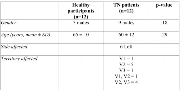
Table 1. Group characteristics Healthy participants (n=12) TN patients (n=12) p-value
Gender 5 males 9 males .18
Age (years, mean ± SD) 65 ± 10 60 ± 12 .29
Side affected - 6 Left -
Territory affected - V1 = 1 V2 = 5 V3 = 1 V1, V2 = 1 V2, V3 = 4 -
Table 2. Patients characteristics Patient
number Age (yrs)/ gender Affected side territory Affected (daily doses) Medications
1 67/ ♂ Left V2 Gabapentine 1200 mg BID
Pregabalin 300 mg BID Naproxen 500 mg PRN
2 69/ ♂ Right V2, V3 Pregabalin 150 mg BID
3 78/ ♂ Left V2 Oxcarbazepine 150 mg BID
4 66/ ♂ Left V2, V3 Gabapentine 300 mg QID
Carbamazepine 200 mg TID Topiramate 100 mg TID
5 59/ ♂ Right V2 Carbamazepine 1200 mg TID
Acetylsalicylic acid 325 mg QD
6 53/ ♂ Left V2 Gabapentine 300 mg TID
7 65/ ♂ Left V1 Oxcarbazepine 600 mg TID
8 59/ ♂ Right V2, V3 Oxcarbazepine 900 mg BID
Baclofen 20 mg TID
9 42/ ♀ Left V2, V3 Oxcarbazepine 300 mg TID
10 65/ ♂ Right V1, V2 Oxacarbazepine 600 mg TID
11 33/ ♀ Right V3 Carbamazepine 1200 mg BID
Pregabalin 75 mg BID
12 59/ ♀ Right V2 Oxacarbazepine 150 mg BID
Table 3. Heart rate (mean ± SD)
Healthy participants
(n=12) TN patients (n=12) t-score p-value
Resting heart rate
(BPM) 66 ± 10 67 ± 12 -0.29 .78
Heart rate during
CPT (BPM) 71 ± 11 70 ± 12 0.17 .87
Change in heart rate (CPT – rest; BPM)
5 + 3 3 ± 4 1.38 .18
Table 4. Baseline HRV values Healthy participants (n=12) TN patients (n=12) t-score p-value Resting sympathetic activity (LF)
(normalized unit, mean ± SD)
63 ± 27 48 ± 23 1.52 .14
Resting parasympathetic activity (HF)
(normalized unit, mean ± SD)
Table 5. Relationships between HRV measures and clinico-demographic variables
Change in sympathetic activity p-value Change in parasympathetic activity p-value
Age† r = -0.09 .69 r = -0.09 .67
Sex†† t = -0.88 .39 t = 0.82 .42
Disease duration (month) † r = -0.26 .40 r = 0.74 .11
Average number of daily pain paroxysms in the preceding week†
r = -0.58 .049 r = 0.42 .18
Mean pain intensity of pain paroxysms experienced in the preceding week (0 -100) †
r = 0.49 .11 r = 0.26 .41
Figure captions
Figure 1. Mean change in low frequency (LF) between baseline (resting condition) and CPT. Both healthy controls and TN patients had an increase in sympathetic activity (LF) during CPT. TN patients showed a greater increase in sympathetic activity when compared to healthy controls (* p < .05).
Figure 2. Mean change in high frequency (HF) between baseline (resting condition) and CPT. Both healthy controls and TN patients showed a decrease in
parasympathetic activity (HF) during CPT. TN patients showed a greater decrease in parasympathetic activity when compared to healthy controls (* p < .05).
References
1. Scrivani SJ, Mathews ES, Maciewicz RJ. Trigeminal neuralgia. Oral Surg Oral
Med Oral Pathol Oral Radiol Endod. 2005;100:527-38.
2. Zakrzewska JM. Diagnosis and differential diagnosis of trigeminal neuralgia. Clin J
Pain. 2002;18:14-21.
3. Rose FC. Trigeminal neuralgia. Arch Neurol. 1999;56:1163-4.
4. Cole CD, Liu JK, Apfelbaum RI. Historical perspectives on the diagnosis and treatment of trigeminal neuralgia. Neurosurg.Focus. 2005;18:E4.
5. Burchiel KJ. Trigeminal neuropathic pain. Acta Neurochir Suppl. 1993;58:145-9. 6. Turp JC, Gobetti JP. Trigeminal neuralgia versus atypical facial pain. A review of the literature and case report. Oral Surg Oral Med Oral Pathol Oral Radiol Endod. 1996;81:424-32.
7. Blumenfeld H. Brain and environs: Cranium, ventricles and meninges. In: Blumenfeld H, editor. Neuroanatomy through Clinical Cases. Sunderland: Sinauer Associates; 2002. p. 121-210.
8. Dandy WE. Surgery of the brain. In: Lewis D, editor. Practice of Surgery. Hagerstown: WF Prior; 1945. p. 177-200.
9. Jannetta PJ. Arterial compression of the trigeminal nerve at the pons in patients with trigeminal neuralgia. J Neurosurg. 1967;26:Suppl-62.
10. Jannetta PJ. Microsurgical approach to the trigeminal nerve for tic douloureux.
Prog Neurol Surg. 1976;7:180-200.
11. Sindou M, Howeidy T, Acevedo G. Anatomical observations during
microvascular decompression for idiopathic trigeminal neuralgia (with correlations between topography of pain and site of the neurovascular conflict). prospective study in a series of 579 patients. Acta Neurochir. 2002;144:1-12.
12. Ishikawa M, Nishi S, Aoki T, et al. Operative findings in cases of trigeminal neuralgia without vascular compression: Proposal of a different mechanism. J Clin
Neurosci. 2002;9:200-4.
13. Hardy DG, Rhoton AL,Jr. Microsurgical relationships of the superior cerebellar artery and the trigeminal nerve. J Neurosurg. 1978;49:669-78.
14. Kress B, Schindler M, Rasche D, et al. [Trigeminal neuralgia: How often are trigeminal nerve-vessel contacts found by MRI in normal volunteers]. Rofo. 2006;178:313-5.
15. Adams CB. Microvascular compression: An alternative view and hypothesis. J
Neurosurg. 1989;70:1-12.
16. Obermann M, Katsarava Z. Update on trigeminal neuralgia. Expert Rev
17. Noguchi I, Hasegawa J, Kobayashi K, et al. Pain relief by stellate ganglion block in a case with trigeminal neuralgia caused by a cerebellopontine angle tumor. Anesth
Prog. 2002;49:88-91.
18. Kowacs PA, Piovesan EJ, Tatsui CE, et al. Symptomatic trigeminal-autonomic cephalalgia evolving to trigeminal neuralgia: Report of a case associated with dual pathology. Cephalalgia. 2001;21:917-20.
19. Sjaastad O, Pareja JA, Zukerman E, et al. Trigeminal neuralgia. clinical manifestations of first division involvement. Headache. 1997;37:346-57. 20. Strittmatter M, Grauer MT, Fischer C, et al. Autonomic nervous system and neuroendocrine changes in patients with idiopathic trigeminal neuralgia. Cephalalgia. 1996;16:476-80.
21. Manahan AP, Malesker MA, Malone PM. Sphenopalatine ganglion block relieves symptoms of trigeminal neuralgia: A case report. Nebr Med J. 1996;81:306-9.
22. Kranzl B, Kranzl C. The role of the autonomic nervous system in trigeminal neuralgia. J Neural Transm. 1976;38:77-82.
23. Simms HN, Honey CR. The importance of autonomic symptoms in trigeminal neuralgia. clinical article. J Neurosurg. 2011;115:210-6.
24. Heart rate variability: Standards of measurement, physiological interpretation and clinical use. task force of the european society of cardiology and the north american society of pacing and electrophysiology. Circulation. 1996;93:1043-65.
25. Headache Classification Commitee of the International Headache Society. The international classification of headache disorders: 2nd edition. Cephalalgia. 2004;24 Suppl 1:9-160.
26. Bernardi L, Leuzzi S, Radaelli A, et al. Low-frequency spontaneous fluctuations of R-R interval and blood pressure in conscious humans: A baroreceptor or central phenomenon? Clin Sci (Lond). 1994;87:649-54.
27. Cevese A, Gulli G, Polati E, et al. Baroreflex and oscillation of heart period at 0.1 hz studied by alpha-blockade and cross-spectral analysis in healthy humans. J Physiol. 2001;531:235-44.
28. Bloomfield DM, Zweibel S, Bigger JT,Jr, et al. R-R variability detects increases in vagal modulation with phenylephrine infusion. Am J Physiol. 1998;274:H1761-6. 29. Akselrod S, Gordon D, Ubel FA, et al. Power spectrum analysis of heart rate fluctuation: A quantitative probe of beat-to-beat cardiovascular control. Science. 1981;213:220-2.
30. Burr RL. Interpretation of normalized spectral heart rate variability indices in sleep research: A critical review. Sleep. 2007;30:913-9.
31. Lotufo PA, Valiengo L, Bensenor IM, et al. A systematic review and meta-analysis of heart rate variability in epilepsy and antiepileptic drugs. Epilepsia. 2012;53:272-82.
32. Hilz MJ, Dutsch M. Quantitative studies of autonomic function. Muscle Nerve. 2006;33:6-20.
33. Chalaye P, Lafrenaye S, Goffaux P, et al. The role of cardiovascular activity in fibromyalgia and conditioned pain modulation. Pain. 2014;155:1064-9.
34. Goadsby PJ, Lipton RB. A review of paroxysmal hemicranias, SUNCT syndrome and other short-lasting headaches with autonomic feature, including new cases. Brain. 1997;120 ( Pt 1):193-209.
35. May A. Trigeminal autonomic cephalalgias: Diagnosis and management. Pain
Clinical Updates. 2012;20:1-8.
36. Tousignant-Laflamme Y, Goffaux P, Bourgault P, et al. Different autonomic responses to experimental pain in IBS patients and healthy controls. J Clin
Gastroenterol. 2006;40:814-20.
37. Cohen H, Neumann L, Shore M, et al. Autonomic dysfunction in patients with fibromyalgia: Application of power spectral analysis of heart rate variability. Semin
Arthritis Rheum. 2000;29:217-27.
38. Vogel T, Gradl G, Ockert B, et al. Sympathetic dysfunction in long-term complex regional pain syndrome. Clin J Pain. 2010;26:128-31.
39. Eze-Nliam CM, Quartana PJ, Quain AM, et al. Nocturnal heart rate variability is lower in temporomandibular disorder patients than in healthy, pain-free individuals. J
Orofac Pain. 2011;25:232-9.
40. Tassorelli C, Micieli G, Osipova V, et al. Combined evaluation of pupillary and cardiovascular responses to cold pressor test in cluster headache patients.
Cephalalgia. 1998;18:668-74.
41. Furlan R, Colombo S, Perego F, et al. Abnormalities of cardiovascular neural control and reduced orthostatic tolerance in patients with primary fibromyalgia. J
Rheumatol. 2005;32:1787-93.
42. Martinez-Lavin M, Hermosillo AG, Rosas M, et al. Circadian studies of autonomic nervous balance in patients with fibromyalgia: A heart rate variability analysis. Arthritis Rheum. 1998;41:1966-71.
43. Raj SR, Brouillard D, Simpson CS, et al. Dysautonomia among patients with fibromyalgia: A noninvasive assessment. J Rheumatol. 2000;27:2660-5.
44. Stein PK, Domitrovich PP, Ambrose K, et al. Sex effects on heart rate variability in fibromyalgia and gulf war illness. Arthritis Rheum. 2004;51:700-8.
45. Chalaye P, Goffaux P, Bourgault P, et al. Comparing pain modulation and autonomic responses in fibromyalgia and irritable bowel syndrome patients. Clin J
Pain. 2012;28:519-26.
46. Reyes del Paso GA, Garrido S, Pulgar A, et al. Autonomic cardiovascular control and responses to experimental pain stimulation in fibromyalgia syndrome. J
47. Khurana RK, Watabiki S, Hebel JR, et al. Cold face test in the assessment of trigeminal-brainstem-vagal function in humans. Ann Neurol. 1980;7:144-9.
48. Heath ME, Downey JA. The cold face test (diving reflex) in clinical autonomic assessment: Methodological considerations and repeatability of responses. Clin Sci
(Lond). 1990;78:139-47.
49. Rang HP, Dale MM, Ritter JM, Flower RJ, Henderson G. Rang and dale's pharmacology. 7th ed. China: Elsevier Churchill Livingstone; 2012.