O
pen
A
rchive
T
OULOUSE
A
rchive
O
uverte (
OATAO
)
OATAO is an open access repository that collects the work of Toulouse researchers and
makes it freely available over the web where possible.
This is an author-deposited version published in :
http://oatao.univ-toulouse.fr/
Eprints ID : 20324
To link to this article : DOI: 10.1111/jmi.12713
URL :
https://doi.org/10.1111/jmi.12713
To cite this version : Payre, Bruno and Gontier, Etienne and Jarray,
Ahmed and Martinez, Yves and Laugier, Jean Pierre and Delalleau,
Alexandre and Gaillard-Martinie, Brigitte and Anselme, Isabelle and
Goudouneche, Dominique and Fourquaux, Isabelle and Hemati,
Mehrdji and Gerbaud, Vincent and Delisle, Marie Bernadette and
Guilbeau-Frugier, Céline A new HPF specimen carrier adapter for
the use of high-pressure freezing with cryo-scanning electron
microscope: two applications: stearic acid organization in a
hydroxypropyl methylcellulose matrix and mice myocardium. (2018)
Journal of Microscopy, 00. 1-11. ISSN 0022-2720
Any correspondence concerning this service should be sent to the repository
administrator:
[email protected]
doi: 10.1111/jmi.12713
A new HPF specimen carrier adapter for the use of high-pressure
freezing with cryoscanning electron microscope: two applications:
stearic acid organization in a hydroxypropyl methylcellulose
matrix and mice myocardium
B . P A Y R E∗ , E . G O N T I E R †, A . J A R R A Y ‡, Y . M A R T I N E Z k, J . P . L A U G I E R #, A . D E L A L L E A U∗∗, B . M . G A I L L A R D ††, I . A N S E L M E ‡‡, D . G O U D O U N `E C H E∗, I . F O U R Q U A U X∗, M . H E M A T I ‡,
V . G E R B A U D ‡, M . B . D E L I S L E∗, § & C . G U I L B E A U - F R U G I E R∗
∗Centre de Microscopie Electronique Appliqu´ee `a la Biologie, Facult´e de M´edecine Rangueil, Toulouse III, P. Sabatier University, 31062, Toulouse, France
†Bordeaux Imaging Center - UMS 3420-Universit´e Bordeaux / CNRS / INSERM, F-33000, Bordeaux, France ‡LGC, INP, ENSIACET, 4 All´ee Emile Monso, 31432, Toulouse, France
§CHU Toulouse and INSERM U 1037, Toulouse CEDEX, 31059, France
kCNRS, FR3450, Federation de recherche Agrobiosciences, Interactions, Biodiversit´e, BP 42617 Auzeville, F-31326, Castanet Tolosan, France #Centre Commun de Microscopie Appliqu´ee, Universit´e de Nice-Sophia Antipolis, Nice, France
∗∗Pixience, 12 rue Louis Courtois de Vic¸ose, 31100, Toulouse, France
††INRA, Centre Clermont-Ferrand - Theix, UMR1019, University of Clermont-Ferrand 1, UFR M´edecine, UMR101, F-63000, Clermont-Ferrand, France ‡‡Centre de Microscopie Electronique St´ephanois - CMES-Saint Etienne, Universit´e de Lyon, Saint-Etienne, France
Key words. Cryogenic scanning electron microscopy, Cryogenic fracture, Cryogenic freezing, High-pressure freezing, HPF specimen carrier adapter.
liquid concentration and acid stearic occupation surface (only for homogeneous solution). For biological samples as my-ocardium, cytoplasmic structures of cardiomyocyte are easily recognized with adequate preservation of organelle contacts and inner cell organization. We expect this new HPF spec-imen carrier adapter would enable more SEM-studies using HPF.
Introduction
Cryoscanning electron microscope (cryo-SEM) examination of fully hydrated cryofixed chemical and biological specimens for morphological studies has been extensively used (Wergin
et al.,1993; Nakatomi et al., 2005; Nguyen et al., 2015; En-gstr¨om et al., 2016).
For the past decade, cryo-SEM observation has experi-enced a wonderful development thanks to the improvement of cryo-observation modules. However, cryopreparation of sam-ples is key to a cryo-observation of quality and can be very challenging.
Tissue which has been cryofixated remains hydrated and specimen components are rapidly immobilized, which is not the case using critical point drying by carbon dioxide (Polliack
et al., 1973) or hexamethyldisilazane (HMDS) (Hazrin-Chong & Manefield, 2012). Cryofixation involves immersing fresh
Summary
Cryogenic transmission electron microscopy of high-pressure freezing (HPF) samples is a well-established technique for the analysis of liquid containing specimens. This technique en-ables observation without removing water or other volatile components. The HPF technique is less used in scanning elec-tron microscopy (SEM) due to the lack of a suitable HPF speci-men carrier adapter. The traditional SEM cryotransfer system (PP3000T Quorum Laughton, East Sussex, UK; Alto Gatan, Pleasanton, CA, USA) usually uses nitrogen slush. Unfortu-nately, and unlike HPF, nitrogen slush produces water crystal artefacts.
So, we propose a new HPF specimen carrier adapter for sample transfer fromHPF system to cryogenic-scanning electronic mi-croscope (Cryo-SEM). The new transfer system is validated us-ing technical two applications, a stearic acid in hydroxypropyl methylcellulose solution and mice myocardium. Preservation of samples is suitable in both cases. Cryo-SEM examination of HPF samples enables a good correlation between acid stearic Authors M.B. Delisle and C. Guilbeau-Frugier contributed equally to this manuscript.
Correspondence to: B. Payre, Centre de Microscopie Electronique Applique´e a` la Biologie, Faculte´ de Me´decine Rangueil, Toulouse III, P. Sabatier University, 31062 Toulouse, France. Tel/fax: +330562889035; e-mail: [email protected]
and hydrated samples into a liquid cryogen. This technique is commonly followed by freeze fracture and freeze drying (Pieniazek & Messinat, 2016).
The technique of nitrogen slush cryofixation is also based on rapid freezing of the sample. However, the nitrogen liq-uid calefaction phenomenon resulting from this technique in-duces slow cooling by freezing and results in water crystals formation due to nucleation and ice crystal growth (Studer
et al., 1989). Usually, nitrogen slush fixation is used in SEM
systems fitted with cryopreparation chamber.
Many cryofreezing techniques are proposed as nitrogen slush alternative. Studies have performed for freon (Lyer et al., 2005), ethane (Issman & Talmon, 2012), propane (Inou´e & Koike, 1989), propane jet (Walther, 2003), and high-pressure freezing (HPF) (Walther, 2003; Fujita et al., 2012) cryofixation.
Unlike the techniques above, HPF enables the freezing of specimen up to a thickness of about 200 µm without the use of cryoprotectant (Walther, 2003) (although preserved frozen sample thickness is dependent of sample kind (Studer
et al., 1995)). The absence of cryoprotectant is essential for
sublimation after cryofracture, because cryoprotectant slows down sublimation.
HPF uses high pressure and very low temperatures to achieve high cooling rates (Bald & Robard, 1978). HPF is a technique to produce vitreous ice which preserves hydrated samples (Dubochet et al., 1983; Moor, 1987; Studer et al., 1992). This technique prevents cryofixation artefacts and has been recommended for better preservation of hydrated sam-ples (Dubochet et al., 1988; Vanhecke et al., 2008).
After HPF, sample cryofracture and freeze etching can be done before SEM examination. This freeze etching cor-responds to a sublimation of frozen water (ice) at low tem-perature and high vacuum. Sublimation of surface ice un-der vacuum reveals details of the fractured face that were originally hidden (Echlin, 2009). After deep etching, struc-tures protruding from the remaining ice are generally not col-lapsed. This is probably a result of the retention of unfreezable water (also known as ‘hydration’ or ‘bound’ water) (Given, 1991).
Each step of cryopreparation can be challenging and is key to a suitable examination of samples. That is why many parts of cryopreparation system have been designed specifically to answer specific needs (Inou´e & Koike, 1989; Lyer et al., 2005; Issman & Talmon, 2012)
One of the limits for the use of HPF for SEM observation is linked to the lack of well adapted modules for transfer of HPF specimen carrier from HPF systems (EM-ICE, Leica, Nanterre, France, or, HPM100, Leica, Nanterre, France) to the SEM cry-otransfer systems (Alto, Gatan, Pleasanton, CA, United States or PP300 and PP3010T Quorum, Laughton, East Sussex, UK). In order to allow this HPF specimen carrier transfer it is necessary to implement an adapter. However, this adapter has to be developed within specific constraints. Firstly, it is
essential that the HPF specimen carrier sandwich is firmly fixated to the adapter, as it will be necessary to apply pres-sure on the sandwich to open it and to create a fracture using the scalpel. Secondly, the HPF carrier sample fixation has to be done in liquid nitrogen. Finally, the HPF specimen carrier adapter has to be locked to the specimen shuttle (this specimen shuttle is designed by Quorum and Gatan cryopreparation chamber).
Taking all these constraints into account, we designed an HPF specimen carrier adapter. To validate its use we tested two specimens: a chemical one [hydroxypropyl methylcellulose (HPMC) with stearic acid (SA)] and a biological one (mice myocardium).
Cellulose derivatives, such as HPMC, are often used in gran-ulation processes as a protective colloid to coat hydrophobic particles such as SA (Mahato & Narang, 2011). When dis-persed in water, hydrophobic SA particles tend to minimize their interaction with the water and thus form large aggre-gates, whereas HPMC can be dissolved in water. HPMC poly-mer is able to form a gel network when mixed with water. Upon dispersing SA in the HPMC-water blend, the HPMC gel will entrap SA particles and prevent their aggregation, thus producing stable SA particles (Jarray et al., 2015). Particle dispersion is known to be stable when the particles remain dispersed over a long time (i.e. the total number of particles remains constant over time). As, HPMC-SA is hydrated, it is a good model to test cryofixation quality. It can be verified if, by examination, the HPMC-SA correlated to reality. To this purpose, we have calculated SA-surface in total surface and compared it to real SA concentration.
For biological sample, we chose mice myocardium, because cardiomyocyte morphology and inner cell structure organi-zation are well established. It is therefore possible to evaluate technical quality of the technique thanks to the examination of the SEM-myocardial ultrastructure.
Materials and methods
HPMC-SA sample preparation
Aqueous polymeric solutions of HPMC-SA are prepared by dispersing SA in a solution of HPMC and water at 85°C, under agitation for 30 to 60 mins with a rotor-stator homogenizer (Ultraturrax T25, Janke and Kunkel, Staufen, Germany). The solution is then crystallized by cooling the suspension to room temperature. Solutions are thereafter degassed at 50 mbar for 2 h and stored at 5°C for at least 24 h.
Mice myocardial preparation
Studies were performed on 2-month-old male C57BL/ 6JOlaHsd mice (purchased from Envigo). Experimental animal protocols were carried out in accordance with the European and French regulation guidelines for animal experimentation
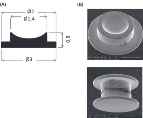
Fig. 1. HPF specimen carrier. In HPF (HPM100 or ICE, Leica, Nanterre, France) samples are frozen in HPF specimen carrier. (A) manufacturer diagram of this carrier with size of each component (in mms). (B) shows the cavity where the sample is inserted and the (C) shows the sandwich which allows to freeze the sample between two HPF specimen carrier. This technique allows a fracture when separating the two carriers and a sample protection from any water contamination.
and Figs. 3 and 4) and settle down on the specimen shuttle (AL200077B, Quorum) which is slipped into the cryostage of cryochamber preparation or of SEM cryochamber (Fig. 2). The technical diagram, the picture and the 3D graphic of the new HPF specimen carrier adapter are shown in Fig. 3. The imen shuttle, HPF specimen carrier adapter, and HPF spec-imen carrier constitute a combined unit (Fig. 4A). The HPF specimen carrier adapter is organized into two areas, a de-posit area and a fixative area (Fig. 4B). Fixation of the HPF sandwich specimen carrier was achieved by slipping it onto slide channels 0.26 mm thick (Fig. 4B, C). This easy fixa-tive method can be used in nitrogen liquid. Moreover, HPF sample fixative area is designed with same principle to ac-cept two specimens reducing the whole procedure duration (Fig. S2).
Transfer of the HPF prepared sample
HPF specimen carrier adapter (Fig. 3), is first fixated by a screw to the specimen shuttle (Quorum; Figs. 2 and 4) and dried carefully. The transfer occurs in a liquid nitrogen dewar. A large slusher pot full of liquid nitrogen is used to place the HPF specimen carrier (Leica) on the new HPF specimen carrier adapter. The following steps to introduce the sample in the cryopreparation chamber are well established and follow user manual steps (PP3000T, Quorum).
and were approved by the local ethical committee. Mice were euthanized through intraperitoneal injection of pentobarbital sodium (160 mg/kg).
Immediately after removal, the heart is plunged in cold PBS during a few seconds thus stopping immediately heart beating. Then, the heart was cautiously slicedwith razor blade (fewmm thick) on ice bed without any torsions or pressures.
HPF-Cryofixation of HPMC or HPMC-SA or mice myocardium
The HPMC or HPMC-SA is inserted between two HPF specimen carriers dedicated for cryofracture (Leica) to create a ‘sand-wich’ (Fig. 1), and it is loaded immediately into a HPM100 HPF machine (Leica). The sample is fixated and frozen within 5 ms at 2050 bar and samples are then transferred to cryovials under liquid nitrogen.
For mice myocardium, each myocardial piece is briefly im-merged in hexadecane solution, and then inserted between two HPF specimen carriers, loaded into a HPM100 HPF ma-chine (Leica) and fixated following the same procedure as HPMC.
New HPF specimen carrier adapter
The HPF specimen carrier adapter, made in aluminum, is de-signed to receive the HPF specimen carrier sandwich (Fig.1
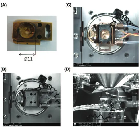
Fig. 2. Specimen shuttle. This shuttle is used for cryopreparation chamber (ref AL200077B, Quorum PP3000T, Laughton, East Sussex, UK). It has a hole of 11 mm (A) in its centre and a screw that allows stubs binding suitable for various samples. It uses to perform samples freezing in nitrogen slush. Specimen shuttle is adapted for SEM cold stage, picture B shows the top view without the specimen shuttle and C with it. (D) overview of SEM chamber with the implement of the specimen shuttle.
Sample cryofracture
The fracture of the sandwich specimen carrier HPF is pro-duced in a PP3000T cryochamber by contact with scalpel blades (Fig. 5A); the whole surface of the sample is fractured (Fig. 5B). If the separation of the sandwich specimen carrier occurs without fracture, it is possible to perform the fracture directly by contact of the sample with a scalpel blade, which will induce a small fracture area (Fig. 5C).
Freeze drying of the surface of the sample
Ice sublimation is necessary for surface sample observation. Ice is directly transformed into water vapor at -95°C in 30 mins. Sublimation is performed in the cryostage of cryopreparation chamber (Quorum).
Examination with scanning electron microscope
Samples are metalized in cryostage of Quorum cryochamber preparation with Platinium for 60 s at 10 mA. The platinium layer is thinner than 1 nm but allows examination at high volt-age with better resolution. Sample was introduced into SEM chamber Quanta 250 FEG FEI SEM (Thermo Fisher, Hillsboro,
OR, USA) and was observed at 1–5 kV with spot size 2 and 0.9–1,1 cm working distance. We used secondary electrons detector located in SEM chamber, so the optimal working dis-tance recommended is around 1 cm. The temperature was kept below −140°C during SEM imaging.
SA counting by image analysis
The density of the SA in the HMPC mixture is measured with the ImageJ image processing software (Rasband, 1997). We chose this software because previous studies validated it in a similar context: Hermana & Walz (2015) to determine the size distribution of adsorbed latex nanoparticles in colloid disper-sion; Ellison and collaborators (2014) for the calculation of the mean particle diameter of coated silver nanoparticles; and Baghbanzadeh et al. (2015) to measure the surface roughness of membranes.
In ImageJ, in order to remove smooth continuous back-grounds from SEM images of HPMC-SA dispersions, we applied a ‘rolling ball’ of 30 pixels. To improve the counting and selection of high intensity objects, median (radius 2 pixels) and high pass (radius 0.7 pixel) filters are applied. These filters remove the background noise in the images and increase the intensities of the particle signals in the dispersions. A
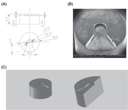
Fig. 3. HPF specimen carrier adapter. This adapter is designed to be locked in the specimen shuttle and to fasten HPF the specimen carrier. (A) HPF specimen carrier adapter manufacturer technical diagram. (B) SEM picture of this adapter (C) 3D overview diagram and cutting diagram.
Fig. 4. Assembly of technical parts (specimen shuttle, HPF specimen carrier adapter, HPF specimen carrier). (A) overview of every parts assembled. HPF specimen carrier is fastened in HPF specimen carrier adapter, itself placed in the specimen shuttle. (B) SEM picture of HPF specimen carrier adapter areas. Deposit area is used to easily place the HPF specimen carrier. Slide channels enable to slip HPF specimen carrier to the fixative area. (C) 3D-overview and cutting diagrams depict HPF specimen carrier adaptor with HPF specimen carrier in fixative area.
Results
Sample transfer with our HPF specimen carrier adapter
Sample transfer is easy but some precautions need to be taken. A screw must be used to fixate the HPF specimen carrier threshold is applied to the images to select objects to be
counted to those with intensity between 90 and 255 in grey levels (8 bit images), with a save of the count mask of the outlines of the counted objects. A second count mask is applied to select the counting area on the flat regions.
Fig. 5. Specimen cryofracture by removing HPF specimen carrier sandwich top in cryochamber preparation with scalpel blades. (A) (top picture) sandwich HPF specimen carrier in HPF specimen carrier adaptor in the cryochamber PP3000T. (middle picture) scalpel blade pushing sandwich HPF specimen. (bottom picture): HPF specimen carrier bottom remaining after sample fracture and after removing the top of the HPF specimen carrier. (B) sample cryofractured due to HPF specimen carrier dissociation. (C) without sample cryofracture after HPF specimen carrier dissociation the cryofracture is produced manually with scalpel blade.
adapter on the specimen shuttle. It is very important to dry the specimen carrier adapter before nitrogen liquid immersion to prevent water crystals in the slide channel. Ten minutes are necessary for the system to reach liquid nitrogen temperature; this is characterized by boiling around the end of the piece. The HPF Specimen carrier transfer from the HPF (or cryotube stockage) to the HPF specimen carrier adapter, occurs in an adapted box containing liquid nitrogen. Cryoelectron microscopy tweezers at liquid nitrogen temperature are used to put HPF specimen carrier on the deposit area of the HPF specimen carrier adapter and to push it on the slide channels to the fixative area. This is technically very simple, however the sliding of the carrier could be blocked by water crystals if this system isn’t well dried or if the sample overflows from the HPF specimen carrier during HPF. Under these conditions, the specimen carrier can slide if two tweezers are used for a strong push. Increasing the size of the slide channel would be a technical solution to avoid crystal blocking but this solution increases the risk to drop the HPF specimen carrier during transfer.
The use of two HPF specimen carriers during HPF provides a specimen carrier sandwich. Specimen carrier sandwich sep-aration on the SEM cryotransfer chamber is very easy with the scalpel blade. Therefore, the whole surface of the sample is cry-ofractured without contamination by water crystals. Should
the fracture not occur, it has to be done manually in SEM cryotransfer chamber. However, extraction of the plastic sup-port for HPF can sometime result in a premature cryofracture, which can be responsible for pollution of the specimen by the emergence of water crystals. This pollution can be removed in the SEM cryotransfer chamber by directly fracturing the specimen again with the scalpel blade or by sublimation.
HPMC and HPMC-SA examination
HPMC does not contain any perforations and has a smooth surface, meaning there is no water crystal artefacts. Some pleating can be observed (Fig. 6A). This data confirms the suitability of cryofreezing by HPF, compared to nitrogen slush which induces holes ‘honeycomb-like structure’ (Fig. S1).
When HPMC is mixed with SA, white crystals appear. Those crystals have been previously described as SA crystals (Jarray, 2015, 2016) and never occur in ‘pure HPMC-water’ (Fig. 6).
The general appearance of HPMC-SA mixtures in water at SA 2% (Fig. 6B) or at SA 10% (Fig. 6C) is quite same, even if the number of SA crystals increase significantly for the SA 10% mixture. White SA crystals are distributed in the HPMC–water with a size below 1 µm. Some of the SA crystals are outside (or partially outside) the HPMC-amorphous phase, whereas others are covered with the HPMC-water phase but are still
Fig. 6. SEM examination fractured surface of various SA concentration in HPMC. (A) HPMC in water [10% (w/w)] exhibits a smooth, sometime pleating surface. HPMC-SA in water under different SA concentrations; (B) HPMC-SA 2%, SA is spread in HPMC, inset: small SA crystal (*). (C) HPMC-SA 10%, SA crystals are numerous but still scattered, inset: small SA crystals (*). (D) HPMC-SA 20%, HPMC cannot keep isolated SA crystals and SA agglomerates appear. Inset: high magnification of an SA agglomerate (*). HPMC: hydroxypropyl-methylcellulose; SA: stearic acid. (Scale bar 5 µm).
artefacts and enough etching to distinguish cell inner struc-tures details. As expected, HPF-myocardium is well preserved and cryofracture enables a near 3D-examination of cardiomy-ocytes organization in myocardium (Fig. 8A). Cardiomyocyte cytoplasm is more homogeneous with HPF than with nitro-gen slush freezing, with fewer water crystals artefacts (Fig. S1). Contractile apparatus of the inner cardiomyocyte is discerned. Specific organization of interfibrillar mitochondria along con-tractile apparatus can be observed (Fig. 8B). At high magni-fication, Z lines appear and can be measured (Fig. 8C), inner mitochondrial structures are well seen and cristae can be de-tected (Fig. 8D).
Discussion
Cryo-SEM examination practices increased tremendously over the last decade. The crucial step for cryo-SEM observation of hydrated samples is cryopreparation. There are various techniques of cryofixation available. HPF is a cryofixation validated and adequate technique for hydrated sample par-ticularly, samples size are preserved on around 200µm of thickness. Furthermore, this technique does not entail the use distinguishable from the swollen HPMC–water surface. There
are no big agglomerates.
At SA 20% (Fig. 6D) the structure of the HPMC-SA mixture changes; the SA crystal distribution becomes polydispersed and it becomes difficult to distinguish SA crystals in the aggre-gates. There is also formation of big agglomerates of SA with different morphologies (Fig. 6D).
Image analysis
Quantification of total HPMC-water surface covered by SA crystals (compared to total surface and expressed in per-centage), with the scientific image analysis program ImageJ (Rasband, 1997) demonstrates an increase of this SA surface correlated with SA concentration. SA crystals surface calcu-lated is well correcalcu-lated with SA concentration until SA 10% (Fig. 7). At SA 20%, the surface calculated is underestimated because of emergence of aggregates.
Mice myocardium examination
SEM frozen tissue examination can be sensitive because this technique needs to optimize freezing without water crystals
Fig. 7. Measure of area covered by SA in HPMC at various SA concentra-tions. SA surface is measured from SEM pictures by imageJ and expressed as percentage of SA area compared to total area. There is a good correla-tion at SA 2% and SA 10% between SA concentracorrela-tion in HPMC mixture and the SA area measured. At SA 20%, SA area measure underestimated compared to the real SA concentration.
of any cryoprotectant. Cryoprotectants are known to prevent a correct examination of the sample after cryofracture and after a long sublimation. Indeed, cryofracture allows observation of the cell surface and inside the inner cell, a near 3D-estimation of tissue level organization (depending of the random fracture) and is acclaimed for morphological examination. However, the transfer of cryosample in cryo-SEM can be challenging be-cause of the lack of suitable HPF-cryotransfer system. In this study, we designed a new HPF specimen carrier adapter for HPF transfer sample.
Various technical constraints have been considered in the design of the carrier, linked to the steps of the cryoprepara-tion and linked to the design of the existing specimen carrier or specimen shuttle currently produced by manufacturers. First, the HPF specimen carrier adapter should be locked to the specimen shuttle and must not show significant expan-sion under the action of low temperature. That is why we have chosen aluminum material because its shape doesn’t change at low temperature. Its diameter is 10 mm to be adapted to 11 mm of specimen shuttle hole. Then the sample is frozen between two HPF specimen carriers. This ‘sandwich’ should
Fig. 8. SEM examination fractured of ultrastructure mice myocardium. (A) At low magnification, cardiomyocytes can be examined in the myocardium with a 3D view (sarcolemma is apparent: arrow). (B) intracellular structures of cardiomyocyte are easily recognized: contractile apparatus (Z) and interfibrillar mitochondria (m) are well distinguished. Intercellular space (is) is noticeable. (C) at high magnification, mitochondria (m) are well visualized and also their organization in the cardiomyocyte, between contractile apparatus. Z-line can be measured (Z; double arrow). (D) mitochondria are suitable cryofixated and cristae organization can be observed.
SA with different morphologies, meaning that HPMC becomes unable to stabilize SA. Our data are in accordance with liter-ature (Jarray et al., 2016). Overall, in our study, there is no water crystal perforation and the structure of the SA crystals in the HPMC polymer gel is well preserved. To confirm our observations, we counted SA crystals with ImageJ. Our data indicates that SA crystals are well dispersed in the HPMC– water mixture. They also show that the HPF technique is able to preserve the structure of dispersed particles. At SA 20% (w/w), we obtain 15.65% ± 1.85 of SA surface on the sample total surface. This value, lower than the real SA concentration in HPMC-SA (i.e. 20% (w/w)), can be explained by a polydis-persity in the mixture; at high polydispolydis-persity the number of chosen images treated by ImageJ become less representative of the distribution of the particles, but also by SA-aggregates. That’s why a high calculation error may be obtained.
Heart is a biological sample SEM-examined for more than 30 years and cardiomyocyte inner structures are well known. However, studies of 80’s with various freezing techniques and more recent (Ashraf M, 1982; Dalen H et al., 1983; M ¨uhlfeld
et al., 2006), demonstrated that it can be difficult to observe myocardium without water crystals artefacts. Therefore test-ing the new HPF specimen carrier adapter with mice my-ocardium seemed challenging enough. Despite the challenge the result is that the HPF-myocardium is adequately preserved, without any water crystscals artefacts, allowing a good ex-amination of sarcolemma, contractile apparatus, interfibrillar mitochondria with cristae details and also allows if necessary to measure sarcomeres length.
Conclusion
The HPF specimen carrier adapter we designed,is available for transfer of HPF specimen carrier. It can be used with cry-otransfer system (Gatan et Quorum) and therefore can be used with cryo-FIB provided with these system.
The tests performed with this adapter, with HPF cryofreezed HPMC without and with SA, confirm that aqueous samples can be frozen and transferred adequately to allow examina-tion but also quantitative analysis. Same quality is obtained with myocardium extending the adapter practice to biological samples. It has to be however noted that every tissues being different the protocol might not be relevant for other organs. We, however, hope that the HPF specimen carrier adapter will allow future biological tests.
Acknowledgements
The authors thank the ‘Mission pour l’interdisciplinarit´e’ of Centre National de la Recherche Scientifique via the R´eseau National des Centres Communs de Microscopie (RCCM) for supporting this work. The work was partly supported by fund-ing from the Minist`ere de l’Enseignement Sup´erieur et de la Recherche (ANR-10-INBS-04 FranceBioImaging), Contrat be fixated firmly to the HPF specimen carrier adapter and this
fixation must be performed in liquid nitrogen. The two parts of sandwich should be separated creating a sample-fracture. This operation requires some manual pressure on the system and therefore implies a stress on the fixation system. That is why we have chosen a fixation system in slides which allows fixation in immersion in the liquid nitrogen and which also firmly fixates the HPF support carrier on the HPF support car-rier adapter.
Use of our HPF specimen carrier adapter is easy without any damage or any disadvantage requiring redesigning the adapter. A few small difficulties, such as the separation of the two parts of the sandwich without generating fracture, are easily resolved by fracturing manually with the scalpel blade.
To validate our HPF specimen carrier adapter the author tested it in ‘real’ condition using two samples: a chemical and aqueous sample (HPMC) and a biological sample (mice myocardium). HPMC is a nonionic cellulose ether, which is soluble in water and has thermal gelation properties. Because of its mechanical properties, HPMC is largely used in phar-maceutical industries. It forms a film that can be used as a matrix containing various substances (e.g. nanoparticles or SA) (De Moura et al., 2008). Its aqueous nature makes it a good candidate for cryotests. For biological sample, we chose myocardium because its morphology is well established and inner cardiomyocytes structures are well described. After cry-ofracture, ice etching is performed at −95°C during 30 min. At this temperature, amorphous ice does not present major modification due to recrystallization because artefactual mod-ifications are not apparent at the magnification the samples are examined. According to DJ Prior data (Prior et al., 2015), we can suppose that if ice grains are present, their size is smaller than 1 nm, and they are therefore not detectable. Any-way, these data concern water and dirty water freezing. For biological samples, the removal of water by etching leads arte-factual morphological modifications due to the shrinking and collapsing structures (Walther, 2003) which hide recrystal-lization artefacts.
The superior quality freezing with HPF compared to nitro-gen slush freezing needn’t be demonstrated (Yong et al., 2014), and as expected we observed a suitable quality of cryofixation with HPF. After HPF, HPMC is homogenous and white crys-tals of SA are easily distinguishable. At high pressure, water is more viscous than at atmospheric pressure, which drasti-cally reduces the nucleation and thus the growth rate of water crystals. Therefore our HPF test permits observation of HPMC structural details similar to its native state. This first step vali-dates our adapter confirming no disturbance in transfer of the HPF cryosample.
Thereafter we have tested the stability of HPMC freezing by mixing HPMC with SA at different concentrations. As ex-pected, at low SA concentrations in the HPMC-SA mixture, SA crystals are stable with good preservation of the morphology of the blend. But at SA 20%, we observed big agglomerates of
Plan Etat R´egion Midi Pyr´en´ees (CPER) et Fonds Europ´een de D´eveloppement R´egional (FEDER) Midi Pyr´en´ees (Microscopie Electronique pour la biologie, la sant´e et les agro biosciences en midi Pyr´en´ees). We thank company Eclisse for HPF carrier adapter machining.
We want to greatly thank Miss Bartlett Juliette for her English reviewing.
References
Ashraf, M. (1982) Preparation of normal and ischemic myocardial tissue for scanning electron microscopy. Scan Electron Microsc. (Pt 2), 697– 707.
Baghbanzadeh, M., Rana, D., Matsuura, T. & Lan, C.Q. (2015) Effects of hydrophilic CuO nanoparticles on properties and performance of PVDF VMD membranes. Desalination. 369, 75–84.
Bald, W.B. & Robard, W. (1978) A device for the rapid freezing of biolog-ical specimens under precisely controlled and reproducible conditions.
J. Microsc. 112, 3–15.
Dalen, H., Scheie, P., Myklebust, R. & Saetersdal, T. (1983) An ultrastruc-tural study of cryofractured myocardial cells with special attention to the relationship between mitochondria and sarcoplasmic reticulum.
J. Microsc. 131(Pt 1),35–46.
De Moura, M.R., Avena-Bustillos, R.J., McHugh, T.H., Krochta, J.M. & Mattoso, L.H. (2008) Properties of novel hydroxypropyl methylcellu-lose films containing chitosan nanoparticles. J. Food Sci. 73, N31–37. Dubochet, J., Adrian, M., Chang, J., Homo, J., Lepault, J., McDowall, A.W.
& Schultz, P. (1988) Cryo-electron microscopy of vitrified specimens.
Quart Rev. Biophys. 21, 129–228.
Dubochet, J., McDowall, A.W., Menge, B., Schmid, E.N. & Lickfeld, K.G. (1983) Electron microscopy of frozen-hydrated bacteria. J. Bacteriol.
155, 381–390.
Echlin, P. (2009) Handbook of Sample Preparation for Scanning Electron
Microscopy and X-Ray Microanalysis. Springer, Boston.
Ellison, J., Wykoff, G., Paul, A., Mohseni, R. & Vasiliev, A. (2014) Efficient dispersion of coated silver nanoparticles in the polymer matrix. Colloids
Surf. A Physicochem. Eng. Asp. 447, 67–70.
Engstr¨om, M.T., Karonen, M., Ahern, J.R., Baert, N., Payre, B., Hoste, H. & Salminen, J.P. (2016) Chemical structures of plant hydrolyzable tannins reveal their in vitro activity against egg hatching and motility of haemonchus contortus nematodes. J. Agric. Food Chem. 64, 840–851. Fujita, A., Fujimoto, T., Ozato-Sakurai, N. & Suzuki, H.A. (2012) method for efficient observation of intracellular membranes of monolayer cul-ture cells by quick-freeze and freeze-fraccul-ture electron microscopy.
J Electron Microsc. (Tokyo). 61, 441–446.
Given, P.S. Jr. (1991) Molecular behavior of water in a flour-water baked model system. Adv. Exp. Med. Biol. 302, 465–483.
Hazrin-Chong, N.H. & Manefield, M. (2012) An alternative SEM drying method using hexamethyldisilazane (HMDS) for microbial cell attach-ment studies on sub-bituminous coal. J. Microbiol. Methods. 90, 96–99. Hermana, D. & Walz, J.Y. (2015) Adsorption and stabilizing effects of highly-charged latex nanoparticles in dispersions of weakly-charged silica colloids. J. Colloid Interface Sci. 449, 143–151.
Inou´e, T. & Koike, H. (1989) High resolution low-temperature scan-ning electron microscopy for observing intracellular structures of quick frozen biological specimens. J. Micosc. 156, 137–147.
Issman, L. & Talmon, Y. (2012) Cryo-SEM specimen preparation under controlled temperature and concentration conditions. J. Micosc. 246, 60–69.
Jarray, A., Gerbaud, V. & Hemati, M. (2015) Prediction of solid – binder affinity in dry and aqueous systems: work of adhesion approach vs. ideal tensile strength approach. Powder Technol. 271, 61– 75.
Jarray, A., Gerbaud, V. & Hemati, M. (2015) Stearic acid crystals sta-bilization in aqueous polymeric dispersions. Res. Design. 110, 220– 232.
Jarray, A., Gerbaud, V. & Hemati, M. (2016) Structure of aqueous colloidal formulations used in coating and agglomeration processes: mesoscale model and experiments, Powder Technol. 291, 244–261.
Lyer, R., Arunagirinathan, M.A., Prabhu, C.S. & Bellare, J. (2005) An im-proved specimen loader for cryo-scanning electron microscopy.
Scan-ning. 27, 141–146.
Mahato, R.I. & Narang, A.S. (2011) Pharmaceutical Dosage Forms and Drug
Delivery. 2nd edn. CRC Press, Boca Raton.
Moor, H. (1987) Theory and practice of high pressure freezing. Cryotechniques
in Biological Electron Microscopy(ed. by R.A. Steinbrecht & K. Zierold), pp. 175–191. Springer, Berlin, Heidelberg.
M ¨uhlfeld, C. & Richter J. (2006) High-pressure freezing and freeze sub-stitution of rat myocardium for immunogold labeling of connexin 43.
Anat Rec A Discov Mol Cell Evol Biol. 288(10),1059–1067.
Nakatomi, R., Hayashida, T., Fujimoto, K., Tohyama, K. & Hashikawa, T. (2005) Cryo-SEM and subsequent TEM examinations of identical neural tissue specimen. Brain Res. Prot. 14, 100–106.
Nguyen H.H., Payr´e, B., Fitremann, J., Lauth-de Viguerie, N. & Marty, J.D. (2015) Thermoresponsive Properties of PNIPAM-Based Hydrogels: Effect of Molecular Architecture and Embedded Gold Nanoparticles.
Langmuir. 31, 4761–4768.
Pieniazek, F. & Messinat, V. (2016) Scanning electron microscopy com-bined with image processing technique: analysis of microstructure, texture and tenderness in semitendinous and gluteus medius bovine.
Scanning. 9999, 1–8.
Polliack, A., Lampen, N., Clarkson, B.D., De Harven, E., Bentwich, Z., Siegal, F.P. & Kunkel, H.G. (1973) Identification of human B and T lymphocytes by scanning electron microscopy. J Exp Med. 138, 607– 624.
Prior, D.J., lilly, K. & Seidemann, M. (2015) Making EBSD on water ice routine. J Microsc. 259, 237–256.
Rasband, W.S. (1997) ImageJ, National Institutes of Health, Maryland, USA. http://imagej.nih.gov/ij/ (accessed 2013).
Studer, D., Muller, M. & Michel, M. (1989) High pressure freezing comes of age. Scanning Microsc. 3, 253–269.
Studer, D., Hennecke, H. & M ¨uller, M. (1992) High-pressure freezing of soybean nodules leads to an improved preservation of ultrastructure.
Planta. 188, 155–163.
Studer, D., Michel, M., Wohlwend, M., Hunziker, E.B. & Buschmann, M.D. (1995) Vitrification of articular cartilage by high-pressure freezing.
J. Microsc. 179, 321–332.
Vanhecke, D., Graber, W. & Studer, D. (2008) Close-to-native ultrastruc-tural preservation by high pressure freezing. Method Cell Biol. 88,151– 164.
Walther, P. (2003) Recent progress in freeze-fracturing of high-pressure frozen samples. J Microsc. 212, 34–43.
Wergin, W.P., Sayre, R.M. & Erbe E.F. (1993) Use of low temperature scanning electron microscopy to observe frozen hydrated specimens of nematodes. J. Nematol. 25, 214–226.
Yong, W., Jing, L., Kim, R., Tseng-Ming, C. & Matthew, L. (2014) Ex-tracellular matrix reorganization during cryo preparation for scan-ning electron microscope imaging of staphylococcus aureus biofilms.
Microsc. Microanal. 20, 1348–1355.
Supporting Information
Additional supporting information may be found online in the Supporting Information section at the end of the article. Supplementary method: Cryofixation of HPMC, HPMC-SA or
mice myocardium by pasty nitrogen. The sample is immerged into pasty nitrogen slusher pot of workstation. Under vaccum conditions, (primary pump) liquid nitrogen (−196°C) is trans-formed into pasty nitrogen (−210°C approximately). Sample transfer to column mounted preparation chamber PP3000T Quorum Technologies is performed by a cryotransfer-rod. Cry-ofracture is produced by scalpel blades in the cryochamber. Sublimation is performed until - 95°C.
Fig. S1. SEM micrographs of HPMC-SA in water (10%–10% (w/w) and myocardium using nitrogen slush.
A: cryofixation using nitrogen slush is a slow freezing pro-cess that generates ice crystals, consequently, HPMC exhibits large pores after sublimation (arrow). SA crystal can indeed be observed (star). Bar scale: 2 µm.
B: examination of cardiomyocyte nitrogen slush freezing is possible and enables to observe endoplamic reticulum and mi-tochondria tight contacts. Contractile apparatus is noticeable. Anyway, some crystallization artefacts and some distortion of structures can be distinguished (arrow). Bar scale: 2 µm.
Fig. S2. HPF specimen carrier adapter for two HPF specimens carrier.The adapter shown in this manuscript, is designed with the same technical principle to accept two specimens. A: HPF specimen carrier adapter manufacturer technical diagram. B: SEM picture of this HPF specimen carrier adapter with two HPF specimens carrier.

![Fig. 6. SEM examination fractured surface of various SA concentration in HPMC. (A) HPMC in water [10% (w/w)] exhibits a smooth, sometime pleating surface](https://thumb-eu.123doks.com/thumbv2/123doknet/2972201.82678/8.892.212.698.165.618/examination-fractured-surface-various-concentration-exhibits-pleating-surface.webp)
