This manuscript has been reproduced from the microfilm master. UMI films the text directly from the original or copy submitted. Thus, some thesis and dissertation copies are in typewriter face, while others may be from any type of computer printer.
The quality of this reproduction is dependent upon the quality of the copy submitted. Broken or indistinct print, colored or poor quality illustrations and photographs, print bleedthrough, substandard margins, and improper alignment can adversely affect reproduction.
ln the unlikely event that the author did not send UMI a complete manuscript and there are missing pages, these will be noted. Also, if unauthorized copyright material had to be removed, a note will indicate the deletion.
Oversize materials (e.g., maps, drawings, charts) are reproduced by sectioning the original, beginning at the upper left-hand corner and continuing from left to right in equal sections with small overlaps.
ProQuest Information and Leaming
300 North Zeeb Road, Ann Arbor, Ml 48106-1346 USA 800-521-0600
Identification of Cellular Targets of the Adenovirus
ElB 55-kDa Protein
par
April Colosimo
Département de Microbiologie et Infectiologie
Mémoire présenté
àla Faculté de médecine
en vue de l'obtention du grade de
maîtrise sciences (M.Sc.)
Acauisitions and
Bibliographie Services Acquisitions et services bibliographiques 385 Welinglan Street Otla.-ON K1AON4 canada 395, rue Welinglon on.a.ON K1AON4 c.r.da
The
author bas granteda
non-exclusive licence allowing the
National Library of Oanada
toreproduce, Joan, distnbute or
sen
copies of this thesis
in
microform,
paper or
electronic formats.
The author retains ownersbip of the
copyright
in thisthesis. Neither the
thesis nor
substantialextracts ftom
itmay
be printedor otherwise
reproduced
without the author' s
permission.
L'auteur a
accordéune licence non
exclusive
permettant àla
Bibliothèque nationale du
Canadade
reproduire, prêter, distribuer ou
vendre
des
copies de
cettethèse sous
la
forme de microfiche/film,
de
reproduction
surpapier ou sur fonnat
électronique.
L'auteur conserve
la
propriétédu
droit
d'auteurqui
protège cettethèse.
Ni
la
thèse
ni des
extraitssubstantiels
de celle-ci ne doivent
êtreimprimés
ou autrement
reproduitssans son
autorisation.
•
Table of Contents
Page
•
List of Tables ii•
List of Figures iii•
Abbreviations iv•
Introduction 1•
Materials and methods 21•
Results 32•
Discussion 54•
Appendix A - Yeast and Bacteria Media 69•
Appendix B - List of Solutions 71•
Acknowledgements 74•
References75
•
•
•
List of Tables
Page • Table 1: DNA tumor viruses and associated cancers 2 • Table 2: Classification schemes for human adenovirus 3 • Table 3: S. cerevisiae proteins positive for interaction with Ad2 ElB 55-kDa 36 • Table 4: Human proteins positive for interaction with Ad2 ElB 55-kDa 41
•
•
•
List of Figures
Page
•
Figure l: Ad2 genomic map of the early proteins and their mRNAs 6•
Figure 2: Schematic diagram illustrating the functional domains in theAd ElB 55-kDa protein. 7
•
Figure 3: Scheme of the yeast two-hybrid system used for library screening 12•
Figure 4: Adenovirus infection leads to disruption of PODs 20•
Figure 5: Method used to screen for Ad2 ElB 55-kDa-interacting proteins 24•
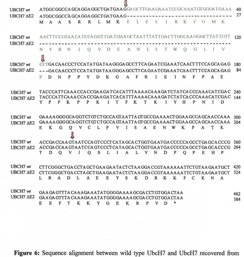
Figure 6: Sequence alignment between wild type UbcH7 and UbcH7recovered from the Hela cDNA library 44
•
Figure 7: Purification ofUbcH7 and UbcH7~E2 45•
Figure 8: DA.XX interacts with the C-terminus of Ad ElB 55-kDa proteins49
•
•
Figure 9: PML and DA.XX localization in cells expressing Ad E 1 B 55-kDaPro teins 50
•
Figure 10: Cellular localization of PML in cells expressing Ad ElB 55-kDaProteins 53
•
Figure 11: Localization of Ad 12 E 1 B 55-kDa and DA.XX in G40 l CC3 cells 53•
•
•
Abbreviations
3-A T - 3-amino-1,2,4-triazole 5-FOA - 5-fluoroorotic acid aa - amino acid(s)
Ad - adenovirus
AD - activation domain Amp - ampicillin
APL - acute promyelocytic leukemia bp - base pair(s)
BD - binding domain
BLAST - basic local alignment search tool DAPI - 4' ,6' -diamidino-2-phenylindole HAT - histone acetyl transferase
HDAC - histone deacetylase kb - kilo base pair(s)
kDa - kilo Dalton
NLS - nuclear localization signal ORF - open reading frame
PBS - phosphate-buffered saline PI - propidium iodide
POO - PML oncogenic domain RT - room temperature
SD - synthetic dropout
U AS - upstream activating sequence Ub - ubiquitin
•
•
•
Abstract
The adenovirus (Ad) ElB 55-k.Da protein is vital to viral growth and complete transformation of adenovirus-infected cells. The detailed mechanisms of E 1 B action are not yet clear. ln order to shed light on the functions of this viral oncoprotein it is essential to determine its cellular targets. The yeast two-hybrid method was utilized to screen the Saccharomyces cerevisiae genomic library and the human Hela cONA library with the Ad2 ElB 55-kDa protein as hait. Partial screening of the S. cerevisiae genomic library (106 transformants) has revealed several yeast proteins that bind to El B. Among
these are: UFOl, involved in the ubiquitin fusion degradation pathway, a major pathway for selective protein degradation in eukaryotes, RIS 1, a member of the S Wl/SNF2 family of DNA-dependent ATPases, and Bdt2 which contains two copies of the evolutionarily conserved bromodomain, homologous to the C-terminal half of mammalian T Af 11250. Human proteins positive for Ad2 ElB 55-k.Da binding were identified through partial screening of the Hela cONA library (7 x 10⁵ transformants). Among them are: UbcH7, a
ubiquitin-conjugating enzyme involved in the ubiquitin-mediated degradation of the tumor suppressor protein p53, known to be stabilized and inactivated by Ad ElB 55-kDa. SUM0-1, a ubiquitin-like protein which covalently modifies a limited number of proteins in a manner similar to ubiquitination, ubiquitin itself, and DAXX, implicated in Fas-induced apoptosis .
The identification of a OAXX/Ad ElB 55-kDa interaction has interesting implications for the role of E 1 B in cell transformation. The pro-apoptotic ability of DAXX appears to be dependent on the physical interaction of OAXX with a tumor suppressor protein, PML. The binding of OAXX by PML results in the localization of DAXX to nuclear bodies, known as PML oncogenic domains (POOs). Ad ElB 55-k.Da binds to the C-terminus of OAXX, required for PML-binding and POO localization. Immunofluorescence co-localization experiments establish that in cell lines expressing Ad ElB 55-kDa proteins there is disruption of PML-DAXX association. There is also disruption of POO formation in these cells. These observations suggest that another mechanism by which the Ad ElB 55-k.Da protein contributes to cell transformation may be by interfering with an apoptotic pathway that acts through OAXX ..
•
•
•
Abstrait
La protéine d'adénovirus (Ad) ElB 55-kDa est essentielle à la croissance virale et à la transformation complète des cellules infectées par adénovirus. Les mécanismes détaillés de l'action d'ElB ne sont pas encore bien connus. Afin déterminer les fonctions de cette oncoprotéine virale, il est essentiel de déterminer ses cibles cellulaires. La méthode de deux-hybride de levure a été utilisée pour cribler la bibliothèque genomique de Saccharomyces cerevisiae et la bibliothèque d' ADNc Hela humaine avec la protéine d'Ad2 ElB 55-kDa. Le criblage partiel de la bibliothèque genomique de S. cerevisiae (106 transformants) a indiqué plusieurs protéines de levure qui se lient à ElB. Parmi
elles: UFD l, impliqué dans la voie de dégradation de fusion par ubiquitination, une voie importante pour la dégradation sélective des protéines dans les eukaryotes, RIS 1, une membre de la famille SWI/SNF2 d' ADN-dépendente ATPases, et Bdf2 qui contient deux copies du bromodomaine, domaine hautement conservé durant l'évolution et qui est homologue à la moitié C-terminale de TAFII250 de mammifères. Un criblage partiel de la bibliothèque d' ADNc de Hela humaine (7 x 10⁵ transformants) a permis d'identifier
des protéines humaines positives pour l'association avec Ad2 El B 55-kDa. UbcH7 est l'une d'entre elles. UbcH7 est une enzyme d'ubiquitination-conjugaison, impliquée dans la dégradation du suppresseur de tumeur p53, celui-ci reconnu pour être stabilisé et inactivé par Ad ElB 55-k.Da. Il y a aussi ubiquitine elle-même, SUM0-1, une protéine semblable aux protéines d'ubiquitination, qui modifie d'un nombre limité de protéines par un procédé similaire à l'ubiquitination, et DAXX, impliquée dans l'apoptose induite par Fas.
L'identification d'une interaction entre DAXX et Ad E 1 B 55-kDa propose des implications intéressantes pour le rôle d'E l B dans la transformation de cellules. Les capacités pro-apoptotiques de DAXX semblent dépendre de l'interaction physique de DAXX avec une suppresseur de tumeur, PML. La liaison de DAXX par PML provoque la localisation de DAXX dans des corps nucléaires, connus sous le nom de domaines oncogènes de PML (PODs). La liaison d'Ad ElB 55-kDa à la portion C-terminale de DAXX est requise pour la liaison a PML et la localisation dans les PODs. Les expériences de co-localisation par immunofluorescence ont démontré que dans des lignées de cellulaires exprimant les protéines Ad ElB 55-k.Da il y a dissociation du complexe PML-DAXX. La formation des PODs dans ces cellules est aussi perturbée. Ces observations suggèrent un autre mécanisme par lequel la protéine d'Ad E 18 55-kDa contribuerait à la transformation cellulaire: celles-ci pour interférer avec un processus apoptotique qui agit par l'intermédiaire de DAXX.
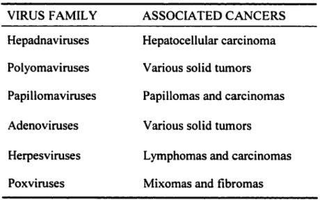
General Background on Adenoviruses
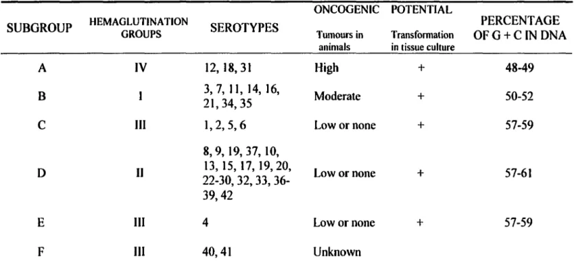
Adenoviruses (Ad) are a broad family of viroses with double-stranded DNA genomes of -35 kilo base pairs (kb). There are greater than 40 adenovirus serotypes classified into subgroups based on their percentage of G + C in DNA, hemaglutination properties, and tumorigenicity (listed in table 1 ). Adenovirus infections are quite common although most are asymptomatic. They have been associated with respiratory illness, conjunctivitis, and gastroenteritis in children. Adenoviruses are lytic in their natural host but are included within a heterogeneous group ofDNA tumor viruses for their ability to induce tumors in experimental systems. Other DNA tumor virus families include hepatitis B viruses, simian virus 40 (SV40) and polyomavirus, papillomaviruses, herpesviruses, and poxviruses. These are listed in table 2 along with their associated cancers. Certain DNA tumor viruses have been directly implicated in human malignancies, such as human papillomavirus type 16 (HPV-16) and HPV-18, the causative agents of cervical cancer and other anogenital cancers. Like adenoviruses, polyomavirus and SV40 are not natural transforming agents. However, these viruses are useful for studying the transformation process. The first report of a pathogenic human virus that could cause cancer in animais was the discovery that adenovirus type 12 could induce malignant tumor formation in rodents (Trentin et al., 1962). This sparked extensive study into the mechanisms of transformation by adenoviruses and other viruses. The application of molecular biology to research on the relationship between DNA tumor
•
•
•
viruses and cancer bas led to the identification of different cellular tumor suppressors, contributing to the understanding of human cancer development.
Table 1: DNA tumor viruses and associated cancers.
VIRUS F AMIL Y ASSOCIA TED CANCERS Hepadnaviruses Hepatocellular carcinoma Polyomaviruses V arious solid tumors Papillomaviruses Papillomas and carcinomas Adenoviruses V arious solid tumors
Herpesviruses Lymphomas and carcinomas Poxviruses Mixomas and fibromas
Modifiedfrom: T. Benjamin and P. K. Vogt. 1991. Cel! transformation
by viruses. ln: B. N. Fields, D. M. Knipe, R. M. Chanock, M. S. Hirsch,
J. L. Melnick, T. P. Monath, and B. Roizman, eds. Fundamental Virology, second edition. New York: Raven Press 291-341
•
•
•
Table
2: Classification se hem es for human adenovirus.
ONCOGENIC POTENTIAL
SUBGROUP HEMAGLUTINATION GROUPS SEROTYPES Tumours in Transformation OFG+C IN DNA PERCENT AGE animais in tissue culture
A IV 12, 18, 31 High + 48-49
B 21, 34, 35 3, 7, 11, 14, 16, Moderate + 50-52
c
III 1,2,5,6 Low or none + 57-598, 9, 19, 37, 10,
D Il 22-30, 32, 33, 36-13, 15, 17, 19,20, Lowornone + 57-61
39,42
E III 4 Lowornone + 57-59
F III 40,41 Unknown
Modified.from: M. S. Horwitz. 1991. Adenoviridae and their replication. /11: B. N. Fields, D. M. Knipe, R. M. Chanock, M. S. Hirsch, J. L. Melnick, T. P. Monath, and B. Roizman, eds. Fundamental Virology, second edition. New York: Raven Press 291-341
Adenovirus-Induced Cell Transformation
Normal cells maintain strict control of cell growth and differentiation, govemed by the requirements of the organism. ln response to adverse stimuli, such as environmental stress, DNA damage, and viral infection, the products of tumor suppressor genes can signal growth arrest or programmed cell death. In contrast, transformed, immortalized cells undergo continuous, unregulated growth. Ali adenovirus serotypes are capable of transforming cells in tissue culture. The transforming genes of adenoviruses, like other DNA tumor viruses, are uniquely viral. Cellular equivalents of these genes do not exist, in contrast to retroviruses, the only RNA viruses with oncogenic potential, which either integrate near a cellular oncogene or carry one in their genomes. Adenovirus oncogenes are expressed early, prior to the onset of viral DNA replication. They encode proteins that interfere with the actions of cellular tumor suppressor proteins and at various points in signaling pathways of apoptosis. This results in the deregulation of cell growth control.
Proteios lnvolved in Adeoovirus-Mediated Cell Transformation
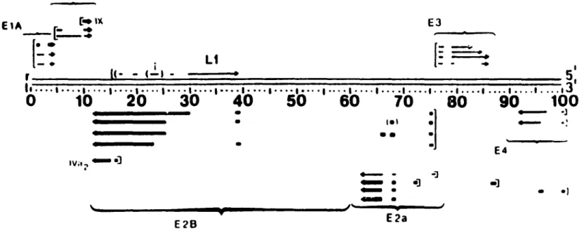
Complete transformation by adenoviruses requires the early region El that covers two transcription blocks, ElA and ElB (figure 1). ElA proteins (overlapping proteins of 26- and 32-kDa) force cells to enter S phase by targeting the retinoblastoma tumor suppressor protein, pRb. The ElB 19-kDa protein is a homologue of Bcl2 and
•
•
•
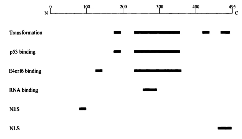
prevents viral-induced apoptosis. The oncoprotein that is the focus of this research is the large adenovirus ElB protein. The ElB 55-kDa protein inhibits the normal biological functions of p53 (Marcellus et al., 1996; Steegena et al., 1995; Yew and Berk, 1992). Figure 2 illustrates the functional domains of ElB 55-kDa including the p53 binding domain. Other viral oncoproteins that interfere with p53 functions include the SV40 large T antigen (Farmer et al., 1992) and E6 of hurnan papillomavirus (Scheffner et al., 1990). p53 is a potent turnor suppressor found mutated in more than 50% of human cancers. ln response to various stimuli, p53 up-regulates genes involved in growth suppression at G l and 02. A major component that contributes to p53-induced cell cycle arrest is p2 l, a cyclin-dependent kinase (CDK) inhibitor. lnhibiting CDK prevents pRb phosphorylation and leads to growth arrest at the G 1 /S boundary. p53 can also promo te apoptosis by up-regulating proteins such as Bax. The ElB 55-kDa protein associates with p53 and interferes with p53 transactivation of its target genes (Sarnow et al., 1982; Yew and Berk, 1992). lt may inhibit p53 function by more than one mechanism. First, the carboxyl-terminal region of EIB 55-kDa seems to act as a direct transcriptional repressor that could be coupled to p53 (Martin and Berk, 1998). Second, ElB 55-kDa specifically inhibits acetylation of p53 by P/CAF at lysine 320 (Liu et al., 2000). P/CAF acetylation of the p53 C-terminus stimulates its sequence-specific DNA-binding ability (Gu and Roeder, 1997) .
•
•
E1B E1A ~- c:i~ EJ-(:.: _..
f=
==-- • . 1 L1 : _ .. 1 r • -
c-s •
51 ,, •••• ' ••• • I • ••• I • ••• ' •••• , •••• ' •••• ' •••• 1 • ••• 1 •••• 1 ••• • , •••• 1 •••. 1 ••• ••••• • • .••• 1 ••• • I •••• 1 •••. 1 •••• 130
10
20
30
40
50
60
70
80
90
100
• •
• IVil., - • ).
E2B..,
~]
••
.,__
-
-]...
•] ~. ~. E2aFigure 1:
Ad2 genomic map of the early proteins and their mRNAs.
- ·J
-
·:
E4 •] • •l•
Thin lines indicate mRNAs detected at early times post-infection in the absence of protein synthesis. Thick lines represent intermediate mRNAs most easily detected at late times. Arrowheads show the 3' end. Tentative promoter sites are indicated by [-. From: M. S. Horwitz. 1991. Adenoviridae and their replication. ln: B. N. Fields, D. M. Knipe, R. M. Chanock, M. S. Hirsch, J. L. Melnick, T. P. Monath, and B. Roizman, eds. Fundamental Virology, second edition. New York: Raven Press 291-341
•
•
Transformation
-
-p53 hinding
-E4orf6 hinding
-RNA hinding
NES
-NLS
Figure 2: Schematic diagram illustrating the functional domains in the Ad EIB 55-kDa protein.
E 1 B 55-kDa consists of 495 amino acids, represented ahove. Black rectangles represent known functional
domains. NLS, nuclear localization signal; NES, nuclear export signal.
•
Acknowledging the contnbution of p53 inactivation to adenovirus-mediated cellular transformation, there is evidence that points towards mechanisms of E 18 55-kDa action that do not involve p53. This includes experiments performed by Goodrum and Omelles (1997; 1998) with Ad5 ElB 55-k.Da mutants. The presence or absence of p53 did not predict the ability of the cell line to support growth of the EIB 55-kDa mutant virus. In the absence of a correlation between mutant virus replication and p53 status, it is possible that there are other cellular regulatory factors that promote virus replication in the wild type adenovirus infection. Additionally, expression of the tumor suppressor protein, PML, inhibits Ad5 ElA/ElB-mediated focus formation of primary rat cells suggesting that modulation of PML activity plays a role in viral transformation (Nevels et al., 1999).
Research Objectives
The major objective of this research is to identify cellular proteins that bind to Ad ElB 55-k.Da. If we take examples from other DNA tumor viroses, we see that their oncoproteins are multifunctional but contain no intrinsic enzyme activity. The~e proteins exert their effects through binding to numerous cellular proteins. ElB 55-kDa is also a multifunctional protein. Aside from the role that ElB 55-kDa plays in the transformation process, it plays an important role in viral growth. EIB 55-kDa mutations that result in its instability hinder viral DNA synthesis (Mak and Mak, 1990). It is also required for the selective transport of viral mRNA from the nucleus to the cytoplasm (Leppard and
Shenk, 1989). Late viral mRNAs are differentially exported by a protein complex that includes ElB 55-kDa and E4orf6. It has been suggested that the viral RNA transporter of this complex is E4orf6 (Dobbelstein et al., 1997). Recently, however, it has been shown that ElB 55-kDa itself is a good candidate for an adenoviral transponer. It actively shuttles in virus-infected cells (Dosch et al., 2001 ), with a C-terminal nuclear localization signal (NLS) as well as a N-terminal leucine-rich nuclear export signal (NES) (Kratzer et al., 2000), as first suggested by Liao et al. (1999). ElB 55-k.Da also has RNA-binding activity. It contains a domain at residues 249-288 homologous to the RNP domains that mediate direct interaction with RNA (Horridge and Leppard, 1998). It is clear that E 1 B 55-kDa plays many rotes in viral replication; however, the detailed mechanisms of its action are not yet fully understood.
With ail of the knowledge that has accumulated on adenoviruses, surprisingly few EIB 55-kDa cellular targets have been identified. Recent reports on associating proteins include PCAF (Liu et al., 2000), and histone deacetylase (HDAC) 1 (Punga and Akusjarvi, 2000). lf its binding partners are resolved, this could lead to elucidation of the functions of ElB 55-kDa. The intended focus is EIB-binding proteins involved in cell cycle regulation or apoptosis in order to investigate ElB 55-kDa acti~n in cell
transformation. Determining how the targeting of cellular proteins by ElB 55-kDa contributes to transformation by adenovirus may provide new directions for understanding normal cell growth control and cancer development.
•
•
•
Experimental Approacb
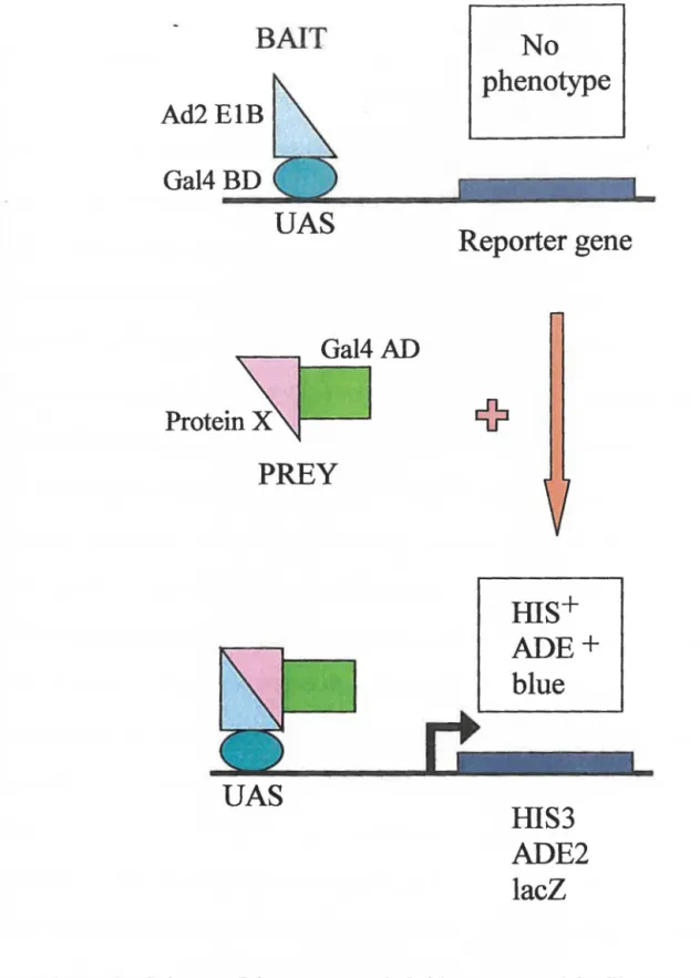
The yeast two-hybrid method was employed to find Ad ElB 55-kDa interacting proteins. With the use of the two-hybrid method, expression libraries can be screened for interaction with a protein of interest, as illustrated in figure 3. The two-hybrid method is a sensitive means of detecting protein-protein interactions, measured by the re-assembly of a functional transcription factor in yeast, which in this case is Ga14. The Gal4 DNA-binding domain (BD) is separated from the Gal4 transcription activation domain (AD) and each can be fused to a protein. The BD recognizes specific upstream activating sequences (UAS) in the promoter of Gal4 target genes. white the AD stimulates transcription initiation from the UAS by directing the assembly of the transcription complex. For this particular screen, Ad2 E 1 B 55-kDa is fused to the Gal4 BD and this is referred to as the bait hybrid. A protein encoded by a library insert from either the
Saccharomyces cerevisiae genomic library or the human Hela cDNA library is fused to
the Gal4 AD and this is referred to as the prey hybrid. If EIB 55-kDa binds to a library protein, the Gal4 BD and AD are brought into close proximity and functional Gal4 is generated. This leads to transcription of reporter genes downstream of the UAS. If the two proteins do not interact then the reporter genes are not transcribed. ln the host yeast strain utilized there exist three reporter genes: his3, ade2, and lacZ. This allows for nutritional selection (his3, ade2) as well as color based assays (lacZ). A different Gal4 promoter drives each reporter gene. The his3 reporter provides the highest sensitivity for growth selection and the ade2 promoter is the most stringent. The numbers of false
positives are greatly reduced with this host strain when compared to earlier strains containing a single promoter element (James et al., 1996).
After determining which cellular proteins associate with Ad ElB 55-kDa through library screening, these interactions can then be verified by alternative means such as immunoprecipitation, GST pull down and immunofluorescence co-localization experiments. The yeast two-hybrid method can be employed to map the domains responsible for the interactions and identify possible E lB-binding domains. Further experiments can then be performed to determine whether El B 55-kDa interferes with the cellular functions of each positive protein. For example, ElB-binding may prevent the protein from interacting with other cellular proteins or block domains required for its activity or cellular targeting. Approaches to studying biological significance would be determined based on the protein of interest.
BAIT
Ad2 ElB
Gal4 BD
UAS
Gal4AD
PREY
UAS
No
phenotype
Reporter gene
ms+
ADE+
blue
HIS3
ADE2
lacZ
Figure 3: Scheme of the yeast two-hybrid system used for library screening.
Bait: Ad2 ElB 55-kDa fused to the Gal4 DNA-binding domain (BD). Prey: human HeLa cDNA or S. cerevisiae genomic DNA library protein (protein X) fused to the Gal4 activation domain (AD). In the absence of an interaction between Ad2 ElB 55-kDa and protein X, neither the hait nor the prey hybrid can activation transcription of reporter genes.
Expected Results
If the human adenovirus oncoprotein does behave as oncoproteins of other DNA tumor viruses, then screening the Hela cDNA library with ElB 55-kDa as bait should yield a large number of interacting proteins. These proteins are expected to be involved in nucleocytoplasmic transport, transcription, and cell cycle regulation. A drawback to screening the Hela cDNA library is the amount of false positives that would be encountered. False positives would consist of human proteins associating with either the Gal4 UAS or the Gal4 BD irrespective of the presence of ElB 55-kDa. Performing the same yeast two-hybrid screen with the S. cerevisiae genomic library should not yield as many true or false positives. Screening the S. cerevisiae library with the Ad2 ElB
55-kDa protein is appropriate because we know that it is biologically active in yeast.
T nduced expression of the E 1 B protein fused to a NLS in S. cerevisiae cells inhibits cell
growth (Liang et al., 1995). NLS-EIB is localized to the yeast nucleus and leads to accumulation of nuclear poly(A)+ RNA. It is expected that screening the S cerevisiae genomic library will reveal possible human proteins that will bind to ElB 55-kDa. Human proteins containing the same evolutionarily conserved domains of the yeast proteins or functional human homologues may also interact with El~ 55-kDa.
Simultaneously screening both the Hela cDNA library and the S. cerevisiae genomic library increases the opportunity of finding hwnan ElB 55-kDa interacting proteins.
We also expect that screening expression libraries with an ElB 55-kDa fragment of hwnan Ad2 may further our understanding of the effects of EIB 55-kDa of
highly oncogenic Adl2. Proteins positive for Ad2 ElB 55-kDa binding would also be tested for binding to Adl2 ElB 55-kDa. which proved too toxic for the host yeast to be used as bait. Expressing Adl2 EIB 55-kDa had a detrimental effect on yeast doubling time. Most of the knowledge that has accumulated on the large ElB protein has corne from studies with non-oncogenic Ad2 or Ad5 of the same subgroup. There is strong sequence similarity between the ElB 55-kDa proteins of serotypes 12 and 2/5, with the greatest differences in the N-terminal third. Downregulation of MCH class l by ElA proteins of Adl2 but not Ad2 or Ad5 is believed to play a role in Adl2 oncogenic potential in rodents (Pereira et al., 1995; Schouten et al., 1995).
Introduction to Some ElB-binding Proteins Identified
Screening the Hela cDNA library and the S. cerevisiae genomic library identified several ElB 55-kDa-interacting proteins. These include proteins involved in the ubiquitin (Ub) fusion degradation pathway, a major pathway for selective protein degradation in eukaryotes, such as Ub and UbcH7 from the human library and yeast Ufdl. Post-translational conjugation to Ub marks a protein for degradati,on by the proteasome. UbcH7 is a Ub-conjugating enzyme responsible for Ub-mediated degradation of p53 (Ciechanover, et al., 1994) and c-fos (Stancovski et al., 1995) and processing of the NficB (nuclear factor-KB) precursor p105 (Orian et al., 1995). Ufdl is involved at a post-ubiquitination step of the Ub-proteasome pathway and is highly conserved from yeast to humans (Johnson, et al., 1995; Novelli et al., 1998). SUM0-1
[also called UBL 1 (Shen et al., 1996), PIC l (Boddy et al., 1996), GMP l (Matunis et al., 1996) or sentrin (Okura et al., 1996)] was also found as a result of Hela cDNA library screening to interact with Ad2 ElB 55-kDa. SUM0-1 is a Ub-like protein which covalently modifies a limited number of proteins in a manner similar to ubiquitination (Kamitani et al., 1997). During the process of writing this work it was discovered by Endter et al. (2001) that Ad5 ElB 55-kDa could be modified by SUM0-1 in vivo.
Other interesting proteins identified include yeast Bdt2, functionally redundant with Bdfl and homologous to the carboxyl-terminal half of mammalian T AFn250, the largest subunit of TFIID (Matangkasombut et al., 2000). Bdfl, Bdt2, and T AFu250 each contain two copies of the evolutionarily conserved bromodomain. There is also human Clk l which phosphorylates SR proteins and may play a role in the altered splicing patterns during the switch from early to late phase of adenovirus replication (Duncan et al., 1997), and transcription repressor DAXX, implicated in Fas-induced apoptosis (Yang et al., 1997; Torii et al., 1999). The discovery that human DAXX binds to Ad2 ElB
55-kDa inspired further investigation into the nature of this interaction. The fact that DAXX is suspected to be a pro-apoptotic protein is of particular interest given the involvement ofElB 55-kDa in cellular transformation.
Introduction to DAXX
DAXX contains no homology to other known proteins. Murine DAXX (mDAXX) was originally identified as binding to the intracellular domain of Fas (also named APO- l and CD95) in a two-hybrid screen by Yang et al. ( 1997). They showed that the overexpression of mDAXX and Fas together enhanced Fas-mediated apoptosis through the Jun N-terminal kinase (JNK) pathway. Fas is a member of the tumor necrosis factor (TNF) receptor superfamily and is activated when immune effector cells deliver Fas ligand (FasL) to the receptor. Conflicting stories have emerged regarding the role of DA.XX in Fas-induced apoptosis. In 1999, Torii et al. showed that sensitivity to apoptosis induced by Fas was enhanced in cells overexpressing the human homologue of mDAXX. This cellular response was specific to Fas. Apoptosis induced by other TNF-family death receptors was not atîected. They reported accelerated activation of caspases but no increase in JNK. activity. Surprisingly, DA.XX may play an anti-apoptotic role in mouse development (Michaelson et al., 1999). A deficiency in DAXX results in extensive apoptosis and embryonic lethality. A recent report claims that Fas-mediated cell death does not involve DAXX (Villunger et al., 2000). A dominant-interfering mutant of DAXX was used in these experiments although they admitted 1:hat higher levels of the mutant might have had an effect on FasL-induced apoptosis.
Torii et al. (1999) also showed that DAXX is in fact a nuclear protein that localizes to structures called PML oncogenic domains (PODs) but does not bind Fas. PODs are essentially accumulations of proteins and these multiprotein complexes
associate with the nuclear matrix (Ascoli and Maul, 1991). There are approximately 10-30 PODs per cell nucleus. In normal cells, PML is concentrated within these structures and is critical to the formation of PODs (lshov et al., 1999). Apart from PML and DAXX, other proteins found in PODs include: p53 (Fogal et al., 2000), co-activator and histone acetyl transferase, CBP (LaMorte et al., 1998), SplOO recognized by autoantibodies from patients with primary biliary cirrhosis (reviewed in Stemsdorf et al., 1997), BML, the RecQ helicase missing in Bloom's syndrome (Zhong et al., 1999), and pRb (Alcalay et al., 1998) (illustrated in figure 4). The localization of ail other proteins to PODs is dependent on SUM0-1 modification of PML (Zhong et al., 2000a). The fonction of these nuclear bodies in the cell is still not clear but there is evidence for their involvement in DNA replication, transcriptional activation and apoptosis (reviewed in Hodges et al., 1998). PML is a cell growth and tumor suppressor protein essential for multiple apoptotic signais (Wang et al., 1998). These include IFN, TNF, Fas, and ceramidc, normally employed to induce apoptosis upon DNA damage or neoplastic transformation. PML is found fused to the retinoic acid receptor-alpha (RARa) in acute promyelocytic leukemia (APL) due to a distinct chromosomal translocation, t(15; 17) (reviewed in Melnick and Licht, 1999). The resulting PMLIRARa fusion protein contributes to the pathogenesis of APL. PMLIRARa can heterodimerize
witb
PML and protect cells from apopt_osis, thus acting in a dominant negative manner (Wang et. al. 1998). Also, due to the presence of the fusion protein, PML can no longer mediate assembly of PML nuclear bodies (Dyck et al., 1994; Weis et al., 1994).DAXX interacts physically with PML and localizes to PODs in mitogen-activated splenocytes (Zhong et al., 2000b ). The sequestering of DA.XX in PODs by PML is necessary for its pro-apoptotic function (Torii et al., 1999). A mutant DA.XX that could no longer localize to PODs was unable to enhance Fas-induced apoptosis. By sequestering DAXX in PODs, the PML protein also inhibits the repressor fonction of DAXX (Li et al., 2000a). DAXX possesses strong transcriptional repressor activity (Hollenbach et al., 1999; Torri et al., 1999) and so it is possible that DA.XX plays a role in programmed cell death by modulating the transcription of certain genes. This repressor fonction may result from the recruitment of HDACs by DA.XX (Li et al., 2000b). HDAC inhibitor, TSA, blocks DAXX repressor activity. DAXX interacts with transcription factor ETS 1 and prevents transactivation of at least two genes regulated by ETS l: MMP 1, involved in the process of tumor invasion and metastasis, and Bcl2, which is anti-apoptotic (Twasaka et al., 1996; Li et al., 2000b).
PODs increase both in size and number following interferon (IFN) treatment of cells and may be sites for antiviral defense mechanisms (Hodges et al., 1998). IFNs are growth regulatory cytokines with antiviral activity (reviewed in Jokik, 1991 ). IFN treatment also dissociates DA.XX from centromeres and results in the association of DAXX with PODs (Everett et al., 1999). Several viruses express proteins that specifically modify PODs, including adenoviruses, herpes simplex virus type 1, cytomegalovirus, and hwnan T-cell leukemia virus. ElB 55-kDa itself localizes to PODs shortly after adenovirus infection, along with E4orf3 (Doucas et al., 1996). PODs then undergo a dramatic morphological change from spherical structures to fibrous-like
tracks. Host factors are recruited, along with ElB 55-kDa. from the PODs to viral inclusion bodies, which are sites of adenovirus DNA replication and late RNA transcription (illustrated in figure 4 }. E4orf3 remains behind in tracks with PML. Experiments with adenovirus deletion mutants point to E4orf3 as the viral protein responsible for POD reorganization, with E lB 55-kDa contributing to the sequential redistribution of POD proteins. However, immunofluorescence co-localization experiments perforrned here show that ElB 55-kDa itself is capable of disrupting the interaction between DAXX and PML, as well as the formation of PODs. These results suggest that another mechanism by which ElB contributes to cellular transformation may be by preventing the apoptotic response triggered by DAXX following adenovirus infection.
CBP/ PML-SUM0-1 pRB
p53-SUM0-1
BML
NDP55 HP 1/Sp100-SUM0-1DAXXI PML-SUM0-1
Figure 4:
Adenovirus infection leads to disruption of PODs.Shortly after adenovirus infection EIB 55-kDa and E4orf3 associate with PODs, which undergo a dramatic morphological change into fibrous-like tracks, illustrated here. Host factors are recruited :from the PODs to viral inclusion bodies. A single POD and known protein components is also illustrated.
•
•
•
Materials and Methods
Y east Two-Hybrid Assays
Yeast two-hybrid assays were performed with Ad2 ElB 55-k.Da (aa 155-495) fused to the Gal4 BD in plasmid pGBDU-Cl along with either Saccharomyces cerevisiae genomic library fragments in plasmid pGAD-C 1, or human Hela cDNA library fragments in plasmid pGAD GH, fused to the Gal4 AD (pre-made MA TCHMAKER plasmid libraries, CLONTECH Laboratories, Inc. ). Two-hybrid assays were also performed with the carboxyl-terminal of DAXX (aa 621-740) fused to the Gal4 AD, isolated from library screening (described below) and several Ad ElB 55-kDa fragments (Ad2 ElB aa l-437, 155-495, and 437-495; Ad12 ElB full length, aa 1-408, and 1-204) fused to the Gal4 BD, cloned into pGBDU-C(X) plasmids. Adenovirus E 1 B constructs were cloned into plasmids by restriction endonuclease digestion and ligation. The yeast strain used for all two-hybrid assays was PJ69-4A (MATa trpl-190 leu2-3,112 ura3-52 his3-200 gal4LJ
ga/80LJ LYS2::GALJ-HIS3 GAL2-ADE2 met2::GAL7-lacZ) outlined in James et al.
(1996).
Co-transformation of plasmids in yeast was done by the basic lithium acetate (LiOAc) method. Several host yeast colonies were used to inoculate synthetic dropout (SD) liquid medium lacking lysine (lys). Cultures were grown overnight at 30°C to an OD6oo between 0.8 and 1.0, centrifuged at 2,600 rpm for 5 min at RT then washed 2x
LiOAc solution for each 50 ml of culture. Next, 0.1 ml of the suspension was added to a 1.5 ml eppendorf containing 10 µg of each plasmid DNA. To test for the presence of contaminants, a control without DNA (H20) was perfonned for each round of
transformation. The mixture was left at RT for 10 min, then 0.28 ml of 50% filtered PEG/LiAc solution was added. The tubes were placed at 30°C for 1 hr. 43 µI DMSO was added and the samples were heat shocked at 42°C for 5 min. After washing 3x with sterile H20, the pellet was resuspended in 1 ml sterile H20. 2 volumes of the suspension
(0.15 ml and 0.3 ml) were plated onto SD lysleu-ura- to select for both the pGAD plasmid which contains the gene for leucine (leu2) and the pGBDU plasmid which contains the gene for uracil (ura3) and incubated 72 hr at 30°C. Where water was used in place of plasmid DNA, no colonies should appear after three days incubation. A single yeast colony was picked from each plate and streaked again onto SD lys-leu-ura-. Plates were incubated at 30°C for 48 hrs after which single colonies could be picked and streaked on different SD plates. Protein-protein interactions were recognized by: /. growth on SD his- + agar in the presence of 5 mM 3-amino-1,2,4-triazole (3-AT) to inhibit hi:d reporter gene auto-activation, 2. growth on SD ade-, and 3. detection of
P-galactosidase activity in the presence of X-Gal (5-bromo-4-chloro-3-indolyl-P-D-galactopyranoside ).To perform the J3-galactosidase assays, single colonies (triplicate for each co-transfonnation) were spread in a straight line on SD lysleu-ura-+ agar and placed at 30°C ovemight. A mixture of 5 ml 1 % agarose in H20, 5 ml heated sodium phosphate buffer,
solidification plates were incubated at 37°C for l hr. By this time, enzyme-producing yeast colonies appear blue. The interaction between Ad2 ElB 55-kDa (aa 155-495) in pGBDU and full length p53 in pGAD was used as a positive control in f3-galactosidase assays as well as growth on selective media. Negative controls consisted of replacing one of the co-transformants with empty pGAD or with empty pGBDU.
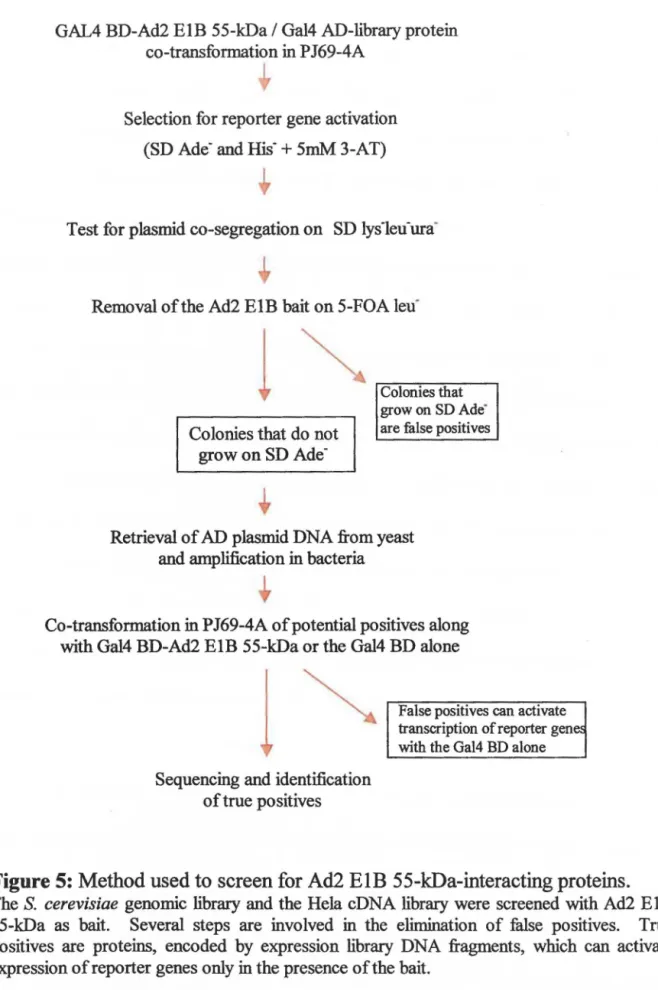
Library Screening
Figure 5 outlines the steps involved in the identification of Ad2 ElB 55-kDa-interacting proteins. Ad2 ElB 55-kDa (aa 155-495) in pGBDU-Cl (hait) and expression library fragments in pGAD-Cl or pGAD GH (prey) were co-transformed into yeast as described above. Between 5,000 and 8,000 colonies were obtained for each plate. Yeast colonies possessing both plasmids of interest were replica plated onto SD bis- + 5 mM 3-AT and SD ade-. Those colonies that grew on both plates were picked and grown on SD lysleu-ura-ade· to select for protein-protein interaction and ensure plasmid co-segregation. Colonies were then grown on SD leu- + agar with the addition of
5-fluoroorotic acid (5-FOA) (selects for the loss of the pGBDU/Ad2 ElB plasmid). These colonies were replica plated onto SD ade- plates. Colonies that grew on SD ade- + agar in the absence of Ad2 ElB 55-kDa were considered false positives and discarded. The majority of colonies were lost at this step. Recovery of potential positives consisted of isolation of plasmid DNA from yeast and amplification in E. co/i.
GAIA BD-Ad2 ElB 55-k:Da / Gal4 AD-library protein
co-transformation in PJ69-4A
Selection for reporter gene activation (SD Ade- and
rus·
+
5mM 3-AT)•
Test for plasmid co-segregation on SD lys1eu·ura·
Removal of the Ad2 ElB hait on 5-FOA leu·
1
Colonies that do not grow on SD
Ade-Colonies that grow on SD Ade· are fàlse positives
Retrieval of AD plasmid DNA from yeast and amplification in bacteria
•
Co-transformation in PJ69-4A ofpotential positives along with Ga14 BD-Ad2 ElB 55-k:Da or the Gal4 BD alone
1
Sequencing and identification oftrue positives
False positives can activate transcription of reporter gene~
with the Gal4 BD alone
Figure 5: Method used to screen for Ad2 ElB 55-kDa-interacting proteins.
The S. cerevisiae genomic library and the Hela cDNA library were screened with Ad2 EIB 55-k:Da as hait. Several steps are involved in the elimination of false positives. True positives are proteins, encoded by expression library DNA fragments, which can activate expression of reporter genes only in the presence of the hait.
---•
•
•
In order to extract plasmid DNA from yeast, 0.1 ml of liquid SD lys-leu- was inoculated with one positive yeast colony. The culture was grown at 30°C to an 00600
between 0.8 and 1.0 and centrifuged for 5 min at 2,600 rpm (here the pellet could be frozen for later use). Cells were washed 2x with sterile H20 and 1 x with LETS buffer. The pellet was resuspended in 0.3 ml LETS and transferred to a 15 ml tube. 0.3 ml of phenol was added followed by glass beads to the surface of the liquid ( 1 eppendorf of beads). Next, the tube was vortexed 6x 30 sec at maximum speed alternating with 30 sec on ice. The supernatant was transferred to an eppendorf and the glass beads were washed lx with 0.15 ml LETS and lx with 0.15 ml chloroform, collecting the liquid each time in the eppendorf. One volume of phenol/chloroform was added to the eppendorf which was then vortexed for several seconds and centrifuged at 12,000 rpm for 5 min at RT. The DNA in the upper phase was removed and precipitated with 95% ethanol and salt for l 0 min at -20°C. The pellet was washed with 95% ethanol and resuspended in 0.3 ml H20.
5 µl RNase A (10 mg/ml) was added and the sample was incubated at 37°C for 10 min. A second phenol/chloroform extraction and ethanol precipitation was performed at this step and the pellet was resuspended in 20 µl sterile H20.
Y east DNA (3 µl) was analyzed on a 0.8% agarose gel and later amplified in E.
coti (XL 1 Blue) by electroporation. 1-5 µl of DNA was mixed with 40 µl of competent cells and electroporated at 1.8 kV (Bio-Rad Gene Pulser, 25 µF with the pulse controller set to 200 ohms). Following electroporation, 0.2 ml of YT was added to the cuvette and the cells were plated on YT
+
agar with 1 OO µg/ml ampicilin (Amp ). Plates were•
•
•
incubated at 3 7°C overnight. A single colony was used to inoculate 2 ml YT + Amp . After shaking overnight at 3 7°C, 0.1 ml of this culture was used to inoculate 250 ml YT +
Amp and again grown overnight at 3 7°C. Bacterial cells were harvested by centrifugation at 6,000 rpm for 15 min at 4°C and resuspended completely in 10 ml solution I. The cell suspension was transferred to a 50 ml centrifuge tube. 10 ml solution II was added and the tube was inverted 4-6 times to mix and incubated at RT for 5 min. 10 ml cold solution III was added to the lysate and mixed by inversion followed by 30 min incubation on ice. After 30 min centrifugation at 15,000 rpm at 4°C, the supernatant was divided into two clean 50 ml centrifuge tubes and each was mixed with 11 ml isopropanol. Tubes were centrifuged immediately at 11,000 rpm at 4°C for 30 min. The DNA pellet was washed with 5 ml 70% ethanol, air-dried and resuspended in 0.5 ml distilled HiO by vortexing. The DNA solution was transferred to an eppendorf. 5 µl RNase A (10 mg/ml) was added and the sample was incubated at 37°C for l hr. Extraction with 0.5 ml phenol consisted of vortexing for several seconds, centrifugation at full speed for 5 min and transferring the upper phase to a clean eppendorf. The extraction was repeated with 0.5 ml phenol/chloroform. 50 µl 3 M NaOAc (pH 5.3) and 0.42 ml isopropanol were added to precipitate the DNA followed by centrifugation at full speed for 10 min. The pellet was washed lx with 0.25 ml 70% ethanol and lx with 0.25 ml 95% ethanol, dried and resuspended in 0.2 ml TE. The DNA concentration was determined by measuring the OD 260 (1 OD26o = 50 µg DNA/ml).
pGAD vectors containing the fragments of interest were then co-transformed back into yeast along with either Ad2 ElB 55-kDa in pGBDU-Cl or the pGBDU-Cl plasmid
alone. Those library proteins possessing intrinsic DNA-binding ability or those that could activate transcription of reporter genes, ade2, his3, and !acZ, through association with the Gal4 BD alone were eliminated at this step. Only those positive interactions dependent on the presence of Ad2 ElB 55-k.Da were considered true. True pos!tives were sequenced using the DNA sequencing procedure originally described by Sanger and Coulson (1975). The primer used for sequencing was 5'-gatgatgaagatacccc-3', present in the Gal4 AD. The proteins were identified via an on-line BLAST search at the National Center for Biotechnology Infonnation web site.
Protein Expression and Purification
The coding regions of UbcH7wt and UbcH7 âE2 were generated by PCR, tagged with FLAG at the 5' end and 6X His at the 3' end, and cloned into pET22b( + ). The proteins were expressed in E. coli BL2l(DE3) upon IPTG induction. To do this, a single
colony containing the plasmid of interest was picked to inoculate 1 OO ml YT + Amp and grown ovemight in a 37°C shaker. This culture was diluted 1:10 (25 ml in 250 ml YT + Amp) and incubated at 30°C until the 00600 was 0.6. IPTG was added .to a final
concentration of 0.4 mM and flasks were placed at 30°C for 4 hrs. The proteins were then purified using nickel-nitrilotriacetic acid (Ni-NT A) agarose. Cells were harvested by centrifugation at 8,000 rpm at 4°C for 10 min. The pellet was resuspended in 12 ml ice-cold His binding buffer and transferred to two 50 ml tubes. Each was sonicated (at
at 4°C, and filtered. Resin was prepared by adding 10 ml His binding buffer to 2 ml Ni-NTA agarose. After 3 min centrifugation at 1,200 rpm, 1 ml of resin remained. 10 ml His binding buffer was added and this was placed at 4°C for 2 hrs, rotating. The resin was spun down and washed 4x with lO ml His washing buffer before eluting with 5x 1 ml His elution buffer. lO µI of each elution was examined on a 12% SOS PAGE gel run at 150 volts in electrophoresis buffer. The gel was stained with Coomassie Brilliant blue for l 0 min at RT and incubated ovemight in de-stain solution. Eluted proteins were dialyzed by transferring the liquid into dialysis tubing secured with clamps. These were placed in dialysis buffer, stirring, ovemight at 4°C. The protein concentrations were determined by preparing three dilutions of bovine serum abumin (BSA) as a protein standard. Standard and sample solutions in 0.8 ml H20 were added to 0.2 ml of Bio-Rad
dye reagent, vortexed, and the absorbance measured at 595 nm. A standard curve was plotted for BSA and used to determine sample concentrations. Ad2 E 1 B 55-kDa was expressed in insect cells and affinity purified with Ni-NT A agarose.
Immunoprecipitation
UbcH7 L\E2 (200 ng) purified from E. co/i was incubated with monoclonal antibody, 2A6, against À.d2 ElB 55-kDa (0.15 ml of hybridoma supematant), or with both 2A6 and Ad2 EIB 55-kDa (100 ng) for l br at 4°C, rotating. 15 µlof protein G-agarose beads
was
added and incubated for l hr at 4°C, rotating. Following centrifugation at full speed for 2 min at 4°C, the beads were washed with 1 ml of wash•
•
•
buffer 1. This step was repeated with wash buffer 2 and wash buffer 3. Beads were then resuspended in 20 µl SOS loading buffer and placed at 1
oo
0c for three min. Theprecipitates were separated on a 12% gel by SOS-PAGE and the proteins were transfcrred to a nitrocellulose membrane (Ply Screen PVDF transfer membrane) for 2 hrs at 150 volts at 4°C in transfer buffer to be analyzed by Western blot. The membrane was blocked for lhr at RT with 5% milk in Tris-buffered saline (TBS) and 0.1% Tween 20 and probed with monoclonal antibody against the FLAG epitope ( 1: 1000 dilution in blocking buffer) for 1 hr at RT. Following washes with TBS-Tween 20 (3x 10 min at RT), the membrane was incubated with secondary antibody, goat anti-mouse for 1 hr (1: 1000 dilution in blocking buffer). The membrane was treated with enhanced luminol reagent (NEN Life Science Technologies) for 60 sec and exposed to film .
Cell Culture
Three different cell lines were cultured: G401, a rhabdoid kidney tumor cell line, 0401 CC3, a derivative of G401 expressing Adl2 ElB 55-kDa protein and 293, human embryonic kidney cells expressing AdS ElA and ElB proteins. 293, G401, G401 CC3, cells were grown in DMEM medium supplemented with 10% fetal bovine serum. Added to the 0401 and 0401 CC3 media were 15 µg/ml hypoxanthine, and 10 µg/ml thymidine. G401 CC3 medium also contained 250 µg/ml G418. Cells were incubated in a humidified chamber at 3 7°c with a 5% C02 environment .
•
•
•
lmmunofluorescence Microscopy
The localization patterns of PML and DAXX were examined in three different cell lines: G40l, 0401 CC3, and 293. Cells were grown ovemight on a glass coverslip to approximately 70% confluence, fixed with 4% paraformaldehyde for 20 min at room temperature (Rn and permeabilized with 0.2% Triton X in PBS for 15 min at RT. After blocking for one hour in blocking buffer (2% fetal bovine serum, 0.1 % sodium azide, 0.1 % Tween 20 in PBS) the cells were incubated with both a rabbit anti-DAXX antibody
(1 :500 dilution in blocking buffer) and a mouse anti-PML antibody (l :250 dilution in blocking buffer) for 2 hrs. The cells were washed 3x with TBS and then incubated for l hr with a mixture of fluorochrome-labeled secondary antibodies: goat anti-rabbit conjugated to Texas red and FITC-conjugated goat anti-mouse (each diluted 1:100 in blocking buffer). The cells were washed again 3x with TBS followed by staining for l 0 min with 4',6'-diamidino-2-phenylindole (DAPI) (0.2 µg/ml in PBS) at RT. Localization of both DAXX and Ad12 EIB 55-kDa was examined in the 0401 CC3 cell line. Cells were prepared as above with primary antibodies consisting of mouse anti-DAXX antibody (1:500 dilution in blocking buffer), and rabbit alpha-ElB (1:1000 dilution in blocking buffer). Fluorochrome-labeled secondary antibodies used were Texas red-conjugated goat anti-mouse, and FITC-red-conjugated goat anti-rabbit ( each diluted 1: 1 OO in blocking buffer).
PML cellular localization was also examined in 0401, G401 CC3 and 293 cell lines. In place of DAPI, cells were stained with propidium iodide (Pl). Cells were
incubated with a mixture of primary antibody, mouse alpha-PMI., and RNase A (0.3 mg/ml in PBS) for 2hrs at 37°C. The secondary antibody used was FITC-conjugated goat anti-mouse. After washing 3x with TBS-Tween 20 the cells were stained for 10 min with PI (1.0 µg/ml in PBS) at RT.
ln ail cases coverslips were mounted and viewed using an Olympus IX-70 scanning laser biological microscope. An Omnichrome ion laser power supply and NEC gas laser power supply were connected to the Olympus Fluoview system. The laser line tilter turret was set at 568 and 488 to allow argon and krypton laser combination. The laser intensity was set at 0.6 and the confocal aperture at 0.2. Cells were examined with the 40X objective (LCPlanFI) with oil. A Lambda l 0-C optical filter changer was used to choose the appropriate filters. Images were captured using Fluoview software (version 2. l.37) and stored as tif files.
•
•
•
Results
Potential Ad2 ElB SS-kDa Cellular Targets
The yeast two-hybrid method was employed for screening the Saccharomyces
cerevisiae genornic library and the Hela cDNA library to identify proteins that associate with Ad E 1 B 55-kDa. The first step concemed choosing an E 1 B construct that was not toxic to cells, did not activate transcription alone, and tested positive in yeast two-hybrid assays against a protein that it was known to interact with. This last step would help to assure that the EIB/Gal4 DNA-binding domain (BD) fusion protein was properly folded. Ad2 ElB 55-kDa (aa 155-495) fused to the Gal4 BD was the hait hybrid used for library screening. lt associates with itself and with p53 fused to the Gal4 transcription activation domain (AD) in two-hybrid assays (data not shown). Proteins encoded by pre-made MATCHMAKER plasmid libraries were fused to the Gal4 AD. The insert size range was 0.4 - 2.0 kb with an average insert size of 1.5 kb.
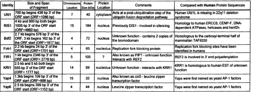
The rationale behind screening a yeast library with the large human adenovirus E 1 B protein was that hurnan homologues of yeast proteins positive for E 1 B-binding, or evolutionarily conserved functional domains, could be identified. The complete DNA sequence of the S. cerevisiae genorne has been determined and made available on the web. Altematively, the biological effects of interactions could be studied in yeast where many cellular processes are less complex. Partial screening of the S. cerevisiae genomic library (106 transformants) has revealed 8 different yeast proteins positive for Ad2 EIB
•
•
•
55-k.Da binding. These proteins are listed in table 3 along with their functions in yeast and comparison to human sequences.
Potential S. cerevisiae Ad2 ElB 55-k.Da-interacting proteins include UFDl, for ybiquitin (Ub) fusion Q.egradation, a 40-k.Da protein essential for cell survival in yeast (Johnson et al., 1995). UFD 1 bas not been fully characterized but it is implicated at a post-ubiquitination step of the Ub-fusion degradation pathway. UFD 1 is also found complexed to Np 14, implicated in nuclear transport (Meyer et al., 2000). A human homologue ofUFDl, called UFDlL, has been described as a potential housekeeping gene (Novelli et al., 1998). UFD 1 L haploinsufficiency contributes to the phenotype seen in 22q 11 deletion syndrome (Y amagishi et al., 1999). Next is RIS l (also known as DIS 1 ) . RIS 1 interferes with silencing and may act by increasing accessibility of silenced DNA. It is a member of the SWl/SNF2 family of DNA-dependent ATPases (Zhang and Buchman, 1997) and shares sequence homology with a number of human proteins including DNA-dependent A TPases, helicases, repair protein ERCC6, and centromere protein, CENP-F. The region covered by both RIS 1 cDNA fragments identified by library screening shows CENP-F homology. CENP-F is a cell cycle specific nuclear autoantigen that associates with both the nuclear matrix and centromeres (Liao et al., 1995). To determine whether it binds to Ad2 ElB 55-k.Da, CENP-F was cloned into the pGAD plasmid. Gal4 AD/CENP-F interacted with the Gal4 BD/Ad2 ElB 55-k.Da in yeast two-hybrid assays but not with Gal4 BD alone (data not shown). Next is Bdf2, functionally redundant with Bdfl and homologous to the carboxyl-terminal half of mammalian TAFu250, the largest subunit of TFIID (Matangkasombut et al., 2000).
•
•
•
Bdfl, Bdf2, and TAFu250 each contain two copies of the bromodomain, a 110 amino acid module conserved from yeast to human (Jeanmougin et al., 1997). T Af 11250 may consist of two separate proteins in yeast, Bdfl/Bdf2 and Tafl45 which possesses acetyltransferase activity. The bromodomain is found in other histone acetyltransferases (HATs) including P/CAF, p300/CBP and in the ATPase subunit of SWI/SNF and their homologues. Despite widespread conservation the functional significance of the bromodomain remains unknown although evidence suggests that it may play a role in chromatin remodeling. lt was demonstrated that the bromodomain has specificity for acetyl-lysine (Dhalluin et al., 1999) and interacts with histones H3 and H4 (Owen et al., 2000 and references therein). A fragment of Bdfl that includes one of the two bromodomains has binding affinity for H3 and H4 (Pamblanco et al., 200 l ). Also, a recent study indicates that the bromodomain of p300 mediates stable association with chromatin (Manning et al., 2001). Next is Fobl, shown to be required for recombination hot spot activity and replication fork blocking within ribosomal RNA genes (rDNA) (Kobayashi and Horiuchi, 1996; Kobayashi et al., 1998). This block has been linked to the generation of circular species of rDNA and aging in yeast (Defossez et al., 1999). Next is FlRl. Relatively little is known about FIRl except that it interacts with REF2 in yeast two-hybrid assays (Russnak et al., 1996). REF2 is responsible for the 3' end cleavage of yeast mRNA prior to the addition of a poly-A tail. Another hit resulting from screening the yeast library was KRll. KRll interacts with KRRl ( containing a KRR motif) to fonn a complex that is required for formation of 40S ribosome subunits in the nucleolus (Sasaki et al., 2000). Both proteins are conserved among eukaryotes. Two Yap transcription factors, 33-kDa Yap4 (also known as CINS) and 44-kDa Yap6, were also
•
•
•
found to bind Ad2 ElB 55-kDa in yeast two-hybrid assays. Yap proteins are AP-l factors with distinct DNA-binding specificity (Fernandes et al., 1997). There are a total of eight Yap family members defined by particular residues within the basic region of the bZIP domain. Y ap4 differs from all other Y ap pro teins in that Y ap4 mutations increase chromosome instability. Unknown open reading frames not listed in table 3 are: YHR134w, YHR073w, YPL138C, and YPL277C .
•
•
•
Table 3: S.
cerevisiae
proteins positive for interaction with Ad2 E1B 55-kDa
ldentity Size and Span ofFraament Chromosome Protein Location SizelkDa Location Protein Comments Compared with Human Protein Sequences Ufd1 700 bp begins 438 bp 3' of the ORF start CORF=1086 bp) 7 40 cytoplasm Acis at a post-ubiquitination step of the Human Ufd1 L is missing in 22q11 deletion ubiauitin-fusion dearadation oathwav svndrome
4 kb and 900 bp both begin
Previously DIS1 - involved in silencing Homology to human ERCC6, CENP-F, DNA-RIS1 1005 bp 3' of the ORF start 15 184 nucleus dependent ATPases, helicases and hsnf2h
lORF=4860 be)
2. 7 kb beglns 576 bp 3' of the Unknown function - contains 2 copies of Homologous to the carboxyl-terminal half of Bdf2 ORF, 3 kb beglns 162 bp 3' of 4 72 nucleus the bromodomain mammalian TAFll250
the ORF start lORF=1917 bo\
Fob1 2.2 kb begins 24 bp 3' of the ORF start CORF=1701 bp) 4 65 nucleolus Replication fork blocking protein Replication fork blocking sites have been identified in humans FIR1 1 kb begins 2346 bp 3' of the ORF start lORF= 2778 be) 5 105 ? Also known as PIP1 - unknown function REF2 is involved in 3' end polyadenylation lnteracts with REF2
2.5 kb and 5 kb both begin
Unknown function- interacts with KRR1 KRR1 is homologous to human EST of unknown KRl1 555 bp 3' of the ORF start 14 69 nucleolus function
'ORF= 1777 be)
YaP4 1.3kb beglns 109 bp 3' of the ORF start l0RF=888 bp) 15 33 nucleus Also known as cin5 - leucine zipper transcriction factor Yaps were first named as yeast AP-1 factors Yap6 2.5 kb begins 399 bp 3' of the ORF start l0RF=1152 bo) 4 44 nucleus Leucine zipper transcription factor Yaps were first named as yeast AP-1 factors
•
•
•
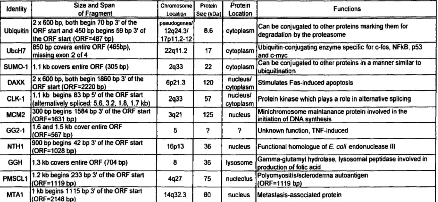
Human proteins positive for Ad2 ElB 55-kDa binding were detennined through partial screening of the Hela cDNA library (700 000 transformants). A total of 11 different proteins have been identified. These are listed, along with their functions and other features, in table 4. Excluded from tables 3 and 4 are "nonsense' peptides, cloned out of frame or in the reverse orientation. As a result of Hela cDNA library screening Ub was found to bind Ad2 E 1 B 55-kDa. Ub is a highly conserved protein consisting of 76 amino acid residues. Covalent ligation to Ub targets abnormal or short-lived proteins for degradation by the 26S proteasome. The minimum signal for efficient proteasomal targeting is a tetra-Ub chain (Thrower et al., 2000). There are three classes of enzymes involved in ubiquitination: the E 1, or Ub-activating enzyme, leads to activated Ub which is covalently linked to E 1 via a thioester bond. Activated Ub is transferred to the E2, or Ub-conjugating enzyme, which may donate Ub directly to proteins or may require an E3, or protein ligase, thought to play a role in substrate recognition. The human Ub-conjugating enzyme, UbcH7 (also called E2Fl) was picked up from library screening.
UbcH7 is a 17-kDa protein responsible for Ub-mediated degradation of the tumor suppressor protein, p53 (Ciechanover, et al., 1994) and oncoprotein c-fos (Stancovski et al., 1995). Proteolysis of p53 and c-fos by this degradation pathway allows for rapid changes in protein level. UbcH7 is also responsible for processing of the NfKB precursor p 105 (Orian et al., 1995). p 105 is degraded to the p50 subunit of the transcription factor NfKB which targets genes involved in cell growth and differentiation, inflammation, lymphocyte activation, and the acute-phase response. SUM0-1, an acronym for §.mail Ub-like modifier, was also found to bind to Ad2 ElB 55-kDa as a result of library screening. SUM0-1 covalently modifies a limited number of proteins in a manner
•
•
•
similar to ubiquitination, referred to as sumolation (Kamitani et al., 1997). It consists of 101 amino acid residues, 18% identical and 48% similar to Ub. Unlike Ub, however, the post-translational conjugation of SUM0-1 to various proteins does not target them for degradation. A growing list of cellular proteins known to be modified by SUM0-1 includes p53 (Gostissa et al., 1999), RanGAPl (Matunis et al., 1996), and PML (Boddy et al., 1996). The conjugation of SUM0-1 to p53 has been reported to result in an increase in p53 transactivation activity by some (Gostissa et al., 1999; Rodriguez et al., 1999) but contradicted recently by Kwek et al. (2001). Sumolation localizes RanGAPl to the nuclear pore complex (NPC) (Matunis et al., 1996). RanGAPl is a regulator of the Ran GTP/GDP cycle and therefore the bi-directional transport of proteins and ribonucleoproteins across the NPC (Melchior et al., 1993; Moore and Bio bel. 1993 ) . Three molecules of SUM0-1 are conjugated to PML at three separate sites (Kamitani et al.. 1998) whereas only one molecule is conjugated to other SUM0-1 protein targets. Sumolation of PML is a prerequisite for POD (PML oncogenic domain) formation. affecting the localization of PML and thus ail other POD components to the nuclear structures (Zhong et al., 2000a). Adenoviruses specifically modify PODs, with El B 55-kDa itself localizing to PODs shortly after infection (Doucas et al., 1996). During the preparation of this manuscript it was reported by Endter et al. (2001) that Ad5 ElB 55-kDa could be modified by SUM0-1 in vivo at a single lysine residue (Kl04). Sumolation affects El B subcellular localization and is required for inhibition of p53 transactivation function and transformation by Ad5 ElB kDa. Next in the list of potential ElB 55-kDa binding partners is DAXX, a transcription repressor implicated in Fas-induced
•
•
•
apoptosis (Torii et al., 1999; Yang et al., 1997). DAXX interacts with SUM0-1-modificd PML and localizes to PODs (Torii et al., 1999).
Aside from Ub, UbcH7, SUM0-1, and DAXX, other potential human Ad2 El B 55-k.Da-interacting proteins include CLK l, which phosphorylates serine/arginine-rich (SR) proteins. SR proteins are essential splicing factors (Caceres et al., 1994; Fu, 1993) thought to be regulated by phosphorylation (Xu and Manley, 1997). CLKl may play a role in the alternative splicing patterns during the switch from early to late phase of adenovirus replication. Overexpression of CLKl kinase shifted the splicing of El Apre-mRNA from multiple Apre-mRNA products, 9S, l2S and l3S, to the 9S RNA isoform characteristic of the late phase of infection by adenovirus (Duncan et al.. 1997). Next is MCM2, required for initiation of DNA replication (Todorov et al., 1994) and implicated in transcription by RNA polymerase II (Yankulov et al., 1999). MCM2 is a mini-chromosome maintenance protein, of which there are at least six (MCM2-7). ln ail eukaryotes MCM2-7 ensure that DNA replicates only once in each cell cycle (for a review on MCM2-7 see Tye, 1999). During the transition from G 1-phase to S-phase MCM2-7 are loaded onto replication origins as part of the pre-replication complex. Phosphorylation of MCM2 alters the conformation of the MCM complex and leads to melting of origin DNA. Next is GG2-l, induced by TNF but of unknown function (Horrevoets et al., 1999). GG2-l shows no homology at the nucleotide, amino acid, or structural level with any protein or gene present in the combined NCBI and EMBL databases. Another potential ElB partner is NTHl, a functional human homologue of E . coli endonuclease