HAL Id: tel-01124341
https://tel.archives-ouvertes.fr/tel-01124341
Submitted on 6 Mar 2015HAL is a multi-disciplinary open access archive for the deposit and dissemination of sci-entific research documents, whether they are pub-lished or not. The documents may come from teaching and research institutions in France or abroad, or from public or private research centers.
L’archive ouverte pluridisciplinaire HAL, est destinée au dépôt et à la diffusion de documents scientifiques de niveau recherche, publiés ou non, émanant des établissements d’enseignement et de recherche français ou étrangers, des laboratoires publics ou privés.
Parasite genetic factors implicated in cerebral malaria
Talleh Almelli
To cite this version:
Talleh Almelli. Parasite genetic factors implicated in cerebral malaria. Parasitology. Université René Descartes - Paris V, 2014. English. �NNT : 2014PA05P605�. �tel-01124341�
UNIVERSITY OF PARIS DESCARTES
FACULTY OF PHARMACEUTICAL AND BIOLOGICAL SCIENCES
DOCTORAL SCHOOL OF MEDICINE, TOXICOLOGY, CHEMISTRY,
ENVIRONNEMENT
Year: 2014 N°:
Speciality: Molecular Biology
Host Laboratory: UMR 216 "Mother and Child Facing Tropical Infections”
Presented by
Talleh Almelli
In fulfillment of the requirements for the degree of
PhD from University of Paris Descartes
Parasite Genetic Factors Implicated in
Cerebral Malaria
Defended [27/05 /2014]
Thesis committee:
M. George Snounou DR2-CNRS Reviewer M. Frédéric Ariey CR1- Pasteur Institut Reviewer Mme. Marie-José Butel Pr-UPD President M. Benoît Gamain DR2- CNRS Examiner Mme. Rachida Tahar CR1-IRD Advisor
2
Table of Contents
Acknowledgment Summary Chapter (1) 1. Introduction1.1. Malaria history at a glance 7
1.2. Malaria burden worldwide 8
1.2.1. Malaria burden in Benin 9
1.3. Human Plasmodium species 11
1.3.1. Plasmodium falciparum life cycle 11
1.4. Selected clinical outcomes of P. falciparum malaria infection 14 1.5. Cytoadherence property of P. falciparum infected erythrocyte 17 1.5.1. Receptors contribute to cytoadherence of infected erythrocyte 20
1.5.2. Cluster Differentiation 36 (CD36) 21
1.5.2.1. Role of cd36 gene polymorphism in malaria 22 1.5.3. Inter Cellular Adhesion Molecule-1 (ICAM-1 CD54) 22
1.5.3.1. Role of icam-1 gene polymorphism in malaria 23 1.5.4. Platelet Endothelial Cell Adhesion Molecule-1 (PECAM-1 CD31) 25
1.5.5. Complement Receptor-1 (CR1) 25
1.5.6. Endothelial Protein C Receptor (EPCR) 25
1.6. Plasmodium falciparum genome
1.6.1. Composition and organization of genome 26
1.6.2. Chromosome Structure 26
1.7. Variant Surface Antigens (VSA) 27
1.7.1. STEVOR and RIFIN 28
1.7.2. Var genes and PfEMP-1 29
1.8. Mutually exclusive expression and switching properties 36 1.8.1. Mutually exclusive expression property of P. falciparum var genes 36
1.8.2. Switching property of P. falciparum 37
3
2. Aims of the study 42
Chapter (2)
Manuscript (1) Gene transcriptomic pattern of Plasmodium falciparum in children with
cerebral malaria and asymptomatic carriers 44
Chapter (3)
Manuscript (2) Cytoadherence phenotype of P. falciparum infected erythrocytes is associated with specific Pfemp-1 expression in parasites from children with cerebral malaria 90
Chapter (4)
Discussion and perspectives 104
References 112
Annex 132
Informed Note Informed Consent
Recruitment File (Uncomplicated malaria) Recruitment File (Cerebral malaria)
4
Acknowledgment
First of all and from the depth of my heart I should thank those, who were always beside me despite the huge distance between us. Without your spiritual support and encouragement I would never ever attain this goal. Thank you my lovely family. I would like to thank my advisor, Dr. Rachida Tahar who provided me with necessary knowledge that I needed for my work.
I wish to express my sincere thanks to my thesis committee: Dr. George Snounou, Dr. Frédéric Ariey, Dr. Marie-José Butel and Dr. Benoît Gamain, who have accepted my invitation to be members of my thesis jury.
Thanks to Dr. Nicaise Ndam Tuikue, who helped me a lot during my missions in Benin and with whom the discussion was always interesting.
I am grateful to Dr. Philippe Deloron, who welcomed me to his laboratory and without his advices I could not be able to accomplish the statistical analysis in the second part of my thesis.
I place on record my gratitude to all the unit members in France and Benin for the nice moments we spent together especially during “Pot de Noël”:
Célia, Alexandra, Justin, Samad, Sèm, Firmine, Kossiwa, Azzizzath, Charles, Simon, Florentin, Pierre, Gwladys, Violetta, Géraud, Julie, Laure, Kadiatou, Emmanuelle, Aicha, Adrian, Sayeh, Pascale, Murielle, Jean-Gerard, Anaïs, Jérôme, Audrey, Nadine, Florence, Roman, Valérie, Michel, Isabelle, Jean Yves, Achille, André, Gilles, Magalie, Brigitte, Carmen, Ibrahim, Komi, Odilon, Aurax, Rodolphe, Atikath, Adicath, Pépin, Rafiou, Carine, Nadia, Bienvenu, Darius, Bienvenu, Manfred, Aziz, Honoré, Silvestre, Patrick, Erasme, Silvain, Paulin, Sévérin, Joseph, Jacques, Pépin, Ginette and Sophie. Immense thanks to all the patients and their parents who made this thesis possible.
5
Summary
Plasmodium falciparum is the most deadly species, causing broad spectrum of disease
manifestations ranging from asymptomatic infection to symptomatic outcomes, including uncomplicated and severe malaria. This latter comprises various clinical forms, notably cerebral malaria. The biological processes involved in the heterogeneity of these clinical outcomes are not well understood. We hypothesized that differential gene expressions contribute to phenotypic variation of parasites leading to specific interaction with the host which subsequently induces several clinical categories of malaria. In order to verify this hypothesis, we investigated the transcriptomes of parasites isolated from Cameroonian children with asymptomatic (AM), uncomplicated (UM), and cerebral malaria (CM). We also analyzed the transcriptomes of the 3D7 line and the selected 3D7-Lib line. The latter has been selected by several rounds of incubation with anti-IgG of Liberian hyper-immune plasma. It has the ability to express group A var and neighboring rif genes, which have been described in several studies to be associated with severe disease and cerebral malaria.
Our results of transcriptomic study revealed the up-regulation of several genes in CM-associated isolates and the 3D7-Lib line compared to AM-CM-associated isolates, and the 3D7 line. Gene Ontology term analysis shows that a majority of differentially expressed genes are mainly implicated in pathogenesis, cytoadherence to host endothelial tissues, and erythrocyte aggregation. Among up-regulated genes in CM-associated isolates compared to AM-associated isolates, there were genes encoding for surface antigens, Maurer’s cleft proteins, transcriptional factors, and antigens involved in protein transport to the host cell surface. In addition, an important variation in the expression of variant surface polymorphic families like var, rif and phist genes has been observed. The most remarkable outcomes were the up-regulation of UPS A and UPS B var genes containing architectural Domains Cassettes DC4, DC5, DC13, and DC8 and their neighboring rif genes in isolates from CM and in the 3D7-Lib line compared with isolates from AM and the unselected 3D7 line, respectively.
Several studies have shown that group A and B var genes containing DC13 and DC8 are implicated in severe malaria, including cerebral malaria. However, the transcriptome profiles were similar in CM and UM isolates, and between AM and UM parasites. The lack of differences in genes expression between CM and UM parasites may be due to similar transcription patterns between isolates of these two clinical groups, or to the inability of microarrays to detect trivial variations. Therefore, in order to confirm the specific transcription profiles of these genes in CM isolates, we examined by RT-qPCR the expression abundance of UPS A1, A3, and B1 var groups found to be highly up-regulated in the CM group, using new samples collected from Beninese children with CM or UM. These genes
6 were significantly highly transcribed in CM compared to UM group. The involvement of these genes in the parasite virulence rises from the ability of their encoded proteins to mediate the cytoadherence of infected erythrocytes to post capillary endothelial cells. Cytoadherence is essential for parasite life to evade spleen-dependent clearance. However, the clinical consequences of this cytoadherence differ among infected individuals. Several endothelial receptors that are involved in the cytoadherence of IEs to venular endothelia have been identified, and few studies attempted to correlate a particular binding phenotype of parasites with a given clinical presentation of malaria. Of these receptors, CD36 and Inter Cellular Adhesion Molecule-1 (ICAM-1) were found as the most commonly used by patient parasites. The implication of these two receptors carried out along with PfEMP-1 ligands in the pathogenesis of cerebral malaria needs to be more elucidated. We examined the adhesive phenotype and the transcription patterns of Pfemp-1 variants of fresh isolates from Beninese children with cerebral malaria and uncomplicated malaria by static binding assay and RT-qPCR respectively, to identify a relationship between the Pfemp-1 variants expressed by the parasites and their cytoadherence phenotype to a specific receptor. The binding levels of IEs were investigated to CD36, ICAM-1 and CSPG (CSA) and the transcription level was assessed for groups A, B, var2, var3, DC8, and DC13, up-regulated in CM isolates in our transcriptomic study.
Our findings reported for the first time that isolates from CM patients have a significant preference for CD36-binding compared with those from UM cases. No differences were observed in binding levels to ICAM-1 or CSPG between these two groups. Furthermore, cerebral malaria isolates transcribed groups A, B, var2, var3, DC8 and DC13 of var genes to higher levels than UM isolates. Interestingly, the high transcription levels of group B in CM parasites correlated with their raised level of binding to CD36, supporting the implication of this receptor along with group B in cerebral malaria pathogenesis. In contrary, the expression profiles of a specific var group and the binding phenotype of isolates to ICAM-1 and to CSPG were not correlated.
The molecular basis of interaction between CD36 and PfEMP-1 variants of group B need to be further investigated for better understanding of cerebral malaria pathogenesis. This interaction could be targeted by specific antibodies or suitable agonist molecules to prevent
P. falciparum severe complicated malaria as cerebral malaria.
In addition to PfEMP1 group A1, A3 and B1 subset, other genes encoding exported proteins as well as non-exported proteins, were up-regulated in CM-associated parasites. The potential role of these genes in CM pathogenesis deserves to be investigated and may allow the identification of new targets for malaria control.
7 Chapter 1
1. Introduction
1.1. Malaria History at a glance
Malaria or mal’ aria “spoiled air” is caused by parasites which have existed with us since the dawn of time. The earliest Plasmodium falciparum DNA has been detected in two Pharaonic mummies aged more than 3200 years B.C.
Hippocrates was one of the first to notice and describe malaria symptoms such as poor health, fever and enlarged spleens in the 5th century B.C.
The first documented treatment against malaria was brought from Peru in the 1600s A.C by the Jesuits. The natives at that time were using the Cinchona barks as a traditional treatment which then had been transported to Europe under the name of “Jesuit’s powder” to treat patients experiencing fever “agues” (Fig. 1).
Fig. 1: Rawhide Bag of Cinchona bark, namely “Seron” used to store and transport Cinchona barks, brought from Peru in 1777 by Hipolito Ruiz Lopez and Antonio Pavon y Jimienz who had been sent by Spanish monarch Charles III to discover the region. Source: Science and society, 2014. (http://www.scienceandsociety.co.uk)
In 1820, Pelletier and Caventou, French pharmacists, were able to successfully isolate the quinine and the quinidine from Cinchona barks that were used later by French workers in Senegal and in the United States of America.
The discovery of malaria parasite was made in 1880 by Alphonse Laveran, a French surgeon in the French army posted in Constantine city, Algeria. He observed parasites in blood samples of patients with fever that were absent in healthy individual blood. He also noticed the disappearance of parasites after administration of quinine. These observations
8 encouraged Laveran to identify and describe the causative agent of malaria, “the parasite”. For this precious finding, Laveran has been attributed Nobel prize of medicine in 1907. In 1885, Marchiafava, an Italian physician, named the genus of malaria parasites “Plasmodium”, and this was followed by the identification of P. vivax and P. malariae by Camillo Golgi and P. falciparum by Marchiafava. The exflagellation property of male gametocytes and the ability to enter inside female gametocyte of P. falciparum parasites has been recognized by MacCallum in 1897. The fourth species of human malaria parasites P.
ovale had been identified in 1922 by Stephens.
The incrimination of mosquito in malaria transmission was made in 1897, when Ronald Ross in India observed different stages of development such as oocytes in the stomach wall as well as sporozoites in the salivary glands inside mosquitoes that had fed on infected birds, and independently by Grassi in Italy who incriminated Anopheles for the transmission of P.
falciparum. Ross was was awarded the Nobel Prize of medicine in 1902.
The sporogonic cycle of Plasmodium parasites inside the female mosquito was observed in 1899 by Grassi, Bastianelli and Bignami. Using blood samples from naïve volunteers infected with mosquito fed on malarial patients, they were able to describe the life cycle inside the anopheles and the biological cycle of P. falciparum, P. vivax and P. malariae. In 1948, Shortt and Garnham discovered the pre-erythrocytic stage of Plasmodium parasites, which takes place inside the human host. Finally, in 1982, Krotoski was able to characterize the
hypnozoite or the latent phase of P. cynomolgi and then of P. vivax (Krotoski et al. 1982). The discovery of natural infections by a 5th species, Plasmodium knowlesi, came late in 2007 after cross infection from Macaca monkey to human being. During the past years, hundreds of cases had been diagnosed with malaria caused by P. knowlesi infection. This latter morphologically resembles to P. malariae, However, P. knowlesi infections, unlike those of P.
malariae, can be fatal to humans (Cox et al. 2010).
1.2. Malaria burden worldwide
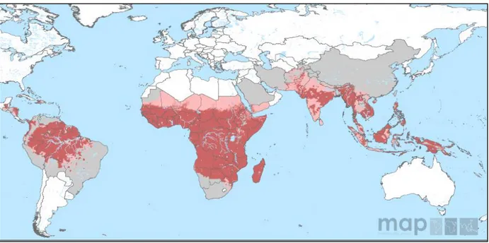
Malaria, the implacable enemy, is one of the most serious health problems in the world (Fig. 2). In 2010, the World health organization estimated 216 million acute cases of malaria in 106 endemic countries and up to 655,000 deaths annually (WHO 2011). The highest rate of malaria cases (81%) and deaths (91%) took place in sub-Saharan Africa, though other continents including Latin America, the Middle East and Asia are at risk as well (Murray et al. 2012). Children and pregnant women in endemic regions bear the brunt. While the majority of deaths occur among children under five years of age, most surviving patients suffer from grave complications, mainly severe anemia. Pregnancy associated malaria (PAM) has undesirable birth consequences such as premature delivery, spontaneous abortion, still birth and low birth weight (Steketee et al. 2001). This disease affects negatively on the economic growth rate of endemic areas, where it is estimated to decrease by 1.3% annually compared
9 to that in malaria-free countries (Sachs and Malaney 2002). Thus, malaria has social and economic impact on needy people and indigent populations who are not able to afford treatment costs or have restrained access to public health care (Orem et al. 2012). Insecticidal bed nets and indoor residual spraying (pyrethroids-vector control) and anti malarial drugs against parasites (notably artemisinins) have significantly favored the reduction of malaria cases and death rate (WHO, 2011). However, these strategies have been thwarted by the emergence of resistance in both mosquitoes and parasites, and by human migration and the recent financial crisis (Leach-Kemon et al. 2012). All these causes are imminent threats to individuals living in endemic regions (WHO 2010). Another strategy to fight against malaria is a vaccine, which, if available, could decrease the malaria incidence by protecting the population at high risks (children and women) from the serious complications of disease; thereby it would reduce the medical expenses and support the reconstructive and developmental projects in the affected countries. However, all the vaccine candidates tested during the last decade were not sufficiently efficient (Snounou and Renia 2007).
Fig. 2: P. falciparum malaria risk defined by Annual Parasite Incidence in 2010. Carmine color refers to stable-transmission geographical zone where annual parasite incidence PfAPI ≥ 0.1%, whereas pink color represents unstable-transmission zone with PfAPI < 0.1%. Light grey mentions to free malarial areas. Source: Malaria Atlas Projects (Map), 2010.
1.2.1. Malaria burden in Benin
The republic of Benin is localized in West Africa, bordered by Nigeria on the East, Togo on the West, Burkina Faso and Niger on the north and the Atlantic Ocean on the south (Fig. 3). The surface area of Benin is 112,600 km² with a population density estimated to 10, 100,000 habitants (WHO, 2013). The climate is tropical in the south and soudanian in the north with
10 two distinct dry and rainy seasons. The rainfall is not stable; it varies between regions and over the years with a range between 1400 mm in the south and around 800 mm in the north (Fink et al, (9) MPETUS Atlas du Bénin; http://www.impetus.uni-koeln.de/fr/impetus-atlas/impetus-atlas-benin).
In Benin, 100% of people are at risk of malaria (WHO, 2013). In 2006, the estimated death rate in infants under 5 years was 125 deaths/1000 live births (Demographic and Health Survey, 2006; http://dhsprogram.com/pubs/pdf/FR197/FR197.pdf).
Plasmodium falciparum is the principal cause of malaria, transmitted by three major
mosquito vectors: An. gambiae and An. melas and An. funestus (Akogbeto and Romano 1999; Moiroux et al. 2012). The epidemiological studies have been concentrated in the coastal areas south of Benin, where the economic and the administrative capitals are situated. Cotonou is the economic capital, and it is characterized by two rainy seasons from April to July and from September to November during which malaria infection is exclusively caused by P. falciparum parasites with approximately 33 infected bites per individual annually (Akogbeto, 1995) (Fig. 3). In this city, the average parasite density in children is from 3000 to 6000 parasites/mm3 and less than 1000 parasites/mm3 in adults (Chippaux et al. 1991). A study conducted at CNHU hospital in Cotonou, showed that 20% of medical visits was due to malaria and 44% of cases were children between 6-23 months (Boulard et al. 1990). Another study reported that the mean number of fever episodes occurred every year in Beninese children under 3 years was 2.4 per children and 33% of those cases were infected with P. falciparum with an estimated mortality rate of 8/1000 cases yearly (Velema et al. 1991).
Part of my thesis work was carried out in Cotonou where field isolates had been collected from three hospitals located in this city, as it is described in the Materials and Methods in the second manuscript.
11
Fig. 3: Global map showing the localization of Benin in west Africa (Top-left), its neighbors and the two capitals, Cotonou and Porto-Novo. Source: Modified from President’s Malaria Initiative (PMI). (http://www.pmi.gov/countries/profiles/benin.html)
1.3. Human Plasmodium species
The etiological agent of malaria is a eukaryotic protozoan parasite of the genus Plasmodium belonging to the apicomplexan family. Parasites are transmitted to human beings by
Anopheles mosquitos’ bites. To date, more than 400 species of anopheline mosquitoes have
been distinguished, but only 40 are recognized as malaria vectors (Service 1993). Some of these mosquitoes e.g. Anopheles gambiae, prefer human being to animals so-called “anthropophilic”, making them highly active malaria transmitters to human hosts.
There are five common species of Plasmpdium that could infect humans: P. falciparum (Welch 1887) (Schaudin 1802), P. vivax (Grassi and Feletti 1890), P. malariae (Grassi and Feletti 1892), P. ovale (Stephens 1922) and P. knowlesi which has recently been identified in Malaysia as the 5th human infecting species after cross infection from Macaca monkey
(Sabbatani et al. 2010).
Among these species, P. falciparum is the most virulent species since it is associated with severe malaria and high mortality in sub-Saharan Africa.
1.3.1. Plasmpdium falciparum life cycle
The Plasmodium falciparum life cycle is complex, and the parasite needs to develop in two different hosts to complete its cycle. The infection is initiated when infected female mosquito bites an individual and inoculates sporozoites with the saliva into the skin. Within
12 one hour the parasites move to the liver and invade hepatocytes. The sporozoites spend 6-10 days inside hepatocytes during which they differentiate and undergo asexual replication yielding upto ~40,000 merozoites that then burst from the infected liver cell into the blood stream. The merozoites invade erythrocytes and go through several morphological stages, beginning with the ring and followed with the trophozoite stages, where the parasites ingest part of erythrocyte cytoplasm and degrade its hemoglobin into amino acids to be used for protein synthesis. Schizonts are produced after multiple round of nuclear multiplication resulting in 4-32 daughter merozoites that are released into the circulation after the infected erythrocyte (IE) bursts. Each merozoite is able to infect a new erythrocyte within 30 seconds and reinitiates the cycle. The duration of the intra-erythrocytic stage of P. falciparum is 48 hours, and clinical symptoms of malaria, fever and chills correlate with IEs bursting. After 10-11 days, another form of parasites can be found in the peripheral blood, namely gametocytes. In fact, some of erythrocytic parasites do not develop into schizonts and instead they differentiate into sexual forms, male and female gametocytes, which are not harmful to human host, but are infectious to mosquitoes (Mouchet et al. 2004; Tilley et al. 2011). Once in the mosquito midgut, following a blood meal, the parasites encounter environmental change, low temperature, modified pH and blood digestive factors such as chymotrypsins and serine proteases. After 30 minutes, the gametocytes mature, where the males transform into eight-motile microgametes and the females into round shaped macrogametes that then emerge from IEs and fertilize to produce a zygote. At this stage the genome of the parasite is diploid and recombination events can lead to an extensive genetic diversity. The sexual life cycle continues inside the mosquito and the zygote pursues its development, as it becomes motile ookinete that enters the stomach wall and resides between basal lamina and epithelial cells. This is followed by the differentiation into an oocyte containing thousands of parasites harboring haploid genomes.
The mature oocyte bursts and releases thousand of sporozoites that then migrate to the mosquito salivary glands where they are ready to be injected into a human host (Cox et al. 2010; Vizioli et al. 2001).
13
Fig.3: Life cycle of Plasmodium falciparum. (1) Sporozoites injected by mosquito, migrate through the circulatory system to the liver, where they invade hepatocytes, multiply then released to blood stream as merozoites which are able to invade erythrocytes. Parasites enter asexual intraerythrocytic multiplications (2), which take 48 hours for P. falciparum. Each schizont gives birth to 30 daughter merozoites. Once erythrocyte ruptures, these merozoites infect other erythrocytes. Some merozoites differentiate to male and female gametocytes, which are taken by mosquito with blood meal, to be fertilized, differentiate and replicate resulting in sporozoites stages ready to infect human host. Source: Modified from (Bousema and Drakeley 2011).
14 1.4. Selected clinical outcomes of P. falciparum malaria infection
The clinical manifestations of P. falciparum malaria vary from asymptomatic infection to uncomplicated malaria and severe malaria including cerebral malaria. The disease symptoms range from fever to severe complications.
Mild form of malaria is mainly manifested as fever and sometimes is accompanied with gastro-intestinal troubles, while severe or complicated malaria consists of life threatening disease. The most vulnerable populations are individuals with non-developed immunity such as children and travelers in endemic areas, or people with reduced acquired immunity like primigravidae women. In malaria endemic regions, asymptomatic infections with low parasite densities are prevalent.
The clinical consequences of malaria take place during the asexual blood cycle of parasite life cycle. The clinical manifestations of malaria are classified as uncomplicated malaria (UM); severe malaria (SM) including severe malaria anemia (SMA) and cerebral malaria (CM). Malaria infection in pregnant women is considered as complicated malaria and known as pregnancy associated malaria (PAM).
During my PhD thesis i have been interested in P. falciparum genetic factors implicated in the pathogenesis of falciparum malaria. For that, i used isolates from Cameroun and Benin collected by our team from children less than six years of age. These children presented distinct clinical manifestation such as CM and UM. A group of children with asymptomatic infections had been collected and used as well.
Uncomplicated malaria (UM)
The most frequent features of uncomplicated malaria infection are chills, fever, sweat, headache, asthenia, weariness, myalgia, nausea and vomiting due to the liberation of hemozoin and the toxic bilirubin followed IE rupture. In case of delayed or subcurative treatment, the high temperature and the high parasite density would worsen the clinical case.
Treatment of uncomplicated malaria aims to eliminate the parasites from the body as rapidly as possible in order to prevent the progression of disease to severe form. An adequate treatment of the infection can attenuate its transmission to others and reduce the emergence of resistance to antimalarial agents.
The first line treatment for infants and young children with UM is artemisinin-based combination therapy (ACTs). A combination of two or more antimalarial medicines with different mechanisms of action and contrasting pharmacokinetics such as artemether / lumefantrine, artesunate / amodiaquine, artesunate / mefloquine, artesunate / sulfadoxine-pyrimethamine, and dihydrortemisinin / piperaquine is the treatment of choice for
15 uncomplicated falciparum malaria. Alternative therapy such as artesunate / tetracycline or doxycycline or clindamycin, quinine plus tetracycline or doxycycline or clindamycin should be administered for seven days (WHO, 2010).
Severe malaria (SM)
In tropical endemic areas, severe malaria occurs primarily in children under 5 years of age whereas in industrialized countries, non-immune adults returning back from endemic zones are susceptible to develop severe malaria (Hatz et al. 2001; Leder et al. 2004). Several predisposing factors participate in this type of malaria and may lead to death, involving age more than 65 years, naïve non immune cases, female sex (especially pregnant), lack of prophylaxis, delay in medical treatment, degree of disease severity at admission and coexisting other health problems (Bruneel et al. 2003; Schwartz et al. 2001).
Severe P. falciparum infections cover a broad spectrum of complications, defined by the WHO as coma; generalized convulsions; cardio-vascular collapse; hemorrhagic syndrome; severe anemia (Hb<6 g/dl); pulmonary edema; hypoglycemia (<2.2 mmol/l); hypouresis (<400 ml/day); hemoglobinuria; and acidosis (PH <7.25). Of these, severe anemia, respiratory distress and cerebral malaria are the major grave outcomes of severe malaria (WHO, 2000 S1). In the absence of appropriate treatment, any of these complications could evolve rapidly and become fatal within hours or days.
Severe malaria anemia (SMA)
It is the most common clinical complication of severe P. falciparum malaria and the main cause of hospitalization of children in endemic areas. Severe anemia is characterized by arthralgia, paleness and low hemoglobin level <5 g/dl with parasite density > 10.000 p/µl (WHO, 2000 S1). This is probably a consequence of multiple causes such as erythrocyte hemolysis, suppressed synthesis of erythrocytes (Phillips et al. 1986) and increased spleen clearance for both uninfected and infected erythrocytes (Dondorp et al. 1999).
Respiratory distress (RD)
This outcome is possibly due to fluid retention and it is described by unusual or frequent deep breathing as a result of metabolic acidosis. This latter is enhanced by the low level of oxygen as a consequence of anemia and decreased microvascular blood flow arising from parasite sequestration and rosetting (Kaul et al.1991; Marsh et al. 1996).
Cerebral malaria (CM)
Patient with this form is characterized by last long unrousable coma accompanied with multiple seizures; as a result of blood brain barrier deterioration (Adams et al. 2002) and fever that may surpass 41°C. In the case of cerebral malaria, careshould be taken to exclude other causes of coma, notably meningitis. This form is responsible for 15-20% of infant
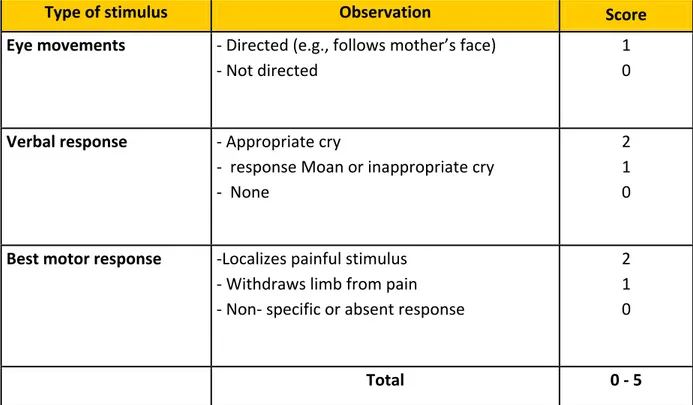
16 mortality and is considered as the principle cause of long-term neurocognitive impairments in African children (Idro et al. 2010). In order to evaluate the state of consciousness of infected patient, the World Health Organization has adopted Glasgw coma scale (score between 6-7) for adult infected patients and Blantyre coma scale for children< 5 years of age. Blantyre score ranges from 0-5 and children with score ≤ 2 are diagnosed as cerebral malaria cases (Table 1, WHO 2000).
Table 1. A coma scale for children, Blantyre coma scale
Type of stimulus Observation Score Eye movements - Directed (e.g., follows mother’s face)
- Not directed
1 0
Verbal response - Appropriate cry
- response Moan or inappropriate cry - None
2 1 0
Best motor response -Localizes painful stimulus - Withdraws limb from pain - Non- specific or absent response
2 1 0
Total 0 - 5
The main goal of the treatment of severe malaria is to prevent death. In cerebral malaria cases, it aims to prevent neurological deficit.
In the case of cerebral malaria, parenteral full doses of any effective antimalarial drugs should be given as rapidly as possible. For adults and children artesunate IV or IM is indicated for a minimum of 24 hours. If it is not available, artemether or quinine is acceptable. After that, treatment should be completed with one of the following agents: ACT; artesunate plus clindamycin or doxycycline; or quinine plus clindamycin or doxycycline. A pre-referral treatment such as rectal artesunate, quinine IM or artesunate IM could be given, and then patient should be referred immediately to an appropriate health care for further treatment (WHO, 2010)
17 Pregnancy associated malaria (PAM)
In spite of the acquired immunity before pregnancy; pregnant women in malaria endemic zones are highly susceptible to malaria infections particularly during the first pregnancy where immunity decreases temporarily. During this period the placenta of pregnant woman offers a convenient environment for parasites with high affinity to placental receptors. P.
falciparum parasites via VAR2CSA, the predominant parasite surface-expressed PfEMP1
protein in PAM, have an elevated capacity to adhere to chondroitin sulfate A (CSA) in the syncytiotrophoblasts of the placenta (Achur et al. 2008), provoking blood flow obstruction and disturbing the key source of nutrition for the fetus (Dorman et al. 2002). This phenomenon results in maternal anemia which itself has undesirable consequences as maternal death, premature delivery, spontaneous abortion, still birth and low birth weight (<2.500g) (Steketee et al. 2001).
The principal objective of treatment in pregnancy associated malaria case, is to save the life of the mother. During the first trimester, administration of quinine plus clindamycin for seven days has been indicated by the World Health Organization. In case of treatment failure, a combination of artesunate plus clindamycin for 7 days is indicated. During the second and third trimesters, sulfadoxine/pyrimethamine combination is recommended as a profylaxis. However, a treatment artemesinin combination therapy (ACT) is indicated.
The virulence of P. falciparum is partially due the capacity of IEs to cytoadher to endothelial cells of microvasculature of deep organs leading to obstraction of these vessels and organs disfunctions.
1.5. Cytoadherence property of P. falciparum
In order to pursue its erythrocyte life cycle, P. falciparum parasite shows an amazing ability to trick the host immune system and evade spleen-dependent clearance. One such phenomenon is called “cytoadherence” (Udeinya et al. 1981). It occurs once the erythrocytic parasites reach tthe mature stages (i.e. late-trphozoite and schizont), where they disappear from the peripheral blood and adhere to different venular endothelial cells of various organs including the brain (MacPherson et al. 1985; Pongponratn et al. 1991).
This sequestration of parasitized erythrocytes has sequels. It enhances the release of soluble mediators and activates the caspase pathways which in turn lead to vascular, neuronal and tissue apoptosis and blood brain barrier damage (BBB) in some cases of malaria (Pino et al. 2003; Wilson et al. 2008). In addition, the accumulation of infected erythrocytes results in microvascular clogging, ischemia, hypoxia, lactic acidosis and activation of inflammatory cytokines which increase endothelial cell adherence molecules (eCAM) and accelerate the adherence of additional populations of infected erythrocytes (van der Heyde et al. 2006).
18 However, the impact of sequestration on the host depends on host immunity, the affected tissues and parasite density.
During pregnancy-associated malaria the target of parasite sequestration is the placenta. Histological analysis of parasitized placenta showed inflammatory lesions with fibrinoid deposition leading to chronic intervillositis. This correlates with negative consequences such as spontaneous abortion, decreased intrauterine growth and increased fetal death (Rota et al. 2006).
In cerebral malaria the preferred niche of IEs is the brain microvasculature. There is mounting evidence that cytoadherence of IEs to brain microvascular endothelia is linked to cerebral malaria, which was confirmed by postmortem studies (Molyneux 1990). Analysis of brain tissues from 24 Thai patients who died of malaria revealed that the post capillary venules of the brain were packed with IEs compared to the other parts of the body including the lungs and the heart (Pongponratn et al. 1991). Similarly, another post mortem study of 21 Vietnamese individuals reported that accumulation of IEs within the microvascular endothelium of the brain was correlated with microvascular congestion and that was associated with premortem coma and shorter time to death (Ponsford et al. 2012).
The most important mediators of cytoadherence are variant surface antigens (VSA), mainly PfEMP-1 members of VAR family. These proteins are exposed at the surface of IEs allowing them to cling to various post capillary endothelial cells (Baruch et al. 1995).
The P. falciparum parasite has different ways to cytoadhere within the host. For that new terms have been coined to describe these mechanisms. The first one is known as “Rosetting”, where the infected erythrocytes bind spontaneously to uninfected erythrocytes and form rosettes resembling aggregates (Udomsangpetch et al. 1989). Rosettes in blood group A or B infected blood are larger and stronger that those in group O blood (Rowe et al. 2007; Udomsangpetch, 1993). Several lines of evidence have indicated that rosetting phenomenon is associated with severe malaria in sub-saharan Africa (Carlson et al. 1990; Treutiger et al. 1992), but not in South East Asia (Ho et al. 1991).
A comparative study of 79 Malian isolates from children with different clinical complications of severe malaria, has reported a contribution of rosetting in severe malaria pathogenesis compared to 91 patients with uncomplicated malaria (Doumbo et al. 2009). Similarly, another study of 111 isolates of Kenyan children with severe malaria and mild malaria found an increased capacity to form rosettes and giant rosettes among isolates from children with severe malaria compared to mild cases (Heddini et al. 2001). Horata et al identified a semi-conserved feature represented by a position of limited variability named (cyc/PolV) that is located in DBLα of PfEMP-1 and associated with high rate of rosettes formation in both severe malaria and mild malaria groups (Horata et al. 2009).
19 The second term is “Clumping” where infected erythrocytes bind to platelets and form clumps (Pain et al. 2001). In vivo, this phenomenon could promote blood vessels occlusion and it is found to be associated with severe malaria (Mayor et al. 2011), particularly cerebral malaria since platelets express CD36 at their surfaces and induce the parasites binding even in absence of venular endothelial CD36 in the brain, thus they act as a bridge between brain endothelium and IEs. This mechanism of sequestration probably reorients the cytoadherence property of parasites and takes part in cerebral malaria pathogenesis (Wassmer et al. 2004). It has been suggested that thrombocytopenia could confer a protective mechanism for the host during cerebral malaria by decreasing platelet-mediated clumping of IEs (Wassmer et al. 2008).
Additional terms to express the distinct mechanisms of cytoadherence ability of infected erythrocyte comprise “binding to cells of immune system” such as plasma B cells, IgM, lymphocytes, monocytes, and dendritic cells (Donati et al. 2004; Ockenhouse et al. 1989; Urban et al. 1999)
20
Fig. 5: Sequestration phenomenon of infected erythrocyte. Schematic cytoadherence mechanisms of
P. falciparum infected erythrocytes (a). Cytoadherence of infected erythrocytes to endothelial cells (b),
Rosetting (c) and Clumpling (d). Source: (Rowe et al. 2009).
1.5.1. Receptors contribute to cytoadherence of infected erythrocyte
A diverse array of endothelial receptors that are likely to participate in the adherence of IE to microvasculature endothelia has been identified. Several investigations attempted to describe a specific binding phenotype of isolates from severe malaria patients that is less frequent in uncomplicated malaria parasites. So far, no definitive candidate has been identified apart from chondroitin sulfate proteoglycan (CSPG) also known as CSA. It is believed that CSA is the main receptor of the VAR2CSA protein that is expressed at the surface of IE and can adhere to the syncytiotrophoblasts leading to placental sequestration of IEs during malaria in pregnant women (Fried and Duffy. 1996; Salanti et al. 2003).
21 Other potential receptors are: Thrombospondin (Roberts et al. 1985); Cluster Differentiation 36 (CD36) (Barnwell et al. 1989; Oquendo et al. 1989); Inter Cellular Adhesion Molecule-1 (ICAM-1) (Berendt et al. 1989) ; Vascular Cell Adhesion Molecule-1 (VCAM-1) (Ockenhouse et al. 1992); Complement Receptor-1 (CR-1) (Rowe et al. 1997); Platelet endothelial Cell Adhesion Molecule-1 (PECAM-1) (Treutiger et al. 1997); Integrin αvβ3 (Siano et al. 1998); P-selectin (Ho et al. 1998); Heparan sulfate (Vogt et al. 2003); Fractalkine (Hatabu et al. 2003); Fibronectin (Eda and Sherman. 2004); Neural Cell Adhesion Molecule (NCAM) (Pouvelle et al. 2007); gC1qR-HABP1-P32 (Biswas et al. 2007) and Endothelial Protein C Receptor (EPCR) (Turner L. et al. 2013). Most of these receptors have not been intensively explored and the role of P. falciparum ligands with each receptor in severe malaria is not well established. Among these candidates, CD36 and ICAM-1 have been extensively investigated and were shown to be the most commonly used by parasite isolates (Udomsangpetch et al. 1996). However, our knowledge about the implication of these receptors in cytoadherence of IE comes from the human genetic polymorphisms associated with malaria severity as well as from cytoadherence assays under static or flow conditions by employing recombinant proteins coated on plastic dishes, cell line, receptor-bound beads or fluorescently labeled proteins.
1.5.2. Cluster Differentiation 36 (CD36)
This transmembrane glycoprotein (88-KD) is encoded by cd36 gene on chromosome 7 and expressed on various cell types including phagocytes, microvascular endothelia; platelets; hepatocytes; retinal pigment epithelium; adipocytes and specialized epithelia of breast, kidney and gut. Thus, a broad spectrum of biological processes would be expected. It acts as anti-angiogenic factor; cellular receptor for thrombospondin, collagen, modified low-density lipoproteins and long-chain fatty. It is also involved in sensory perception (Silverstein and Febbraio 2009). The binding site in CD36 for P. falciparum ligands is located within 97-110 and 139-184 amino acids regions, as suggested by monoclonal antibodies and peptides against this protein (Baruch et al. 1999; Daviet et al. 1997). The parasite ligands for this receptor are PfEMP-1 variants (Baruch et al. 1996) encoded by group B and C of var genes through their N-terminal CIDR1α domain (Miller et al. 2002; Robinson et al. 2003).
CD36 is considered as the major host receptor for P. falciparum field isolates (Chaiyaroj et al. 1996; Newbold et al. 1997), though its pivotal role in malaria pathogenesis is still unclear. Whereas several laboratory assays did not note differences in CD36-binding patterns between field isolates from severe and mild malaria cases living in Africa (Heddini et al. 2001; Mayor et al. 2011; Rogerson et al. 1999), others have supported a correlation between uncomplicated malaria and binding to CD36 (Newbold et al. 1997; Ochola et al. 2011). Conversely, researchers in Asian endemic regions have shown that CD36 may play a part in malaria severity (Ho et al. 1991; Udomsangpetch et al. 1996) as it facilitates the binding of
22 high proportion of parasites throughout the body, promoting systemic infection that contributes to the severity of the disease.
The role of CD36 in the interaction between IEs and host immune system is elusive. While, adherence of IEs to macrophages via CD36 induces phagocytosis and enhances IEs clearance, (McGilvray et al. 2000), binding to dentritic cell (DC) via CD36 yields to DC dysfunction decreasing the adaptive immune response, thus exacerbating the infection (Urban et al. 2001). However, other finding suggests that IEs-binding to CD36 is not necessary to inhibit DC maturation; instead high parasite density can induce immunosuppression (Elliott et al. 2007).
It has been shown that CD36-binding is not involved in the rosetting process (Rowe et al. 2000); however, it is essential but not sufficient for the clumping phenomenon (Arman et al. 2013).
1.5.2.1. Role of cd36 gene polymorphism in malaria
Several studies suggested an important role of human gene polymorphism in the malaria severity but these investigations have reported contradictory conclusions.
In Thailand, a correlation between cd36 polymorphisms and protection against severe malaria has been detected. The frequencies of 14T to C allele in the upstream promoter region and 53G to T in the downstream promoter region of exon 8, were significantly higher in mild malaria than those of cerebral malaria cases, suggesting that such cd36 polymorphism may protect against cerebral malaria. Similarly, the frequency of repeat 12TG in intron 3 [in3 (TG) 12], which is linked with CD36 deficiency, was higher in uncomplicated malaria than in cerebral malaria cases. This polymorphism could alter the binding affinity of CD36 isoform to IEs, leading to decrease the risk of cerebral malaria (Omi et al. 2003).
In Africa however, researches led to confounding outcomes. Non-sense substitution mutation of T to G in cd36 gene at nucleic acid 188 of exon 10 leading to CD36 deficiency was found to bestow protection against severe malarial anemia (SMA), probably through decreased binding of IEs to microvascular endothelia (Chilongola et al. 2009; Pain et al. 2001). However, this finding contradicted with another study of African and Middle Eastern populations in which no correlation between CD36 deficiency and protection from severe malaria was observed (Fry et al. 2009).
These results concluded that the existence of distinct cd36 mutations in Africans and Asians populations might be related to alternative or additional selection pressures on cd36 and co-evolution between human host and parasite.
1.5.3. Inter Cellular Adhesion Molecule-1 (ICAM-1 CD54)
It is a transmembrane glycoprotein (90-115-KD) and a member of the immunoglobulin like superfamily. It is constituted of five immunoglobulin (Ig)-like domains. This protein is found as a dimer at the surface of cells and its N-terminal Ig-like domain displays binding sites for
23 fibrinogen, the Lymphocyte Function-associated Antigen-1 (LFA-1), cell-entrance site of human rhinouvirous and P. falciparum PfEMP-1. This latter is situated on the BED side in the core of ICAM-1 dimer (Fig. 6) (Jun et al. 2001; Ockenhouse et al. 1992). Thus, ICAM-1 takes part in recognition and adherence between cells.
The parasite ligands for this receptor are PfEMP-1s via DBLβ-C2 of group B, C and DBLβ3 of group A (Bengtsson et al. 2013; Howell et al. 2008; Oleinikov et al. 2009). ICAM-1 is expressed at a restricted level on several types of tissues such as leucocytes, vascular endothelial cells, mucous epithelia of the tonsils, hepatic sinusoids lining cells, spleen, thymus epitheliums, and some fibroblasts (Dustin et al. 2011). Upon pro-inflammatory cytokine stimulation e.g. TNF-α, IL-6 and INF-y, ICAM-1 is up-regulated in various cell lineage including vascular endothelial cells, macrophages and leucocytes. The activated leucocytes bind to ICAM-1/LFA-1 and transmigrate in to the sites of inflammation (Caldenhoven et al. 1994).
Postmortem brain histopathology examination has reported high levels of ICAM-1 and co-localaization with IEs in cerebral blood vessels (Turner G.D. et al. 1994). Furthermore, during malaria infection, ICAM-1 was found to be up-regulated on endothelial cells (Turner G.D. et al. 1998). However, in vitro cytoadherence assays led to contradictory outcomes about the implication of this receptor in malaria pathology. While several outcomes did not note any difference in binding pattern of ICAM-1 between parasites form severe and mild cases (Heddini et al. 2001; Rogerson et al. 1999), others pointed to an association between disease severity and binding to ICAM-1 (Newbold et al. 1997; Ochola et al. 2011).
However, it has been shown that clinical malaria isolates including from cerebral malaria, bound to CD36 10-fold higher than to ICAM-1; and 80% of ICAM-1-selected IEs bind to CD36 suggesting a crucial role of CD36 in IEs sequestration (Udomsangpetch et al. 1996). There is rising evidence that CD36 and ICAM-1 act in synergy to strengthen IEs sequestration in post capillaries endothelial cells (Heddini et al. 2001; McCormick et al. 1997; Yipp et al. 2000). ICAM-1 promotes rolling and static binding of infected erythrocytes whereas CD36 provides stronger stationary adherence, mentioning to possible complementary role between these receptors (Cooke et al. 1994).
1.5.3.1. Role of icam-1 gene polymorphism in malaria Polymorphism analyses of icam-1 gene, found on chromosome 19, and its relation with malaria are inconclusive and contradictory. Fernandez-Reyes et al have identified a mutation in the N-terminal domain of ICAM-1 (known as ICAM-1Kilifi) with a gene frequency >30% associated with an increased risk of developing cerebral malaria in Kenya (Fernandez-Reyes et al. 1997). This polymorphism involves A to T substitution at position 179 resulting in a lysine to methionine replacement at position 29 in the BC-loop that interacts with rhinoviruses, lymphocytes, and infected erythrocytes. Similarly, Amodu et al found that the presence of the G allele at ICAM-1 exon 6 increase the risk of severe malaria in Nigerian children by 3.6 fold (Amodu et al. 2005). Conversely, three studies did not agree with those
24 findings since no correlation between ICAM-1Kilifi polymorphism frequency and severe malaria has been detected (Bellamy et al. 1998; Fry et al. 2008; Ohashi et al. 2001). Surprisingly, a study in Gabon has shown that 55% of children with mild malaria were carriers to ICAM-1Kilifi whereas only 39% of those with severe malaria were carriers to this mutation and 52% of healthy children were ICAM-1Kilifi carriers, suggesting a protective role for this polymorphism (Kun et al. 1999).
Finally, mutagenesis analysis of ICAM-1 binding site for P. falciparum targeting alanine residues in the binding region resulted in 25 mutant proteins, e.g ICAM-1S22/A where alanine residue was replaced by serine at position 22 (Tse et al. 2004). In this study three different parasite lines (ItG, JDP8, A4) with different binding ability to wild type ICAM-1, (ICAM-1ref) were used to evaluate their adherence-capacities to a panel of mutant ICAM-1 proteins under flow and static conditions. The outcomes revealed that these parasite lines shared a similar binding region BED of ICAM-1, but they used different residues of ICAM-1 and showed variable binding phenotypes. This might be due to the variant PfEMP-1 expressed on the surface of each parasite line.
Fig. 6: The crystal structure of N-Terminal Ig-like domain of human ICAM-1 shown with fibrinogen, LFA-1 and P.
falciparum binding sites. The binding site of Rhinovirus includes residues dispersed throughout Ig-like domain.
The strands of the β-barrel are named from A to G. Some residues have been numbered for wild type ICAM-1. Source: (Tse et al. 2004).
25 1.5.4. Platelet endothelial Cell Adhesion Molecule-1 (PECAM-1 CD31)
Human PECAM-1 gene maps to chromosome 17, and the 130-kD protein is extensively expressed on monocytes, platelets and endothelial cells where it participates in endothelial cell-cell junction stabilization and mediates the transmigration of leucocytes into sites of inflammation (Gumina et al. 1996). It has been demonstrated that 54% of Kenyan isolates of cerebral malaria patients bound to PECAM-1 (Heddini et al. 2001), but in another study no association of this binding with severe malaria had been found (Newbold et al. 1997). The parasite ligand is PfEMP-1 through its CIDRα and DBL2δ domains (Chen et al. 2000), which bind to the N-terminal segment of Ig-like domains (the first four domains) (Treutiger et al. 1997).
Among the four existing polymorphisms at loci 125 and 563 [125 valine (V)-563 asparagine (N); 125V-563 serine (S); 125 leucine (L)-563N; and 125L-563S], The frequency Valine to asparagine (125 V/V-563 N/N) in PECAM-1 was found to raise the risk of cerebral malaria in Thailand (Kikuchi et al. 2001), and Leucine to Valine polymorphism at codon 125 was not found to confer protection against severe malaria in Kenya or Papua New Guinea (Casals-Pascual et al. 2001).
1.5.5. Complement Receptor-1 (CR1)
Complement regulatory protein-1 (160-250kD) is expressed on infected erythrocytes, leucocytes and dentritc cells (Khera and Das 2009). In vivo, CR1 binds to complement components such as C3b and C4b resulting in several essential functions such as phagocytosis and elimination of immune complexes from the blood circulation (Ahearn and Fearon 1989). CR1 has been defined as rosetting-mediating receptor because in the absence of CR1, Infected erythrocytes were unable to form rosettes (Rowe et al. 1997). Moreover, monoclonal antibodies against this receptor inhibited rosettes formation (Rowe et al. 2000). The parasite ligand is PfEMP-1 through its DBLα and the binding site is C3b of CR1 (Rowe et al. 2000). CR1 gene is harbored on chromosome 1 and it has been shown that Thai individuals with T-T genotype in promoter region are protected from cerebral malaria by enhancing the expression of erythrocyte-CR1 which in turn increases the clearance of immune complexes (Teeranaipong et al. 2008). By contrast, CR1 deficiency of human erythrocyte decreased the rosetting in Papua New Guinea and protected against severe malaria (Cockburn et al. 2004).
1.5.6. Endothelial Protein C Receptor (EPCR)
EPCR has recently been reported as a new mediator of infected erythrocytes adherence to microvascular endothelial cells via CDRα1 of domain cassette 8 and DC13 of PfEMP-1. It is encoded by proc gene on chromosome 20 and expressed primarily on arteries and veins endothelia and at low level on brain capillaries endothelia (Simmonds and Lane 1999). This
26 receptor (46-kD) provides anti-inflammatory and cytoprotective effects to endothelial cells by the assistance of activated protein C (APC)-EPCR signaling pathway. Further, antithrombotic activity and conservation of vascular integrity are also promoted by the same pathway supported by this protein (Mosnier et al. 2007). However, disruption of EPCR expression in the brain has been observed in cerebral malaria cases which gave rise to localized microvascular thrombosis and inflammation at site of sequestration (Moxon et al. 2013). Interestingly, it has been shown that PfEMP1 binds to EPCR near or at the same region of activated protein C leading to abortion of cytoprotective effects mediated by APC-EPCR on endothelial cells (Turner L. 2013). These findings are shedding the light on a novel therapeutic strategy.
1.6. Plasmodium falciparum genome
1.6.1. Composition and organization of genome
The nuclear genome of P. falciparum is haploid with 23 mega bases (Mb) dispersed throughout 14 chromosomes that vary in size from 0.643-3.29 Mb. Up to now, 5542 protein-coding genes have been identified. The average length of intonless genes is 2.3 Kb, with introns of an average size of 950 bp found in 54% of the genes (Gardner et al. 2002).
This genome is distinct from the other eukaryotes as it rich in adenine (A) and thymine (T) bases that represent 80.6% of parasite genome. This confers instability to the genome and makes DNA arduous to handle. The genome holds 43 transfer RNAs (tRNA) and a few genes of ribosomal RNA (rRNA) that are classified according to the expression stage of parasite life cycle. Type A rRNAs are mainly expressed in human host whereas type S rRNAs are preferentially expressed in the Anopheles host (Waters 1994).
Apart from its nuclear genome, P. falciparum parasite has a small mitochondrial (6-kb) and an apicoplast (35-kb) genome. The latter is indispensable for parasite existence, since this organelle contributes to fatty acids, isoprenoids and haeme metabolism. In addition, about ten percent of proteins encoded by nuclear genes are targeted to the apicoplast (Gardner et al. 2002).
1.6.2. Chromosome structure
One of the properties of P. falciparum genome is the complexity of sub-telomeric regions that are composed of 5 blocks (SB 1-5), the localization and the composition of these blocks promotes mitotic recombination between telomers (Gardner et al. 2002). The parasite chromosomes carry conserved and polymorphic domains. The conserved domains are located within the centromeric regions where the house-keeping genes are dominant. The polymorphic domains are situated in subtelomeric blocks, rich in adenine and thymine and so likely to play a role in recombination, gene conversion between telomere proximal genes and chromosome pairing (de Bruin et al. 1994).
27 At least three variant gene families (rif, stevor, var) are implicated in the antigenic variation phenomenon. They are preferentially found in the sub-telomeric regions and separated from the telomers by many conserved tandem repeats from 1-6 namely telomere-associated repetitive elements (TARE) each with 21 base pair (Repititives- 20) (Oquendo et al. 1986) (Fig. 9).
Sixty percent of P. falciparum genes code for hypothetical proteins with unknown function. A considerable proportion of products with specific function are proteins dedicated for antigenic variation followed by restricted repertoire of membrane transporters and metabolic enzymes (Gardner et al. 2002).
1.7. Variant surface antigens (VSA)
The spectacular ability of P. falciparum parasite to fool the host immune system, thus to ensure a chronic infection is associated with a set of variant surface antigens. VSA is a term used to describe variable surface antigens that are encoded by multigenic families and expressed at the surface of infected erythrocyte. These VSA are implicated in antigenic variation as well as in cytoadherence of IEs to host cells that lead to severe malaria under certain circumstances.
However, in malaria endemic regions, immunity to non-cerebral severe malaria is acquired after few malarial episodes (Gupta et al. 1999) and it appears to be directed against P.
falciparum VSA. Indeed, parasites associated with severe malaria in non-immune young
children, expressed limited and conserved subset of VSA which are recognized by IgG from adults more than the VSA expressed by parasites causing mild or asymptomatic infections (Nielsen et al. 2002).
So far, five families of VSA have been identified var (~60), rif (~150), stevor (~30), Pfmc2TM (~11) and surfin (~10) (Baruch et al. 1995; Blythe et al. 2009; Kyes et al. 1999; Lavazec et al. 2006; Winter et al. 2005) each with variable members of genes, predominantly localized in subtelomeric sites, the chromosomal regions subjected to gene recombination (Hernandez-Rivas et al. 1996; Freitas-Junior et al. 2000). Furthermore, var genes family is the best-characterized VSA member and is found to play a crucial role in malaria pathogenesis. 1.7.1. STEVOR and RIFIN
Although these two hyper variable families share sequence homology, stevor genes seem to be more conserved than rifin ones (Gardner et al. 1998). The 3D7 genome holds approximately 150 rif genes; most of which are organized head to head with var genes, and about 30 stevor genes. The majority of stevor genes are localized at the chromosome ends; contrary to var and rif which can be found in the center of parasite chromosomes (Gardner et al. 2002).
The proteins encoded by these genes, RIFIN and STEVOR, are small polypeptides ranging from 25 to 45 kD and characterized by putative signal sequence, followed by a
semi-28 conserved domain, an hyper variable region (HVR) and a conserved C-terminal domain (Cheng et al. 1998). These proteins are exported to the surface of infected erythrocytes (Blythe et al. 2008; Kyes et al. 2001) and display highly polymorphic amino acid sequences in
P. falciparum isolates (Albrecht et al. 2006; Blythe et al. 2008), suggesting a potential
implication in antigenic variation and hence in evading the defense mechanism (Fernandez et al. 1999; Niang et al. 2009).
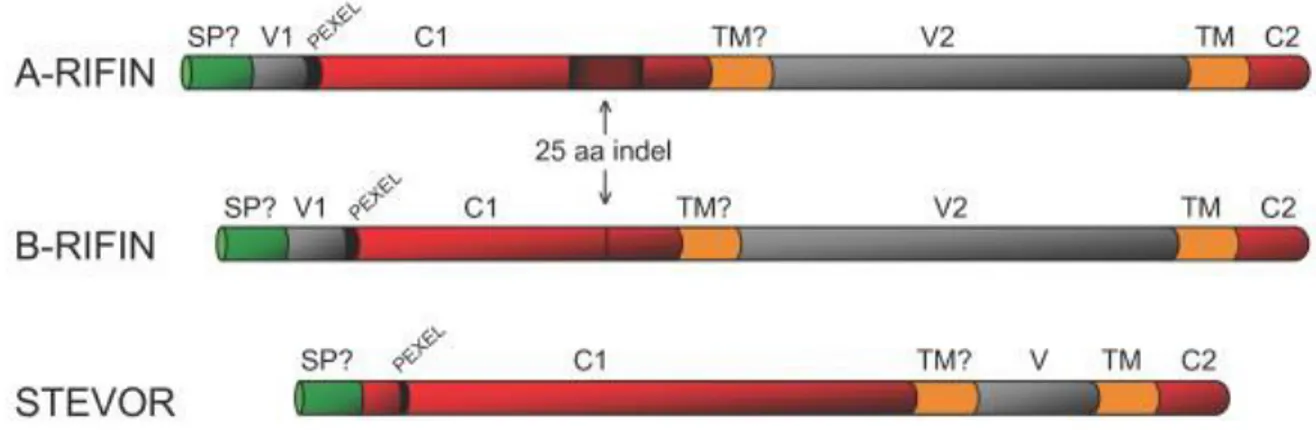
The RIFIN family members have been classified into two sub-groups, type-A and type-B which further could be divided into B1, B2, B3 clusters and can be transcribed simultaneously with type-A (Joannin et al. 2008; Petter et al. 2007). Type-A RIFINs are larger than those of type-B due to the presence of 25 amino acids motif within the semi-conserved region (C1) of type-A. Indeed, this motif is situated at approximately 66-amino acids downstream of Plasmodium export element motif (PEXEL), but it is not found in B-type RIFINs (Fig. 7). Moreover, while type-A group contains 10 conserved cysteine residues, only 6 of them are located in type-B RIFINs (Joannin et al. 2008). Furthermore, solely type-A RIFINs are trafficked to the surface of parasitized erythrocytes via Maurer’s clefts whereas type-B RIFINs are retained within the parasites (Petter et al. 2007).
A comparison analysis of transcript abundance of rif and var using P. falciparum 3D7 and NF54 clones showed a distinct rif transcript pattern during gametocytes and sporozoites stages comparing to rif and var expression profiles during the asexual stages. In addition, a single variant of B1-type RIFIN dominated the sexual stages of P. falciparum 3D7 and NF54 suggesting an important role of this gene in the adherence during this stage and shedding the light on this protein as a potential transmission blocking vaccine candidate (Wang et al. 2010).
As RIFIN proteins, STEVOR antigens display two transmembrane domains; one of which is hypothetical (Fig. 7). Antibodies directed against STEVORs found that these proteins are expressed at the surface of the IE at schizogony, resulting in antigenic variability during this stage (Niang et al. 2009). These antigens are found in the rhoptries and at the surface of merozoites as well as among released component in the invading merozoites, suggesting a potential role during invasion and probably in immune evasion in the erythrocyte invasion process (Khattab et al. 2008).
Transcription profiles of rif-A, rif-B and stevor at different stages of parasite life cycle in the clinical isolates cultivated in vitro compared to 3D7 clone had been investigated by real time PCR (Bachmann et al. 2012. The results revealed two peaks of transcription of rif-A and
stevor during the ring and trophozoite stages. In addition, clinical isolates showed
tremendous transcript abundance of these variant surface antigens comparing with 3D7. Immunoflouresence analysis of these isolates detected the localization of RIFIN and STEVOR at young trophozoites, shiozonts and merozoites. The findings led to propose a complex model of the expression of these polymorphic families throughout the asexual life cycle of
29 parasite which might be in relation with clinical outcomes of infection (Bachmann et al. 2012).
Fig. 7: Schematic organization of STEVOR, type-A and type-B RIFIN proteins. (Indel): insertion/deletion of 25 aminoacid conecnsus located in typ-A RIFIN, but not in type-B; (SP?): Potential signal; (TM): transmembrane domain; (V, V1, V2) are variable regions and (C1 and C2) are conserved regions. Source: (Joannin et al. 2011).
1.7.2. Var genes and PfEMP-1
The proteins encoded by the hyper variable var genes family are Plasmodium falciparum erythrocyte membrane proteins 1 (PfEMP-1) that range in size from 200-400 kDa (Baruch et al. 1995; Smith et al. 1995; Su et al. 1995). These antigens are trafficked to the surface of infected erythrocyte where they are anchored into electron dense protrusions, namely knobs (Igarashi et al. 1987) with the assistance of other proteins, mainly Knob Associated Histidine Rich Proteins (KAHRP) (Pologe et al. 1987). These antigens play a role of mediator between IEs and endothelial receptors, leading to sequestration of IEs throughout the body organs and bringing about severe complications. PfEMP-1 proteins are major targets of host defense. Pre-exposure to anti PfEMP-1 antibodies promotes selection and expression of specific antigens during a novel infection (Bull et al. 1998). Each individual parasite harbors ~ 60 copies of var genes dispersed throughout 14 chromosomes with a majority localized in the subtelomeric regions and the rest in the center of chromosomes. All these genes consist of two exons separated by a conserved intron. The larger one, exon I (3.5-9 kb) encodes the polymorphic extracellular region implicated in cytoadherence phenomenon which includes N-terminal segment (NTS); multiple domains of duffy binding like domain (DBL), one or two cysteine rich inter domain regions (CIDR) and a transmembrane domain (TM), while exon II (1-1.3 kb) codes for a semi-conserved cytoplasmic tail with an acidic terminal segment (ATS) (Fig. 8) (Gardner et al. 2002; Su et al. 1995).
30
Fig. 8: General characteristics of var genes. All members of var gene family consist of two exons separated by conserved intron. The variable extra cellular domain encoded by exon 1 and involves: N-terminal segment (NTS); Duffy binding like domains (DBL); cysteine rich inter domain regions (CIDR) and transmembrane domain (TM). The intracellular domain (exon 2) encodes a semi conserved amino acid terminal segment (ATS) Source: Modified from (Sherf, 2008).
Based on sequence homology of coding and non coding regions, chromosomal location, direction of transcription and domain structure of encoded proteins, var genes have been categorized into three major groups A, B and C, three conserved groups var1, var2 and var3, and intermediate groups B/A & B/C (Kraemer and Smith 2003; Lavstsen et al. 2003). All group A var genes (10 genes) possess ups A flanking sequence, situated in the sub telomeric sites and transcribed toward the telomere in the opposite direction of group B var genes (22 genes) which have a ups B sequence and are found near the telomere and their transcription is headed to the centromere. Group C var genes (13 genes) are flanked by ups C, located near the centromere and oriented toward the telomere. The intermediate groups B/A (4 genes) and B/C (10 genes) have upstream B sequence followed by the coding regions of group A or C respectively. Var1 ups D and var2csa ups E are highly conserved groups each with a unique sub telomeric gene; var3 is a conserved subfamily of group A with three small genes (Fig. 9) (Lavstsen et al. 2003).
31
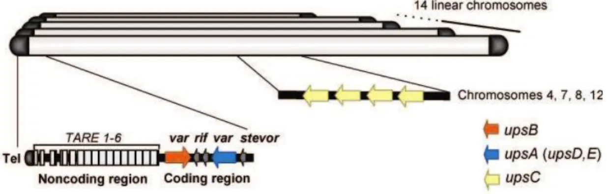
Fig. 9: Genomic organization and nuclear position of P. falciparumvar genes.
Global organization of subtelomeric regions of the 14 linear chromosomes shows that they all share non coding repeats of various sizes called TARE 1–6 (telomere associated repeat elements) situated beside telomere repeats. Subtelomeric site is followed by a member of the group B var genes, usually accompanied by another group A var genes transcribed in the opposite direction. Group C var genes are clustering in the chromosome center and rif genes are often interspersed with var genes.
Source: Modified from (Scherf et al. 2008).
The extra cellular parts of PfEMP-1 proteins are composed of multiple Duffy binding-like domains (DBL) and one or two cysteine rich inter domain regions (CIDR), each of 300-650 amino acids. Each copy of var gene has its own length according to the number and types of domains including in its sequence (3.9-13 kb). However, four proteins, VAR2CSA and VAR3 PfEMP-1 members, do not belong CIDR domains (Rask et al. 2010; Smith et al. 2000) (Fig. 10).
Based on sequence similarity, DBL domains are categorized into DBL α, β, ϒ, ϵ, δ ,ζ and five smaller distinct classes (four DBL domains of VAR2CSA and a DBL α/ ζ of VAR3 ) (Rask et al. 2010; Smith et al. 2000). CIDR domains are classified into five distinct domains α, β, ϒ, δ, and pam (Rask et al. 2010; Smith et al. 2000). These subclasses can be further sub categorized into 147 subtypes (e.g. DBLα 1.1). In general, the subtypes of these domains are located in the N-terminal part of PfEMP-1 (e.g. DBLαCIDRαDBLβ/DBLϒ) and they are associated with ups of var groups, which is not the case of domains associated with C-terminal part (e.g. DBLδCIDRβ/ϒDBLϵDBLζ) (Rask et al. 2010).