COMPUTED TOMOGRAPHIC FINDINGS IN
35
DOGS WITH
NASAL ASPERGILLOSIS
JIMMY
H. SAUNDERS,
DVM, JEAN-LUC
ZONDERLAND,
DVM, C ~ C I L E
CLERCX,
DVM, PHD,
INGRID
GIELEN,
DVM, MS, F R ~ D ~ R I C
R. SNAPS,
DVM, PHD,
MARTIN
SULLIVAN,
DVM, PHD,
HENRI
VANBREE,
DVM, PHD, ROBERT
F. DONDELINGER,
DVM,
PHD
The purpose of this study was to describe the computed tomographic (CT) features of nasal aspergillosis in dogs. Initial (n = 35) and follow-up (n = 12) CT images were available from 35 dogs. The most commonly encountered CT findings were (1) moderate to severe cavitary destruction of the turbinates with presence of a variable amount of abnormal soft tissue in the nasal passages, (2) non-specific thickening of the mucosa adjacent to the inner surface of bones of the frontal sinus, maxillary recess and nasal cavity and, (3) thickened reactive bone. The findings were consistent with a disease initially affecting one nasal cavity then progressing into the ipsilateral frontal sinus, the contralateral nasal cavity and the contralateral frontal sinus. Two dogs with associated nasal foreign body had a more localized invasion of the nasal cavity. Attenuation values and contrast enhancement were not specific. With follow-up examinations, a reduction in the amount of abnormal soft tissue was observed in all dogs except one, but this reduction could not be quantified. Veterinuy Radiology & Ultrasound, Vol. 43, No.
I , 2002 pp 5-9.
Key words: aspergillosis, canine, computed tomography, nasal cavity.
Introduction
UNGAL RHINITIS is a common disease in dogs and ac-
F
counts for 7 to 34% of dogs with chronic nasal dis- ease. Aspergillus fumigatus is the most frequently iso-lated organism in canine nasal and paranasal sinus infec- t i o n ~ . ~ In most dogs, the disease tends to be invasive and slowly destroys the turbinates.'
Diagnosis is established with help of clinical history, physical examination, radiography, computed tomography (CT), rhinoscopy, serology, cytology, histology, and myco- logic c u l t ~ r e . ~ ' ~ Conventional radiography permits differen- tiation between nasal neoplasia and nasal aspergillosis in the majority of dogs.' The main radiologic features of nasal aspergillosis are turbinate loss with punctate lucencies of the supporting bones, increased radiolucency rostrally and increased opacity caudally in the nasal cavity, and increased ~.
From the Department of Medical Imaging, Faculty of Veterinary Medi- cine, Ghent University, Salisburylaan 133, 9820 Merelbeke, Belgium (Saunders, Gielen, vanBree); Departments of Small Animals (Zonderland, Clercx) and Medical Imaging (Snaps), Faculty of Veterinary Medicine, University of Likge, Belgium; Department of Veterinary Clinical Studies, University of Glasgow, Scotland (Sullivan); Department of Medical Im-
aging, University Hospital Center, University o f Likge, Belgium (Don-
delinger).
Address correspondence and reprint requests to Jimmy H. Saunders, Department of Medical Imaging, Faculty of Veterinary Medicine, Ghent University, Salisburylaan 133, 9820 Merelbeke, Belgium. Email address: Jimmy.Saunders~ru_9.ac.be
Received September 20, 2000; accepted for publication July 28, 2001.
opacity in the frontal sinus with thickened and mottled fron- tal bone.8
CT is a reliable, non-invasive technique for the evalua- tion of chronic nasal disease in dogs and is also useful to define the extension of lesions for radiation therapy plan- ning.'-'
'
CT is superior to radiography for identification of unilateral vs. bilateral nasal cavity disease and to define the extension of lesions into adjacent structures.' I , ' * However, to date there is no detailed study about the specific CT features of canine nasal aspergillosis. The aim of our study is to describe the pre- and post-treatment CT features of nasal aspergillosis in dogs and to relate these to the spread of the disease.Materials and Methods
The C T images of 35 dogs with nasal aspergillosis seen at the Department of Medical Imaging, Ghent University (n = 11) and the Small Animals Clinic, University of Liege (n = 24 including all follow-up scans) between August 1997 and January 2000 were used for this study. Age ranged from 9 months to 10 years (mean age = 4.3 years). Fifteen different breeds were represented (8 Rottweiler, 5 Labrador Retriever, 5 Golden Retriever, 3 Belgian Shepherd dogs, 2
Doberman, 2 Afghan Wolfhound, 2 mixed breed dogs, 1
Newfoundland, 1 Alaskan Malamute, 1 Basset Hound, 1 Dachshund, 1 Bull Terrier, 1 German Shepherd dog, 1 Pyre- nean Shepherd, 1 Bobtail). There were 20 males and 15
females.
6 SAUNDERS ET AL. 2002
A physical examination was performed on each dog. Then. anesthesia was induced with droperidol
+
fentanyl* ata dose of 0.08 mglkg body weight IV, penthotal? at a dose
of 5-15 mg/kg body weight 1V and maintained with halo-
thane$ ( 1 5 2 % ) . Radiographic, CT and rhinoscopic exami- nations were performed during this anesthetic procedure. Swabs, cytobrush, and biopsies were taken during rhinos- copy for mycologic culture, cytology, and histology. Blood samples were also taken for serology. Previously estab- lished guidelines were used to consider a definite diagno- sis."
CT examinations were performed with either a 3'd gen- eration conventional CT scanners at the Ghent University
( 1 20 kV, 100 mA, slice thickness 2 mm) or a 4'h generation helical CT scanner" at the University of Libge (1 10 kV, 125 mA, pitch
1.5,
index 3, slice thickness 3 mm) with the dogs in ventral recumbency. Transverse slices were acquired from the caudal level of the frontal sinus to the nostrils. Pre- and post-contrast examinations using a bolus IV injection of700 mglkg of body weight of non-ionic iodine contrast me- dium# were performed. Reformatted dorsal plane images were also obtained. Regions of interest (ROI) were drawn on mucosal thickening and on abnormal soft-tissues in the nasal cavity andlor frontal sinus and, time-density attenua- tion data were measured before and after contrast medium administration.
The CT criteria that were evaluated were based on a previous study,14 complemented with our experience. Major CT criteria were: extension of the lesions (rostral, middle, caudal nasal cavity, frontal sinus, uni- or bilateral); presence or absence of abnormal soft-tissues andlor mucosal thick- ening (nasal passages classified as absent = no abnormal soft tissue-mild = abnormal soft tissue in 113 of the nasal cavity-moderate = 2/3 or severe = all, maxillary re- cesses, caudal recesses, retrobulbar space, pharynx, frontal sinuses, presence of a focal rounded soft tissue accumula- tion); presence of lysis (cribriform plate, turbinates classi- fied as absent = no turbinate lysis-mild = turbinate lysis in 113 of the nasal cavity-moderate = 213 or severe = all, maxillary recesses, bones associated with the caudal re- cesses, nasal, frontal, palatine, ethmoid, maxillary and vomer bones) and presence of hyperostosis or reactive new bone. A pattern was defined as generalized when the entire nasal cavity and frontal sinus were affected on at least one side and as localized when restricted to the nasal passage.
Hard copies were printed with a bone window (window width (WW) 3500-window level (WL) 500) and a soft- tissue window (WW 340-WL 25) for each dog. These
*Thalamonala, Janssen-Cilag, Beerse. Belgium. tPhenobarbitaP, Ahbott, Illinois.
$Fluothane@, Zeneca, Delaware.
$Pace CT, GE Medical Systems, Milwaukee. "Picker 6000, Picker International Inc., Ohio. #Omnipaque 300, Nycomed, Brussels, Belgium
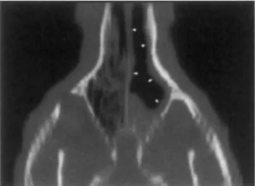
FIG. 1. Transverse image (window width = 3500, window level = 500) of the frontal sinus and nasal cavity at the level of the second maxillary premolar of a 12-year-old rottweiler. There is hyperostosis of the lateral wall of the left frontal sinus (large arrowheads) with mucosal thickening along this inner surface of the bone (small arrowheads). Lysis of the frontal crest (large arrow) is also present. Presence of abnormal mft-tissue is noted
in the left the nasal cavity (small arrows).
copies were reviewed jointly by two of the authors (JHS, FRS). When necessary, the WW and WL were adjusted on the computer monitor for visualization of other structures.
All dogs were treated with a noninvasive intranasal infu- sion of enilconazole 1%.** The treatment was repeated each
6 weeks until there was no clinical or rhinoscopic evidence
of infection. The follow-up images were acquired one month after clinical resolution with the same scanning pro- tocol and were reviewed using the same methodology as used previously. Special attention was given to potential regrowth of the conchae, bone healing and effect of sam- pling on turbinate appearance.
Results
The lesions were generalized in 26 dogs (26135 = 74%) with the contralateral side affected in 19 dogs (19126 =
73%). The lesions were localized in 9 dogs (9135 = 25%) with the contralateral side affected in 4 dogs (419 = 44%). Lesions were bilateral in 23 of the 35 dogs (23/35 = 65%). Frontal sinus abnormalities were seen in 26 dogs (26135 = 74%) and included mucosal thickening (Fig. I ) in 19 dogs (19/26 = 73%), complete obliteration of the sinus by soft tissue in 4 dogs (4126 = 15%) and presence of abnor- mal soft tissue without mucosal thickening in 3 dogs (3126
= 1 I %). In addition, the presence of a large amount of abnormal soft tissue associated with the sino-nasal ostium was noted in 24 dogs (24126 = 92%). The frontal bone was affected in 24 dogs (24/26 = 92%), unilaterally in 13 dogs
( I 3/24 = 54%) and bilaterally in 11 dogs (1 1/24 = 46%).
In 10 of these 11 dogs, there was frontal crest lysis and extension to the contralateral side (Fig. 1). The frontal bone
VOL. 43, NO. 1 CT FEATURES OF NASAL ASPERGILLOSIS 7
pattern was predominantly hyperostotic in 10 dogs (10/24
= 42%), predominantly lytic in 2 dogs (2/24 = 8%) and mixed in 12 dogs (12/24 = 50%).
Nasal passage abnormalities (Fig. 2) were present in all 35 dogs (35’35 = 100%). On the most affected side, the
turbinate destruction was severe in 26 dogs (26/35 = 74%), moderate in 8 dogs (8/35 = 23%) and mild in 1 dog (1/35 = 3%). On the less affected side, the turbinate destruction was severe in 2 dogs (2/35 = 5%), moderate in 7 dogs (7/35
= 20%), mild in 10 dogs (10/35 = 29%) and absent in 16 dogs (16135 = 46%). On the most affected side, the amount of abnormal soft tissue was severe in 15 dogs (15/35 =
43%), moderate in 9 dogs (9135 = 26%) and mild in 1 1
dogs ( 1 1/35 = 31%). On the less affected side, it was severe in 2 dogs (2/35 = 5 % ) , moderate in 5 dogs (5/35 = 15%), mild in 10 dogs (10/35 = 29%), and absent in 18 dogs (1 8/35 = 5 I %). Cribriform plate destruction and re- placement of fatty retrobulbar tissue with soft tissue were present in 2 dogs (2/35 = 5%). Abnormal soft tissue was present in the caudal recesses in 15 dogs (15/35 = 42%)
and bony lesions in 5 dogs ( Y 3 5 = 14%). For the maxillary recesses, mucosal thickening and abnormal soft tissue were
observed in 14 dogs (14/35 = 40%) and a mucosal thick-
ening alone in 10 dogs (10135 = 28%). Bone lysis of the ethmoid bone (orbital lamina) was visible in 17 dogs (1 7/35 = 48%). The maxillary bone was affected in 16 dogs (16/35 = 45%). The nasal septum was deviated in 7 dogs (7/35 =
20%), destroyed in 8 dogs (8/35 = 23%) or both in 11 dogs
(1 1/35 = 31%). Lysis of the vomer bone was present in 11
dogs ( I 1/35 = 3 1 %) and of the nasal bone in 2 dogs (2/35
= 5%). On dorsal and/or sagittal reconstructions a cavitat- ing process was present in all dogs, even apparent in dogs with a severe accumulation of soft tissue (35/35 = 100%) (Fig. 3). The term cavitating process describes an aggressive lytic pattern where the affected turbinates are completely destroyed leaving a homogeneously black empty air filled space. An extramucosal mass-like soft tissue accumulation could be distinguished in 2 dogs (2135 = 5%).
Follow-up examinations in dogs having clinical resolu- tion were characterized by a decrease in soft-tissue in 1 1 of the 12 follow-up examinations (1 1/12 = 91%) including a reduction of the mucosal thickening in the nasal cavity in 6 dogs (6/12 = 50%). Bony changes, conchae regrowth or changes related to sampling were not observed.
FIG. 2. Transverse images (window width = 3500, window level = 500) of the nasal cavity illustrating the different patterns that may be encountered in dogs with nasal aspergillosis. ( A ) Complete turbinate lysis, mucosal thickening (arrowheads) and, maxillar bone hyperostosis (arrow) and lysis (open arrow) in the right nasal cavity. (B) Localized turbinate lysis, thickening and distortion of the turbinates in the right nasal cavity. (C) Turbinate lysis dorsally, mucosal thickening, abnormal soft tissue and complete filling of the maxillary recess (arrow) in the right nasal cavity. Abnormal accumulation of Soft tissue densities are also visible in the left nasal cavity. (D) Localized turbinate l y s k (bilaterally), severe amount of abnormal soft-tissue and mucosal thickening in the left maxillary recess (arrows) in the left nasal cavity.
8 SAUNDERS ET AL. 2002
FIG. 3. Dorsal image, reformatted, of a 5-year-old rottweiler. There is destruction of the turbinates and mucosal thickening (arrowheads) in the left nasal cavity.
Reliable attenuation values could not be measured in the nasal cavity, except when the ventral nasal conchae were present (n = 12). These conchae had pre- (mean 74.1 HU
2 21.5 HU) and post contrast (mean 230.4 HU
-+
87.5 HU)values comparable to normal and other pathologic nasal tissue.
The abnormal mucosal thickening in the frontal sinus (n
= 19) had pre-contrast attenuation values between 10 and 70 HU (mean 37.1 HU
+-
20.9 HU) and was not enhanced.Discussion
There is no specific study of the CT features of canine nasal aspergillosis. However, a few studies about chronic
canine nasal disease included dogs with a s p e r g i l l ~ s i s . ~ ~ ' ~ ~ ~ ' Unilateral loss of turbinates, local nasal mucus accumula-
tion and regional mucosal swelling were d e s ~ r i b e d . ~ , ' ~ ~ ~ ' In another study on 12 dogs with nasal aspergillosis, turbinate destruction, and paranasal fluid accumulations were evi- dent.22 A cavitating lesion was again a significant feature.22 In our study, the most common CT findings were: (1) mod- erate to severe destruction of the turbinates with a variable amount of abnormal soft tissue in the nasal passages; (2) non-specific thickening of the mucosa along the bones of the frontal sinus, maxillary recess and nasal cavity; and (3) thickened reactive bone (reactive bone dorsolaterally and localized frontal crest lysis). It should be noted that the cavitated appearance, due to turbinate lysis, was still detect- able in dogs with a large amount of abnormal soft tissue in the nasal passages. When the frontal sinus was involved, there was mostly a severe amount of abnormal soft tissue around the sino-nasal ostium.
N o dog had a frontal sinus infection without a concurrent abnormality in the nasal cavity. This finding suggests that aspergillosis in these dogs presented as an aggressive (de-
structive), slowly progressive process, affecting the nasal turbinates of one nasal cavity, then extending into the con- tralateral nasal cavity and/or ipsilateral frontal sinus and finally via frontal crest lysis into the contralateral frontal sinus.
Because of its cross-sectional images, CT has been proven to be superior to conventional radiography in dem- onstrating the extent of lesions and in differentiating infec- tious rhinitis and nasal neoplasia in both dogs, cats and humans.] ',15,lfJ Dorsal reconstructions permit accurate as-
sessment of the integrity of the cribriform plate.17 Dorsal reconstructions were also the most appropriate to assess the full extension of the cavitating process in this study.
Eleven examinations were obtained with a conventional CT and 24 with a helical CT. The advantages of helical vs conventional CT are a significantly reduced acquisition time with consequent optimization of dynamic contrast studies, improved detection of solitary lesions and the quality of post-processing 2- and 3-D reconstructions.'8-20 In this study, the authors found no appreciable difference in image quality or reconstructions.
In man, CT permits differentiation between the aerogenic and dental forms of sinonasal aspergillosis." Imaging char- acteristics suggestive of fungal sinusitis are non-specific soft tissue changes (rim of soft-tissue attenuation of varying thickness along the bone) in the sinus with reactive bone and localized areas of osteomyelitis, and the association of inflammatory sinus disease with involvement of the adja- cent nasal In CT densitometric examinations, organic masses containing calcium phosphate or carbonate can be seen. These represent endogenous fungal products, which indicate an aerogenic pathogenesis of sinusal as- pergillosis.Is Such calcified masses were not observed in our dogs.
Attenuation measurements are susceptible to a variety of errors, particularly in a pathologic nasal cavity that com- prises many complex structures of different physical densi- ties (bone, cartilage, mucosa, air).25 In this study, measure- ments of attenuation values in the nasal cavity were not reliable, with the exception of the ventral nasal conchae where pre- and post-contrast attenuation values comparable to that measured in normal nasal cavity or in nasal tumors were found." This is in agreement with previous findings, and may be explained by the high vascularity even in nor- mal nasal tissue.
'
'
Attenuation values in the frontal sinuses were between 10 and 70 HU, corresponding to the measurements in humans with sinusal a s p e r g i l l o s i ~ . ~ ~ . ~ ~ A differentiation between mucoid secretions and mycetoma could not be made on the basis of the attenuation values. In man, the attenuation val- ues of mucoid secretions vary with time from 10-25 HU for acute secretions to 30-60 HU for chronic secretions and the HU of a mycetoma falls within the same range mak- ing definite distinction i m p o s ~ i b l e . ~ ~ Two extramucosal
VOL. 43, No. 1 CT FEATURES OF NASAL ASPERGILLOSIS 9
masslike soft tissue accumulations observed on rhinos- copy could not be found on subsequent C T examina- tion. Contrary to observations made in the nasal cavity, no degree of enhancement of the mucosal thickening was ob- served in the frontal sinus. The authors suggest that the absence of enhancement may be caused by hypertrophic and/or hyperplastic, and fibrotic changes of the sinusal mucosa.
A possible etiopathogenesis of aspergillosis was not es- tablished but it was associated with foreign bodies in two dogs. An impacted canine tooth in a 10-month-old Belgian Shepherd dog and a bullet in a 9-month-old Labrador Re- triever. Foreign bodies have been described as potential predisposing factors to nasal aspergillosis, but without sup- portive evidences2'
The characteristic CT features of canine nasal aspergillo- sis noted in this study were not typical of neoplasia. The CT findings with nasal neoplasia are patchy areas of increased soft tissue opacity, destruction of part of all of the ethmoid bone, abnormal soft tissue in the retrobulbar spaces, de- struction of one or both of the lateral maxillae, destruction
of the nasal bone or the rostra1 dorsal maxillae, or hyper- ostosis of the lateral maxilla.'
With follow-up examinations, a reduction in the amount of abnormal soft tissue was observed in all dogs except one, but this reduction could not be quantified. Reduction of mucosal thickening was present in half of the dogs in the nasal cavity but not in the frontal sinus. Regrowth of the conchae, bone healing or resolution of bone lysis were not observed. However, these changes require a long time to occur while our follow-up examinations were performed one month after clinical resolution.
To conclude, the CT findings most commonly encoun- tered in nasal aspergillosis in dogs include cavitated-like turbinate lysis, a rim of soft tissue of variable thickness along bones in the frontal sinus, maxillary recess and nasal passages, the abnormal presence of soft tissue accumulation and thickened, reactive bony changes. Nasal aspergillosis should be considered a primary differential diagnosis when cavitated-like lysis and diffuse thickening of the nasal inu- cosa along the bones of the nasal cavity and frontal sinus are present.
REFERENCES I . Lane JG, Clayton-Jones DG, Thoday KL, Thomsett LR. The diag- nosis and successful treatment of Aspergillus Fumigatus infection of the frontal sinuses and nasal chambers of the dog. J Small Anim Pract 1974: 15:79-83.
2. Harvey CE, O'Brien JA. Nasal aspergillosis-penicilliosis. In Kirk RW (ed): Current Veterinary Therapy. WIII. Philadelphia, WB Saunders, 1983, pp 236-240.
3. Tasker S, Knottenbelt CM, Muiiro EAC, Stonehewer J , Simpson JW, Mackin AJ. Aetiology and diagnosis of persistent nasal disease in the dog: a retrospective study of 42 cases. J Small Anim &act 1999;40:473478.
4. Sharp NJH, Harvey CE, Sullivan M. Canine nasal aspergillosis and penicilliosis. Comp Cont Educ 1991;13:41-48.
5. Sharp NJH. Aspergillosis and Penicilliosis. In Greene G E (ed): In- fectious diseases of the dog and cat. 2"" ed. Philadelphia. WB Saunders, 1998, pp 404409.
6. Colas G. Pathologie des cavitis nasales et sinusales chez le chien et le chat. Prat Med Chir Anim Comp 1996;31:479499.
7. Sullivan M, Lee R, Skae CA. The radiological features of sixty cases of intra-nasal neoplasia in the dog. J Small Anim Pract 1987;28:575-586.
8. Sullivan M, Lee R, Jakovljevic S, Sharp NJH. The radiological features of aspergillohis of the nasal cavity and frontal sinuses in the dog.
J Small Anim Pract 1986;27:167-180.
9. Thrall DE, McLeod DA, Bentel GC, Dewhirst MK. A review of treatment planning and dose calculation in veterinary radiation oncology. Vet Radiology I 989;30: 194-22 I .
10. McEntee MC, Page RL, Heidner GL, Cline JM, Thrall DE. A ret- rohpective study of 27 dogs with intranasal neoplasms treated with cobalt radiation. Vet Radiology 1991;32: 135-139.
I I . Codner EC, Lurus AG, Miller JB, Gavin PR, Gallina A, Barbee DD. Comparison of computed tomography with radiography as a noninvasive diagnostic technique for chronic nasal disease in dogs. J Am Vet Med Assoc l993;202:1 106-1 110.
12. Thrall DE, Robertson ID, McLeod D, Heidner GL, Hoopes PJ, Page LR. A comparison of radiographic and computed tomographic findings ill
3 I dogs with malignant nasal cavity tumors. Vet Radiology 1989;30:59-66. 13. Sharp N, Sullivan M. Harvey CE. Treatment of canine nasal as- pergilloai\. I n Practice 1992;14:27-31.
14. Burk RL. Computed tomographic imaging of nasal disease in 100
dogs. Vet Radiology & Ultrasound 1992;33: 177-180.
IS. Krenrnair G, Lenglinger F. Maxillary sinus aspergillosis: Diagnosis
and differentiation of the pathogenesis based on computed tomography densitometry of sinus concretions. Journal of Oral Maxillo-Facial Surgery 1995;53:657-663.
16. Forrest LJ. The head: excluding the brain and orbit. Clin Techn Small Anim Pract 1999;14:170-176.
17. Koblik PD, Berry CR. Dorsal plane computed tomographic imaging of the ethinoid region to evaluate chronic nasal disease in the dog. Vet Radiology 1990;3 I :92-97.
18. Bonaldi VM, Garcia P, Coche EE, Sarazin L, Bret PM. Spiral com- puted tomography: a new CT imaging modality. La Presse MCdicale 1996;
25:1109-1114.
19. Ney DR, Fishman EK, Kawashima A, Robertson DD, Scott WW. Comparison of helical and serial C T with regard to three-dimensional imaging of musculoskeletal anatomy. Radiology 1992; l85:865-869.
20. Remy-Jardin M, Rimy J, Giraud F, Marquette CH. Pulmonary nod- ules: detection with thick-section spiral CT versus conventional. Radiology 1993; 187:s 13-520.
21. Schwarz T. Comparison of sensitivity and specificity of conven- tional X-ray and computed tomography (CT) in nasal tumors and mycoaes in dogs. Vet RadIology 1995;36:428.
22. Mathews KG, Koblik PD, Richardson EF, Davidson AP, Pappagi- anis D. Computed tomographic assessment of noninvasive intranasal infu- sions in dogs with fungal rhinitis. Vet Surg 1996;25:309-3 19.
23. Centeno RS, Bentson JR, Mancuso AA. CT. scanning in rhinocer- ehral mucormycoais and aspergillosis. Radiology 198 I ; 140:383-389.
24. Zinreich SJ, Kennedy DW, Malat J, Curtin HD, Epstein JI, Huff LC. Kumar AJ, Johns ME, Rosenbaum AE. Fungal sinusitis: diagnosis with CT and MR imaging. Radiology 1988; 169:439444.
25. Williams G. Bydder GM, Kreel L. The validity and use of computed tomography attenuation values. Britisch Med Bull 1980;36:279-287.
26. Fellows DW, Zinreich SJ. The paranasal sinuses and nasal cavity. In: Lee SH, Rao KCVG, Zimmerman R A ed. Cranial MRI and CT. 4"' ed. McGraw-Hill, New York. 1999; pp. 831-836.
27. Som PM, Curtin HD. Chronic inflammatory sinonasal diseases in-
cluding fungal infections: the role of imaging. Radio1 Clinics North America 1983;31:3344.
28. Suter PF. Diseases of the nasal cavity, larynx and trachea. In: Suter PF ed. Thoracic radiography. A text atlas of thoracic diseases of the dog and cat. Wettswil, Switi.erland. 1985; pp 206-223.