pour obtenir le grade de
DOCTEUR DE L’UNIVERSITÉ DE TOULOUSE
Délivré par : Université Toulouse III-Paul Sabatier
UFR Sciences de la Vie et de la Terre (SVT)
Spécialité : Biosciences Végétales
présentée et soutenue par
Muhammad IRSHAD
le 9 juillet 2008
**********************************************
DYNAMIQUE DES PROTÉINES PARIÉTALES
AU COURS DE L'ÉLONGATION CELLULAIRE
DANS DES HYPOCOTYLES ÉTIOLÉS
D'ARABIDOPSIS THALIANA :
**********************************************
E l d l S i E l i Vé é i i A i Bi i é i i
APPROCHES PROTÉOMIQUE ET TRANSCRIPTOMIQUE
Directeurs de thèse : Dr. Elisabeth JAMET et Pr. Dr. Rafael PONT-LEZICA **********************************************
**********************************************
Ecole doctorale: Sciences Ecologiques, Vétérinaires, Agronomiques et Bioingénierie Unité de recherche: Surfaces Cellulaires et Signalisation chez les Végétaux, UMR 5546,
UPS, CNRS, Pôle de Biotechnologies Végétales, 24, chemin de Borde Rouge, BP 42617 Auzeville, 31326 Castanet-Tolosan, FRANCE
JURY
**********************************************
Directeur de thèse Directeur de recherche CNRS
Rapporteur Chargée de recherche INRA, Versailles
Rapporteur Directeur de recherche INRA, Orléans
Président du jury Professeur UPS
Dr. Elisabeth JAMET
Dr. Samantha VERNHETTES Dr. Gilles PILATE
Prof. Dr. Chantal TEULIERES
MEMBRE INVITÉ
Directeur de thèse Professeur UPS
Prof. Dr. Rafael PONT-LEZICA
THESIS
THESIS
for obtaining the degree of
DOCTORATE FROM UNIVERSITÉ DE TOULOUSE
Delivered by: Université Toulouse III-Paul Sabatier
UFR Sciences de la Vie et de la Terre (SVT)
presented and defended by
Muhammad IRSHAD
UFR Sciences de la Vie et de la Terre (SVT)
Specialization: Biosciences Végétales
**********************************************
on July 9, 2008
DYNAMICS OF CELL WALL PROTEINS DURING CELL ELONGATION
IN ETIOLATED HYPOCOTYLS OF ARABIDOPSIS THALIANA
AS SHOWN BY PROTEOMIC AND TRANSCRIPTOMIC SURVEYS
**********************************************
AS SHOWN BY PROTEOMIC AND TRANSCRIPTOMIC SURVEYS
Supervisors:Dr. Elisabeth JAMET and Prof. Dr. Rafael PONT-LEZICA
**********************************************
**********************************************
Doctoral school: Sciences Ecologiques, Vétérinaires, Agronomiques et Bioingénierie Research Lab: Surfaces Cellulaires et Signalisation chez les Végétaux, UMR 5546,
UPS, CNRS, Pôle de Biotechnologies Végétales, 24 chemin de Borde Rouge, BP 42617 Auzeville, 31326 Castanet-Tolosan, FRANCE
JURY ********************************************** JURY Supervisor Directeur de recherche CNRS Dr. Elisabeth JAMET Reporter Chargée de recherche INRA, Versailles
Dr. Samantha VERNHETTES
Reporter Directeur de recherche INRA, Orléans
Dr. Gilles PILATE
President of the jury Professeur UPS
Prof. Dr. Chantal TEULIERES
INVITED MEMBER
Supervisor Professeur UPS
“If you detect any mistakes of mine, I rely on your
superior knowledge to excuse them; for who has ever avoided
errors in the wide-extended field of Nature? Who is furnished
with sufficient stock of observations? I shall be thankful for
your friendly corrections, I have done what I could myself”.
Linnaeus,
Father of modern Taxonomy
Dedication ___________________________________________________________________________
___________________________________________________________________________ II
DEDICATION
I dedicate this humble effort of mine
to my loving parents, my brother Israr and
my sweet wife Lubna, without the love, devotion,
sincerity, guidance and encouragement of whom,
I would have been lost.
ACKNOWLEDGEMENTS
I would like to thank Dr. Simon HAWKINS, Dr. Gilles PILATE, Prof. Dr. Chantal TEULIERES and Dr. Samantha VERNHETTES for evaluating this work.
Sincere thanks are due to Prof. Dr. Rafael PONT-LEZICA for accepting me in his group to accomplish this work.
I offer special thanks to my supervisors Dr. Elisabeth JAMET and Prof. Dr. Rafael PONT-LEZICA for their prudent and companionate guidance, sincere advices, constant encouragement, kind and inspiring behavior, critically reviewing the manuscript and help up to the last moment, without which it would have been impossible to accomplish this work.
I would like to reflect my gratitude to Dr. Gisèle BORDERIES, Miss. Carole PICHEREAUX and Dr. Michel ROSSIGNOL for their help and cooperation during MALDI-TOF analysis.
I am highly grateful to Dr. Hervé CANUT, Mr. David ROUJOL, Miss. Hélène SAN CLEMENTE, Mrs. Ludivine SOUBIGOU-TACONNAT and Dr.Jean-Pierre RENOU who in addition to my supervisors, contributed to the analyses of proteomic and transcriptomic data or experimental work during this study.
I would like to reflect my heartily gratitude to my colleagues Cécile, Georges, Martine, Geneviève, Tan, Philippe and the whole group of Jean-Philippe for their help, encouragement, valuable suggestions, technical help and criticisms during this study.
I must acknowledge the “services communs” of UMR 5546 for their sincere help and full cooperation during my work in the lab.
I am highly grateful to Higher Education Commission of Pakistan, Islamabad for grant of scholarship, to SFERE, France for their wholehearted and sincere cooperation and to Government of NWFP, Higher Education Department, Peshawar, for grant of study leave.
Last but not least, I extend my respect, love and thanks to my family members, especially my parents and my brother Israr without whose affection, whole hearted wishes and help in administrative works, it would have been impossible to complete this work. How can I forget the patience and sacrifice of my wife Lubna and my son Talha while I have been working
Contents ___________________________________________________________________________ ___________________________________________________________________________ IV
CONTENTS
LIST OF ABBREVIATIONS ………...……… 1 CHAPTER 1. INTRODUCTION Chapter summary (French) ...………...…………...………. 41.1. Cell wall components ...………...……...………. 7
1.1.1. Polysaccharides ………...………….. 7
1.1.1.1. Cellulose …………...………...………….. 7
1.1.1.2. Cross-linking glycans ……… 8
1.1.1.3. Pectic matrix ………. 8
1.1.2. Cell wall proteins ………...………... 9
1.1.2.1. Structural proteins ……… 10
1.1.2.2. Arabinogalactan proteins (AGPs) ………...…...…….. 11
1.1.2.3. Other proteins ……….. 11
1.1.3. Other cell wall components ………...………. 12
1.1.3.1. Lignins ………..………….. 12
1.1.3.2. Lipids ………...……….…….. 13
1.1.3.3. Suberin ………...….…..…….. 14
1.1.3.4. Inorganic salts ………...…….…….. 14
1.2. Cell wall in relation to cell elongation and growth ………...….…….. 15
1.3. A. thaliana etiolated hypocotyls as a model for cell elongation ……...……….. 17
1.4. Objectives of the work ……….. 18
CHAPTER 2. MATERIALS AND METHODS 2.1. Materials ………..………….…….. 19
2.1.1. Plant material ……….. 19
2.1.2. In vitro culture for obtaining etiolated hypocotyls ……….……….… 19
2.2. Methods ………..……….….. 19
2.2.1. Isolation of cell walls from hypocotyls ………..………..….. 19
2.2.2. Sequential extraction of proteins from purified cell walls …………..……….….. 20
2.2.3. Protein fractionation by cation exchange chromatography ……...………….. 20
2.2.4. 1D-E (SDS-polyacrylamide gel electrophoresis) and staining procedures …….... 21
2.2.4.2. Staining with silver nitrate ………...………... 22
2.2.4.3. Staining with Amido Black ………..……….. 22
2.2.4.4. Staining with Gelcode® Glycoprotein staining kit ………..……... 22
2.2.5. Transfer of proteins from gel on PVDF membrane ………..……... 23
2.2.6. Protein identification by mass spectrometry ………..….... 23
2.2.7. Extraction of RNAs ………..……….. 24
2.2.8. RT-PCR and semi-quantitative PCR ………..……….... 25
2.2.9. Microarray analysis ……….……….. 26
2.2.10. Statistical analysis of microarray data ………..…………... 26
2.2.11. Data Deposition ……….…………..….. 27
2.2.12 Bioinformatic analysis ………..……...….. 27
CHAPTER 3. EVALUATION OF CELL WALL PREPARATIONS FOR PROTEOMICS: A NEW PROCEDURE FOR PURIFYING CELL WALLS FROM ARABIDOPSIS HYPOCOTYLS Chapter summary (French) ……….………..…………...….. 29
Abstract ………...………...…... 34
Background ………...………...………....…... 35
Results and discussion ………...……..………... 35
Analysis of early methods ………...…………... 35
A modified method to prepare plant cell walls ………..…... 39
Sequential salt extraction of proteins from cell walls ………..…...…... 43
Conclusion ……….…….….. 43
Methods ………...…………...………... 44
Plant material and isolation of cell walls ………...….…... 44
Sequential proteins extraction and identification ………...…….... 44
Additional material (List) ………...……... 45
References ………...………..……... 45
CHAPTER 4. A NEW PICTURE OF CELL WALL PROTEIN DYNAMICS IN ELONGATING CELLS OF ARABIDOPSIS THALIANA: KNOWN PLAYERS AND NEW COMERS Chapter summary (French) ………..………... 47
Contents ___________________________________________________________________________ ___________________________________________________________________________ VI Keywords ………...………..………..…... 55 Abbreviations ………...………...………...……...……... 55 Introduction ………...…...…………..………...…... 56
Material and Methods ………...………...………... 58
Plant material ………...……….……….…. 58
Cell wall purification and protein extraction ………...……...…….…... 58
Protein separation by cationic exchange chromatography ………...……... 58
Protein separation by mono-dimensional electrophoresis (1D-E) and identification . 59 Semi-quantification ………...………...……..………... 59
Bioinformatic analyses ………..………...………... 59
Results ……….………...………...…..….. 61
Establishment of methods for efficient proteomic analysis of hypocotyl cell walls .. 61
Proteins identified in cell wall extracts of Arabidopsis etiolated hypocotyls …... 62
Semi-quantitative comparative analysis of CWPs ………...………....……….. 63
Discussion …..………...………....…...……….. 66
Additional data files (List) ………...………...………….. 70
References …………..………...……….…….. 71
CHAPTER 5. CELL WALL BIOGENESIS OF ARABIDOPSIS ELONGATING CELLS: TRANSCRIPTOMICS COMPLEMENTS PROTEOMICS Chapter summary (French) ………...……….. 77
Abstract ………...……….…...………...……….. 82
Background ………..…...……….………..……….. 83
Results and Discussion ………...……….………..……….. 84
Level of transcripts of cell wall genes (CWGs) during hypocotyl elongation …….. 84
Genes encoding secreted proteins with high or moderate level of transcripts in etiolated hypocotyls ………...……….…………..…...…….. 86
Are there variations in the level of transcripts between half- and fully-grown hypocotyls? ………...……….………..…….. 87
Transcriptome vs. proteome ………...……….……….. 89
Conclusions ………...……….………....……….. 91
Methods ………...……….………...….……….. 92
Plant material ………...………...……….….. 92
Transcriptome studies ………...………...……….. 93
Statistical analysis of microarray data ………...……...….……….. 93
Data deposition ………...……….……….……….. 94
Semi-quantitative PCR ………...………...………...….. 94
Bioinformatic analyses ………...……… ………... 94
References ………...………..……….……….. 96
Additional data files (List) ………...………...….….. 103
CHAPTER 6. CONCLUSIONS AND PERSPECTIVES French version ………...……….…...….. 104
English version ………...………...………….…….. 108
LITERATURE CITED ………...……….…...…….……….. 111
ANNEXES AND SUPPLEMENTARY DATA Annexes chapter 2 ………...……….…..….……... 125
Supplementary data chapter 3 ………...……….…..………..…...… 128
Supplementary data chapter 4 ………...……….…..………..…...… 144
Abbreviations ___________________________________________________________________________
___________________________________________________________________________ 1
List of abbreviations
1D-E/2D-E: one or two dimensional electrophoresis 4CL: 4-coumarate CoA ligase
ACN: acetonitrile AG: arabinogalactan
AGP: arabinogalactan protein
AGP-PRP: arabinogalactan protein–proline-rich protein
AGRIKOLA: Arabidopsis genomic rnai knock-out line analysis BSA: bovine serum albumin
BY2: bright yellow (cv of Nicotiana tabacum) C3H: p-coumarate 3-hydroxylase
C4H: cinnamate 4-hydroxylase
CAD: cinnamyl alcohol dehydrogenase
CATMA: complete Arabidopsis transcriptome microarray CBB: Coomassie brilliant blue
CCoAOMT: caffeoyl-CoA O-methyltransferase CCR: hydroxycinnamoyl-CoA reductase
cDNA: complementary DNA CESA: cellulose-synthase
Col 0: ecotype Columbia 0 of A. thaliana
COMT: caffeic acid/5-hydroxyferulic acid O-methyltransferase CSC: cellulose-synthesizing complex
CSL: cellulose-synthase like CWG: cell wall gene
CWP: cell wall protein
dNTP: desoxynucleotide triphosphate (dATP, dCTP, dGTP, dTTP) DTT: dithiotheritol
EDTA: ethylenediamine tetra acetic acid EGase: endo-1,4-β-D-glucanase
Endo-PG: endopolygalacturonase ER: endoplasmic reticulum F5H: ferulate 5-hydroxylase
FPLC: fast protein liquid chromatography FWER: Family Wise Error Rate
G: guaiacyl (unit of lignin) GalA: galacturonic acid GAX: glucuronoabinoxylan GFP: green fluorescent protein GH: glycoside hydrolase
GPI: glycosylphosphatidylinositol GRP: glycine-rich protein
GST: gene-secific sequence tag GT: glycosyl transferase
H/PRP: hydroxyproline/proline-rich protein H: p-hydroxyphenyl (unit of lignin)
HCT: p-hydroxycinnamoyl-CoA:quinate shikimate p-hydroxycinnamoyl-CoA transferase HG: homogalacturonan
HGA: homogalacturonic acid HRGP: hdroxyproline-rich protein kDa: kilo Dalton
LAE: late-abundant embryogenesis protein LB medium: Luria-Bertani medium
LC: liquid chromatography
LC-MS/MS: liquid chromatography-tandem mass spectrometry LRR: leucine-rich repeat
LRX: leucine-rich repeat extensin LTP: lipid transfer protein
MALDI-TOF: matrix-assisted laser desorption ionization-time of flight MS medium: Murashige and Skoog medium
MS: mass spectrometry
nptI: neomycin phosphotransferase
PAGE: polyacrylamide gel electrophoresis PAL: phenylalanine ammonia-lyase PCR: polymerase chain reaction PG: polygalacturonase
Abbreviations ___________________________________________________________________________
___________________________________________________________________________ 3
PGIP: poly galacturonase-inhibiting protein PL: pectate lyase
PL: polysaccharide lyase PM: plasma membrane PME: pectin methylesterase PRP: proline-rich protein
PTM: post-translational modification PVDF: polyvinylidene fluoride QI: semi-quantitative index RG-I: rhamnogalacturonan-I RG-II: rhamnogalacturonan-II ROS: reactive oxygen species rpm: round per min
RT-qPCR: reverse transcription-quantitative PCR S: syringyl (unit of lignin)
SDS: sodium dodecyl sulphate SPG: secretory pathway gene TE buffer: Tris EDTA-buffer
Tris: Trishydroxymethylaminomethane, or 2-amino-2-hydroxymethyl-1,3-propanediol U: unit
UHQ water: Ultra High Quality Water XG: xyloglucan
CHAPTER 1
Chapter 1 ___________________________________________________________________________
___________________________________________________________________________ 4
Chapter summary (French)
L’existence d’une paroi autour des cellules végétales est l’un des caractères qui les différencie des cellules animales. Cette paroi joue des rôles importants au cours du développement des plantes, et au cours de leurs interactions avec l’environnement qu’il s’agisse de stress biotiques ou abiotiques. Elle est également essentielle pour le port dressé des végétaux et permet la circulation de molécules signal entre cellules distantes. La croissance des végétaux fait intervenir deux processus cellulaires, qui sont la division cellulaire et l’augmentation de volume qui peut-être être soit anisotrope (grossissement), soit directionnelle (élongation). Ce travail porte sur la recherche de protéines importantes pour l’élongation des cellules végétales, et plus particulièrement pour les modifications des parois au cours de ce processus.
La première partie de cette introduction décrit les constituants pariétaux principaux : les polysaccharides (cellulose, hémicelluloses, pectines), les protéines, les lignines, les lipides, les cires et les ions. La description des protéines est volontairement succinte puisqu’elle reprend l’état des connaissances avant le début de mes travaux de thèse, i.e. avant l’essor des approches protéomiques qui ont permis d’avoir une vue globale des protéines pariétales.
Dans les deux parties suivantes sont présentés successivement les protéines dont le rôle dans l’élongation cellulaire est connu ainsi que le mécanisme particulier de l’élongation cellulaire dans les hypocotyles d’Arabidopsis thaliana. En effet, ces hypocotyles présentent la particularité de s’allonger environ 100 fois en un temps très court par simple élongation cellulaire.
Enfin, les objectifs de ma thèse sont développés. Il s’agissait d’avoir une vue globale de la régulation des gènes impliqués dans la biogenèse des parois au cours d’un processus d’élongation cellulaire chez A. thaliana. Le modèle expérimental retenu a été celui des hypocotyles étiolés. Les questions posées étaient les suivantes :
• Quelles sont les protéines présentes dans les parois des hypocotyles étiolés à deux
stades de leur développement (croissance active vs croissance terminée) ?
• Existe-t-il des différences entre les protéomes pariétaux à ces deux stades
développement ?
Deux approches complémentaires ont été menées : une approche protéomique impliquant la mise au point de protocoles de purification de parois (chapitre 3) et de séparation des protéines pariétales (chapitre 4) ; et une approche de transcriptomique (chapitre 5). L’ensemble des résultats obtenus a été interprété grâce à la bioinformatique non seulement pour la prédiction de la localisation sub-cellulaire des protéines, mais encore pour celle de domaines fonctionnels.
Chapter 1 ___________________________________________________________________________
___________________________________________________________________________ 6
Land plants are among the largest organisms. This achievement is based largely on the growth, and mechanics of their cell wall, a structure that encases cells of plants, algae, fungi and bacteria like an armour. Plant cell wall is remarkable complex and dynamic entity and is one of the most sticking features that differentiate plants from animals and other eukaryotic cells. Cell wall of higher plants is of high economic importance to human especially as raw material of human and animal food, textiles, wood, paper, thickeners, biofuels and other products.
Cell wall was first observed by Robert Hooke in 1665 when he examined thin slices of cork under his microscope. In fact, he found small tiny boxes that he called cells and being dead material, consisted only of cell walls.
Cell wall of higher plants is a complex but organized molecular composite that may comprise many different polysaccharides, lignin, suberin, wax, proteins, aromatic substances, calcium, boron and water. Owing to the diversity of cell shapes and functions, the molecular composition and arrangement of cell wall exhibits a great diversity. Generally the cell wall consists of three parts: middle lamella, primary wall and secondary wall. Middle lamella is the first layer formed between the adjacent cells at the time of cytokinesis. Primary wall is simultaneously laid inner to the middle lamella, and is present in all type of cells and allows changes in cell size and shape. Secondary wall is impregnated on the inner surface of the primary cell wall in some type of cells (e.g. conducting cells) after achieving their final shape and size.
The cell wall of higher plants performs a variety of functions during growth and development as well as in plant defense, including maintenance of the osmotic pressure, water movement, rate and direction of cell growth, cell differentiation, intercellular communication and signalling, structural support and morphology of plant, cohesion among the cells of a tissue, storage, protection against pathogens and abiotic stresses. They also contribute to the functional specialization of cell types.
Here a detailed description of the cell wall components, their properties, structural organization and interactions among them, will be given. In addition, the involvement of different cell wall components especially the proteins in growth and development will be elaborated. Finally, we will discuss how cell wall proteins contribute to cell elongation by interacting with other cell wall components which makes the subject of this work.
Synthesis of cellulose microfibrils and callose occurs at the plasma membrane surface. Synthesis and glycosylation of cell wall proteins and wall-modifying enzymes occur at the rough ER while synthesis of non-cellulosic polysaccharides at the Golgi apparatus. From here, the materials destined for cell wall are transported to cell surface in the form of secretory vesicles formed at the trans-Golgi network.
Chapter 1 ___________________________________________________________________________
___________________________________________________________________________ 7
1.2. Cell wall components
About 10% of the plants genome is devoted to cell wall biogenesis (http://cellwall.genomics.purdue.edu/). Cell wall biogenesis has been divided into six distinct stages: substrate generation, polymer synthesis, secretion, assembly, rearrangement during development, and disassembly. These steps are taking place both inside and outside the cell.
Figure 1.1 illustrates the cell compartment where synthesis and processing or modifications of major cell wall components occur.
The molecular composition and arrangement of wall polymers differ among species, among tissues of a single species, among individual cells, and even among regions of the wall around a single cell (Buchanan et al., 2000). Following are the major cell wall components.
1.1.1. Polysaccharides 1.1.2.1. Cellulose
Cellulose is the most abundant plant polysaccharide that accounts for 15-30% of the dry mass of the primary cell wall and even more in the secondary walls. In plants, cellulose occurs in paracrystaline assemblies, i.e. microfibrils of about 36 parallel arranged
(1→4)β-D-Glc chains hydrogen-bonded to one another along their length (Figure 1.2). Many microfibrils
combine to form a cellulose fiber laid down on the cell surface in several layers distinguished by the different orientation of their fibers (Buchanan et al., 2000). Cellulose synthesis (reviewed in Reiter (Reiter, 2002); and Cosgrove (Cosgrove, 2005) occurs at rosette-like structures, the cellulose-synthesizing complex (CSC) that consists of six
hexagonally-arranged subunits (CESA) that are embedded in the plasma membrane (Figure 1.3). Different
CESA genes are assumed to be required to make a functional CSC, and different sets of genes are involved in the formation of the primary and secondary walls. CESA1, CESA3 and CESA6 or CESA6-like (CESA2, CESA9) are required for biosynthesis of the primary wall, whereas CESA4, CESA7 and CESA8 are required to form secondary walls. The most convincing proof of this comes from the study on the CESA1 and CESA3 mutants that result in gametophyte lethality and CESA6 mutant causing cellulose deficiency and growth defects (Persson et al., 2007). In other such studies, the catalytic domain of cotton CesA was immunolocalized at and near the CSCs (Kimura et al., 1999) and the mutant atcesA1 (rsw1) was shown to cause disassembly of CSC, reduced cellulose accumulation and accumulation of non-crystalline (1→4)β-D-Glc (Arioli et al., 1998). CSCs probably contain other proteins. KORRIGAN is assumed to have some role in polymerization or crystallization of microfibrils
d
e Figure 1.2: Structure of cellulose microfibrils
(after Buchanan et al., 2000)
a. Helically-winded microfibrils around an elongating cell
almost transverse to the long axis of cell; b. Freeze-fracture replica of an elongating maize root cells showing arrangement of cellulose microfibrils as impression through the plasma membrane; c. A single microfibril; d. Cross-section of microfibril showing spacial arrangement of glucan chains in microfibril showing spacial arrangement of glucan chains in the core of the microfbrill; e. X-ray diffraction showing arrangement of atoms in the unit structure of a microfibril.
Rosette subunit
(1→4)β-D-Glc chain cellulose microfibril
c
CESA Rosette subunit Rosettes
Figure 1.3: The cellulose-synthesizing machinery of the cell (after Cosgrove 2005) a Immunogold labelling of CESA localized to hexameric cellulose synthesizing complex (CSC) in a. Immunogold labelling of CESA, localized to hexameric cellulose synthesizing complex (CSC) in the plasma membrane. The black circles represent gold nanoparticles that are attached to antibody against CESA. Scale bar, 30 nm; b. A model showing how three different CESA proteins (shown in three different colours: orange, brown, green) might be organized into subunits and then into a hexameric CSC; c. A model of how CESA complexes synthesize a cellulose microfibril. Each CESA protein can synthesize a single (1→4)β-D-Glc chain. In this model, microfibril is shown to consist of
Figure 1.4: Structure of xyloglucan(after Reiter 2002) XLFG type xyloglucan of Arabidopsis where the solid arrows indicate linkages that are always present, where as dashed arrows denote substitution patterns.
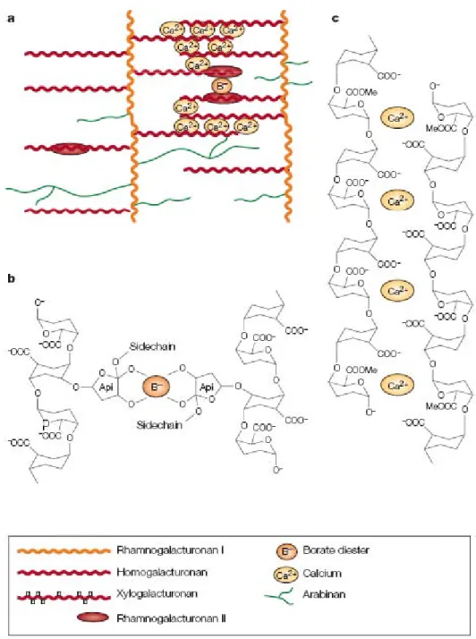
Figure 1.5: Structure of pectins(after Willats et al., 2001)
Simplified schematic diagram to indicate some of the features of the three major types of pectins: HGA, RG-I and RG-II. Oligosaccharide epitopes recognized by anti-HGA monoclonal antibody PAM1 and anti-RG-I monoclonal antibodies LM5 and LM6 are also indicated.
or in recycling of sitosterol (Nicol et al., 1998). COBRA is thought to link the complexes to nearby microtubules for guidance along the membrane (Roudier et al., 2005). KOBITO1 is assumed to take part in the cellulose synthesis machinery or to play a role in the coordination between cell elongation and cellulose synthesis (Pagant et al., 2002).
Cellulose is present in cell wall of all types, provides strength and rigidity to cell wall, and prevents the swelling of the cell wall and rupture of the plasma membrane that might occur when osmotic conditions favour water entry into the cell.
1.1.1.2. Cross-linking glycans
The cell wall polysaccharides that can hydrogen-bond to cellulose microfibrils are classified as cross-linking glycans. Most of them are also called hemicelluloses. In type I cell wall, characteristic of dicots, the major cross-linking glycans of primary cell wall are xyloglucans (XGs). XGs typically consist of a (1→4)β-D-Glc backbone carrying (1→6)α-D-Xyl moieties on three consecutive Glc residues. As shown in Figure 1.4, the (1→6)α-D-Xyl residues attached to the second and third Glc residues of the backbone can carry D-Gal in (1→2)β-linkage, and the second of these Gal residues is usually substituted by L-Fuc in (1→2)α-linkage (Reiter, 2002) to make XLFG xyloglucan according to the nomenclature of Fry et al. (Fry et al., 1993). The structure and distribution of the side branch (chains) vary in different tissues and species (forming XXFG, XXXG, XXLG, XLLG or XLXG) which seem important for bonding to cellulose. Glucuronoabinoxylans (GAXs) are the major cross-linking polymers of type II cell wall, characteristic of commelinoide monocots and some dicots. The structure of GAXs varies considerably with respect to degree of substitution and position of attachment of α-L-Ara residues (Carpita and Gibeaut, 1993). The other types of cross-linking glycans are “mixed linkage” (1→3, 1→4)β-D-glucan, glucomannans, galactoglucomannans and galactomannans (Carpita and Gibeaut, 1993).
1.1.1.3. Pectic matrix
Pectins (reviewed in Ridley et al. (Ridley et al., 2001) and Willats et al. (Willats et
al., 2001)) are a mixture of heterogenous, branched and highly hydrated polysaccharides
(Figure 1.5). They are rich in polygalacturonic acid (PGA) and account for 30% total cell wall mass (Carpita and Gibeaut, 1993). GalA occurs in two major structural features (homo and heteropolymers) that form the backbone of three polysaccharide domains found in almost all
Figure 1.6: Formation of pectin networks by covalent and ionic bonds (after Cosgrove 2005)
a. A model of how the different pectins may be covalently linked together to form a
macromolecular pectin network. In this model, RG-I serves as the backbone and the other pectin domains are attached as branches; b. RG-II chains are cross-linked to form dimers through a borate ester bond; c HGA forms stiff gels through Ca2+-mediated crosslinking of its carboxyl groups through ionic and coordinate bonds.
(RG-II). These three types of GalA covalently bond to each other to form a pectic network throughout the primary cell wall matrix and middle lamellae. HGA is a linear, unbranched (unsubstituted) homopolymer containing about 100–200 GalA residues, and is called smooth region of pectins. HGA is an abundant and widespread kind of pectin which is synthesized in the Golgi apparatus and deposited in the cell wall. It has 70–80% GalA residues methylesterified at the C-6 carboxyl. The demethylesterified HGA in the cell wall matrix can
be cross-linked by Ca2+ to form supramolecular assemblies and gels (Figure 1.6). Other
modifications and substitutions of HGA are not as widespread as methyl esterification. RG-I (known as the hairy region of pectin) is an acidic pectic domain consisting of as many as 100 repeats of the disaccharide L-Rha-D-GalA. RG-I is abundant and heterogenous and generally glycosidically-attached to HGA domains. Arabinogalactan and arabinan side chains commonly include Gal and L-Ara residues. Type I arabinogalactans are (1→4)β-linked D-Gal with non-reducing terminal-Ara (t-Ara) substituted at the O-3 of some of the D-Gal units (Carpita and Gibeaut, 1993). Arabinogalactans of type II with (1→3)β- and (1→6)β-linked-D-Gal residues also occur on pectic backbones. Arabinans can become branched by links through O-2 and O-3. Another major, widespread highly conserved pectic domain is RG-II which is similar to HGA by possessing homopolymer backbone, but is branched like RG-I. It consists of around 9 GalA residues backbone substituted by 4 heteropolymeric side chains each consisting of eleven different sugars including Api, aceric acid and 2-keto-3-deoxy-D-manno-octulosonic acid (kdo). RG-II can dimerize by means of borate ester links through Api residues.
In plants, several enzymes like pectin methylesterases (PMEs) (Micheli, 2001)
polygalacturonase (PGs) (Tanaka et al., 2002) and pectate lyases (PLs) (Marin-Rodriguez et
al., 2002) modify or degrade pectins in relation with changes in cellular adhesion and plasticity of the cell wall, a prerequisite to elongation and development.
1.1.2. Cell wall proteins
Until the last decade cell wall was considered to be made mainly of polysaccharides and structural proteins. But recent studies, especially proteomic studies have reported the presence of many other proteins including enzymes that perform a wide range of functions. The present study has further contributed to this area. Here cell wall proteins (CWPs) will be treated as have been considered before this study: structural proteins, that can form networks
a Ara Gal Ara b c d
Figure 1.7: Cell wall structural proteins, motifs and glycosylation (after Buchanan et al., 2000).
a. Tomato extensin possessing Ser (Hyp)4or related motif with high glycosylation with tetra-Ara as well as Gal at Ser. Isodityrosine linkage is also shown on the Tyr-Lys-Tyr motif, a likely position for this linkage; b. Maize extensin like Thr-rich moderately-glycosylated protein; c. A Soybean PRP that lacks contiguously hydroxylated Ser, Thr, glycosylated protein; c. A Soybean PRP that lacks contiguously hydroxylated Ser, Thr, and Hyp residues (a likely position for glycosylation with Ara) are not heavily glycosylated; d. Petunia GRP with no glycosylation site.
to response to biotic and abiotic stresses. All CWPs possess a signal peptide and are targeted to secretory pathway (Buchanan et al., 2000).
1.1.2.1. Structural proteins
Although all types of cell wall are mainly composed of polysaccharides, cellulose-crosslinking glycans network embedded in pectic matrix, another network is formed by cell wall structural proteins. Cell wall structural proteins can be grouped into three principal classes on the basis of their sequence rich in some particular type of amino acids: the extensins, hydroxyproline/proline-rich proteins (H/PRPs) and glycine rich proteins (GRPs).
Examples of such proteins are given in Figure 1.7. All these proteins are encoded by large
multigene families and are developmentally-regulated. Extensins and H/PRPs are assumed to be cross-linked to make the wall less extendable when the cells achieve their final size. Their relative amount varies among tissues and species.
Extensins are well studied-structural proteins, which are rod-shaped distinguished by their amino acid sequence rich in Pro, Tyr, Lys, Ser, His, Val where Pro may reach up to
50%. They possess the repeating motif Ser-(Pro)n (n ≥ 3) and Tyr-Lys-Tyr sequences that are
important for their secondary and tertiary structure making inter- and intra-molecular bonding. They are highly O-glycosylated and have basic pIs (Cassab, 1998; Sommer-Knudsen et al., 1998; Buchanan et al., 2000). Normally extensins are O-glycosylated with chains of Ara on contiguous Hyp residues and Gal on Ser (Kieliszewski and Lamport, 1994; Kieliszewski, 2001).
H/PRPs represent another large multigene family of structural proteins. Because of their similarity to extensins they are also thought to be rod-shaped (Buchanan et al., 2000).
H/PRPs contain the repetitive pentamere motif (Pro-Hyp-Val-Tyr-Lys)n or its variants but
lack Ser and are O-glycosylated (Cassab, 1998).
GRPs (reviewed in Ringli et al. (Ringli et al., 2001)) that belong to another major cell wall structural protein family, should be distinguished from intracellular GRPs that are assumed to bind RNAs. Cell wall GRPs have repetitive sequences which may contain more than 60% Gly arranged in short repeating units. The GRP protein sequences often follow the
Figure 1.8: Structural complexity of an arabinogalactan (Seifert and Robert 2007)
Figure 1.8: Structural complexity of an arabinogalactan (Seifert and Robert 2007)
The first complete structure of an arabinogalactan glycan, derived from synthetic green flourescent protein (GFP):(Ala-Pro)51, expressed in tobacco BY2 cells.
motif varies (Ringli et al., 2001). Beside the (Gly-X)n motif, higher-order repetitive sequences
that are rarely perfect are sometimes found and were proposed to be important for the formation of the secondary structure of the proteins. GRPs are believed to form plate-like structure rather than rod-shape conformation, on the plasma membrane-cell wall interface. They are thought to crosslink to cell wall polysaccharides are therefore difficult to extract.
1.1.2.2. Arabinogalactan proteins (AGPs)
AGPs (reviewed in Seifert and Roberts (Seifert and Roberts, 2007)) consist of a core protein of highly varying length and domain complexity, and one or more arabinogalactan
(AG) side chains (Figure 1.8). They often contain a glycosylphosphatidylinositol (GPI) lipid
anchor (Borner et al., 2005). The relative ratio of glycan to protein is sometimes higher than 9, but can vary strongly for the same AGP core protein isolated from the same tissue. AGPs are precipitated by treatment with the β-Yariv reagent. In some AGPs, the peptide backbones are 10-13 residue-long and are called AG peptides. Most AGP sequences consist of a single central domain rich in Pro, Ala, Ser, and Thr. Most AGPs are O-glycosylated at one or more Hyp residues by AG type II (found in dicots) groups. These consist of (1→3) and (1→6)β-linked Gal chains connected to each other by (1→3, 1→6)-(1→6)β-linked branch points, O-3 and O-6 positions substituted with terminal Ara residues. Type I AGs (characteristic of monocots and some dicots) are (1→4)β-linked D-Gal with non-reducing terminal-Ara (t-Ara) substituted at the O-3 of some of the Gal units (Carpita and Gibeaut, 1993). AGPs are involved in a variety of functions, e.g. embryonic and post-embryonic patterns, pollen tube guidance, growth, secondary wall deposition, abscission and interaction with growth regulators and microbes.
1.1.2.3. Other proteins
Recently it has become clear that cell wall is very dynamic and contains many different enzymes and other agents (reviewed in Cosgrove (Cosgrove, 1999) and Fry (Fry, 2004)) conferring it dynamic properties like plasticity and extensibility. Among these proteins were expansins (e.g. α- and β-expansins), hydrolases (e.g. cellulases, xylanases, PMEs, PGs), transferases (e.g. xyloglucan endotransglycosylase hydrolases (XTHs), lyases (e.g. pectate lyases) oxido-reductases (e.g. peroxidases and laccases). These non-structural proteins perform a wide range of biological roles. A few examples of these functions are cited below while others, concerning especially the modification of cell wall and regulation of cell elongation are given in section 1.2.
Figure 1.9: Lignin biosynthesis in dicots(after Boudet et al., 2003)
The figure shows a simplified view of the most favoured lignin biosynthetic pathway in angiosperms where the implication of laccases in the oxidation of monolignols is still not known.
Abbreviations: CAD, cinnamyl alcohol dehydrogenase; CCoAOMT, caffeoyl-CoA O-methyltransferase; C3H, p-coumarate 3-hydroxylase; C4H, cinnamate 4-hydroxylase; CCR, hydroxycinnamoyl-CoA reductase; 4CL 4-coumarate CoA ligase; COMT caffeic acid/5-hydroxycinnamoyl CoA reductase; 4CL, 4 coumarate CoA ligase; COMT, caffeic acid/5 hydroxyferulic acid methyltransferase also known as AldOMT, 5-hydroxyconiferaldehyde O-methyltransferase; F5H, ferulate hydroxylase also known as Cald5H, coniferaldehyde 5-hydroxylase; HCT, p-hydroxycinnamoyl-CoA:quinate shikimate p-ydroxycinnamoyl-CoA transferase; PAL, phenylalanine ammonia-lyase; SAD, sinapyl alcohol dehydrogenase.
Inside cells, proteases are involved in all aspects of the plant life cycle (Schaller, 2004), but their role in cell wall is not very clear. Outside cells, some are thought to be involved in cell-cell communication by generating local-signals (Matsubayashi and Sakagami, 2006). The subtilisin-like serine protease Stomatal Density and Distribution 1 (SDD1) is thought to control stomata distribution and density in Too Many Mouths (TMM) dependent way (Berger and Altmann, 2000; Nadeau and Sack, 2003). The cell wall HRGP,
Root-Shoot-Hypocotyl-Defective (RSH), is essential for normal embryo development in A. thaliana and
the mutant fails to develop normal embryo because mal-positioning of the cell plate at the time of division leading to the formation of abnormal embryo (Hall and Cannon, 2002).
Reviewing the role of the cell wall in embryogenesis (Malinowski and Filipecki, 2002), the
authors have highlighted that chitinases, XTHs, and peroxidases participate in embryogenesis regulation by involvement in signal transduction, and by influencing cell shape and division plane. AGPs are considered source of signals in a variety of ways (Seifert and Roberts, 2007). They may bind directly to a receptor that activate a signal transduction cascade or may release lipid signals by cleavage of GPI anchor by phospholipase or oligosaccharide as signals by endoglucanases. Inflorescence Deficient in Abscission (IDA) gene encoding an AGP playing a role in the abscission where its mutant delayed floral organ abscission and its over-expression produced opposite effect. Pectate lyases play important role in softening ripening fruits
(Marin-Rodriguez et al., 2002). Peroxidases that rigidify cell wall by cross-linking of wall
compounds are expressed to cope with biotic and abiotic stresses like wound, pathogen interaction, and climatic aggression (Passardi et al., 2004; Passardi et al., 2005). Polygalacturonase-inhibiting proteins (PGIPs) protect plant against fungal attack by inhibiting
fungal PGs (De Lorenzo et al., 2001).
1.1.3. Other cell wall components 1.1.3.1. Lignins
Lignins (reviewed in Boerjan et al. (Boerjan et al., 2003), Boudet et al. (Boudet et
al., 2003) and Davin and Lewis (Davin and Lewis, 2005)) are the 2nd most abundant plant substances in vascular plants. They are usually deposited in the secondary cell wall with few lignins exceptionally occurring in primary cell wall (Buchanan et al., 2000). Lignins are assumed to be obtained by the oxidative polymerization of monolignols in the cell wall by peroxidases and laccases. The first step of lignin biosynthesis is the deamination of phenylalanine (dicots) or tyrosine (monocots) by ammonia lyases, yielding cinnamic or
p-Figure 1.10: Lignin structure(after Evtuguin and Amado 2003, in Davin and Lewis 2005) An example of putative primary sequence structure of a syringyl lignin-derived hexamer fragment from Eucalyptus globulus. The radical-radical coupling linkages between the sinapyl alcoholyp g p g g py monomers are shown in red.
Figure 1.11: Plant cuticle (after Heredia-Guerrero et al., 2008)
Current microscopic model of plant cuticle that shows the epicuticular wax crystals deposited on an amorphous and dense matrix of cutin polymer, containing some intracuticular waxes.
Figure 1.12: Major monomers of plant cutin (after Heredia-Guerrero et al., 2008) Chemical structure of the major monomers present in plant cutin. These monomers are derived from C16saturated fatty acid and from C18unsaturated fatty acids. Some plants have mainly C16 family of monomers, whereas others have a mixture of both C16and C18families of monomers.
formation of hydroxycinnamoyl-CoA thioesters, reduction of hydroxycinnamoyl-CoA thioesters to hydroxycinnamaldehydes, and reduction of hydroxycinnamaldehydes lead to three p-hydroxycinnamyl alcohols (monolignols), p-coumaryl, coniferyl, and sinapyl alcohols which are transported from the cytosol to the apoplast. When reaching the apoplast, monolignols undergo dehydrogenative polymerization via oxidases, forming lignins. They comprise two major components namely guaiacyl (G), derived from coniferyl alcohol, and
syringyl (S), derived from sinapyl alcohol (Figure 1.10) and one minor component
p-hydroxyphenyl units (H), derived from p-coumaryl alcohol. Although polymers of only three types of monomers, the composition and structure of lignins vary significantly among different plants or within the same plant (Billa et al., 1998).
1.1.3.2. Lipids
Lipids constitute the impermeable hydrophobic outer portion of the cell wall called cuticle, a continuous layer that covers aerial parts of leaves, fruits and young non-woody
stems. Cuticle weight ranges from 2000 µg/cm2 (in fruits) to 450-800 µg/cm2 (in leaves).
Forty to 80% of this is constituted by cutin, the extracellular lipids that are interconnected by ester bonds and can be studied after hydrolysing the polyesters (Heredia, 2003). Waxes are soluble extracellular lipids and can be extracted with organic solvents. The structure and
composition of the cuticle vary in different plants, plant organs and growth stages but
basically comprise a cutin matrix developed on the epidermal plant cell wall with waxes. Waxes are solid, partially crystalline aggregates at room temperature (Schreiber, 2005). They appear either embedded in the matrix (intracuticular) or deposited on its surface (epicuticular)
as shown in Figure 1.11 (Riederer and Müller, 2005). Epicuticular wax may exist as a smooth
film in some species or as wax crystals in other species (Buschhaus et al., 2007). Chemically,
cutin is a polymeric networkof polyhydroxylated C16 and C18 fatty acids cross-linked by ester
bonds (Figure 1.12). Waxes are generally described as mixtures of homologous long-chain
aliphatic compounds, like alkanes, alcohols, aldehydes, fatty acids and esters with the addition of varying proportions of cyclic compounds like triterpenoids and hydroxycinnamic acid derivatives (Heredia, 2003).
Cuticle controls non-stomatal water loss, protects plants against ultraviolet radiation. It does not allow the water to stand easily on surface of the plant thus minimizing accumulation of dust, pollen and air pollutants. In addition, surface wax is thought to play
Chapter 1 ___________________________________________________________________________
___________________________________________________________________________ 14
2003). Surface wax has also been shown to participate in a variety of plant-insect interactions (Eigenbrode and Espelie, 1995). For example epicuticular lipid extracts and individual lipid component enhance or deter oviposition, movement, and feeding.
1.1.3.3. Suberin
Suberin is a biopolymer making a barrier between plants and the environment in specialized plant tissues (e.g. periderm, bark and tuber skin) that protects the internal living tissues from dehydration, injuries, and pathogens (Soler et al., 2007). Suberized cells are also found in the epidermis and hypodermis of roots, the endodermis and the bundle sheath of grasses (Graca and Santos, 2007). Suberin is a complex polyester made of glycerol and long-chain diacids and hydroxyacids (Schreiber et al., 1999; Groh et al., 2002). In suberized cells, suberin represents up to 50% of the mass of the cell wall (Pereira, 1988).
In the outermost tissues of plants, suberized cells play a vital role affording protection against environmental aggressions and pathogens, and controlling temperature and water loss (Schreiber et al., 1999; Groh et al., 2002).
1.1.3.4. Inorganic salts
Calcium and boron are the two major minerals localized in the cell wall. Calcium is also an essential plant nutrient and is required for various structural roles in the cell wall and membranes. Plants take up calcium from soil through roots and calcium is delivered to the shoots via xylem. Calcium enters the plant cells through specific ion channels in their plasma
membranes (White and Broadley, 2003). In cell wall, Ca2+ participates in cross-linking the
demethylesterified HGA to form supramolecular assemblies and gels (see Figure 1.6) (Willats
et al., 2001) that modify cell wall physical and chemical properties.
Boron is an essential nutrient for vascular plants. In cell wall, it cross-links RG-II to
form dimers by borate ester bond between two apioses (see Figure 1.6) (Ridley et al., 2001).
Boron deficiency makes the tissues brittle or fragile, while plants grown on high boron levels may have unusually flexible or resilient tissues (Loomis and Durst, 1992). Boron helps establishing an effective legume–Rhizobium symbiosis (Bolanos et al., 1994) and is required for the maintenance of nodule cell wall structure (Bonilla et al., 1997).
membrane, whereas hemicelluloses and pectins, which compose the matrix polysaccharides, are synthesized in the Golgi apparatus and are deposited to the wall surface by vesicles. Xyloglucans are tightly bound to microfibrils by hydrogen bonds and form cross-links between them, thus constituting a load-bearing network. For clarity, the hemicellulose–cellulose network is shown on the left part of the cell wall without pectins, which are emphasized on the right part of the figure. In most plant species the main hemicellulose is xyloglucan (blue), while hemicelluloses such as arabinoxylans (grey) and mannans (not shown) are found in lesser amounts. The main pectin polysaccharides include
h l t I d h l t ith ll t f l l t bi
rhamnogalacturonan I and homogalacturonan, with smaller amounts of xylogalacturonan, arabinan, arabinogalactan I (not shown) and rhamnogalacturonan II. Pectin domains are believed to be covalently linked together and to bind to xyloglucan by covalent and non-covalent bonds. Neutral pectin polysaccharides (green) are also able to bind to cellulose surfaces. pH of the cell wall and the action of different enzymes (not shown) like expansins, endo-1,4-β-D-glucanases, XTHs, PMEs and peroxidases on the cell wall components modify the properties of cell wall resulting in wall loosening or rigidification.
Chapter 1 ___________________________________________________________________________
___________________________________________________________________________ 15
1.2. Cell wall proteins in relation to cell elongation and growth
Growth can be described as an irreversible increase of volume. In plants, growth is the outcome of cell division, enlargement of the new cells and their differentiation into different types of tissues. These processes of growth are accompanied by permanent change in size (usually an increase in length or volume) and an increase in dry mass of the growing part.
Enlargement of cells may be polarized, expanding more in one axis than the other (elongation) or it may be uniform in all directions as in isodiametric cells. Furthermore polarized growth takes place in two ways. In the first case, called tip growth, in which the growth is focused on a single specialized region, the apex of the tip growing cell. This type of growth is found in pollen tube, root hair and fungal hyphae. The other type is called diffuse growth and takes place at any point of a meristematic cell mostly in the cell wall parallel to the axis of elongation. This latter type of elongation generally occurs in the apical meristem of root and shoot or the intercalary meristem. This study is focused exclusively on diffuse polarized cell growth.
Before maturity, plant cells usually enlarge 10- to 1000-fold in volume by the process of vacuolation and irreversible cell wall expansion (Cosgrove, 1997). According to this review, there are two major concepts that account for the molecular basis of expansion. The first concept couples wall expansion to biosynthesis and secretion of wall polymers but did not explain the wall stress relaxation which is essential expansion of the cell via water uptake. The second concept considers wall expansion as result of biochemical loosening of the wall which permits turgor-driven extension of the wall polymer network. Though convincing, this latter neglects the need for integration of new polymers into the expanding wall.
To explain cell wall expansion based on rheological properties of the growing wall several potential mechanisms of stress relaxation and expansion can be imagined. Figure 1.13 represents the organization of various cell wall components and the various types of coordinated interactions between them that allow the cell to elongate but maintain cell wall integrity as intact structure. These interactions are described below in slight detail.
The acid-growth theory of cell wall expansion (Hager, 2003), which states that
auxin, in an activated form (~A) activates a H+-pumping ATPase at the plasma membrane (PM) which utilizes respiratory energy (ATP) to raise the proton concentration in the
apoplast. This triggers cell elongation by activating cell wall enzymes. The transport of protons into the cell wall is compensated by a flow of cations into the cytoplasm, which maintains turgor pressure during cell elongation.
In elongating cells, the cellulose microfibrils are deposited perpendicular to the axis of elongation, forming a spring-like structure (Green, 1962). Such an arrangement reinforces lateral walls on one side and allows directional expansion (elongation) of the cell on the other hand through loosening of cellulose-hemicellulose networks. Expansins that can restore acid-induced creep of denatured walls, also participate this process. In fact, expansins are thought to transiently displace short stretches of hemicelluloses that are bonded to the surface of cellulose microfibril. It makes polymers to creep dragging along other structural components
if the wall is in tension (Cosgrove, 1998).
Endo-1,4-β-D-glucanases (EGases) are assumed to cleave the β-1,4-glycosidic bonds between glucose of xyloglucans relaxing wall polysaccharides to move apart during cell elongation.
XTHs also perform cell wall modifications required for the process of wall assembly and cell expansion, by either wall loosening or incorporating new xyloglucan chains into extending walls (Chanliaud et al., 2004). They cleave xyloglucans and rejoin the newly generated reducing ends to others by acting on the xyloglucans attached to cellulose microfibrils (Vissenberg et al., 2005).
PMEs can cause wall loosening or wall stiffening according to their mode of action
on HGs (Micheli, 2001). Once integrated into the cell wall, they may act randomly (as in
fungi) or linearly (as in plants) on HGs. When PMEs act randomly on HGs (at acidic pH), the demethylesterification releases protons that promote the action of endopolygalacturonases (Endo-PGs) and contribute to cell wall loosening. When PMEs act linearly on HGs (at basic
pH), PMEs give rise to blocks of free carboxyl groups that could interact with Ca2+, so
creating a pectate gel. Because the action of Endo-PGs in such a gel is limited, this action pattern of PMEs contributes to cell wall stiffening.
Figure 1.14. Changes in thickness of cell walls of dark-grown hypocotyls (after Derbyshire et al., 2007)
Cell wall thicknesses of different tissues [outer epidermis (OE), inner epidermis (IE), outer cortex (OC), inner cortex (IC), and endodermis (EN)] of hypocotyls freeze-fractured at their mid-points at different developmental stages of dark-grown hypocotyls:
different developmental stages of dark grown hypocotyls:
stage I: at the embryo stage prior to germination stage II: at the onset of germination
stage III: at 50% of their final length
residues, monolignols, suberin units and ferulic acids. The radicals produced by the peroxidative cycle, if linked to other polymers can cross-link cell wall polymers like extensins or lignins that block any further wall loosening and hence cell expansion. Inversely, by hydroxylic cycle they produce reactive oxygen species (ROS) like ·OH that can cleave various polysaccharides non-enzymatically and contribute to wall loosening. Wall loosening
is also caused by regulating H2O2 concentration by both peroxidative and hydroxylic cycles.
Listed above are the proteins potentially involved directly or indirectly in cell wall elongation or its arrest, but still a huge investment is needed in this field to completely understand the process.
1.3. A. thaliana etiolated hypocotyls as a model for cell elongation
To understand the mechanism of cell elongation, hypocotyls of A. thaliana are widely
used as model (Gendreau et al., 1997). During hypocotyl elongation, almost the entire cell machinery is devoted to synthesize, export and reorganize cell walls. The cell wall remains dynamic in real sense provided with the required plasticity and elasticity. Etiolation of hypocotyls makes them to elongate more than hypocotyls of light-grown seedlings. In dark-grown seedlings, hypocotyls elongate along a spatially and temporally steep acropetal gradient. This growth takes place mainly by cell elongation (100-fold elongation as compared to embryo cells) and does not involve significant cell divisions. Elongation follows an acropetal gradient and takes place in two distinct, time-separated phases: synthesis and deposition of new cell wall polymers and addition and re-organization of the existing ones (Gendreau et al., 1997; Derbyshire et al., 2007). During the first 3 days after germination, synthesis and deposition of cell wall material are the main processes, producing cells with thick walls. In the following days (until 7 days), hypocotyls grow mainly by extensive polymer disassembly and rearrangement resulting in thiner cell walls (Figure 1.14). Furthermore they respond to growth hormones as normal plant parts do. The model plant A.
thaliana have the advantage of the existence of mutants with altered hypocotyl growth
facilitating gene functional study (Mouille et al., 2003). Owing to these characteristics, etiolated hypocotyls of A. thaliana could be considered an ideal model for the study of cell elongation.
Chapter 1 ___________________________________________________________________________
___________________________________________________________________________ 18
1.4. Objectives of the project
As discussed above, CWPs contribute to several physiological functions. But these correspond to only a small part of the cell wall genes and role of most of the CWPs is still hidden. This project is aimed at identifying cell wall genes involved in cell elongation or its arrest. Here we have tried to identify the members of the multigene families known to be involved in elongation or elongation arrest in etiolated hypocotyls of A. thaliana as well as other proteins that could contribute to these processes.
The questions raised during this work are:
i) Which are the proteins present in cell wall of elongating hypocotyls and
fully-grown hypocotyls that have stopped elongation?
ii) Do there exist differences between the cell wall proteomes of elongating
organs and those that have stopped elongation? What are these differences?
iii) What are the functions of these proteins?
To achieve these goals, comparative cell wall proteomics was used as primary approach in etiolated hypocotyls of A. thaliana: a fast growing stage (5-day-old) and a stage when there is no more elongation (11-day-old) will be compared. Since previous studies showed no defined correlation between the mRNAs and protein concentrations (Moritz and Meyer, 2003), working directly with proteins was preferred here. Proteomics has been defined as a global qualitative analysis of complex protein mixtures, including the post-translationally modified proteins as well as those encoded by alternatively spliced transcripts, and appears as a complementary approach to genomic and transcriptomic studies (Hunter et al., 2002). Comparative proteomics provides an overview of the proteome and detects proteins which are altered between different stages (Moritz and Meyer, 2003) like the two physiological stages in this study. In addition, a comparative transcriptomic study was performed on the same material. Microarrays are a powerful, sensitive, versatile, and easy-to-use genomic tool that can simultaneously determine expression levels for thousands of genes at reasonable cost (Meyers et al., 2004; Trevino et al., 2007). Furthermore, combining the two types of data will give information about the post-transcriptional regulations of genes encoding CWPs. Here a global view of the functions of different genes representing these two physiological stages will be given.
CHAPTER 2
Chapter 2 ___________________________________________________________________________ ___________________________________________________________________________ 19 2.1. Materials 2.1.1. Plant material
Arabidopsis thaliana ecotype Columbia 0 (Col 0) was used for in vitro and in vivo
cultures for obtaining the hypocotyls, other plant material, and for transformation with the desirable constructs.
2.1.2. In vitro culture for obtaining etiolated hypocotyls
One hundred and thirty mg seeds of A. thaliana were weighed in an Eppendorf tube, soaked for 2-3 h in 1 mL tape water, sterilized by treating with 4 times diluted Javel (sodium hypochlorite) for 45 min. To remove the sterilizing solution, the seeds were washed 6 times with 1 mL of sterilized ultra high quality (UHQ) water each time. Finally, the seeds were sowed in Magenta boxes containing 50 mL of Murashige and Skoog (MS) medium (Murashige and Skoog, 1962) pH 5.8, supplemented with 2% (w/v) sucrose and 0.8-1.2% (w/v) agar. Synchronization of germination was obtained by 2-4 days chill treatment and 4 h light treatment of the seeds. Seedlings were grown at 23°C in the dark for 5 or 11 days.
2.2. Methods
2.2.1. Isolation of cell walls from hypocotyls (after Feiz et al. (Feiz et al., 2006))
For one experiment, hypocotyls were collected from 18 and 36 Magenta boxes for the 11 day-old and 5 day-old samples respectively. Hypocotyls of A. thaliana were collected by cutting the seedlings with scissors below the cotyledons and above the crown measuring ~0.7 cm for 5 day-old and ~1.2 for 11 day-old seedlings. The hypocotyls thus obtained were transferred to 5 mM sodium acetate buffer (pH 4.6), 0.4 M sucrose in a large Petri dish kept on ice. To remove the cut cotyledons and testa, hypocotyls were extensively washed on a 2 mm mesh, with 0.8-3.0 L of 5 mM sodium acetate buffer (pH 4.6), 0.4 M sucrose depending
on the weight of the hypocotyls. Afterwards, the hypocotyls were transferred to a Moulinex®
-type grinder containing 600 mL of the same buffer supplemented with 1 mL of protease
inhibitor cocktail (Sigma Aldrich, Saint Louis, USA) and Polyclar® at the rate of 1 g/10 g
fresh weight of hypocotyls in order to complex the phenolic compounds (Charmont et al.,
2005). The mixture was ground in cold room at full speed for 5 min. Cell walls were
separated from soluble cytoplasmic materials by centrifugation for 15 min at 1000 × g at 4°C using the Beckman J2-HC centrifuge and a JA 14 rotor. They were further purified by two additional centrifugations in 150 mL per tube of 5 mM sodium acetate buffer (pH 4.6) containing 0.6 M and 1 M sucrose respectively. The cell wall containing pellet was hold at
each step. To ensure the removal of cytosolic proteins and other soluble material, the pellet was extensively washed on nylon net (25 μm pore size) with 3-5 L of 5 mM sodium acetate buffer (pH 4.6). Finally it was ground in liquid nitrogen with mortar and pestle in order to obtain very fine powder and to have intimate contact with the protein extraction buffer. The cell wall fraction was lyophilized and stored at -20°C.
2.2.2. Sequential extraction of proteins from purified cell walls
Generally, 0.6-0.9 g of lyophilized cell walls were taken per tube for protein extraction. Proteins were extracted in 4 successive steps by using 10 mL/g of salt solutions in
this order: 2 extractions with 6-9 mL 5 mM sodium acetate buffer (pH 4.6), 0.2 M CaCl2,
followed by 2 extractions with 6-9 mL 5 mM sodium acetate buffer (pH 4.6), 2 M LiCl. Protease inhibitor cocktail (Sigma Aldrich, Saint Louis, USA), was added at a concentration of 15 μL/g cell walls, during the first extraction with each salt. Cell walls were resuspended by vortexing during 5-10 min at room temperature and then centrifuged for 15 min, at 40000 × g and 4°C. The protein content of each extract was measured using the Bradford method
(Bradford, 1976) with the Coomassie® protein assay reagent kit (Pierce, Perbio Science,
Rockford, USA) taking bovine serum albumine (BSA) as a standard. Proteins were desalted
using Econo-Pac® 10DG columns (Bio-Rad Laboratories, Inc. Hercules, CA, USA)
equilibrated with 0.2 M ammonium formate. Depending on the volume of each extract and of protein concentration, protein solutions could be concentrated by successive centrifugations
using the Centriprep® centrifugal filter device (YM-10 kDa membrane for volumes greater
than 6 mL or 5 kDa for smaller volumes) (Millipore, Billerica, MA, USA) at 4000 × g. Finally, proteins were lyophilized.
2.2.3. Protein fractionation by cation exchange chromatography
All the lyophilized extracts were combined and redissolved in a total volume of 2 or 3 mL of water. They were again quantified as described above. One mg of proteins was used
for chromatographic fractionation on a 1 mL HiTrapTM SP FF column (Amersham
Biosciences, Uppsala, Sweden) equilibrated with 50 mM MES (pH 5.6m adjusted with
NaOH) operated with an FPLCTM System (Amersham Biosciences, Uppsala, Sweden)
controlled by FPLCdirectorTM version 1.0 (Amersham Biosciences, Uppsala, Sweden). The
Chapter 2 ___________________________________________________________________________
___________________________________________________________________________ 21
the column was accomplished at a flow rate of 0.5 mL/min. A 10 mL unfixed fraction was collected at the same rate. Three mL of first wash with 50 mM MES (pH 5.6) were collected at a flow rate of 1 mL/min. Fixed proteins were eluted by a gradient from 0 to 0.8 M NaCl in 50 mM MES (pH 5.6) and 24 fractions (1 mL each) were collected at a flow rate of 1 mL/min. A modified gradient was applied when enrichment of certain proteins was required. Finally the column was successively washed with 3 mL of 1.2 M and 3 mL of 1.5 M NaCl in 50 mM MES (pH 5.6) at the same flow rate. These washes were also collected as 6 fractions (1 mL/tube). Two µL/mL Protease inhibitor cocktail (Sigma Aldrich, Saint Louis, USA) was added to all the 1 mL collecting tubes. Quantity of proteins in each fraction was measured by the Bradford method (Bradford, 1976). The fractions were combined in groups of 2 or 3 depending on their protein concentration and were desalted as previously described prior to lyophilization.
2.2.4. 1D-E (SDS-polyacrylamide gel electrophoresis) and staining procedures
Each lyophilized chromatography fraction (group of 2 or 3 fractions) was redissolved in 200 µL water and electrophoresis of proteins was performed according to Laemmli (Laemmli, 1970). Samples were loaded on 12 × 15 cm polyacrylamide gel with a concentration of 12.5% and a thickness of 1.5 mm (Annex I). For transfer, 8% polyacrylamide gels of 8 × 6 cm and 1.5 mm thickness were used (Annex II).
Gels were stained with any one of the following staining procedures:
2.2.4.1. Staining with Coomassie Brilliant Blue (CBB)
CBB staining was performed according to Scheler et al. (Scheler et al., 1998). After electrophoresis, the gel was fixed by overnight dipping and gentle shaking in 50% methanol. It was then rinsed 3 times with UHQ water by changing the water after each 30 min. Then, the
gel was sensitized by dipping it in sensitising solution (34% methanol, 2% H3PO4 17%
ammonium sulfate) for 1 h. Finally, the gel was put in the staining solution (34% methanol,
2% H3PO4 17% ammonium sulfate, CBB G (Sigma Aldrich, Saint Louis, USA) 0.66 g/L for
2-3 days. For clearing the background, the gel was rinsed 1-2 times in 2% H3PO4 before