HAL Id: tel-01844247
https://tel.archives-ouvertes.fr/tel-01844247
Submitted on 19 Jul 2018HAL is a multi-disciplinary open access archive for the deposit and dissemination of sci-entific research documents, whether they are pub-lished or not. The documents may come from teaching and research institutions in France or abroad, or from public or private research centers.
L’archive ouverte pluridisciplinaire HAL, est destinée au dépôt et à la diffusion de documents scientifiques de niveau recherche, publiés ou non, émanant des établissements d’enseignement et de recherche français ou étrangers, des laboratoires publics ou privés.
biomedical applications
Mohammed Elmahmoudy
To cite this version:
Mohammed Elmahmoudy. Micro- and nano-patterning of conducting polymers for biomedical appli-cations. Other. Université de Lyon, 2017. English. �NNT : 2017LYSEM027�. �tel-01844247�
1 N°d’ordre NNT: 2017LYSEM027
THESE de DOCTORAT DE L’UNIVERSITE DE LYON
opérée au sein del’Ecole des Mines de Saint-Etienne
Ecole Doctorale
N° 488
Sciences, Ingénierie, Santé
Spécialité de doctorat : Microélectronique
Discipline
: Bioélectronique OrganiqueSoutenue publiquement le 16/10/2017, par:
Mohammed ELMAHMOUDY
Micro- et Nano-patterning de Polymères Conducteurs pour des
Applications Biomédicales
Devant le jury composé de :
De Mello, John Professeur, Imperial College London Président et rapporteur Offenhäusser, Andreas Professeur, Forschungszentrum Jülich Rapporteur
Dufour, Isabelle Professeur, Université de bordeaux Examinatrice Inal, Sahika Professeur assistante, King Abdullah
University of Science and Technology
Examinatrice
Gkoupidenis, Paschalis Group leader, Max Plank Institute for polymer research
Examinateur
Malliaras, George Professeur, Ecole des Mines de Saint-Etienne
Directeur de thèse
Sanaur, Sébastien Professeur assistant, Ecole des Mines de Saint-Etienne
2
3
École Nationale Supérieure des Mines de Saint-Étienne NNT:
Mohammed ELMAHMOUDY
Micro- and Nano-Patterning of Conducting Polymers for Biomedical Applications Speciality: Organic Bioelectronics
Keywords: Bioelectronics, PEDOT:PSS, Nano-patterning, Microelectronics Abstract:
Bioelectronics uses electrical signals to interact with biological systems. Sensors that allow for electrical read-out of important disease markers, and implants/stimulators used for the detection and treatment of pathological cellular activity are only a few examples of what this technology can offer. Due to their intriguing electroactive and mechanical properties, organic electronics or π-conjugated materials have been extensively explored regarding their use in bioelectronics applications. The interest in organic electronic materials stemmed from their soft and flexible nature which dampens the mechanical properties mismatch with tissue. This less “foreign” surface enhances the signal transfer to/from cells in vitro. The attractive mixed electronic/ionic conductivity feature of conducting polymers enables coupling between the electronic charges in the bulk of the organic films with ion fluxes in biological medium. The prototypical material of organic bioelectronics is the conducting polymer poly(3,4-ethylenedioxythiophene) (PEDOT) doped with polystyrene sulfonate (PSS). PEDOT:PSS is commercially available, water-dispersible conjugated polymer complex that can be cast into films of high hole and cation conductivity, good charge storage capacity, biocompatibility, and chemical stability. In the present work we investigate an approach to tailor the mechanical, electrical, and electrochemical properties of PEDOT:PSS and study their impact on the performance of organic electrochemical transistors. In addition, we study the effect of micro-structuring and nano-patterning on the electrochemical impedance of PEDOT:PSS- coated gold electrodes for future neural recordings and stimulation. Moreover we demonstrate the use of micro-patterned PEDOT:PSS in cell adhesion and migration.
4
École Nationale Supérieure des Mines de Saint-Étienne NNT:
Mohammed ELMAHMOUDY
Micro et Nano-patterning de Polymères Conducteurs pour des Applications Biomédicales
Spécialité: Bioélectronique Organique
Mots clés: Bioélectronique, PEDOT:PSS, Nano-patterning, Microélectronique Résumé:
La bioélectronique utilise des signaux électriques pour interagir avec des systèmes biologiques. Les capteurs qui permettent la lecture électrique de marqueurs de maladies importantes et les implants/stimulateurs utilisés pour la détection et le traitement d'activité cellulaire pathologique ne sont que quelques exemples de ce que cette technologie peut offrir. Du fait de leurs propriétés électro-actives et mécaniques fascinantes, l'électronique organique ou les matériaux conjugués π ont été largement exploités dans le domaine de la bioélectronique. L'intérêt pour les matériaux électroniques organiques provient de leur nature douce et flexible qui amortit les désagrégations mécaniques à l’interface avec les tissus. Cette surface moins "étrangère" améliore le transfert de signal vers / en provenance des cellules in
vitro. Le mélange intéressant entre conductivité électronique et ionique de ces polymères
conducteurs permet le couplage entre les charges électroniques présentent dans le volume des films organiques avec les flux ioniques du milieu biologique. Le matériau prototypique de la bioélectronique organique est le polymère conducteur poly(3,4-éthylènedioxythiophène) (PEDOT) dopé avec du polystyrène sulfonate (PSS). Le PEDOT: PSS, disponible dans le commerce, est un complexe de polymères conjugués qui peut se disperser dans l'eau, être coulé sous forme de films biocompatibles très conducteurs comprenant des trous et des cations, doté d’une bonne capacité de stockage de charge et d’une bonne stabilité chimique. Dans ce rapport, nous étudierons une approche pour moduler les propriétés mécaniques, électriques et électrochimiques du PEDOT: PSS et étudier leur impact sur la performance des transistors électrochimiques organiques. Par ailleurs, nous évaluerons l'effet de la micro-structuration et du nano-patterning sur l'impédance électrochimique des électrodes en or recouvertes de PEDOT: PSS utiles pour de futurs enregistrements et stimulations neurales. Enfin, nous démontrerons l'utilisation du PEDOT:PSS à micro-motifs pour l'adhésion et la migration de cellules.
5
Acknowledgment
I would like to start off by thanking George Malliaras, the director of my thesis, for his outstanding and wise supervision of my Phd and his continuous professional and personal support. You gave us, PhD candidates and post-docs, the opportunity to learn and develop without pressuring anyone in anyway. You have created a work environment where everyone felt at home and gave us the necessary motivation to keep on the good work with the freedom to explore and satisfy our curiosity.
Thanks to Sébastien Sanaur for establishing the project and bringing the fund and doing all the necessary work to make it happen. And mostly for giving me the great opportunity to join the amazing Department of Bioelectronics (BEL).
A special thanks to Roisin Owens for her personal and professional support and for leading the department for the last period of my PhD.
I would like to thank all my colleagues from the BEL for being cooperative, and for making the department a very rich place for scientific discussions and exchange. You have definitely made the group a better place, and gave it all the fun and positive energy needed to keep it going and producing excellent work that is recognized worldwide. Names including the visiting professors and students; Xenofon Strakosas, Thomas Lonjaret, Marc Ramuz, Mar FERRO, Rod O’Connor, Alexandra Rutz, Ilke Uguz, Mary Donahue, Paschalis Gkoupidenis, Vincenzo F Curto, Christopher Proctor, Sahika INAL, Usein Ismailov, Donata Iandolo, Yi zhang, Charalampos Pitsalidis, Ana Sanchez, Esma Ismailova, Dimitrios A Koutsouras, Eloïse Bihar, Marcel Brändlein, Anna-Maria Pappa, Shahab Rezaei Mazinani, Jolien Pas, Magali Ferro, Aimie Pavia, Isabel del Agua, Gerwin Dijk, Viviana Fascianella, Loig Kergoat, Carol Chan, Helena Gleskova, David Martin, Jon de Mello, Alberto Salleo, Miriam Huerta, Clemens STOLZ, Jacob Friedlein, david ohayon, Michel Fiocchi, Adam WILLIAMSON, Susan Daniel, Nathan Schaefer, Takamatsu SEIICHI, Gaëtan Scheiblin, Sofia Drakopoulou,
6
Amale Ankhili, Federica MARIANI, Timothée Roberts, Silvia Battistoni, Julie, Patrick FOURNET, Margaret BRENNAN, Yingxin, Manu, Maria, Simon Régal, liza klots, Jonathan Rivnay, and Daniel Corzo. As well as Pierre Leleux from Panaxium startup for the active support in the clean room and fruitful discussions.
I specially thank people who I had the pleasure to work with on common projects; Sahika INAL, Alexandra RUTZ, Ilke UKUZ, Mary DONAHUE, Vincenzo CURTO, Magali FERRO, Rodney O’CONNOR, Adel HAMA, and Isabel DEL-AGUA-LOPEZ. In addition to my collaborators; Martina SCHMIDT from University of Bayreuth, Germany, and Anne CHARRIER from CINAM, CNRS, Aix-Marseille University, France.
I would like to also thank Bastien MARCCHIORI, Mohamed SAADAOUI, and Cyril CALMES from Flexible Electronics Department (FEL) for their help and support. As well as the clean room staff; Gaëlle RONDEAU, Sylvain NOLOT, Jessica MAZUIR, Thierry CAMILLONI, and Jean-Philippe for taking care of the safety and security in the clean room area and their efforts in keeping the machines up and running.
Special thanks to Philippe Lalevée, the director of the Center of Microelectronics Georges Charpak of the Provence, and the platform Micro-Packs, for his great support during my PhD and for his efforts in managing our école and providing a productive environment for work. Not to forget the great people behind the scene, the administration stuff; Véronique VILLAREAL for her great help though out my PhD, as well as Michelle Gillet, and Anaïs BALAGUER. Aurélie and Johanne at the front desk. And Gracien COUNOT and Christophe LECOINTE from the IT office, as well as melodie CAYOL and LAGAIZE Richard from the logistics office.
I would like to also thank the Agence Nationale de la Recherche for supporting my PhD project financially (grant number: ANR-14-CE08-0006).
To my mother, Mervat SALAH, you have been my strength and inner sound of wisdom. You have given me and my siblings, the example of protecting the pure kind human nature along
7
the years and through any conditions. You gave us the example of the strength, the patience, and the strive to success. Being a single mother of four for 26 years, you have seen a lot of challenges, and lot of distressing moments and situations, you have been the super mama who made it through all. You have made everyone impressed by how far you could take us. You have been self-ignoring to provide for us and make us as happy as possible. We love you and appreciate who you are. All what I have acquired from you through the years gave me strength and persistence to be the man I am today, and this has been always a support for me during my PhD work.
To my siblings, Ahmed, Amr, and Rana, you are the rock stars. You have gone so far in your lives, and made me look up to you. You have been always the support system that I could rely on. You gave me and the family all the needed support while I was busy working on my Phd and being so far away. You have created sustained the ecosystem for the family, thank you really and truly. You have been always there for anything I needed. Your presence has made a huge difference, and I wish I can pay you back a little of what you have given me.
Last, but not least, I would like to give very special thanks to my partner Douaa Mckadm. You give me all the emotional support, encouragement, and love to stay focused on my PhD work. At my many very low points, you saw the bright side of things, and you gave me the right analysis, and told me the exact words that kept me moving. You are such a positive adorable cheerful smart person. Even though my PhD drained me out of energy for extended periods of time, and stressed me and changed my moods, you have been always understanding and absorbing. And most of all, you have been always my mirror; you made me learn more things about myself, and understand more who I am and what I really need and want in life. Intelligent strong loving women like you can build nations and civilizations. I love you, and I wish I can make you always happy.
8
Table of Content
Chapter 1 ... 10
PEDOT:PSS for Biomedical Applications ... 10
1.1. Introduction ... 10
1.2. Synthesis and Deposition ... 12
1.2.1 Electrochemical Approach ... 12
1.2.2 Chemical Approach ... 14
1.2.2.1 Synthesis ... 14
1.2.2.2 Deposition ... 15
1.3. Properties ... 18
1.3.1 Film Structure and Mechanical Properties ... 18
1.3.2 Electrical properties ... 22 1.3.3 Stability ... 26 1.4. Applications ... 31 1.4.1 Electrophysiology ... 33 1.4.2 Tissue Engineering ... 44 1.4.3 Biosensing ... 48 1.4.4 Drug Delivery ... 52
1.5. Conclusions and Future Directions ... 57
References ... 59
Chapter 2 ... 67
Tailoring the Electrochemical and Mechanical Properties of PEDOT:PSS Films for Bioelectronics ... 67
2.1 Introduction ... 67
2.2 Results and Discussion ... 70
2.2.1 Effect of the cross-linker on electrical properties ... 70
2.2.2 Cross-linked PEDOT:PSS properties in aqueous environment ... 72
2.3 Conclusion ... 79
2.4 Experimental Section ... 79
References ... 83
Supporting Information ... 85
Chapter 3 ... 89
Facile Nano-Patterning of PEDOT:PSS Thin Films ... 89
9
3.2 Results and Discussion ... 92
3.3 Conclusion ... 101
3.4 Experimental Section ... 101
References ... 104
Supporting Information ... 105
Chapter 4 ... 109
Designing Low Impedance Amorphous PEDOT:PSS Electrodes For Neural Recordings ... 109
4.1 Introduction ... 109
4.2 Results and Discussion ... 112
4.3 Conclusion ... 117
4.4 Experimental Section ... 117
References ... 121
Chapter 5 ... 122
Linear Cell Migration on Micro-Patterned Conducting Polymers ... 122
5.1 Introduction ... 122
5.2 Results and discussion ... 124
5.3 Conclusion ... 133
5.4 Experimental Section ... 134
References ... 138
Supporting Information ... 140
Chapter 6 ... 142
Conclusion and outlook ... 142
Appendix ... 146
10
Chapter 1
PEDOT:PSS for Biomedical
Applications
1.1. Introduction
The field of electronics has had a great impact in the field of medicine. Electronic devices such as glucose monitors, pacemakers, defibrillators, cochlear implants, and deep brain stimulators are resulting into quality of life improvements for millions around the world. Traditionally, electronics have been built from inorganic electronic materials such as silicon and metals, which have drastically different properties (chemical, mechanical, electrical) than those of biological materials. Recently, organic electronic materials are being increasingly investigated for their potential to interface with biological systems.[1,2] Advantages of organics include a chemical structure that is rather similar to that of biological molecules, “softer” mechanical properties than their inorganic counterparts, and the capability to conduct ionic charges.
Conjugated polymers possess alternating single and double bonds that form π molecular orbitals along the polymer backbone.[3] They can be doped to become conducting, resulting into excess holes or electrons in these orbitals that can move along the polymer backbone as well as between adjacent polymer chains. The doping process is described as oxidation or reduction of the conjugated polymer, or as p-type and n-type doping, respectively. At any case, counter ions (dopands) are present in the film to compensate the excess electronic charge. There are several types of conducting polymers, based on polypyrrole, polythiophene, and polyaniline. The applications of these conducting polymers in
11
biomedical applications are extensive.[4] This review will focus on the polymer
poly(3,4-ethylenedioxythiophene) doped with poly(styrene sulfonate) (PEDOT:PSS) due to its ubiquitous use. In this system, PEDOT is the π-conjugated polymer and it is doped p-type by the polyanion PSS (Figure 1).
Figure 1. PEDOT:PSS structure and morphology. The chemical structure of PEDOT:PSS and commonly described microstructure of the conducting polymer system (a) synthesis onto PSS template, (b) formation of colloidal gel particles in dispersion and (c) resulting film with PEDOT:PSS-rich (blue) and PSS-rich (grey) phases. (d) Aggregates/crystallites support enhanced electronic transport. Reproduced with permission.[5] Copyright 2015, Nature publishing group.
PEDOT:PSS is the most commonly used conducting polymer in organic electronics due to its high conductivity, accessibility (easy to synthesize and commercially available), and facile film deposition. Besides these advantages, PEDOT:PSS is cytocompatible, has a lower Young’s modulus than traditional electronic materials, and shows mixed ionic/electronic conductivity, all of which are favorable properties for interfacing with biological systems. PEDOT:PSS can be synthesized by chemical and electrochemical routes, and can be deposited in a variety of ways to yield structures of different form factors including thin films, fibers and 3D porous scaffolds. Many of these deposition methods are inexpensive and rely on equipment that is readily available. Moreover, its properties can be tuned over a wide range through the incorporation of various additives.
12
In this review, we begin by outlining the synthesis and deposition of PEDOT:PSS as these two processes have a large impact on the resulting properties. We will then overview film structure, mechanical and electrical properties, and discuss stability issues. Finally, we will go over the broad range of applications in field such as electrophysiology, biosensing, tissue engineering and drug delivery.
1.2. Synthesis and Deposition
There are two main types of synthetic routes of PEDOT:PSS; chemical polymerization and electrochemical polymerization – both initiate an oxidation reaction of the monomer EDOT. Both have been widely used in biomedical applications, and the application often dictates the choice of fabrication method. Electrochemical polymerization and deposition occur simultaneously, whereas chemically-polymerized PEDOT:PSS results in a aqueous dispersion, which can be deposited in a great variety of ways. Here, we will briefly discuss each synthetic route as well as how PEDOT:PSS from each method is deposited for device fabrication.
1.2.1 Electrochemical Approach
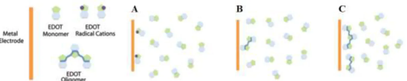
Electrochemical polymerization is an easy and fast technique for depositing PEDOT:PSS on conductive surfaces such as gold. It also allows selective deposition on specific areas where a metallic film is present. This a convenient method to pattern PEDOT:PSS, as opposed to multi-step photolithographic techniques, but also requires a conductive substrate, which can limit its use in some devices. This process is typically performed in an electrochemical cell with a three-electrode configuration. A working electrode, reference electrode and counter electrode are submersed in a solution of the monomer 3,4-ethylenedioxythiophene (EDOT) and poly(styrene sulfonate) sodium salt in deionized water.[6,7] EDOT monomers are oxidized by the metal electrode and form a cation.
13
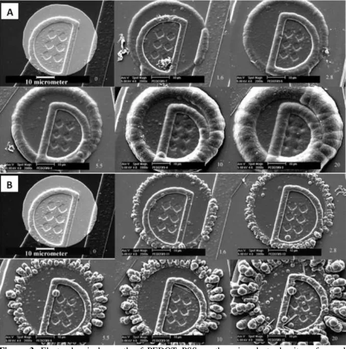
the electrode(Figure 2).[7,8] Polymerization can be done in potentiostatic (constant potential),
galvanostatic (constant current) or potentiodynamic modes. PEDOT:PSS films electrochemically deposited in the galvanostatic mode were found to be more uniform when compared to those deposited in potentiostatic mode (Figure 3).[6] In both modes, an edge effect was observed and this led to thicker deposition along the circumference of the electrodes. The edge effect is due to a non-uniform current distribution on the electrodes and is more pronounced when operating in potentiostatic mode.[6] The deposition time, current density and solution concentration contribute to the thickness of the electrochemically deposited film. For instance, the film thickness is directly proportional to the deposition charge (ex. for 1 µm at 5 µC). In another study, a potentiodynamic mode was used to polymerize PEDOT:PSS by scanning potentials using cyclic voltammetry.[9] When compared
to the films obtained by galvanostatic and potentiostatic modes, the potentiodynamic films were relatively more homogenous.
Figure 2. Electrical polymerization process. (a) The metal electrode oxidizes the EDOT monomers creating radical cations. (b) These radical cations combine, creating dimers, trimers, and higher oligomers. (c) As the molecular weight of the polymer chains increase they become insoluble, precipitating onto the metal electrode surface. Adapted with permission.[8] Copyright 2016, the Royal Society of Chemistry.
14
Figure 3. Electrochemical growth of PEDOT :PSS on the microelectrode sites of neural probes (a) galvanostatic (b) potentiostatic. Numbers on the right corner of each image indicate the deposition charge on each site (in µC). Adapted with permission.[6] Copyright 2003,
Elsevier.
1.2.2 Chemical Approach
1.2.2.1 Synthesis
PEDOT can be chemically synthesized via several synthetic routes[10] with the aid of oxidizing agents such as iron (III) chloride or nitride and peroxodisulfates[11]. Obtaining a stable aqueous dispersion is assisted by the presence of PSS. Due to its polar nature stemming from its sulfonic acid groups, PSS helps solvate PEDOT in the form of
15
PEDOT:PSS complexes.[11,12] The resulting dispersion is very stable and has a long shelf-life,
which has facilitated its commercialization. Several commercialized formulations exist (e.g. Heraeus Clevios™, Agfa Orgacon, Sigma Aldrich), allowing easy processing and reproducible film formation.[12]
The weight ratio between the thiophene groups and sulfonic acid groups for commercially available PEDOT:PSS (Heraeus Clevios™) is about 1:2.5; however, there is only a single charge introduced on every 3-4 thiophene groups. The fact that the charge ratio and mass ratio of chemicals do not match shows that the PSS is always added in excess.[12–14] Although not required for doping/charge balance, excess PSS is required for stabilizing the PEDOT:PSS dispersion by forming PSS shells around the PEDOT:PSS gel-like particles to prevent aggregation through electrostatic repulsion.[12,15]
1.2.2.2 Deposition
Chemically-polymerized aqueous dispersions of PEDOT:PSS can be deposited in a great variety of ways. Deposition methods include conventional coating techniques such as drop casting, spin-coating, solution-shearing, doctor-blading, ink-jetting, electro-spinning and spraying.[12] Every technique might require some optimization of the PEDOT:PSS dispersion content to obtain a uniform film. Additionally, solution viscosity, surface tension, and adhesion to the substrate must also be considered and optimized. Dispersion additives have a direct impact on these parameters and will be discussed in Section 1.3.2.[16,17]
Drop casting is the simplest coating technique, which includes only two basic steps: casting the polymer on a substrate and drying. The greatest advantage of this technique is that no machines are required. However, films obtained by drop casting are relatively thick (depending on the viscosity and substrate wettability) and lack homogeneity along the substrate.
Spin-coating PEDOT:PSS is typically used for thin film applications and often used as part of photolithographic fabrication in which PEDOT:PSS can be patterned with µm-scale
16
resolution. Unlike drop casting, spin-coating yields very homogenous films of up to ~300 nm thick from a single layer and ~800 nm from multiple layers.[12] In contrast to electrochemical deposition, spin coating yields more homogeneous PEDOT:PSS coatings, particularly at the edges of the film. It can be used to coat large substrates, with high reproducibility.[12] In this technique, the substrate is placed on a rotating sample holder and then the solution is drop cast on it before or while the holder is rotating. The thickness of the polymer film decreases with rotation speed (rpm) and increases with solution viscosity.[18] The spin coating step is usually followed by a drying step to remove the excess water content and some solvent additives. This drying step can be done by thermal treatment or by applying vacuum. A drawback of this technique is generating significant material waste since most of the used solution is excessive and is ejected during the process. Further details of polymer spin-coating, in terms of both physical and chemical properties, are described in the following review.[19]
Doctor blading is another technique used for film formation of homogenous thicknesses. In this technique, the blade (or knife) is placed parallel to a substrate with a certain gap that defines the film thickness. Afterwards, the solution is placed in front of the knife which then starts moving along the substrate. In comparison to spin coating, the material loss using doctor blading is negligible, and the range of thicknesses (10 µm to 500 µm) obtained is much higher.
Solution shearing deposition is a similar technique to doctor blading and has been used for organic polymer deposition.[20–22] In this technique, the solution is placed in-between two surfaces. The bottom surface is a heated substrate treated to be hydrophilic, while the upper surface is a silicon blade treated to be hydrophobic. The top plate is designed to move parallel to the substrate at a controlled speed. This movement leads to exposing some solution to air, and with heat provided by the bottom blade, the solvent can evaporate to form the film. The obtained film contains crystallized sites that propagate along with the direction of shearing. This technique was shown to be scalable up to areas of 5 x 5 cm. Shearing speed, temperature,
17
and solution properties are all parameters that can be tuned in this technique. Like spin coating, the deposited thickness using this technique is inversely proportional with the shearing speed. Shearing parameters have been changed to tune film morphology and composition: 20 to 250 nm thicknesses were obtained from shearing speeds of 0.02 to 4 mm/sec.[23] In comparison to spin coating, shearing deposited PEDOT:PSS is more conductive and more transparent.[23]
Inkjet printing is an additive solution deposition technique that has also been used for depositing PEDOT:PSS.[24–26] This technology allows direct pattering of the ink on a variety of substrates including those that are rigid, flexible, or stretchable.[24,27] This technology consists of computer-aided design, ink preparation, printing, and curing by heat or UV light.[25,27] Thickness of inkjet-printed films can be tuned by the number of printed layers. A
thickness of 190 nm was obtained from two layers of PEDOT:PSS, but this is dependent on ink composition and process parameters.[26] In addition to its low cost, other advantages of this technique include minimal material loss and the ability to pattern without expensive masks that are used in photolithography.[28]
Spray deposition techniques such as airbrush spraying, electro-spraying, and ultrasonic spraying have been used for depositing PEDOT:PSS; however, most of their applications were only focused on organic photovoltaic devices, which is out of the focus of this review.[29–34]. Other form factors of PEDOT:PSS include the fabrication of microfibers using wet-spinning,[35,36] brushing onto textiles[37] and scaffolds using freezing templating.[38]
The great variety of deposition methods gives flexibility in fabricating PEDOT:PSS into a variety of forms and device structures. Desired material properties (e.g. morphology, electrical properties, adhesion to substrate), patterned features, and thickness also further dictate deposition method.
18
Figure 1. Comprehensive schematic overview of the proposed models: (A) PEDOT:PSS forms spherical gel particles in aqueous dispersions. Upon drying a solid film consisting of lentil-like shaped grains develops. (B) At low relative humidity, e.g. 23% rH the hydrogen bonds in the PSS rich shell are very strong and therefore external tensile forces lead to a transgranular brittle fracture and therefore smooth fracture surfaces. (C) At higher relative humidity, e.g. 40% rH the hygroscopic PSS takes up water which causes the hydrogen bonds to be weakened and also leads to a swelling of the film. Upon exertion of external tensile forces individual PEDOT:PSS grains can slide by each other and therefore intergranular plastic fracture takes place. The fracture surface is rough with rather spherical grains forming the outer layer. Reproduced with permission.[39] Copyright 2009, Elsevier.
1.3. Properties
The vast majority of work on the determination of properties of PEDOT:PSS has been on spin coated films of chemically-polymerized PEDOT:PSS, owing to the application of this material in a variety of organic electronic devices such as light emitting diodes and solar cells. The properties of electrochemically polymerized PEDOT:PSS, on the other hand, have received less attention.
1.3.1 Film Structure and Mechanical Properties
PEDOT:PSS particles have been repeatedly described as having a “pancake” morphology, as shown in Figure 1, and this also describes the bulk morphology of PEDOT:PSS spin coated thin films.[5,40–42] Experiments combining anisotropic conductivity
19
presence of PEDOT-rich particles of 20-25 nm in diameter and 5-6 nm in height separated by PSS lamellas.[41] These findings were supported by scanning tunneling electron microscopy on PEDOT:PSS thin films (25 nm thick), which revealed the same distribution and sizes of the PEDOT:PSS particles.[40] By using energy dispersive X-ray spectroscopy, the PSS shell around the individual PEDOT clusters was found to be 5-10 nm.[40] Ultraviolet photoelectron spectroscopy has also been used to study the surface morphology of PEDOT:PSS thin films. This spectroscopy method can differentiate between PEDOT and PSS by their binding energies.[43] Authors found that the PSS concentration at the surface is 3 folds higher than expected and that the PSS concentration in the bulk is lower than its concentration at the surface.[43,44]
One of the main advantages of PEDOT:PSS is its soft nature compared to inorganic conductors. This softness allows PEDOT:PSS films to be conformable and even stretchable in some cases.[16,45] PEDOT:PSS films swell when immersed in water or placed in a humid environment, and their thickness increases accordingly.[11,12] Swelling also helps increase ionic mobility in the film.[46] Additionally, the swelling is responsible for a reduction in the electrochemical impedance of PEDOT:PSS-coated metal electrodes, as discussed in detail in Section 1.3.2. For these reasons, swelling is a very important property of PEDOT:PSS films for biomedical application.
A general morphological model for the swelling of PEDOT:PSS films[39] was proposed assuming the films contained PEDOT-rich particles and PSS shells (Figure 4).[40] At low relative humidity, the model states that the hydrogen bonds in the PSS shells are strong, and the film exhibits brittle fracture that propagates through the grains.[40] At a higher humidity, PSS shells absorb water and the inter-distance between the shells increases; therefore, hydrogen bonds between PSS shells diminish and so does the mechanical strength of the film.[40] A few studies have quantified the swelling of PEDOT:PSS films when immersed in water[46,47], and their findings are summarized in Table 1.
20
Several studies have investigated the mechanical properties of PEDOT:PSS using techniques such as tensile testing, peak force quantitative nanomechanical property mapping (QNM) and strain-induced buckling instability (SIEBIMM).[16,35,39,48,49] Table 2 summarizes Young’s modulus values of PEDOT:PSS in different conditions of humidity as well as in water.
Cross-linkers are often added to PEDOT:PSS to increase film stability. Film stability is required for biomedical devices that are immersed in aqueous solutions. Not only must the films maintain their integrity, but also they should have strong adhesion to the substrate. Furthermore, the use of cross-linkers can increase the mechanical properties of films. A few cross-linkers of PEDOT:PSS have been demonstrated to date.
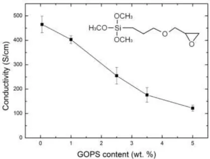
3-glycidoxypropyltrimethoxysilane (GOPS) is often used as a cross-linker for PEDOT:PSS films.[50–52] GOPS allows strong film integrity and strong adhesion to the substrate when immersed in aqueous solutions.[53] Increasing the GOPS concentration in PEDOT:PSS films increases the mechanical strength as shown in Table 2 but at the expense of conductivity.[53] Divinylsulfone (DVS) is another chemical additive that was recently used as a low temperature cross-linker for PEDOT:PSS.[54] In contrast to GOPS, DVS only had a
very small effect on conductivity while enhancing the mechanical properties and film integrity.[54]
Aside from cross-linkers, other additives may be used to change the mechanical properties and swelling. Some researchers have blended polyvinyl alcohol (PVA) with PEDOT:PSS to increase its flexibility, ductility, and durability.[55–57] The Young’s modulus of
pure PVA films was increased from 41.3 MPa to 1.63 GPa by increasing the concentration of PEDOT:PSS from 0% to 50%.[58] The increase in PEDOT:PSS ductility resulting from PVA addition was countered by a decrease in tensile strength, Young’s modulus, and conductivity since PVA is insulating and decreases contact between the PEDOT-rich regions.[55,59]
21 Table 1. The swelling of PEDOT:PSS in water
PEDOT:PSS Form Additives Measurement Technique
Swelling References
Thin film - AFM 660% ± 90% Duc et al.[47]
Thin film 1% GOPS AFM 40% ± 1% Duc et al.[47]
Thin film 1% GOPS AFM 35% ± 4% Stavrinidou et
al.[46]
Table 2. The mechanical properties of PEDOT:PSS thin films, pristine and with additives PEDOT:PSS Form Additives Specific Conditio ns Measuremen t Technique Young’s modulus Referenc es Microfibers - Room temp. Tensile test 1.1 ± 0.3 GPa Okuzaki et al.[35] Cast film 3% EG 50% rH / 25 °C Strain gauge Stress-Strain curves 1.8 ± 0.2 GPa Okuzaki et al.[35]
Pipetted film - 40%rH Tensile test 2.7 GPa Lang et
al.[39,48]
Pipetted film - 50%rH Tensile test 2.0 GPa Lang et
al.[39,48] Pipetted film - 23% rH/ 22 ◦C Tensile test 2.8 ± 0.5 GPa Lang et al.[39] Pipetted film - 40% rH/ 22 ◦C Tensile test 1.9 ± 0.02 GPa Lang et al.[39]
22 Pipetted film - 55% rH/ 22 ◦C Tensile test 0.9 ± 0.2 GPa Lang et al.[39]
Thin film - - SIEBIMM* 0.8 ± 0.1
to 1.02 ± 0.2 GPa Greco et al.[16] Thin film - 23 to 30% rH Buckling method 2.26 ± 0.05 GPa Tahk et al.[60] Electrochemicaly deposited PEDOT thin film - 25 °C PeakForce QNM** 2.6 ± 1.4 GPa Qu et al.[49] Electrodeposited film - - NanoIndentat ion 3.23 ± 1 GPa Baek et al.[61]
Cast film PEDOT:PSS(50%)/ PVA
40 ± 2% rH
Tensile test 1.632GPa Chen et al.[55] * SIEBIMM stands for strain-induced buckling instability for mechanical measurements. ** QNM stands for quantitative nanomechanical property mapping.
1.3.2 Electrical properties
PEDOT:PSS exhibits intrinsic electronic conduction properties.[12] The conductivity of PEDOT:PSS is usually deduced from the sheet resistance measured from a flat film deposited on a non-conducting supporting layer using the 4-points probe technique or the transmission line method.[16,62] The weight ratio of PEDOT to PSS alters the charge density on PEDOT and results in a change in the conductivity.[12] The conductivity of PEDOT:PSS films is highly anisotropic: the lateral conductivity is three times higher than the vertical conductivity.[41] This difference is due to film morphology, as PEDOT-rich particles, of about 20-25 nm in
23
diameter and 5-6 nm in height, are separated by PSS lamellas, which have weak electrical conduction.[41]
The conductivity of commercial, aqueous PEDOT:PSS (pristine) ranges from 10-5 to 101 S/cm and can even exceed 1000 S/cm with certain additives.[12] Several methods have been investigated to enhance the electrical conductivity of PEDOT:PSS films including treatments with light and heat as well as the treatments with ionic liquids, salt solutions, acids, organic solvents, and surfactants.[63] A comprehensive summary of the conductivity values of treated PEDOT:PSS versus pristine PEDOT:PSS was presented in a recent review.[63] Here, we will only highlight the three most widely used solvent additives used for enhancing the conductivity of PEDOT:PSS.[63]
Dimethyl sulfoxide (DMSO) is a high-boiling point, polar solvent used for enhancing the conductivity of PEDOT:PSS.[64–72] DMSO was first added to PEDOT:PSS dispersions in 2002.[73] By adding one-part DMSO to three parts PEDOT:PSS (v/v), the conductivity was increased by 2 orders of magnitude, from ~0.8 S/cm to ~80 S/cm.[73] This increase in conductivity is believed to be due to remaining DMSO that increases the separation of positive and negatives charges on the PSS and PEDOT, respectively, and thereby reduces the electrostatic interactions between the chains (“screening effect”).[73] Since then, many works
have investigated the effect of solvents on the conductivity and morphology of PEDOT:PSS films.[63–69,74–76] By adding DMSO, PEDOT-rich grains become enlarged and were more uniformly distributed in films. The increase in conductivity was attributed to these morphological changes.[65] For inkjet-printed PEDOT:PSS films, the conductivity of
PEDOT:PSS was increased by four orders of magnitude by adding 5% DMSO to the ink.[70,77] The additional increase in conductivity was due to a three-fold increase in PEDOT particle size.[77]
Used in a similar fashion as DMSO, ethylene glycol (EG) is another high-boiling point, polar solvent used for increasing the conductivity of PEDOT:PSS
24
films.[43,45,47,55,56,60,61,66] It was demonstrated that by adding 7% EG, there was an increase of
~3 orders of magnitude in the conductivity of PEDOT:PSS films.[78] Later, a 100-fold enhancement in the conductivity was observed when adding or when treating with EG.[79] The mechanism of this increased conductivity was found to be from a change in the conformation of coiled PEDOT chains: from more compacted coils to either more expanded coils or linear chains. Furthermore, PEDOT chains changed from a benzoid structure (contributing to a coil conformation) to a quinoid structure (contributing to a linear chains) as revealed by Raman spectroscopy. In another study, authors reported an increase of more than 100 fold in the PEDOT:PSS conductivity (from 6 to 800 S/cm) with the addition of 5% EG.[5] They suggested that this increase in conductivity was due to increased aggregation of PEDOT particles and tighter π-stacking.[5] In other groups, researchers suggested that the PEDOT
particle size increased and became more homogenously packed.[75,80] All of these studies relate the increase in PEDOT:PSS conductivity with EG addition to a change in the PEDOT:PSS film morphology, either due to stronger PEDOT particle interactions or due to more linear PEDOT chains.
Another example of the organic solvents used for improving the conductivity of PEDOT:PSS is sorbitol (a sugar alcohol).[75,81–85] With the addition of sorbitol, PEDOT:PSS conductivity was increased by 2 orders of magnitude due to an increase in the PEDOT particle size.[75] In the PEDOT:PSS dispersion, sorbitol undergoes a chemical conversion to 1,6-anhydrosorbitol and the PEDOT:PSS particle size increases, both of which contribute to improving conductivity.[84]
In neural recordings and other electrophysiology applications, the electrode impedance is an important indicator of how well an electrode will capture signals. Therefore, analyzing and understanding the mechanism of charge transport and electrochemical impedance is crucial for designing and optimizing the performance of such devices.[86] PEDOT:PSS coatings are known to dramatically decrease the impedance of metallic electrodes, especially
25
in the low frequency range (by ~2 orders of magnitude).[6,87,88] This drop in the impedance is
due to the mixed electronic and ionic conductivity in PEDOT:PSS; PEDOT-rich regions (electrically-conducting) facilitate and increase opportunities for charge exchange while the high water content of PEDOT:PSS imparts high ionic conductivity.
The complex electrochemical impedance curve of a PEDOT:PSS thin film is typically composed of two regions, a capacitance-dominated region at low frequencies and a resistance-dominated region at high frequencies.[88] The transition between these two regions happens at a characteristic frequency (fc). The fc of a gold electrode shifts to lower values when coated
with PEDOT:PSS (from 10 kHz to 100 Hz for 0.5 x 0.5 mm electrode, 350 nm PEDOT:PSS layer thickness).[87] The impedance curve can be modeled by a simple RC or R(RC) circuit model, where R corresponds to the solution resistance (Rs), and C corresponds to the film
capacitance. Since PEDOT:PSS swells in aqueous media and liquid electrolytes can enter the film, the charge transport can be understood in terms of volumetric capacitance (Cv:
capacitance per unit volume).[89] It was reported that capacitance scales with the volume of PEDOT:PSS films and this causes lower impedance.[88] The volumetric capacitance of PEDOT:PSS can be understood as follows (Figure 5):[90] PEDOT chains carry holes and PSS
chains carry the sulfonate anions that balance these holes.[90] Upon applying a positive bias, the cations from the electrolyte move inside the PEDOT:PSS film and replace the holes (Figure 5a). The volumetric capacitance can be estimated by treating the injected cations and sulfonate anions pairs as elementary capacitors. In Figure 5b, the holes/anion sites are represented as a group of planes perpendicular to the film surface and distanced by an average spacing α.[90] The authors suggest that every plane represents a double layer capacitance
(C’DL=C/A, capacitance per unit area), and they deduced the volumetric capacitance as C*= C’DL/α.[90] This study suggested that the PEDOT volumetric capacitance can be enhanced by bringing the hole/anion sites closer together by adding dopants that are smaller than PSS or by blending with nanomaterials with high charge densities.[90]
26
Figure 5. (a) Schematic showing the dedoping process caused by ion injection, where a is the average site distance. (b) Schematic showing an array of plates against which ions pile up to form double layer capacitors. Adapted with permission.[90] Copyright 2016, John Wiley and Sons.
1.3.3 Stability
As will be discussed more extensively in the following section, PEDOT:PSS-based devices have been utilized with cells and tissues and have been implanted in the body. Unlike standard electronics, these conditions necessitate their performance submerged in an aqueous environment. Differing greatly from a simple salt solution, cell culture media and in vivo conditions are quite complex. Furthermore, operation is at physiological temperature (37 °C), which again differs from the common condition of room temperature. The stark contrast of between traditional electronics and bioelectronics warrants investigation of device performance at such conditions, especially for long periods of time. Parameters such as method of deposition, adhesion with the substrate, operation conditions (stimulation, recording, continuous, pulsed) and environment have a profound influence of stability. In this section, we discuss reports of PEDOT:PSS performance in simulated biological environments as well as in vivo.
In vitro studies with cells and tissues as well as in vivo implantations often require
sterilization, particularly if the studies are carried out over time as opposed to one discrete measurement. Long-term stability measurements of PEDOT:PSS devices, discussed below,
27
should be conducted sterile to avoid any confounding factors, such as bacterial and fungal growth and subsequently, biofouling of electrode sites. Many polymers cannot survive or are deeply affected by clinical sterilization procedures such ethylene oxide, autoclave, and hydrogen peroxide. In a recent study, the effects of various sterilization methods of PEDOT:PSS were studied.[91] No apparent differences in surface roughness of spin coated PEDOT:PSS electrodes were observed with heat steam sterilization. Furthermore, mean impedance at 1 kHz across tested electrodes increased only slightly, and transconductance in OECTs was found to change very little. It is important to note these are annealed (140 °C), cross-linked films, and this processing seems necessary for film integrity to be maintained after autoclave. In another sterilization method, H2O2 gas plasma was devastating to
performance leading to non-functional devices in which PEDOT:PSS delaminated from underlying metal, even though this sterilization method utilizes far lower temperatures than autoclave.[91]
Early work with PEDOT:PSS first investigated the conducting polymer performance in phosphate-buffered saline (PBS) at room temperature for short-term periods in vitro. Electrochemically-polymerized films[92] and coatings on gold[93,94] and PtIr wire arrays[95] as
well as chemically-polymerized PEDOT:PSS on track-etch membranes[96] observed little (~20% loss in electroactivity) to no changes for up to 2 weeks or with 1,000 of cycles of stimulation. Authors studying electrochemically deposited PEDOT:PSS on Pt electrodes did, however, find differences in performance with coatings of varied thickness when continuously stimulated for 2 weeks in PBS.[97] Thirty electrodes of different thicknesses
showed some cracking in the PEDOT:PSS films as well as delamination. Cracking only occurred in the films of intermediate thickness while the only films that delaminated were the thickest ones. The appearance of cracks did occur in the thickest films as well, but at an earlier time point, preceding delamination. The conductivity of PEDOT:PSS also diminished with stimulation time, and delaminated PEDOT:PSS coincided with electrical characterization
28
that were similar or identical to Pt properties. Even electrodes that did not display any obvious signs of damage still showed changes in electrical properties, including decreased charge capacity, peak shift, and peak separation. The authors note peak separation can be from a change in ion diffusivity, which may be a result of microstructural changes in the film.
Others have also investigated the performance of PEDOT:PSS at physiological temperature or at other elevated temperatures in PBS in vitro. These elevated temperatures (~60-70 °C) simulate long-term polymer aging at 37 °C; these protocols are based off standard protocols for assessing polymer degradation in medical devices.[98] At 67 °C for 28 days, which simulates 32 weeks at 37 °C, impedance at 1 kHz increased by 27 kΩ, and the maximum negative voltage in response to 5 µA increased by 350 mV in PEDOT:PSS coated PtIr microwire arrays (75 µm in diameter). However, large, clinical PEDOT:PSS coated Pt electrodes (3x6 mm) were not found to degrade at 13 weeks of elevated temperature, corresponding to 2 years in vivo. It was suggested the delamination could have been the main contributor to the degradation in performance microwires while macrowires remained similar, but this was not tested.[95] In electropolymerized PEDOT:PSS on gold electrodes in a flexible polyimide-based array, stimulation was conducted in PBS at 37 °C. Electrodes were stable for at least 7 weeks of continuous stimulation with no significant change in impedance after 4.2 billion bipolar current pulses. No significant changes were observed in the unstimulated experiments lasting over 10 months.[99] Adhesion to the underlying substrate was cited as an issue in another study of electrochemically-polymerized PEDOT:PSS on ITO or gold.[93] In PBS at 37 °C, all 9 PEDOT:PSS films on ITO delaminated within 2 weeks while only 1 of 9 PEDOT:PSS films on Au had delaminated by 35 days. Interestingly, no delamination was observed when PEDOT:PSS films on ITO were incubated in deionized water or 10 mM H2O2
at 37 °C. Hydrogen peroxide was investigated in order to simulate oxidative stress that can occur at sites of biomaterial implantation.[100] The electroactivity of PEDOT:PSS rapidly declined around 10-15 days in dilute H2O2.[93] The decline was attributed to over-oxidation of
29
PEDOT in which the double bonds of the polymer backbone are broken and result in a loss of conjugation and conductivity. In other long-term stability studies, electrochemically deposited PEDOT:PSS on gold-parylene C neural probes displayed slight increases and variations in the mean impedance over the course of 6 months in artificial cerebral spinal fluid (ACSF), but never exceeded 100 kΩ for 1 kHz.[101] With PEDOT:PSS electrochemically deposited on carbon fibers, the average impedance started at 143 kΩ, but over the course of a simulated time of 172.2 days (over 24 weeks) in PBS at 60 °C, the impedance increased gradually to ~1920 kΩ.[102]
PEDOT:PSS neural electrodes, for both recording and stimulation, have been tested in a few in vivo studies in both mice and rats. On microfabricated silicon shanks (PEDOT:PSS electrochemically deposited onto gold recording sites), there was a decrease in performance (decreasing single unit and multiunit yields, signal amplitude and signal-to-noise ratio) within the first two months but remained stable for the remaining two months of the study.[103] Impedance also fluctuated in the first month, but increased steadily until ~2 MΩ at 1 kHz. In another study of electropolymerized PEDOT:PSS on silicon neural probes, the mean impedance was typically less than 1 MΩ at 1 kHz over the course of over 6 weeks after initial increase in impedance that occurred early within the first few days.[104] This quick initial impedance increase was also observed with PtIr wire arrays with electropolymerized PEDOT:PSS.[95] Starting at ~65 kΩ, impedance at 1 kHz increased over 9 days but then slowly decreased to ~550 kΩ. Importantly, the impedance still remained ~200 kΩ lower then bare metal electrodes.[95]
The above in vivo studies were conducted with conventional neural probes, which are made of inorganic materials that far exceed the stiffness of surrounding brain tissue and therefore are thought to contribute to tissue damage and the prolonged immune response to neural probes. Recent efforts are being made to make smaller and more flexible probes aimed at minimizing tissue damage and improving biocompatibility. These probes will be discussed
30
further in the following sections, but with regards to chronic performance in vivo, flexible probes have recently shown potential for improved performance over conventional probes. In microfabricated neural probes composed of parylene C and PEDOT:PSS coated gold, electrode impedance began at 10-25 kΩ for 1 kHz.[101] Impedance increased by only a factor of 2, and stable recordings were obtained for 30 weeks. Throughout the study, 70-90% of electrodes were functional but varied with time. In another study, the choice of substrate was shown to have an impact on neural recording performance. PEDOT:PSS on glassy carbon outperformed PEDOT:PSS on Pt when fabricated in polyimide microelectrode arrays.[105] Results from these two studies underscore the importance of device properties and material choice that may affect PEDOT:PSS performance, especially long-term.
Cited as potential issues from many of the above studies, PEDOT:PSS delamination can be prevented with strategies to improve adhesion to the underlying substrates, typically metal. Adding the silane cross-linker GOPS in enough quantity to PEDOT:PSS dispersions results in a strong adhesion to glass substrates as well as metal surfaces such as gold. It is important to note that these surfaces are pre-activated by plasma oxygen cleaning or other chemical cleaning methods. As noted in one of the studies above, ITO is a more challenging surface for PEDOT:PSS or PEDOT:PSS:GOPS adhesion. To overcome this, ITO surface functionalization with GOPS is used by sonicating a pre-activated ITO substrate in a solution containing water, ethanol, and GOPS (unpublished results). The created GOPS layer on ITO surface bonds with the GOPS content in the PEDOT:PSS:GOPS blend, forming a stable adhesion. On Pt microwires, PEDOT:PSS was electrochemically deposited after a coating poly(vinyl alcohol)/poly(acrylic acid) inter-penetrating polymer networks. Mechanical and electrochemical stabilities were improved over PEDOT:PSS alone with notably improved adhesion on Pt that prevented delamination.[106] Adhesion of PEDOT:PSS coatings can also be improved to a varierty of substrates (Pt, Ir, ITO) when electrochemical polymerization is conducted with amine-functionalized EDOT to produce a P(EDOT-NH2) layer on the
31
electrode, facilitated by amine-metal adsorption, and onto which additional PEDOT was polymerized and covalently linked.[107] In a method for inducing mechanical rather than chemical adhesion, nanostructured IrOx and Pt improved adhesion of PEDOT:PSS coatings[108].
In summary, PBS does not seem to alter PEDOT:PSS performance nor does increasing temperature or stimulation. Long-term performance of PEDOT:PSS in PBS is obtainable in
vitro. Thickness of coatings, substrate adhesion, and electrode surface area influence
degradation properties mostly through cracking or delamination. Unobservable microstructural changes in the polymer, which change impedance, have yet to be thoroughly investigated. Oxidative environments, mimetic of inflamed tissue, can be devastating to the electrical performance of PEDOT:PSS. Liquid media containing more complex factors, such as hydrogen peroxide, that mimic the in vivo environment need to be investigated further. As well, the potential of PEDOT:PSS on more flexible and more biocompatible neural probes is a very promising avenue and will likely see performance exceeding that of PEDOT:PSS on stiff neural probes due to better tissue compatibility. Certainly, several strategies should be investigated to minimize the drastic impedance increase observed with implanted PEDOT:PSS.
1.4. Applications
PEDOT:PSS has been utilized in several forms for biomedical applications. The first example and the most widely used is the coating of PEDOT:PSS on electrodes for recording as well as stimulation of electroactive cells and tissues. As discussed above, the conducting polymer coating decreases the electrode impedance by increasing the effective area of the electrode and also increases the charge injection limit; thus, recording and stimulation performances are improved. The fabrication of arrays with PEDOT:PSS electrodes have a
32
broad impact in that they can be used to study biology as well as providing therapy or treatment.
PEDOT:PSS has also been utilized in organic electrochemical transistors (OECTs). OECTs have proven most useful for recording low amplitude signals (electrophysiology) or low concentrations of the desired analyte (biosensing) in that these devices can provide high signal amplification. First reported in 1984, the OECT consists of a polymer channel with source and drain electrodes. The channel is in contact with an electrolyte, in which a gate electrode is immersed.[109] Application of a gate voltage drives ions from the electrolyte into the channel and changes its conductivity. This ion-to-electron transduction mechanism makes OECTs useful transducers for biological applications. OECTs and their various applications are discussed at greater length in the following review[110] while this review will specifically
focus on those based on PEDOT:PSS.
One key advantage of using PEDOT:PSS-based devices for recording and stimulation, either through electrodes or OECTs, is the ability to use varying substrates, including those that are cheap, flexible, and biocompatible. Various fabrication methods, as discussed above, lend the way to scale-up and commercialization at low costs. Compared to bare metal electrodes, conducting polymer coatings decrease electrode impedance and afford recording sites of smaller surface areas and therefore, a greater recording site density can be achieved on a device of certain dimensions. Finally, as will be discussed further in examples of tissue interfacing, fabrication of devices on flexible substrates is key for improving biocompatibility. Other, less frequently explored form factors include free-standing films, porous or tubular scaffolds, fibers, nanowires[111] and particles. Films and scaffolds are useful for interfacing electronics with cells. While films can be used for in vitro cell culture, scaffolds provide a three-dimensional environment that is more mimetic of the body. Furthermore, 3D scaffolds are used in tissue-engineered implants. Tubular scaffolds have mostly been proposed as nerve conduits. Alternatively, fibers can serve as soft and flexible electrodes for
33
electrophysiology. PEDOT:PSS particles can serve as sites for nucleation in electrochemical polymerization or in colloidal inks for printed electronics. Here, we discuss applications of all of the above PEDOT:PSS devices in highlighted examples.
1.4.1 Electrophysiology
1.4.1.1. Implantable Devices
Electrophysiology with implantable devices includes neural recordings - electrocorticography ECoG (from surface of brain) and stereoelectroencephalography SEEG (penetrating brain tissue) - as well as intramuscular electromyography EMG. Requirements for implantable electronics vary greatly from those for cutaneous electrophysiology, which is discussed in the next section. Neural electronics are the most widely studied of implantable bioelectronic devices. Neural electrodes serve those in basic science with the important task of uncovering the complexity of how the brain works. For the application of neural electrodes, Brain-Machine Interfaces (BMI) have been proposed and investigated with great success. Recording of brain activity can be translated to cursor control – including for speech and wheelchair control, movement of an artificial limb, and muscle stimulation for movement of a patient’s own paralyzed limb.[112,113] Especially for the brain, the softest tissue in our body,
developing soft, flexible and small probes[114] are of pressing importance to the field of
chronic BMIs, which has traditionally used metal and silicon probes with poor biocompatibility[115,116]. PEDOT:PSS has been used as both a coating of these metal wires and silicon shank probes and more recently, have been used on electrodes on flexible substrates.
In 2003, the ability of PEDOT:PSS to improve biological interfacing of metal electrodes was first demonstrated. PEDOT:PSS was electrodeposited on gold electrodes and was found to decrease the impedance by nearly two orders of magnitude.[94] This study would also include the first demonstration of in vivo neural recordings with PEDOT:PSS, which was used to coat penetrating Michigan-style silicon electrodes for acute measurements in a guinea pig.
34
PEDOT:PSS has also been electrochemically applied to tips of 7 µm diameter carbon fibers (Figure 6).[117] In contrast to traditionally used silicon shanks or metal microwires, these fibers served as an all organic-based fiber providing higher compliance and a smaller device footprint for minimizing brain damage in neural recordings. The coated fibers were one order of magnitude less stiff than silicon neural probes. When compared to uncoated probes, only PEDOT:PSS coated devices were able to detect single unit activity in the motor cortex of rats. The devices demonstrated a high signal-to-noise ratio and were stable over 5 weeks.
Figure 6. PEDOT:PSS coated carbon fibers for ultra-small, implantable neural probes. A) Electrochemical deposition of PEDOT:PSS on carbon fibers B) carbon fibers were coated with poly(p-xylylene) and poly((p-xylylene-4-methyl-2-bromoisobutyrate)-co-(p-xylylene)) (blue) and were finally covalently grafted with poly(ethylene glycol) (red). The fiber was cut to remove insulation and expose the carbon fiber (gray). A recording site was prepared by electrochemically depositing PEDOT:PSS (green) on the exposed tip. C) Scanning electron micrograph of final neural probe. 7 µm diameter. D) Carbon fiber neural probes on top of a10 mm silicon electrode. Scale bar, 50 µm. E) Carbon fibers (arrows) implanted 1.6 mm deep into the cortex of a rat brain. Scale bar, 100 µm. F) Simultaneous neural recordings with and without PEDOT:PSS. Single unit activities were only detected in vivo with PEDOT:PSS coating (SNRs of 12.7 and 4.71 and a noise floor of 23.4 µV). Adapted with permission.[117]
2012, Macmillan Publishers Ltd.
Also towards conformable devices for more biocompatible neural probes, the microfabrication of PEDOT:PSS neural probes with parylene C has been recently developed (Figure 7).[118–121] Conformability is especially important for measurements on the brain since
35
on the surface of the brain, the probe can follow its folds and while inside the brain, the probe can move and flex with micromotions associated with body movement, respiration, and blood flow. Using standard lithography procedures and spin coating of PEDOT:PSS, flexible probes were fabricated to be only 4 µm thick with micrometer-diameter, PEDOT:PSS-covered gold electrodes and OECTs. ECoG measurements on the surface of the brain were performed with these probes consisting of PEDOT:PSS OECTs. Compared to surface electrodes, PEDOT:PSS OECTs showed a superior signal-to-noise ratio than electrodes in measurements in rats as well as the detection of fast and low amplitude signals previously only obtainable with penetrating probes.[121] In later work, the authors reported the unprecedented ability to detect single unit activity using PEDOT:PSS electrodes placed on the surface fo the brain.[119] These electrodes were also successfully used in humans to monitor brain activity in patients who were undergoing surgery for epilepsy treatment.[119] These parylene-based probes have also been presented in a penetrating probe format with the aid of an insertion shuttle. When compared to the standard of silicon probes, parylene electronics showed minimal glial scarring in rats[120]. Thicker parylene probes with electrochemically deposited PEDOT:PSS have also been demonstrated[122], and these did not seem to require an insertion aid for brain
36
Figure 7. Flexible, parylene C neural probes with Au-PEDOT:PSS electrodes. A) Neural probes are microfabricated by photolithography. Recording sties can be in OECT or electrode configurations. B) Schematic (top) and optical micrograph (bottom) of neural probes for delaminating shuttle for insertion into the brain. C-D) Anti-glial fibrillary acidic protein staining of mouse brain after 1-month implantation of silicon (C) and parylene (D) probes. The glial scar is more evident silicon probes. Black arrows indicate example glial cell bodies. Scale bar, 50 µm. A-D) Adapted with permission[120]. 2015, Wiley. E) Parylene ECoGs. When placed on the somatosensory cortex of rats PEDOT:PSS OECTs (pink) outperform PEDOT:PSS (blue) surface electrodes with a superior SNR. Measurements are also compared to penetrating Ir electrodes (black). Adapted with permission.[121] 2013, Macmillan Publishers Ltd. F) A flexible, 256-electrode parylene ECoG (Neurogrid), laid onto an orchid petal. Scale bars 5 mm, 100 µm inset. G) Extracellular action potentials are detected from the surface of the cortex and hippocampus of rats. In another experiment, the probe detects LFPs and spiking activity in epilepsy patients. Adapted with permission.[119] 2014, Macmillan
Publishers Ltd.
Using replica molding with PDMS, an all-polymer microelectrode array was fabricated (Figure 8). Microchannels were made in the PDMS and were filled with PEDOT:PSS to create the electrode paths. Impedance ranged from 400 kΩ to 4 MΩ between 1 Hz and 1 kHz. The average impedance at 1 kHz, used for measuring action potentials, was approximately 40 times higher than metal electrodes; at 1 Hz, used for measuring local field potentials, impedance was 15 times lower. Interesting benefits to such a device include its support for cultured cells and optical transparency for subsequent biological imaging. Furthermore, the device is much more elastic than metal-based devices and this elasticity may be beneficial for some applications, especially those in vivo. The authors evaluated the utility of such devices in vitro and in vivo with various recordings, including cardiac activity from embryonic mice hearts, recordings of individual action potentials from stimulated retinal explants, spontaneous recording of cortico-hippocampal cells for 63 days, and the detection of LFPs in epidural and epicortical configurations on the primary visual cortex of rats.[123]
37
Figure 8. All-polymer microelectrode arrays composed of PEDOT:PSS within PDMS. A1) Transparent microelectrode array (60 electrodes, 49 x 49 mm2) with conductive PEDOT:PSS tracks (blue) comprising contact pads, channels, and recording sites. Fixed with well for cell culture. Scale bar, 1 mm. A2-A3) Higher magnification images of A1. Scale bars, 1 mm. Readable text of paper underneath array illustrates its transparency. B) Array is flexible (array with graphite-PDMS conductors) Scale bar, 1 mm. C) Cross-section of one PEDOT:PSS electrode. Scale bar, 100 mm. D) Cortico-hippocampal rat calls are supported on the device, 38 days in vitro. Optical imaging is facilitated by array transparency. PEDOT:PSS electrodes. Scale bar, 100 mm. E) Immunofluoresence staining of neural network. Beta-tubulin III, green, dendrites and axons. MAP2, red, dendrites. Hoechst stain, blue, nuclei. graphite-PDMS conductors. Scale bar, 100 mm. Adapted with permission.[123] 2011, Elsevier.
In addition to recording, electrodes are also needed for stimulation in the central and peripheral nervous systems as well as in muscle. Stimulating electrodes can be used externally or internally. The applications discussed in this section are for implantable probes, while cutaneous applications are presented in the following section. Stimulation is used in cochlear implants[124] and visual prostheses[125], as well as for pain management[126], Parkinson’s