HAL Id: hal-02477859
https://hal.archives-ouvertes.fr/hal-02477859
Submitted on 13 Feb 2020
HAL is a multi-disciplinary open access
archive for the deposit and dissemination of
sci-entific research documents, whether they are
pub-lished or not. The documents may come from
teaching and research institutions in France or
abroad, or from public or private research centers.
L’archive ouverte pluridisciplinaire HAL, est
destinée au dépôt et à la diffusion de documents
scientifiques de niveau recherche, publiés ou non,
émanant des établissements d’enseignement et de
recherche français ou étrangers, des laboratoires
publics ou privés.
Development of new biocompatible 3D printed graphene
oxide-based scaffolds
Habib Belaïd, Sakthivel Nagarajan, Catherine Teyssier, Carole Barou,
Jonathan Barés, Sébastien Balme, Hélène Garay, Vincent Huon, David Cornu,
Vincent Cavailles, et al.
To cite this version:
Habib Belaïd, Sakthivel Nagarajan, Catherine Teyssier, Carole Barou, Jonathan Barés, et al..
De-velopment of new biocompatible 3D printed graphene oxide-based scaffolds. Materials Science and
Engineering: C, Elsevier, 2020, 110, pp.110595. �10.1016/j.msec.2019.110595�. �hal-02477859�
Development
of new biocompatible 3D printed graphene oxide-based scaffolds
Habib
Belaid
a,b,
Sakthivel Nagarajan
a,
Catherine Teyssier
b,
Carole Barou
a,b,c,
Jonathan Barés
d,
Sebastien
Balme
a,
Hélène Garay
d,
Vincent Huon
d,
David Cornu
a,
Vincent Cavaillès
a,1,
Mikhael
Bechelany
a,⁎,1a Institut Européen des Membranes, IEM – UMR 5635, ENSCM, CNRS, Univ Montpellier, Montpellier, France
bIRCM, Institut de Recherche en Cancérologie de Montpellier, INSERM U1194, Université Montpellier, Montpellier F-34298, France cBiologics 4 life, 84120 Pertuis, France
d LMGC, Laboratoire de Mécanique et Génie Civil, Univ Montpellier, CNRS, Montpellier, France e C2MA, IMT mines d'Alès, France
Keywords: Polylactic acid Graphene oxide Nanocomposite 3D printing Biocompatibility A B S T R A C T
The aim of this work was to develop a bioresorbable, biodegradable and biocompatible synthetic polymer with good mechanical properties for bone tissue engineering applications. Polylactic acid (PLA) scaffolds were gen-erated by 3D printing using the fused deposition modelling method, and reinforced by incorporation of graphene oxide (GO). Morphological analysis by scanning electron microscopy indicated that the scaffold average pore size was between 400 and 500 μm. Topography imaging revealed a rougher surface upon GO incorporation (Sa = 5.8 μm for PLA scaffolds, and of 9.9 μm for PLA scaffolds with 0.2% GO), and contact angle measurements showed a transition from a hydrophobic surface (pure PLA scaffolds) to a hydrophilic surface after GO in-corporation. PLA thermomechanical properties were enhanced by GO incorporation, as shown by the 70 °C increase of the degradation peak (thermal gravimetric analysis). However, GO incorporation did not change significantly the melting point assessed by differential scanning calorimetry. Physicochemical analyses by X-ray diffraction and Raman spectroscopy confirmed the filler presence. Tensile testing demonstrated that the me-chanical properties were improved upon GO incorporation (30% increase of the Young's modulus with 0.3%GO). Cell viability, attachment, proliferation and differentiation assays using MG-63 osteosarcoma cells showed that PLA/ GO scaffolds were biocompatible and that they promoted cell proliferation and mineralization more efficiently than pure PLA scaffolds. In conclusion, this new 3D printed nanocomposite is a promising scaffold with adequate mechanical properties and cytocompatibility which may allow bone formation.
1. Introduction
For decades, bone disease management, for instance in osteoporosis, has been challenging due to the reduced bone self-repair capacity [1]. Therefore, in patients with critical bone loss, fractures are treated by surgical implantation of a passive artificial junction called “scaffold”, used to promote bone growth [2]. In this approach, the scaffold mor-phology, chemical composition and physico-chemical properties play key roles because they must mimic the multi-scale structure of the bone extracellular matrix to allow cell adhesion, proliferation and di ffer-entiation [3,4]. The scaffold mechanical properties, degradation and
biocompatibility are directly influenced by the composition of the used
material [5].
To fabricate biomaterials suitable for bone regeneration, formula-tions based on biodegradable polylactic acid (PLA) polymers [6] have been developed. PLA is made of dextrose extracted from bio-based materials, such as corn or cellulose [7]. It is routinely used for medical applications, for instance in sutures [8] or orthopaedicfixation devices [9]. Unfortunately, biodegradable synthetic materials, such as PLA, are rather brittle and usually display relatively small deformation at break, high rigidity, and low plasticity for small deformations. These me-chanical characteristics (e.g., Young's modulus of about 2–3 GPa and ultimate strength of 53 MPa [10] for bulk material) are often in-compatible with biological applications. Specifically, cortical bone has
⁎Corresponding author.
E-mail address:[email protected](M. Bechelany).
1Co-last authors.
purchased from Honeywell. Tween 20 (CAS 9005-64-5) was purchased from VWR International. Alexa-conjugated anti mouse IgG (Alexafluor 488, A11001) was purchased from ThermoFisher Scientific. MEM alpha medium (Gibco 12571-063), dimethyl sulfoxide (DMSO) (BDH Prolabo 23486.297), foetal bovine serum (FBS) (Eurobio CVFSVF00-01), peni-cillin/streptomycin (Gibco 15140-122) and 0.05% trypsin-EDTA (Gibco 25300-054) were used for cell cultures.
2.2. Preparation of the PLA/GO scaffolds
GO was prepared according to the modified Hummers method [48]. Briefly, 3 g of graphite powder was added to a 9:1 mixture of con-centrated H2SO4/H3PO4under stirring for 5 min. Then, 18 g of KMnO4
was added to the solution containing the graphite and the acid mixture, and stirred for 12 h. Then, 3 mL of H2O2was added to the solution with
magnetic stirring for 1 h, followed by centrifugation at 6000 rpm for 10 min. The precipitates werefirst washed with 30% HCl, then with distilled water, andfinally with ethanol. The purified GO precipitate was dried at 50 °C for 24 h.
The PLA solution (10 mL of 10% (w/v)) was prepared using di-chloromethane as solvent. Different percentages of filler (0.1 to 0.3 wt %) were used. GO was dispersed in acetone (1 mg per mL) and placed in an ultrasonic bath for 15 min. The GO-containing solution was added to the polymer solution under constant magnetic stirring until the solution was homogeneous. The composite polymer solution was poured into a Teflon dish and allowed to dry at room temperature overnight. The obtained dried polymer was afilm and was cut into pieces and in-troduced into a single screw extruder (Noztek pro) at an extrusion temperature of 200 °C. Afilament with a diameter of 1.75 mm was obtained and used for 3D printing. The scaffold was modelled using computer-aided design (CAD) software (Design Spark Mechanical). After deciding the scaffold shape, a STL file was created to be analysed with the Prusa3Dslicer software. Scaffolds were 3D printed using a Prusa Research MK2S printer. All printing parameters are given in Table S1.
2.3. Morphological properties
The scaffold size, morphology, and microstructure were analysed by scanning electron microscopy (SEM) (HITACHI S4800 system). For SEM observation, scaffolds were coated with platinum using a Polaron SC7620 Mini Sputter Coater. The diameters of the struts and of the obtained pores were calculated with the Image J software.
A chromatic confocal rugosimeter (STIL SA) equipped with a CHR1000 sensor was used for the 3D characterization of the topo-graphy of cylinder surface areas of 10 mm of 3D printed PLA and PLA/ GO scaffolds (two different locations of 2 ∗ 2 mm for each scaffold with 5 lateralμm step). Data post-treatment was done withMontainsMap7 (DigitalSurf). The determined roughness parameter was the ar-ithmetical mean height of the surface (Sa).
The contact angles of ultrapure water on 3D printed PLA and PLA/ GO scaffolds was measured using the sessile drop method with a B-CAM-21-BW (CCCIR) monochrome camera and a Led R60 lamp (Conrad). Equilibrium contact angles (considered at 60s) were mea-sured for 5μL droplet volumes in three different locations for each condition. One Touch Grabber and Image J were used to calculate the contact angles.
2.4. Chemical and structural properties
Raman spectra of scaffolds and films were obtained in ambient conditions using a 659.55 nm laser and a Horiba Jobin Yvon Raman spectrometer (model M.F·O). The X-ray diffraction (XRD) patterns of PLA and PLA/GO scaffolds were recorded using CuKα radiation, 2Ɵ range of 10–70° with a scan speed of 2°·min−1, and the PANalytica
Xpert powder XRD system. The Fourier Transform Infrared (FTIR) a modulus of elasticity between 7 and 17 GPa and an ultimate strength
up to 133 MPa, depending on the age. In trabecular bone, the elastic modulus is about 0.44 GPa and the ultimate strength is 6.8 MPa [11].
Moreover, PLA hydrophobicity renders bone cell attachment and pro-liferation difficult [12].
To overcome these problems, PLA scaffold properties could be im-proved by incorporating nanofillers, such as graphene oxide (GO) [13].
Graphene (the elementary structure of graphite) is a single layer sheet composed of sp2-bonded carbon atoms arranged in a flat honeycomb structure. It possesses remarkable properties, such as high mechanical strength and extremely large surface area [14,15]. GO structure is
si-milar with the addition of polar functional groups (such as epoxides, hydroxyl, carboxylic groups) that are crucial for promoting interaction with the polymer matrix [16,17]. Different studies have investigated
PLA reinforcement with GO [18,20]. For example, Pinto et al. showed
that GO addition increases the Young's modulus by 115% and the yield strength by 95% [19]. Other studies demonstrated that GO
reinforce-ment of biopolymers is biocompatible, promotes cell adhesion and proliferation, and improves composite wetting [21–25].
To design scaffolds for bone tissue engineering, different synthesis techniques can be used: solvent casting and particulate leaching [26],
emulsion freeze-drying [27], phase separation [28], or electrospinning
[29]. However, these techniques do not allow controlling efficiently the
morphology and structure of the interconnected pores. On the other hand, various studies demonstrated that the 3D controlled scaffold ar-chitecture significantly affects its mechanical properties [30,31] as well
as bone cell adhesion and proliferation [32,33]. Therefore, recent works
focused on the development of 3D printed scaffolds [34–36] using different techniques, such as stereolithogaphy [37,38], 3D plotting
[39], selective laser sintering [40], bioprinting [41], and fused
de-position modelling (FDM) [42]. FDM is the most widely used additive
manufacturing method and presents several advantages compared with other techniques [43]. Indeed, FDM is cheap, does not require solvents,
and gives great possibilities in polymer handling and processing [44].
Amorphous thermoplastic polymers, such as PLA, are among the most common materials used in this type of process [45–47].
The objective of this work was to create a 3D porous scaffold with controlled architecture, good mechanical properties and adequate composition allowing biocompatibility. To this aim, we developed a PLA/GO nanocomposite material and created by FDM 3D printing a multifunctional scaffold with a customized structure. We then analysed many parameters of these scaffolds (morphology, chemical, structural and mechanical properties, and biocompatibility) to demonstrate their potential usefulness for biological applications.
2. Experimental section 2.1. Materials
PLA pellets were purchased from NatureWorks LLC. Graphite powder (20 μm synthetic, CAS 7782-42-5), dichloromethane (CH2Cl2, < 99.9%, CAS 75-09-2), sulfuric acid (H2SO4, 95.0–98.0%,
CAS 7664-93-9), phosphoric acid (H3PO4, 85 wt% in H2O, CAS
7664-38-2), hydrogen peroxide (H2O2, 30% (w/w), CAS 7722-84-1),
po-tassium permanganate (KMnO4, > 99.0%, CAS 7722-64-7), ethanol
(96% vol, CAS 64-17-5), cetylpyridinium chloride (CAS 6004-24-6), glutaraldehyde (25% in H2O, CAS 111-30-8), 37% formaldehyde (37 wt
% in H2O, CAS 50-00-0), phosphate buffered saline (PBS) (P4417)
ta-blets, Triton X 100 (CAS 9002-93-11), Bovine Serum Albumin (BSA) (≥98%, CAS 9048-46-8), Mowiol 40–88 (CAS 9002-89-5), L-ascorbic acid (CAS 50-81-7), β-glycerophosphate (≥99%, CAS 154804-51-0), Alizarin Red S (CAS 130-22-3), anti-actin antibody (clone CA15, A5441), dexamethasone (≥80%, CAS 50-02-2), Hoechst 33342 (≥98%, CAS 23491-52-3) and 3-(4,5-dimethylthiazol-2-yl)-2,5-di-phenyl tetrazolium bromide (MTT, 98%, CAS 298-93-1) were pur-chased from Sigma-Aldrich. Acetone (≥99% (GC), CAS 67-64-1) was
H
thalpy of fusion,ΔHcfis the enthalpy of cold crystallization, andΔHꝏis
the reference melting enthalpy for the 100% crystalline polymer (ΔHꝏ= 93 J·g−1) [49]. The resulting differential scanning calorimetry
(DSC) curves were analysed to determine the polymer glass transition temperature (Tg), the melting temperature (Tm), the cold temperature crystallization (Tcc) and the crystallinity (Xc). The thermogravimetric analysis (TGA) was performed using a TGA G500 device (TA Instru-ments). About 10 mg of each sample was heated in air atmosphere from room temperature to 900 °C, at a heating rate of 10 °C·min−1. 2.6. Mechanical properties
The mechanical properties of the 3D printed PLA/GO scaffolds were characterized using a modular traction system (1/ME) coupled with a 5 kN force sensor (maker). Samples were printed in the shape of a dog bone (40 mm length, 4 mm width, and 1.5 mm thick). The exact geo-metry is given in Fig. S1. Samples were then clamped between dedi-cated jaws and pulled at a constant speed of 0.01 mm·s−1until they broke. Samples were imaged with a 16 Mb camera (SVS-VISTEK) at 1 Hz. Samples were initially randomly patterned with thin black paint to perform digital image correlation (DIC). Using an already described DIC algorithm dedicated to large deformations [50,51], sample strain changes could be computed without inaccuracy coming from the ma-chine and jaw plays. Linear elastic regions from the stress−strain graphs were then used to calculate the Young's modulus from at least three assays. The stress at which the sample begins to break was also measured.
2.7. Biodegradation
The enzymatic degradation of PLA and PLA/GO(0.2%) by alcalase was monitored during 25 days following a previously reported method for biodegradation analysis [52]. Briefly, PLA and PLA/GO strips were
printed (50 [w] × 150 [l] × 0.126 [h] mm, approximately 0.5 g) and immersed in 25 mL of TRIS buffer (pH 9.5, 60 °C) with 3 mM L-cysteine, 0.05% sodium azide and 50% on weight of fabric enzyme concentra-tion. Weight loss of the nanocomposites was evaluated by determining the dry weight of the samples at 4, 18 and 25 days. The samples were dried at 105 °C for 90 min, cooled in a desiccator, and then weighed in a closed weighing bottle. The percentage weight loss was calculated as follows: Weight loss (%) = (W1 × W2/W1) × 100 where W1 and W2 are the dry weights of the samples before and after biodegradation, respectively.
2.8. Cell viability and adhesion assays
Scaffolds were sterilized with ethanol for 30 min and under UV light (405 nm) for 1 h. MG-63 osteosarcoma cells and MC3T3-E1 pre-osteoblast cells were cultured on the sterilized scaffolds for 7 days. Cell
viability was analysed using the MTT assay. Cells were incubated with 100μL of culture medium containing 0.05 mg·mL−1of MTT solution for 3 h. The obtained purple-coloured formazan crystals, due to MTT re-duction by living cells, were solubilized by addition of 100μL of DMSO and absorbance recorded at 560 nm using a Multiskan microplate spectrophotometer (Thermofisher, USA). For the cell adhesion assay, MG-63 cells werefixed with 4% formaldehyde (500 μL per well) at room temperature for 20 min. After washing with PBS and permeabi-lization with PBS/0.1% Triton X 100 for 15 min, cells were incubated with PBS/1% BSA for 3 h. Then, they were incubated with an anti-actin antibody (to stain the cytoskeleton) at 4 °C overnight, and washed twice with PBS/0.05% Tween 20. After incubation with the Alexa-conjugated anti mouse IgG secondary antibody, nuclei were stained with Hoechst 33342 at room temperature for 1 h. Samples were mounted with Mowiol and images acquired with afluorescent microscope (DM6000 Leica).
2.9. Mineralization assay
MG-63 cells were plated in Petri dishes on the PLA/GO nano-composites and grown until confluence (day 0). Then, they were swit-ched to differentiation medium, supplemented with ascorbic acid (50 mg·mL−1), β-glycerophosphate (5 mM) and dexamethasone (10−8M) that was refreshed every 48 h. Mineralized nodule formation was monitored at day 0, 14 and 21 by staining with Alizarin Red-S. Briefly, cells were rinsed twice with PBS and fixed with 4% for-maldehyde at room temperature for 20 min. Then, cells were rinsed twice with PBS (pH 4.2), and stained with 40 mM Alizarin Red-S (pH 4.2) at room temperature for 20 min, followed by extensive rinsing with water. For quantification, the supernatant absorbance was mea-sured at 540 nm using a microplate reader (Bio-Rad) after extraction with 10% (wt/vol) cetylpyridinium chloride. The background level of OD obtained in the absence of any cells was subtracted from all mea-surements.
2.10. Statistical analysis
All the results are described as means ± standard deviation (SD). The statistical significance between groups were determined with the Student's t-test and were considered significant for *p < 0.05 and **p < 0.005.
3. Results and discussion
3.1. Generation and morphological analysis of PLA/GO scaffolds To reinforce PLA, GOfillers were added to the polymer matrix at different percentages (0.1, 0.2, and 0.3%) and named PLA/GO-films. The composites were then extruded via a single screw extruder and put in shape with a FDM system. To facilitate bone regeneration, it was decided to generate scaffolds with a porous interconnected network and a pore size around 300μm, corresponding to an infill of 70% (see Table S1 for the used printing parameters and Fig. S2 for macroscopic images of the scaffolds).
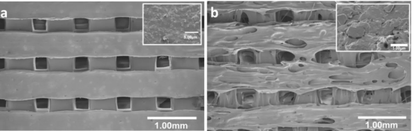
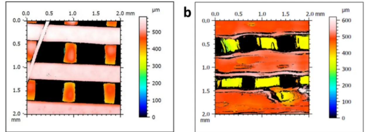
The SEM micrographs shown inFig. 1illustrate the porous mor-phology of scaffolds obtained with PLA alone (Fig. 1a) or with 0.2% GO (Fig. 1b). The pore size (405 ± 20μm and 485 ± 30 μm, respec-tively) and the wall width (380 ± 65μm and 360 ± 130 μm, re-spectively) were comparable in the PLA scaffolds and in the PLA scaf-folds with 0.2% of GO. Similar results were obtained with 0.1% and 0.3% GO (Table 1), showing that GO addition in the PLA matrix had a very moderate effect on pore morphology during the 3D printing step. Analysis of the 3D topography images (Fig. 2) indicated that the surface of PLA scaffolds with 0.2% GO (Fig. 2b) was rougher compared with the smooth surface of PLA scaffolds (Fig. 2a). In agreement, PLA scaffolds with 0.2% GO displayed higher Sa values (Table 1). This could spectrum of PLA and PLA/GO nanocomposites was recorded with the
NEXUS instrument, equipped with an attenuated total reflection ac-cessory in the frequency range of 600–4000 cm−1. The FTIR spectrum
was scanned at 1 cm−1 resolution, and signals were averaged from 32
scans.
2.5. Thermal properties
The different scaffolds were analysed using a differential scanning calorimeter 2920 (maker), equipped with a RCS90 cooling system (maker). Samples were accurately weighed (≈4 mg) in an aluminium TA pan (maker) and sealed. An empty sealed pan was used as reference. Samples were first cooled to 25 °C and then heated to 210 °C with a heating rate of 10 °C min−1. Then, they were cooled again to 25 °C with
nitrogen as purge gas. The degree of polymer crystallinity was calcu-Hf −
∆ ∞
∆Hcf where ΔH
f is the
be attributed to the presence of fillers at the surface and their good dispersions in the polymer matrix [53]. Only in PLA scaffolds with 0.3%
GO, roughness was slightly decreased compared with PLA scaffolds (Table 1), and this could be due GO agglomeration.
In bone tissue engineering, the surface properties of biomedical devices are very important. For instance, the material must display sufficient hydrophilicity to allow the attachment of cells on the scaffold surface, and their proliferation and differentiation for bone regenera-tion [54–56]. Water contact angles (Fig. S3) were smaller for PLA/GO scaffolds compared with PLA scaffolds (Table 1). Pinto et al. [18] re-ported similar results for PLA/GOfilms with a decrease of the contact angle of about 9° after GO addition. Conversely, Zhang et al. found that GO addition had no effect on the contact angle for PLA/GO electrospun fibers [57]. This suggests that GO presence at the scaffold surface in-creases its hydrophilicity, and that the method used to obtain thefinal material directly influences the surface properties. In our study, pristine PLA had a contact angle of 118°, while PLA with 0.3% of GO had a contact angle of 54°. This reduction might be caused by direct inter-actions of the liquids with partially exposedfillers at the PLA surface. Hydrogen bond interactions between oxygen-containing groups in GO and water can explain this behavior. In conclusion, the presence of GO in the 3D PLA matrix decreased the surface hydrophobic properties and, as a consequence, should improve cell attachment and proliferation at the surface of the material.
3.2. Structural characterization of PLA/GO scaffolds
To confirm GO formation from graphite, XRD analysis of the ob-tained powder showed the characteristic GO peak at 11° that corre-sponded to the (002) plane (Fig. 3a). This evidenced graphite exfolia-tion through the oxidaexfolia-tion process. The PLA-film (red in Fig. 3a) showed four characteristics peaks at 2θ = 15, 17, 19 and 23° that de-scribed the alpha form of PLA [58]. PLA crystalline peaks disappeared afterfilament extrusion (green). This might be due to the quenching (air atmosphere) of PLA melting during 3D printing and the high speed of cooling that inhibited the crystalline structure rearrangement. Moreover, it has been shown that natural PLA has the lowest percentage of crystallinity among PLAfilaments (coloured or not) [59]. Hence, the XRD results suggest that PLA crystalline phase was lost during
extrusion.
The PLA/GO biocomposites before extrusion (PLA/GO-film; blue in
Fig. 3a) displayed two characteristic PLA peaks at 2θ = 17 and 23°.
However, these peaks were broader than in the PLA-film, possibly due to micro-stresses induced by GO addition in the polymer matrix. In-deed, the peak corresponding to GO was not detected. This could be related to the low percentage offillers or the good GO dispersion in the matrix [60]. After extrusion, the same behavior was observed for the PLA/GO scaffold (pink,Fig. 3a), with a broad peak between 10° and 25° that corresponded to the polymer amorphous nature.
3.3. Chemical characterization of the scaffolds
To understand PLA organization and GO interactions with the polymer matrix during the nanocomposite fabrication, FTIR spectra were recorded (Fig. 3b). The characteristic GO functions were the bands at 1040 cm−1 (CeO elongation vibrations) and 1740 cm−1 (C]O elongation vibrations of the carbonyl and carboxylic groups). The peak at 1630 cm−1was probably due to skeletal vibration of the graphite domains. The broad peak around 3000–3500 cm−1was attributed to
hydroxyl groups. No difference was observed between the peaks of pure PLA and PLA/GO scaffolds. Indeed, the greater PLA peak absorbance and in the same range than that of GO did not allow detecting the GO peaks in the PLA/GO biocomposites.
Similarly, the D and G-band signature of pure GO powder was de-tected at 1345 and 1590 cm−1respectively, using Raman spectroscopy (Fig. 3c). The vibration of sp2-bonded carbon atoms in a 2D hexagonal lattice gave the G-band, whereas the vibration of carbon atoms with pendent bonds in the plane of the disordered graphite was associated with the D band. The D band is generally correlated with defects from vacancies, grain boundaries, and amorphous carbon species that lead to sp3-hybridized carbon, hence a differentiated band of sp2-hybridized carbon. For the PLA/GOfilm, two peaks were observed at 1345 and 1590 cm−1that appeared as widened bands of weak intensities and corresponded to the presence of GO in the PLA. The Raman spectra of PLA/GO scaffolds were not significantly different from those of PLA scaffolds.
Fig. 1. Scanning electron micrographs showing the architecture of FDM 3D printed (a) PLA and (b) PLA/GO (0.2%) scaffolds. Insets show a magnification of the surface.
Table 1
Pore size, wall width, roughness (i.e., Sa, the arithmetic average height of the surface), and contact angles of ultrapure water for PLA and PLA/GO scaffolds.
Samples Pore size (μm) Wall width (μm) Sa (μm) Contact angles (°) PLA 405 ± 20 380 ± 65 5.8 ± 1.5 118 ± 2 PLA/GO (0.1%) 450 ± 25 350 ± 100 7.5 ± 0.6 70 ± 2 PLA/GO (0.2%) 485 ± 30 360 ± 130 9.9 ± 4.0 69 ± 1 PLA/GO (0.3%) 455 ± 24 400 ± 130 5.5 ± 0.9 54 ± 2
Fig. 2. Scaffold topography. Representative 3D images of the surface roughness of (a) PLA and (b) PLA/GO (0.2%) scaffolds.
Fig. 3. Chemical and structural properties of the nanocomposites. (a) XRD diffractograms, (b) FTIR spectra, and (c) Raman spectroscopy data of the synthesized GO and PLA/GO nanocomposites (films or 3D printed scaffolds, as indicated).
3.4. Thermal analysis of the PLA/GO scaffolds
The influence of GO on PLA thermal stability was monitored by TGA (Fig. 4a). The 1% weight loss for PLA observed below 200 °C was due to the adsorbed water. The second major weight loss observed between 300 and 400 °C was caused by the degradation of the PLA polymer. As shown in the derivative weight curves (Fig. 4b), the maximum de-gradation temperature peak shifted from 298 °C to 366 °C for the na-nocomposites with 0.3% GO. This shift could be explained by the in-terfacial interactions between GO and PLA through hydrogen bonds and/or van der Waals forces, as previously reported [61,62]. The strong interactions with GO led to the improvement of the biocomposite thermal stability, possibly due to a diminution of the chain mobility at the interface with GO [63]. The last weight loss from 400 °C was due to the thermochemical decomposition of the remaining organic content from PLA and from GO due to pyrolysis of labile oxygenated groups when PLA is reinforced with GO [64].
The effects of GO addition on PLA crystallinity and on Tg, Tcc and Tm were evaluated by DSC analyses (Fig. 4c). Enthalpy of fusion, glass transition and melting point were measured and are summarized in
Table 2, together with the calculated crystallinity. For PLA, Tg were 59 °C and 61 °C and Tm were 168 °C and 169 °C before and after ex-trusion, respectively. This indicated that the extrusion process did not affect PLA thermal properties.
After extrusion, PLA showed a cold crystallization peak at 100 °C. Conversely, the cold crystallization peak was suppressed before extru-sion, as reported in a previous study on a PLAfilm [65]. In line with these observations, PLA crystallinity level was lower after than before extrusion, and this change could have been caused by the extrusion process [66]. These results confirmed the XRD observations on the
polymer crystallinity changes during extrusion.
For PLA reinforced with 0.1% GO, the glass transition at 55 °C was followed by an exothermic cold crystallization peak at 112 °C. When the GO content was increased to 0.3%, the Tg at 56 °C was followed by a double peak of cold crystallization at 83 °C. These results are surprising because a previous study reported that the Tg of graphene and GO/ polymer nanocomposites is significantly increased (4 °C or more) when using functionalized nanofillers, due to more interactions with the matrix [67]. Moreover, GO addition should increase the cold crystal-lization temperature because in the presence of enough GO content in the polymer matrix, the motion of PLA chains is confined, the cold crystallization process of PLA is restricted, and consequently the crys-tallization temperature increases [68].
The melting peak was not changed by GO addition to the PLA ma-trix, but a double melting peak appeared at 166 °C when GO loading level was increased to 0.3 wt%. The second melting peak was higher,
Fig. 4. Thermal properties of nanocomposite materials. (a) TGA and (b) derivative TGA curves of 3D printed PLA nanocomposites with different GO percentages. (c) Representative DSC graphs showing Tg (glass transition temperature), Tm (meting temperature) and Tcc (cold temperature crystallization) of PLA and PLA/GO nanocomposites before and after 3D printing.
Table 2
Temperatures, enthalpies of different thermal transitions, and crystallinity of PLA and PLA/GO materials.
Samples Tg (°C) Tm(°C) ΔHf (J/g) ΔHcf (J/g) χ (%) PLA 61 169 30 10 21 PLA-film 59 168 30 – 32 PLA/GO (0.1%) 55 156 27 27 0 PLA/GO (0.2%) 57 171 43 19 26 PLA/GO (0.3%) 56 166 40 24 16 PLA/GO (0.3%)-film 55 169 42 – 45
Tg, glass transition temperature; Tm, melting temperature;ΔHf, enthalpy of
indicating that more crystalline forms were generated with higher GO loading levels [69]. Finally, thermal analysis by TGA and DSC showed that GO presence in the polymer matrix did not affect the filament extrusion. The 3D printing conditions also were not influenced by GO addition. However, GO enhanced the thermal stability of the nano-composite scaffolds.
3.5. Mechanical properties of the 3D printed nanocomposites
Composites used for biomedical implants should withstand high tensile loads. The characterization of the mechanical properties of PLA and PLA/GO nanocomposites (Fig. 5) focused on the region where samples responded elastically to traction. In this region, the elastic modulus was measured as a function of the GO percentage. The Young's modulus for PLA was in good agreement with the literature [70] (i.e., about 2 GPa for a sample with 30% porosity). This value significantly increased to 2.6 GPa after addition of 0.3% GO (improvement of about 30% of the elastic modulus) for a scaffold with 30% porosity (Fig. 5a). Then, samples were loaded until breaking. When GO density was high enough, GO incorporation increased the tensile strength from 34 MPa for pure PLA to 39 MPa for samples with 0.3% GO. Conversely, in samples with lower GO density, tensile strength at break was lower (Fig. 5b). This is due to the fact the GO inducedflaws at the very local scale that made the material weaker. With higher GO densities, this phenomenon is counterbalanced by the fact that GO is intrinsically stronger than PLA, which makes the material stronger. The Poisson's ratio was 0.3 for PLA, which is characteristic for this polymer, and was not changed by GO addition (Fig. 5c). The improved stiffness of the PLA/GO scaffold compared with the PLA scaffold highlighted the
reinforcement of the scaffold by GO addition.
Here, only the influence of GO addition on the polymer mechanical properties was investigated. However, the architecture and percentage of filling inside the scaffold also have an effect on the mechanical properties of the material, particularly on the tensile strength, but these features were beyond the goal of this study.
Several studies have shown the improvement of mechanical prop-erties and biological activity of PLA scaffolds generated by FDM [71,72]. Different fillers were used in compression [73] andflexural studies [74], but to the best of our knowledge, the mechanical prop-erties of PLA-based nanocomposites were never investigated by tensile strength analysis. Overall, ourfindings indicate that GO is a promising filler for improving the mechanical properties of biopolymers made by FDM.
3.6. Biological studies
The scaffold biocompatibility was then investigated using MG-63 cells that were derived from a human bone osteosarcoma and exhibit osteogenic potential (Fig. 6a). Compared with cells grown without scaffolds (control), the statistical analysis showed a higher viability of the cells in the presence of PLA and PLA/GO independently of GO percentages. No significant differences (ns in the figure) were observed between the samples with 0.1 and 0.2% meaning that the addition of GO had no influence on the viability of the cells. These results suggest that GO incorporation as nanofiller for PLA is biocompatible.
MC3T3-E1 cells were used to confirm the biocompatibility of the scaffolds (Fig. 6b). After 4 days of culture, no significant differences were observed between control and the scaffolds. After 7 days of
Fig. 5. Mechanical properties of the scaffolds. (a) Young's modulus, (b) Tensile stress at break, and (c) Poisson's ratio values of the different PLA/GO scaffolds compared with PLA scaffolds.
Fig. 6. (a) MG-63 and (b) MC3T3-E1 cell viability when cultured in the presence of PLA and PLA/GO nanocomposite scaffolds was assessed with the MTT assay at day 4 and 7 after seeding (ns = not significant).
Fig. 7. MG-63 cell proliferation and attachment on PLA and PLA/GO nanocomposite scaffolds. (a) Analysis by DAPI staining of DNA (blue) of MG-63 cell pro-liferation on the PLA and PLA/GO scaffolds. (b) Cell proliferation monitoring using the MTT assay at different time points after seeding (ns = not significant, *p < 0.05, **p < 0.005). (c) Actin immunodetection (red) and DAPI staining of DNA (blue) in a selected area of MG-63 cells attached on the PLA and PLA/GO scaffolds. (For interpretation of the references to colour in this figure legend, the reader is referred to the web version of this article.)
culture, higher viability was observed with addition of 0.2 and 0.3% GO. This suggests that the PLA/GO scaffolds enhanced the viability of these cells.
In some cases, biomaterials implantation in bone tissue may cause an inflammatory response due to the acidic microenvironment induced by the scaffold [75]. However, PLA is a stable polymer with a slow degradation rate which depends on polymer composition and on the tissue considered [76]. In this case PLA is relatively stable with < 1% degradation after 25 days (Fig. S4). The introduction of GO will im-prove the degradation of PLA (Fig. S4). PLA scaffold does not induce high enough acidity in the microenvironment to have a major negative effect on cell behavior. The cells viability study confirmed that PLA does not induce any cytotoxicity.
Similarly, MG-63 cell proliferation on the two scaffolds was mon-itoredfirst by staining cell nuclei with Hoechst 33342. This showed that cells readily proliferate on the scaffolds at 24 h, and reached confluency after 7 days of culture on the PLA and PLA/GO scaffolds (Fig. 7a). Cell proliferation quantification with the MTT assay at day 4 and 7 (Fig. 7b) confirmed these results and showed that GO incorporation promoted cell proliferation compared with scaffolds made only of PLA. Finally, cell attachment to the scaffolds was also analysed by staining cell nuclei with Hoechst 33342 and the cytoskeleton with an anti-actin antibody (Fig. 7c) which demonstrated that cells attached well on the PLA and PLA/GO scaffolds.
Finally, MG-63 cell mineralization was monitored by Alizarin Red-S staining at day 1, 14 and 21 after induction of differentiation. Colorimetric quantification of calcium deposition on the scaffolds by MG-63 cells (Fig. 8) showed that at day 21, it was increased by two-fold in samples with PLA/GO scaffolds compared with PLA scaffolds. This indicates that GO incorporation in the scaffold promotes mineraliza-tion. Altogether, these results demonstrate that PLA reinforced with 0.2% GO is biocompatible and may improve MG-63 cell mineralization activity.
4. Conclusions
In conclusion, GO-reinforced PLA scaffolds were synthesized and thoroughly characterized, particularly their structural and mechanical properties and their biocompatibility. We successfully 3D printed PLA/ GO nanocomposites with controlled morphology and a network of
interconnected pores around 300μm. Altogether, our results demon-strated that GO incorporation 1) increased the surface roughness and hydrophilicity, 2) did not modify the transition temperature, 3) de-creased polymer crystallinity, 4) improved the mechanical properties of the scaffold, and 5) promoted bone cell attachment, proliferation and differentiation. Our data clearly indicate that PLA reinforcement with 0.2% GO might represent a good strategy to obtain 3D printed scaffolds with very attractive mechanical properties and bioactivity, thus pro-viding a promising material that could be used for bone tissue en-gineering applications upon validation using appropriate in vivo models.
Author statement
Conceptualization, C. T., D. C., V. C., M. B.; methodology, H. B., S. N., C. T., D. C., V. C., M. B.; formal analysis, H. B., S. N., C. T., C; B., J. B.; data curation, H. B., S. N., C. T., C. B., J. B., S. B.; writing—original draft preparation, H. B., S. N.; writing—review and editing, H. B., S. N., C. T., J. B., S. B., H. G., V. H., D. C., V. C., M. B.; supervision, C. T., D. C., V. C., M. B.; project administration, V. C., M. B.; funding acquisition, V. C., M. B.
Declaration of competing interest
The authors declare that they do not have any conflict of interest. Acknowledgment
We acknowledge thefinancial support from Indo-French Centre for the Promotion of Advanced Research-Cefipra (Project 5608-1), from CNRS (Project “Osez l'Interdisciplinarité: TraitCancer”) and from University of Montpellier (MUSE“3DTraitCancer”).
Appendix A. Supplementary data
Supplementary data to this article can be found online athttps:// doi.org/10.1016/j.msec.2019.110595.
References
[1] D. Tang, et al., Biofabrication of bone tissue: approaches, challenges and translation for bone regeneration, Biomaterials 83 (2016) 363–382.
[2] H.T. Aro, A.J. Aho, Clinical use of bone allografts, Ann. Med. 25 (1993) 403–412. [3] K. Rezwan, Q.Z. Chen, J.J. Blaker, A.R. Boccaccini, Biodegradable and bioactive
porous polymer/inorganic composite scaffolds for bone tissue engineering, Biomaterials 27 (2006) 3413–3431.
[4] S. Bose, M. Roy, A. Bandyopadhyay, Recent advances in bone tissue engineering scaffolds, Trends Biotechnol. 30 (2012) 546–554.
[5] F.J. O’Brien, Biomaterials & scaffolds for tissue engineering, Mater. Today 14 (2011) 88–95.
[6] A.J.R. Lasprilla, G.A.R. Martinez, B.H. Lunelli, A.L. Jardini, R. Maciel Filho, Poly-lactic acid synthesis for application in biomedical devices—a review, Biotechnol. Adv. 30 (2012) 321–328.
[7] J. Muller, C. González-Martínez, A. Chiralt, Combination of poly(lactic) acid and starch for biodegradable food packaging, Materials (Basel) 10 (1–22) (2017). [8] D.E. Cutright, E.E. Hunsuck, Tissue reaction to the biodegradable polylactic acid
suture, Oral Surgery, Oral Med. Oral Pathol. 31 (1971) 134–139.
[9] R.R.M. Bos, et al., Degradation of and tissue reaction to biodegradable poly (L-lactide) for use as internalfixation of fractures: a study in rats, Biomaterials 12 (1991) 32–36.
[10] K. Madhavan Nampoothiri, N.R. Nair, R.P. John, An overview of the recent de-velopments in polylactide (PLA) research, Bioresour. Technol. 101 (2010) 8493–8501.
[11] V. Karageorgiou, D. Kaplan, Porosity of 3D biomaterial scaffolds and osteogenesis, Biomaterials 26 (2005) 5474–5491.
[12] B.Q. Chen, et al., Investigation of silkfibroin nanoparticle-decorated poly(L-lactic acid) composite scaffolds for osteoblast growth and differentiation, Int. J. Nanomedicine 12 (2017) 1877–1890.
[13] H. Kim, A.A. Abdala, C.W. Macosko, Polymer nanocomposites with graphene, Young, 2010, pp. 1–13, ,https://doi.org/10.1021/ma100572e.
[14] A.K. Geim, Graphene: Status and Prospects. 1530 (2010) 1530–1535. [15] S. Stankovich, et al., Graphene-based composite materials, Nature 442 (2006)
282–286.
Fig. 8. MG-63 cell differentiation on PLA and PLA/GO scaffolds was evaluated using the Alizarin Red-S mineralization assay at different time points after switching to differentiation medium (ns = not significant, *p < 0.05). (For interpretation of the references to colour in thisfigure legend, the reader is referred to the web version of this article.)
[16] Y. Zhu, et al., Graphene and graphene oxide: synthesis, properties, and applications, Adv. Mater. 22 (2010) 3906–3924.
[17] W. Gao, The chemistry of graphene oxide, Graphene Oxide Reduct. Recipes, Spectrosc. Appl. (2015) 61–95,https://doi.org/10.1007/978-3-319-15500-5_3. [18] A.M. Pinto, et al., Biocompatibility of poly (lactic acid) with incorporated
graphene-based materials, Colloids Surfaces B Biointerfaces 104 (2013) 229–238. [19] A.M. Pinto, J. Cabral, D.A.P. Tanaka, A.M. Mendes, F.D. Magalhães, Effect of
in-corporation of graphene oxide and graphene nanoplatelets on mechanical and gas permeability properties of poly (lactic acid)films, Polym. Int. 62 (2013) 33–40. [20] Q. Chen, et al., 3D printing biocompatible polyurethane/poly(lactic acid)/graphene
oxide nanocomposites: anisotropic properties, ACS Appl. Mater. Interfaces 9 (2017) 4015–4023.
[21] D.G. Papageorgiou, I.A. Kinloch, R.J. Young, Mechanical properties of graphene and graphene-based nanocomposites, Prog. Mater. Sci. 90 (2017) 75–127. [22] S.-R. Ryoo, et al., Biomedical applications of graphene and graphene oxide, Acc.
Chem. Res. 46 (2013) 2211–2224.
[23] S.S. Nanda, G.C. Papaefthymiou, D.K. Yi, Functionalization of graphene oxide and its biomedical applications, Crit. Rev. Solid State Mater. Sci. 40 (2015) 291–315. [24] R.K. Kankala, et al., Cardiac tissue engineering on the nanoscale, ACS Biomaterials
Science and Engineering 4 (2018) 800–818.
[25] E. Kolanthai, et al., Graphene oxide-a tool for the preparation of chemically crosslinking free alginate-chitosan-collagen scaffolds for bone tissue engineering, ACS Appl. Mater. Interfaces 10 (2018) 12441–12452.
[26] C. Liao, et al., Fabrication of porous biodegradable polymer scaffolds using a sol-vent merging/particulate leaching method, J. Biomed. Mater. Res. An Off. J. Soc. Biomater. Japanese Soc. Biomater. Aust. Soc. Biomater. Korean Soc. Biomater. 59 (2002) 676–681.
[27] N. Sultana, M. Wang, PHBV/PLLA-based composite scaffolds fabricated using an emulsion freezing/freeze-drying technique for bone tissue engineering: surface modification and in vitro biological evaluation, Biofabrication 4 (2012) 15003. [28] Y.X. Huang, J. Ren, C. Chen, T.B. Ren, X.Y. Zhou, Preparation and properties of poly
(lactide-co-glycolide)(PLGA)/nano-hydroxyapatite (NHA) scaffolds by thermally induced phase separation and rabbit MSCs culture on scaffolds, J. Biomater. Appl. 22 (2008) 409–432.
[29] S. Nagarajan, et al., Design of boron nitride/gelatin electrospun nanofibers for bone tissue engineering, ACS Appl. Mater. Interfaces 9 (2017) 33695–33706. [30] F.S. Senatov, et al., Mechanical properties and shape memory effect of 3D-printed
PLA-based porous scaffolds, J. Mech. Behav. Biomed. Mater. 57 (2016) 139–148. [31] S. Gómez, M.D. Vlad, J. López, E. Fernández, Design and properties of 3D scaffolds
for bone tissue engineering, Acta Biomater. 42 (2016) 341–350.
[32] A. Rogina, et al., Macroporous poly(lactic acid) construct supporting the os-teoinductive porous chitosan-based hydrogel for bone tissue engineering, Polymer 98 (2016) 172–181.
[33] D.H. Rosenzweig, E. Carelli, T. Steffen, P. Jarzem, L. Haglund, 3D-printed ABS and PLA scaffolds for cartilage and nucleus pulposustissue regeneration, Int. J. Mol. Sci. 16 (2015) 15118–15135.
[34] R.K. Kankala, et al., Effect of Icariin on engineered 3D-printed porous scaffolds for cartilage repair, Materials (Basel) 11 (2018).
[35] R.K. Kankala, X.M. Xu, C.G. Liu, A.Z. Chen, S.B. Wang, 3D-printing of microfibrous porous scaffolds based on hybrid approaches for bone tissue engineering, Polymers (Basel) 10 (2018).
[36] S. Bose, S. Vahabzadeh, A. Bandyopadhyay, Bone tissue engineering using 3D printing, Mater. Today 16 (2013) 496–504.
[37] F.P.W. Melchels, J. Feijen, D.W. Grijpma, A review on stereolithography and its applications in biomedical engineering, Biomaterials 31 (2010) 6121–6130. [38] R. Gauvin, et al., Microfabrication of complex porous tissue engineering scaffolds
using 3D projection stereolithography, Biomaterials 33 (2012) 3824–3834. [39] Y. Luo, A. Lode, A.R. Akkineni, M. Gelinsky, Concentrated gelatin/alginate
com-posites for fabrication of predesigned scaffolds with a favorable cell response by 3D plotting, RSC Adv. 5 (2015) 43480–43488.
[40] J.M. Williams, et al., Bone tissue engineering using polycaprolactone scaffolds fabricated via selective laser sintering, Biomaterials 26 (2005) 4817–4827. [41] S.V. Murphy, A. Atala, 3D bioprinting of tissues and organs, Nat. Biotechnol. 32
(2014) 773.
[42] I. Zein, D.W. Hutmacher, K.C. Tan, S.H. Teoh, Fused deposition modeling of novel scaffold architectures for tissue engineering applications, Biomaterials 23 (2002) 1169–1185.
[43] B.N. Turner, R. Strong, S.A. Gold, A review of melt extrusion additive manu-facturing processes: I. Process design and modeling, Rapid Prototyp. J. 20 (2014) 192–204.
[44] F. Ning, W. Cong, J. Qiu, J. Wei, S. Wang, Additive manufacturing of carbonfiber reinforced thermoplastic composites using fused deposition modeling, Compos. Part B Eng. 80 (2015) 369–378.
[45] P.S.P. Poh, et al., Polylactides in additive biomanufacturing, Adv. Drug Deliv. Rev. 107 (2016) 228–246.
[46] T. Serra, J.A. Planell, M. Navarro, High-resolution PLA-based composite scaffolds
via 3-D printing technology, Acta Biomater. 9 (2013) 5521–5530.
[47] T. Serra, M.A. Mateos-Timoneda, J.A. Planell, M. Navarro, 3D printed PLA-based scaffolds: a versatile tool in regenerative medicine, Organogenesis 9 (2013) 239–244.
[48] D.C. Marcano, et al., Improved synthesis of graphene oxide, ACS Nano 4 (2010) 4806–4814.
[49] E.W. Fischer, H.J. Sterzel, G. Wegner, Investigation of the structure of solution grown crystals of lactide copolymers by means of chemical reactions, Kolloid-Zeitschrift Kolloid-Zeitschrift Für Polym 251 (1973) 980–990.
[50] T.-L. Vu, J. Barés, Soft Grain Compression: Beyond the Jamming Point, (2019). [51] T.L. Vu, J. Barés, S. Mora, S. Nezamabadi, Deformationfield in diametrically loaded
soft cylinders, Exp. Mech. (2019),https://doi.org/10.1007/s11340-019-00477-4. [52] S.H. Lee, I.Y. Kim, W.S. Song, Biodegradation of polylactic acid (PLA)fibers using
different enzymes, Macromol. Res. 22 (2014) 657–663.
[53] K. Deshmukh, S.M. Khatake, G.M. Joshi, Surface properties of graphene oxide re-inforced polyvinyl chloride nanocomposites, J. Polym. Res. 20 (2013). [54] Y. Arima, H. Iwata, Effect of wettability and surface functional groups on protein
adsorption and cell adhesion using well-defined mixed self-assembled monolayers, Biomaterials 28 (2007) 3074–3082.
[55] D.P. Dowling, I.S. Miller, M. Ardhaoui, W.M. Gallagher, Effect of surface wettability and topography on the adhesion of osteosarcoma cells on plasma-modified poly-styrene, J. Biomater. Appl. 26 (2011) 327–347.
[56] K. Webb, V. Hlady, P.A. Tresco, Relative importance of surface wettability and charged functional groups on NIH 3T3fibroblast attachment, spreading, and cy-toskeletal organization, J. Biomed. Mater. Res. 41 (1998) 422–430.
[57] C. Zhang, et al., The surface grafting of graphene oxide with poly (ethylene glycol) as a reinforcement for poly (lactic acid) nanocomposite scaffolds for potential tissue engineering applications, J. Mech. Behav. Biomed. Mater. 53 (2016) 403–413. [58] J.M. Campos, A.M. Ferraria, A.M. Botelho Do Rego, M.R. Ribeiro, A.
Barros-Timmons, Studies on PLA grafting onto graphene oxide and its effect on the ensuing compositefilms, Mater. Chem. Phys. 166 (2015) 122–132.
[59] B. Wittbrodt, J.M. Pearce, The effects of PLA color on material properties of 3-D printed components, Addit. Manuf. 8 (2015) 110–116.
[60] M. Gong, Q. Zhao, L. Dai, Y. Li, T. Jiang, Fabrication of polylactic acid/hydro-xyapatite/graphene oxide composite and their thermal stability, hydrophobic and mechanical properties, Journal of Asian Ceramic Societies 5 (2017) 160–168. [61] S. Villar-Rodil, J.I. Paredes, A. Martínez-Alonso, J.M.D. Tascón, Preparation of
graphene dispersions and graphene-polymer composites in organic media, J. Mater. Chem. 19 (2009) 3591–3593.
[62] T. Ramanathan, et al., Functionalized graphene sheets for polymer nanocomposites, Nat. Nanotechnol. 3 (2008) 327–331.
[63] Y. Xu, W. Hong, H. Bai, C. Li, G. Shi, Strong and ductile poly(vinyl alcohol)/gra-phene oxide compositefilms with a layered structure, Carbon N. Y. 47 (2009) 3538–3543.
[64] Y. Shen, et al., Chemical and thermal reduction of graphene oxide and its elec-trically conductive polylactic acid nanocomposites, Compos. Sci. Technol. 72 (2012) 1430–1435.
[65] M.S. Huda, M. Yasui, N. Mohri, T. Fujimura, Y. Kimura, Dynamic mechanical properties of solution-cast poly(L-lactide)films, Mater. Sci. Eng. A 333 (2002) 98–105.
[66] P. Ravi, P.S. Shiakolas, T.R. Welch, Poly-L-lactic acid: pellets tofiber to fused fi-lament fabricated scaffolds, and scaffold weight loss study, Addit. Manuf. 16 (2017) 167–176.
[67] K.H. Liao, S. Aoyama, A.A. Abdala, C. Macosko, Does graphene change T g of na-nocomposites? Macromolecules 47 (2014) 8311–8319.
[68] H.-D. Huang, et al., Improved barrier properties of poly (lactic acid) with randomly dispersed graphene oxide nanosheets, J. Memb. Sci. 464 (2014) 110–118. [69] Z. Xu, et al., Morphology, rheology and crystallization behavior of polylactide
composites prepared through addition offive-armed star polylactide grafted mul-tiwalled carbon nanotubes, Polymer (Guildf) 51 (2010) 730–737.
[70] Y. Song, et al., Measurements of the mechanical response of unidirectional 3D-printed PLA, Mater. Des. 123 (2017) 154–164.
[71] A. Grémare, et al., Characterization of printed PLA scaffolds for bone tissue en-gineering, J. Biomed. Mater. Res. - Part A 106 (2018) 887–894.
[72] A. Gregor, et al., Designing of PLA scaffolds for bone tissue replacement fabricated by ordinary commercial 3D printer, J. Biol. Eng. 11 (2017) 1–21.
[73] C. Esposito Corcione, et al., Highly loaded hydroxyapatite microsphere/PLA porous scaffolds obtained by fused deposition modelling, Ceram. Int. 45 (2019) 2803–2810.
[74] C.E. Corcione, et al., 3D printing of hydroxyapatite polymer-based composites for bone tissue engineering, J. Polym. Eng. 37 (2017) 741–746.
[75] R.K. Kankala, et al., Highly porous microcarriers for minimally invasive in situ skeletal muscle cell delivery, Small 15 (2019).
[76] D. da Silva, et al., Biocompatibility, biodegradation and excretion of polylactic acid (PLA) in medical implants and theranostic systems, Chem. Eng. J. 340 (2018) 9–14.