I
Université de Montréal
Longitudinal assessment of neural activity in Parkinson’s
disease with mild cognitive impairment using task based fMRI
Par
Mohamed Salah A.salam Al-azzawi
Université de Montréal
Faculté de médecine
Thèse présentée à la Faculté des études supérieures en vue de l’obtention du grade de
maîtrise en sciences neurologiques
Ao
ût 2014
Université de Montréal Faculté des études supérieures
Ce mémoire intitulé:
Longitudinal assessment of neural activities in Parkinson’s disease mild cognitive impairment using
task based fMRI
Présenté par:
Mohamed Salah A.salam Al-azzawi
a été évaluée par un jury composé des personnes suivantes :
Dr.Christian Duval (Président rapporteur) Dr Oury Monchi (Directeur de recherche) Dr Richard Camicioli (Membre du jury)
Résumé
La maladie de Parkinson (PD) a été uniquement considérée pour ses endommagements sur les circuits moteurs dans le cerveau. Il est maintenant considéré comme un trouble multisystèmique, avec aspects multiples non moteurs y compris les dommages intérêts pour les circuits cognitifs. La présence d’un trouble léger de la cognition (TCL) de PD a été liée avec des changements structurels de la matière grise, matière blanche ainsi que des changements fonctionnels du cerveau. En particulier, une activité significativement réduite a été observée dans la boucle corticostriatale ‘cognitive’ chez des patients atteints de PD-TCL vs. PD non-TCL en utilisant IRMf. On sait peu de cours de ces modèles fonctionnels au fil du temps. Dans cette étude, nous présentons un suivi longitudinal de 24 patients de PD non démente qui a subi une enquête neuropsychologique, et ont été séparés en deux groupes - avec et sans TCL (TCL n = 11, non-TCL n = 13) en fonction du niveau 2 des recommandations de la Movement Disrders Society pour le diagnostic de PD-TCL. Ensuite, chaque participant a subi une IRMf en effectuant la tâche de Wisconsin pendant deux sessions, 19 mois d'intervalle. Nos résultats longitudinaux montrent qu'au cours de la planification de période de la tâche, les patients PD non-TCL engageant les ressources normales du cortex mais ils ont activé en plus les zones corticales qui sont liés à la prise de décision tel que cortex médial préfrontal (PFC), lobe pariétal et le PFC supérieure, tandis que les PD-TCL ont échoué pour engager ces zones en temps 2. Le striatum n'était pas engagé pour les deux groupes en temps 1 et pour le groupe TCL en temps 2. En outre, les structures médiales du lobe temporal étaient au fil du temps sous recrutés pour TCL et Non-TCL et étaient positivement corrélés avec les scores de MoCA. Le cortex pariétal, PFC antérieur, PFC supérieure et putamen postérieur étaient négativement corrélés avec les scores de MoCA en fil du temps. Ces résultats révèlent une altération fonctionnelle pour l’axe ganglial-thalamo-corticale au début de PD, ainsi que des niveaux différents de participation corticale pendant
une déficience cognitive. Cette différence de recrutement corticale des ressources pourrait refléter longitudinalement des circuits déficients distincts de trouble cognitive légère dans PD.
Abstract
PD was traditionally thought of as purely a movement disorder, now it is considered a multisystem disorder, with multiple non-motor aspects including damages to the cognitive circuits. Mild cognitive impairment (MCI) in PD has been linked with structural gray matter, white matter as well as functional brain changes. Specifically, significantly reduced activity was observed in the ‘cognitive’ corticostriatal loop in patients with PD-MCI vs. PD non-MCI using fMRI. Little is known regarding the course of these functional patterns over time. In this study we present longitudinal follow up of 24 non-demented PD who underwent neuropsychological investigation and were separated in two groups - with and without MCI (MCI n=11, Non-MCI n=13) according to the MDS level 2 recommendation for diagnosis of PD-MCI. Afterwards, each participant underwent an fMRI investigation by performing the Wisconsin Card Sorting Task over two sessions, 19 months apart. Our longitudinal results show that during planning set-shift period of the task, PD Non-MCI patients were engaging the normal cortical resources but they also activated more cortical areas at time 2 that are related to decision-making such as the medial prefrontal cortex (PFC), parietal lobe and the superior PFC, whilst the patients with MCI failed to engage these areas at both time points. The striatum was not engaged for both groups at time 2 and for MCI group at time 1. Furthermore, medial temporal lobe structures (MTLS) were under-recruited overtime for both the MCI and Non-MCI PD patients, and were positively correlated with MoCA scores over time. Parietal cortex, anterior PFC, superior PFC, and posterior putamen were negatively correlated with MoCA scores. These results reveal functional alteration along the basal ganglial-thalamo-cortical axis in early PD, as well as different cortical involvement levels along the course cognitive impairment. This discrepancy in cortical resources recruitment over time might reflect deficient circuitry distinct to cognitive impairment in Parkinson’s disease.
Contents Résumé III Abstract V List of tables IX List of figures X Abbreviations XI
List of annexes XIII
Acknowledgement XV
CHAPTER 1 1
Introduction 1
Parkinson’s disease historical background 2
Etiology 3
Environmental factors 4
Genetic factors 5
Epidemiology 6
Neuropathology and Anatomy 7
Clinical features 9
Motor signs and symptoms 9
Non-motor signs and symptoms 10
Diagnosis 12
Treatment 13
Cognitive dysfunction 15
Dementia 15
Biomarkers of PD-MCI 18
Cerebrospinal fluid 18
Genetic 19
Neurophysiology 19
Neuroimaging 20
Structural Magnetic Resonance Imaging 21
Single photon emission computed tomography and photon emission topography 24
Diffusion Tensor Imaging 26
Functional Magnetic Resonance Imaging 26
Task-based fMRI 27
Memory Task 27
Executive Task 29
CHAPTER 2 31
ARTICLE 31
Evolution patterns of neural activity linked to cognitive process in Parkinson’s disease,
a longitudinal fMRI study 31
Introduction 34
Material and Methods 35
Subjects 35
Neuropsychological assessment 38
Cognitive task during fMRI 39
fMRI scanning 40
MRI data analysis 41
Contrast analysis 41
Results 43 Discussions 47 References 51 Figures 57 Tables 60 CHAPTER 3 69 Discussion 70 CHAPTER 4 78
Conclusion and future directions 78
Bibliography 82
List of tables
-Table 1: Neuropsychological test battery according to cognitive domain 38
-Table 2 A: Parkinson’s disease patient demographics according to cognitive impairment group at
both time points for PD non-MCI group. 61
-Table 2 B: Parkinson’s disease patient demographics according to cognitive impairment group at
both time points for PD MCI group. 61
-Table 3: significant activations in the contrast receiving negative feedback minus control feedback (planning the set-shift) in both PD MCI and PD non-MCI patient groups 62
-Table 4: significant activations in the contrast of matching after negative feedback minus control matching (Executing the set-shift) in both PD MCI and PD non-MCI patient groups. 65
-Table 5: activation peaks with correlation analysis with individual MoCA scores
List of figures
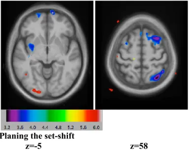
-Figure 1: significant activations in the contrast receiving negative feedback minus control feedback
(planning the set-shift) Time 1 vs. Time 2 59
-Figure 2: significant activations in the contrast of matching after negative feedback minus control
matching (Executing the set-shift) Time 1 vs. Time 2 59
-Figure 3: significant activation peaks with correlation analysis with individual MoCA scores across
List of abbreviations AD – Alzheimer’s Disease
ANOVA – Analysis of Variance
CSF – Cerebrospinal Fluid
DLPFC – Dorsolateral Prefrontal Cortex
DTI – Diffusion Tensor Imaging
H&Y – Hoehn and Yahr
MCI – Mild Cognitive Impairment
MMSE – Mini-Mental State Examination
MNI – Montreal Neurological Institute
MoCA – Montreal Cognitive Assessment
MRI – Magnetic Resonance Imaging
fMRI – Functional Magnetic Resonance Imaging
MTLS – Medial Temporal Lobe Structures
PD – Parkinson’s Disease
PFC – Prefrontal Cortex
SMA – Supplementary Motor Area
SPECT – Single Photon Emission Computed Tomography
UPDRS – Unified Parkinson’s Disease Rating Scale
VBM – Voxel Based Morphometry
List of annexes
-Annex—I: Informed Consent form for patients with Parkinson’s disease participating in a research
project 101
-Annex—II: Preliminary screening for magnetic resonance imaging study (MRI) 108
-Annex—III: Areas of Brodmann 110
XIV
« The dream was marvelous but the horror was great; we must treasure the dream whatever the horror; for the dream has shown that misery comes at last to the healthy man, the end of his life is sorrow. » Enkidu‘s death, Tablet VII « O Mighty King, remember now that only Gods stay in eternal watch. Humans come then go, that is the way fate decreed on the Tablets of Destiny. So someday you will depart, but till that distant day Sing, and dance. Eat your fill of warm cooked food and cool jugs of beer. Cherish the children your love gave life. Bathe away life's dirt in warm drawn waters. Pass the time in joy with your chosen wife. On the Tablets of Destiny it is decreed For you to enjoy short pleasures for your short days. « Siduri to Gilgamesh, Tablet X
-The Epic of Gilgamesh. Mesopotamia, Iraq. Circa 1800 B.C.
Acknowledgment
Completion of the work described in this thesis was possible due to the contribution of several people. I would like to express my gratitude to all of them. First and foremost, I would like to thank my research director, Professor Oury Monchi, for allowing me to be a member amazing team of PCAN (Parkinson’s, Cognition, aging and neuroimaging) and benefiting from his academic input, valuable guidance and cultural knowledge. Professor Monchi gave me the intellectual freedom I needed to write the board and equivalency exams of my medical degree and to proceed with research during the past two years. I will be forever grateful to you Oury, my feet are put and stable in the research field because you opened the door for me.
I would like to thank Dr. Atsuko Nagano-Saito; her help was my primary source of solving technical difficulties of different software and instrumental in obtaining the results. Her words of encouragement, patience and insightful discussions put me through the intractable practical difficulties throughout the two years. She always found time to break through the busy schedule and the piles of articles on her desk to answer even the simplest question I could have. For all this, I am grateful to you Atsuko.
My words will not be enough to thank my friend and colleague Dr. Alexandru Hanganu. His relentless determination and unconditional helpfulness taught me that this curious scientific journey was never to reach its destination without a marvelous companionship of a wise man like him. Alex, you are an achiever, and I owe you a lot for this achievement. I will keep my “todays always better than my yesterdays”.
I am equally thankful to members of the “comité de parrainage”: Dr. Pierre Rainville and Dr.Simona Brambati for their positive inputs during the project presentation in 2013 and 2014. I would like to acknowledge the support and help of the entire CRIUGM members, especially: Clotilde Degroot and France Lainé for their help in scanning the participants, Christophe Bedetti and Jean-Sébastien Provost for solving some fMRIstat puzzles, Carolyn Hurst for her technical support at the unit of fMRI.
I am very much indebted to my family. Dad, your inspiration kept things in perspective for me, working hard and quietly always led to achievement. Mom, your positive spirit, love and faith in me are what make me move mountains. Saif, my back is straight because you hold it. Sura, I know I will not fall as long as I keep looking up to you.
CHAPTER 1
2
1.1. Parkinson’s disease historical background
Ever since first described in 1817 by James Parkinson in his classic publication "an essay on the shaking palsy", Parkinson's disease has been primarily recognized as a motor disorder. At the time, James Parkinson reported six cases of the disease, three of which were not actually examined by him, but rather casually observed in London’s streets. Detailed observations of the patients’ asymmetrical motor symptoms, the insidious onset and the long duration of disease were noted. Parkinson also noted the progression of disease; increase immobility, disturbances of sleep, speech and bodily functions. Though interestingly enough he stated "the senses and the intellects being uninjured".
Nearly 50 years later, Charcot suggested the name "Parkinson's disease," He classified the disorder as "névrose", meaning a neurologic disorder without a known pathologic lesion. Charcot found little benefit from therapies available at the time, including belladonna and ergot products (Goetz 1986). But it was not until 1913, when the patholognomic feature of the disease "Lewy bodies" that the disease was first described. Yet, no progress in dopaminergic treatment was made until the 1960's (Goetz 2000) when Birkmayer and Hornykiewicz’s published their observations of the Levodopa's antikinetic effects on PD patients:
"Bed-ridden patients who were unable to sit up, patients who could not stand up when seated, and patients who when standing could not start walking performed all these activities with ease after L-dopa [levodopa]. They walked around with normal associated movements and they could even run and jump. The voiceless, aphonic speech, blurred by pallilalia and unclear articulation became forceful and clear as in a normal person" (Birkmayer and Hornykiewicz 1961).
3
These discoveries opened the chances for series of subsequent open-label and double-blinded placebo controlled levodopa trials. These trials came only to confirm both short and long-term benefits (Barbeau 1969; Cotzias et al., 1969; Yahr et al., 1969). While cognitive profile of other neurodegenerative diseases such as Alzheimer’s disease were heavily investigated, it was only many years later when researchers have started to look into the cognitive decline in PD and eventually recognize it as an early component of the disease (Foltynie et al 2004).
Now, PD is now regarded as multisystem brain disease in which predisposed neuronal types in specific regions of the peripheral, enteric and central nervous systems become progressively involved by presenting neuronal loss, Lewy bodies and Lewy neuritis (Del Tredici et al., 2002) in the substantia nigra pars impacta which project to the dorsal striatum (McGeer et al., 2004), this neuronal involvement has a widespread and heterogeneous effects on the nervous system resulting in various clinical motor and non-motor expressions (Foltynie et al., 2002). Despite recent advances, much is yet unknown regarding the pathogenesis, characteristics and prognosis of the cognitive impairment in PD.
1.2 Etiology
It is unknown why the loss of nerve cells associated with Parkinson's disease occurs. PD is characterized by the loss of specific subsets of dopaminergic neurons in the substantia nigra pars compacta which project to the dorsal striatum (McGeer et al., 2004). However, PD is considered a multisystemic disorder in which predisposed neuronal types in specific regions of the peripheral, enteric and central nervous systems become progressively involved by presenting neuronal loss, Lewy bodies and Lewy neuritis (Del Tredici et al., 2002). The
4
neuropathology of PD is associated with alpha-synuclein-containing Lewy-Bodies (Sabbagh et al., 2009). Nevertheless, it has now become a fact that PD is much more than just the loss of nigro-striatal neurons, this morphological feature being only one aspect in the whole picture of the disease (Tolosa et al., 2009). A small proportion of cases can be due to known genetic factors such as in familial Parkinson’s disease. Many experts think that the disease is caused by a combination of genetic, endogenous risk factors and environmental factors, which may vary from a patient to another. Yet, the interactions between these factors with their triggering to susceptible genes are yet to be discovered (De Lau et al., 2006).
1.2.1. Environmental factors
Epidemiological researchers have identified several environmental factors such as; exposure to certain toxins or injury, rural living, well water, industrial pollution, manganese, smoking, saturated fatty acid rich diet and pesticides might increase the risk of developing PD, however, cause-effect relationship is yet to be identified. In 2009, the US Department of Veterans Affairs added PD to a list of diseases possibly associated with exposure to a synthetic neurotoxin agent called Agent Orange or MPTP (an agent used in studying laboratory models of PD) which can also cause immediate and permanent Parkinsonism. There is a chemical resemblance between MPTP and some of herbicides and insecticides.
This resemblance suggested that an MPTP-like environmental toxin might be a cause of environmental cases of Parkinson disease, but no specific agent has been yet identified. Nonetheless, it was found that mitochondrial complex I activity is reduced in Parkinson disease, suggesting a common pathway with MPTP-induced Parkinsonism. Thus, environmental etiology in MPTP exposure can be attributed to the dopaminergic loss as in
5
well water study (Gatto et al., 2009). Luckily, cases of MPTP-induced Parkinson’s in the general population are very rare. It is also noted that a simple exposure to an environmental toxin is never enough to cause Parkinson’s. There is an inverse association between smoking and PD, and coffee consumption seems to decrease the risk of PD (Liu et al., 2012). Dietary factors such as diet rich in polyunsaturated fatty acid and low in saturated fatty acid might lower the risk of developing PD; however consistent results are still missing (Kamel et al., 2013).
1.2.2. Genetic factors
PD has been traditionally considered a non-genetic disorder. However, around 15% of individuals with PD have a first-degree relative who has the disease (Samii et al., 2004). At least 5% of people are now known to have forms of the disease that occur because of a mutation of one of several specific genes (Lesage et al., 2009). Our understanding of genetics in PD is limited by several factors; the penetrance of mutations is often unknown, clinical and pathological expressions are variable, and presence of a genetic mutation indicates a risk for developing PD. Furthermore, genetic research though advancing has not been able to establish estimates about timing of disease onset (Stephenson et al., 2009).
Nevertheless, in the last two decades there have been some discoveries in understanding the genetic causality and association with PD. The Contursi kindred family showed for the first time that PD could indeed be inherited (Golbe et al., 1990). Six generation of large family in Iowa with autosomal dominant Parkinson’s disease were studied. The ‘Iowa kindred’ or ‘Spellman-Muenter kindred’ family study (Devine et al., 2011) provided good insight regarding the genetic risk factors predisposing to Parkinson’s disease.
6
The genetic causality of Parkinson’s disease can be outlined by categorizing the genes into two major categories; causal and PD-associated genes. An example of causal genes is alpha-synuclein, which is located on chromosome 4. Normally, the chromosome carries only a single copy of the alpha-synuclein gene, but members of Iowa kindred family with Parkinson’s carried three copies of the gene. This extra dose of alpha-synuclein caused certain family members to develop Parkinson’s at a young age. This study proved the important role that the alpha-synuclein gene plays in PD because the alpha-synuclein protein is the main component of Lewy bodies (Lesage et al., 2009).
1.3. Epidemiology
Following Alzheimer's disease, idiopathic PD is the second most frequent neurodegenerative disorder (Rijk et al., 1997). Studies have shown that PD has increasing prevalence with age, with about 1 % of the population over the age of 60, and about 4 % of people in the highest age groups being affected by PD (De Lau et al., 2006). Furthermore, PD also affects 10 percent of people older than 80 years of age. This disease affects over 4 million people over age 50, and the rates in 2005 were expected to double over the following 2 decades (Dorsey et al., 2007). As a general estimation, PD affects around 1-4 % of the general population more than 60 years of age (De Lau et al., 2006). PD is about 1.5 times more common in men than in women.
Gilberto Levy proposed a model for the relationship between PD and aging; he also noted an interaction between the effects of age and disease on non-dopaminergic structures (Levy et al., 2007); stating that advancing age, rather than disease duration, is the most important determinant of clinical progression (Levy et al., 2007).
7
The Parkinson’s disease society in Canada estimates that more than 100,000 Canadians are thought to have Parkinson’s disease. Due to the aging baby boomer population, this number is expected to rise dramatically. The population aged 65 and over is predicted to rise over 30 years from 11.6% to 23.6% (Parkinson Society Canada fact Sheet, April 2014). The age onset of PD is quite variable; yet, the majority of cases are being diagnosed as late as age of 50 those diagnosed with Parkinson’s under the age of 40 is referred to as “Young Onset.” It has been recently suggested that the age related Alzheimer’s type pathology and the age at which the disease symptoms onset are the two major factors for the PD pathology progression (Halliday et al., 2010).
1.4. Neuropathology and anatomy
Histologically, PD is characterized by the presence of intracellular alpha-synuclein positive inclusions containing Lewy-bodies and Lewy neuritis (Sabbagh et al., 2009). It is also characterized by a subsequent reduction of dopaminergic cells in the substantia nigra pars compacta (Fearnley et al., 1991) and a resultant dopamine deficiency in specific nuclei of the basal ganglia.
The mechanism by which this dopaminergic cellular loss occurs has not yet been well understood. There is speculation of several mechanisms by which the brain cells could be lost (Obeso et al., 2010). However, animal models have shown that the injection of synthetic α-synuclein fibrils in mice induced Lewy body like pathology, progressive dopaminergic cell loss in the substantia nigra, and motor impairments. It has been also suggested that the abnormal accumulation of alpha-synuclein that bound to ubiquitin intracellularily might provide an explanation for the dopaminergic cellular loss, the accumulated insoluble protein
8
forming Lewy bodies throughout subcortical and cortical brain regions (Davie 2008; Schulz-Schaeffer 2010). There are other researchers who have suggested cell-death mechanisms including protosomal and lysosomal system dysfunction and reduced mitochondrial activity. However, none of these studies has been confirmed yet (Obeso et al., 2010). Regardless of the different suggested mechanisms, the resulting dopaminergic deficiency subsequently explains the motor and cognitive dysfunction. Arguably, this explanation does not account for the full clinical picture seen in patients with PD (Rodriguez-Oroz et al., 2009).
Heiko Braak presented a theory, suggesting that the distribution pattern of alpha-synuclein develops in a topographically predictable sequence in six stages, during which Lewy bodies first appear in the olfactory bulb, medullar oblongata and pontine tegmentum in the first 3 stages, with individuals at these stages being asymptomatic. As the disease progresses, Lewy bodies develop in amygdalda and thalamus. According to the Braak model, in the final stages (Braak V-VI) areas in the basal ganglia are affected and the pathological process might reach the sensory association cortex, prefrontal cortex and finally the entire neocortex (Braak et al., 2004; Braak et al., 2006). There are conflicting opinions about this model and it has been criticized because it does not provide convincing evidences for the caudal to rostral temporal progression of the disease and for the possibility of a beginning of the deficits in non-dopaminergic system of the brainstem (Burke et al., 2008). Nevertheless, Braak’s model has been globally confirmed and validated by other investigators (Parkkinen et al., 2008). Furthermore, the neuropathology of synuclein has shown a potentiality to better understand the pathogenesis of PD.
9
Others suggested that in the earlier stages of PD, dopamine depletion is restricted to the putamen and dorsal caudate nucleus, originating from substantia nigra, and only later progresses to the more ventral parts of the striatum and the mesocorticolimbic dopaminergic, this results in decreased inhibition of the internal globus palidus via direct pathway and increased excitation of the internal globus palidus via the indirect pathway. In turn, this results in increased inhibition of the thalamus and reduced excitation or inhibition of the cortex, depends on the task performed (Rosvold 1972; Kish et al., 1988; Swainson et al., 2000; Cools et al., 2001). This explanation can account for the dopamine depletion evolving in a spatiotemporal pattern within the striatum and the terminal distribution of its cortical afferents to explain the cognitive impairment in PD (Monchi et al., 2010).
1.5. Clinical Features
1.5.1. Motor signs and symptoms
The clinical presentation of PD is quite variable (Weiner 2008). The major clinical motor signs of PD are resting tremor, bradykinesia, rigidity, and postural instability (Gelb et al., 1999). These signs are typically unilateral in early PD. The cardinal resting tremor sign is the most common motor symptom in PD occurring in approximately 70% of patients (Jankovic et al., 1990). Tremor in the upper extremity usually starts in the fingers or thumb, as the disease progresses, tremor may spread to lower extremity on the same side or the other upper extremity before it gets generalized, albeit, it remains asymmetrical. PD clinical presentation is very heterogeneous and even presents different motor subtypes according to the pathology and the pattern of disease progression. It has been also found that tremor-dominant patients have a slower rate of progression and better prognosis than akinetic, or
10
non–tremor-dominant patients (Rajput et al., 1993). On the other hand, Postural gait instability is usually observed in later stages of the disease, it is poorly responsive to treatment and represents a common source of impaired balance and frequent falls (Yao et al., 2013) and a poor prognostic factor (Foltynie et al., 2002).
1.5.2. Non-motor signs and symptoms
As discussed earlier, according to Braak’s model PD begins its progression at the base of the brainstem long before it is detected (Braak et al., 2004). By the time patients visit the neurologist clinic for the first time, PD is on an already ongoing progression. The motor deficits present after a 40-60% of dopaminergic neuron loss in the striatum (Obeso et al., 2000). There has been a growing notion that many non-motor symptoms might present earlier than the cardinal motor symptoms. The non-motor symptoms that are thought to be present in early stage of PD are: olfactory dysfunction, autonomic dysfunction such as constipation and bladder dysfunction, sleep disturbances, mood changes and cognitive dysfunction. Many of these could be explained by the Braak hypothesis and give rise to investigate possible signs of pre-symptomatic PD (Tolosa et al., 2009).
Non-motor symptoms are common in PD. In fact, more than 50% of PD patients experience olfactory dysfunction such as anosmia, 35% of patients have severe hyposmia and 14% of patients have moderate hyposmia (Hawkes et al., 1977; Muller et al., 2002). Depression is highly frequent in PD, occurring in up to 45% of cases (Burn 2002). In a longitudinal study, it was suggested that there was an increased risk of developing PD in subjects suffering from depression as compared to not depressed ones (Schuurman et al., 2002). Furthermore, the great majority of PD patients develop sleep disruption, and there is evidence that the process usually starts at early stages of the disease (Chaudhri 2003).
11
Virtually all PD patients suffer from autonomic dysfunction represented by constipation (Magerkurth et al., 2005). Subtle autonomic disturbances that can at least partly be related to the degeneration of the vagal nerve are an early and frequent sign in PD (Micieli et al., 2003). Presence of these “pre-motor” signs and symptoms implicate a possibility of early detection, which might help in early treatment, control of debilitating prognosis and decrease morbidity. However, having screening tools that are cost effective added to the lack of effective therapeutic regimes and knowledge of course PD pathology (Stephenson et al 2009) are what complicate our approach to PD.
Although long-term studies in PD have demonstrated that most patients will eventually develop dementia, the time of onset of dementia and the less sever cognitive deficits is variable (Pedersen et al., 2013). Through the usage and the interpretation of neuroimaging techniques, this research aims at recognizing patterns of neural activation using functional magnetic resonance imaging in order to better characterize and predict cognitive dysfunction. By providing an overview of neuroimaging findings specific to MCI, this research hopes to delineate a better understanding of the course of the disease at early stages. In addition, methodological issues involved in studying this heterogeneous population and future directions to continue to improve our understanding of the clinical subtypes cognitive dysfunction will also be highlighted and discussed. Due to the relevance of understanding cognitive dysfunction in PD, a separate section of this thesis is going to be dedicated to discuss it in depth.
12
1.6. Diagnosis
Diagnosis of Parkinson's disease relies primarily on clinical history and neurological examination. The diagnosis of idiopathic PD is often challenging because the symptoms are generally insidious, and a list of differential diagnosis might be extensive, including other Parkinsonism producing disorders such as AD, Lewy-body dementia, progressive supranuclear palsy and parkinsonian ‘plus’ disorders such as Multiple System Atrophy and Progressive Supranuclear Palsy. On the other hand, as the disease takes its chronic course, the progress might reveal misdiagnosis. For that reason some authorities recommend that the diagnosis be periodically reviewed (National Collaboration Centre for Chronic Conditions in Great Britain 2006).
There are several suggested diagnostic criteria by different authorities; based on the apparent motor signs and the response to dopaminergic treatment. However, this research has adopted the United Kingdom PD society brain bank criteria to diagnose participants in the study. These criteria require presence of slowness of movement as well as resting tremor or rigidity or postural instability. The other differential causes for these symptoms -if present- need to be ruled out as well. The UK PD society criteria also require that more than two of the following are identified during the onset or along the course of the disease; unilateral onset, tremor at rest, progression in time, asymmetric motor symptoms, response to levodopa for at least five years, clinical course of at least ten years and appearance of dyskinesia induced by the intake of excessive levodopa. (Jankovic et al., 2008) The accuracy of diagnostic criteria valuated at autopsy is 75–90%, with specialists such as neurologists having the highest diagnosis rates of accuracy (Jankovic et al., 2008).
13
One of the clinical tools to evaluate the severity of PD and follow its clinical progression longitudinally is known as the Unified Parkinson’s Disease Rating Scale (UPDRS). Following the UPDRS scores over time provides insight into the patient's disease progression. Hoehn and Yahr (H&R) scale is another scaling system that defines PD into five basic stages according to the severity of symptoms. There is a little role of neuroimaging for PD diagnosis; anatomical MRI and CT scans used in clinical settings of PD patients usually appear normal. However, MRI and CT scan may be used to rule out disorders that could have similar symptoms and they considered the corner stone in the research field of PD.
1.7. Treatment
Treatment of PD is not therapeutic. It provides symptomatic relief. It encompasses medications that are useful for the motor symptoms. Dopamine replacement therapy such as Levodopa, several dopamine agonists, Monoamine oxidase (MAO) inhibitors, deep brain stimulation and surgery are examples of PD treatment options. The group of medication chosen to administer relies on the stage of the disease. For example, if the patient presents with initial disabilities that require medical intervention, agents such as MAO-B inhibitors or dopamine agonists are of choice since they provide a good symptoms control with minimal side effects as compared to Levodopa, Levodopa use will be delayed as much as possible. Nonetheless, if the patient presents with motor symptoms, and was already administered Levodopa, and experienced medication side effects such as wearing off and dyskinesia, the treatment will aim to control the dose fluctuation by using multiple regimes. Ultimately, for PD patients suffering from uncontrolled predominantly motor symptoms, surgical options including deep brain stimulation can be used.
14
Further studies have also indicated that Cholinesterase inhibitors might be useful for PD patients with cognitive dysfunction, agents such as Donepezil was associated with improvements in memory monitoring (Leroi et al., 2004). Recent studies have documented benefits from MAO inhibitors. MAO inhibitors have demonstrated potential neuroprotective effects that might slow the progression of the disease, in addition to increase dopamine levels in the brain. Rasagiline, a selective MAO type-B inhibitor, to PD-MCI patients may exert beneficial effects on certain aspects of attention and executive functions for non-demented PD (Hanagasi et al., 2011) and might be beneficial for the with dementia as well (Emre et al., 2004).
1.8. Cognitive Dysfunction
1.8.1. Dementia
Dementia is a common late feature of PD. A person with PD has two to six times the risk of dementia compared to the general population with a dramatic impact on the quality of life and it is considered a risk factor to place PD patients in nursing homes facilities (Aarsland et al., 2001; Caballol et al., 2007). It usually occurs at late stages of the disease and the prevalence of dementia increases with duration of the disease (Caballol et al., 2007). Histologically, PD-dementia has shown a more generalized pattern of Lewy body distribution and an Alzheimer’s like pattern of neurofibrillary triangles and senile plaques (Dickson DV 2007). Typically PD-dementia has an insidious onset, and a slow progression; developing within the context of an already diagnosed PD. PD-dementia usually involves different cognitive domains such as attention, memory, visuospatial, constructional and executive domains.
The diagnosis of PD-dementia is clinical. It relies on medical history, neurological and mental examination. It is important to differentiate PD-dementia from other neurodegenerative disorders such as dementia with Lowy bodies, the latter typical presents with dementia, vivid visual hallucinations and Parkinsonism. This might be quite challenging given the shared features of the two diseases, therefore, some authorities suggest periodic re-evaluations for diagnosis. Dementia is associated with a reduced quality of life in people with PD and their caregivers, increased mortality, and a higher probability of needing nursing home care (Carballo et al., 2007).
1.8.2. Mild Cognitive Impairment (MCI)
MCI was originally a term used for description of the transitional stage between normal cognitive functioning and AD (Petersen 2004). Later on; it has been implemented to categorize non-demented PD patients with cognitive dysfunction that –unlike dementia- does not intervene with their life style. One major challenge when attempting to understand the nature and source of this deficit, is the marked cognitive heterogeneity that exists amongst PD patients. Furthermore, Even among PD-patients presenting significant cognitive deficits, there is great variability with respect to which cognitive domain is affected. This variability can be partially explained by the inhomogeneous dopamine decrease in the basal ganglia and the related circuits (Lewis and Barker 2009; Sawamato et al., 2008). Some of the cognitive domains that are typically affected in PD include; fronto-executive functions (e.g. planning and set-shifting), memory, non-frontal lobe derived dysfunctions such as visuospatial and language function difficulties (Janvin et al., 2003; Mamikonyan et al., 2009).
Single domain impairment is more common than multiple domains and within a single domain; nonamnestic impairment is more common than isolated amnestic deficits (Litvan et al., 2011). Although increasing age is associated with cognitive dysfunction in PD patients, this variable alone, does not account for the higher prevalence of dementia in PD patients compared to the general population (Janvin et al., 2006).
MCI can be diagnosed using neuropsychological assessment tests. However, unified comprehensive neuropsychological testing tools were lacking. For that reason, the movement disorder society (MDS) initiated a task force that included guidelines to diagnose MCI. These guidelines also provide examples of appropriate neuropsychological testing for each cognitive domain (Litvan et al., 2012). The guidelines consist of two main categories of criteria; level II and I.
17
Level I category of the MDS task force is based on an abbreviated cognitive assessment to detect a possible PD-MCI. The requirements for level I are impairment on a scale of global cognitive abilities validated for use in PD or impairment on a limited battery of neuropsychological tests each assessing a domain such as attention and working memory as well as executive, language, memory, and visuospatial functions. Impairment must be present on at least two tests to diagnose PD-MCI by level I criteria. Level I criteria do not allow complete subtyping of PD-MCI and they provide less diagnostic certainty than level II. (Litvan et al., 2012). However, they are more suitable for clinical practice as well as for large-scale studies (Pedersen et al., 2013) when the comprehensive neuropsychological testing is not practical or unavailable.
By level II criteria, the (MDS) task force recommends formal, comprehensive neuropsychological testing that encompasses at least two tests for each of the five cognitive domains previously listed. Impairment should be present on at least two tests, either within a single cognitive domain or across different cognitive domains. The use of two tests in each cognitive domain (minimum of 10 tests) for the level II category addresses all cognitive domains equally, can increase sensitivity, and allow full subtyping of PD-MCI (Litvan et al., 2012).
Some of the validated tests of global cognitive ability in PD are the Montreal Cognitive Assessment, the Parkinson’s Disease-Cognitive Rating scale, Scales of Outcomes of Parkinson’s disease-Cognition and the Blessed Dementia Rating Scale.
1.9. Biomarkers for MCI
Early interventions have the potential to exhibit better outcomes if taken place early enough in the course of PD. Therefore, early detection of MCI can be critical not only in treating but also in initiating a clinical diagnostic design to help predicting the future occurrence of PD dementia as early as possible. Biomarkers for PD-MCI including Cerebrospinal fluid, neurophysiology, genetics and neuroimaging are currently under study, their use in combination, or separately to incorporate them in PD-MCI criteria is to be determined.
1.9.1. Cerebrospinal fluid (CSF)
Proposed CSF biomarkers for PD-MCI include those which have been incorporated into researching criteria for AD-MCI in an effort for early detection and interventions (i.e., low beta-amyloid -42 peptide and elevated total tau or phosphorylated tau levels), because these might be components of an inflammatory or oxidative stress process subsequent to the degenerative disease (Albert et al., 2011). While some suggested that CSF beta-amyloid -42 and tau predict cognitive decline in PD as well (Alves et al., 2010; Montine et al., 2010; Shi et al., 2010; Siderowf et al., 2010); others proposed that progression from very mild MCI to more pronounced MCI was not reflected by these biomarkers deviations (Wallin et al., 2011). It is also concerning that variability between testing laboratories for CSF may limit the utility of these techniques in clinical practice (Zhang et al., 2007). Therefore, whether CSF biomarkers are useful in early stage PD requires further research.
1.9.2. Genetics
Genetic biomarkers that have been identified in PD patients with cognitive dysfunction encompassed polymorphisms related to dopamine regulating enzyme in the prefrontal cortex [catechol-O-methyl transferase gene (COMT Val158Met)], which affect executive function tasks (Foltynie 2004) and microtubule-associated protein tau gene (MAPT) H1/H1 genotype. An inversion polymorphism in the latter was found to be a risk factor for dementia PD patients as compared to normal control (Goris et al., 2007; Seto-Salvia et al., 2011) and associated with greater posterior cortical cognitive impairments (Williams-Gray et al., 2009). These findings tend to support the hypothesis that posterior as compared to frontostriatal cortical cognitive deficit may have a higher risk for developing dementia in PD patients (Ekman et al., 2014). Recent study in our laboratory revealed that VNTR DAT 1 porlymorphism has shown to play a role in cortico-striatal activation and cognition Habak et al., (in press). Yet, more research needs to be conducted to specify the importance genetic markers on the early prediction of dementia in PD.
1.9.3. Neurophysiology
It was reported that there were increases in slow wave activity, and a decreased alpha and fast wave activities at the frontal pole, frontal location specifically in patients with executive dysfunction also increases in the absolute and relative posterior theta amplitude have been reported (Fonseca et al., 2009).
These quantitative electroencephalogram characteristics in PD-MCI might represent an intermediate electrophysiological state along the disrupted cognitive spectrum that could complement neuropsychological testing to detect MCI; however, it does not elaborate on the underlying pathophysiology of the disease. These biomarker findings merit replication in larger samples, with
20
particular attention to PD-MCI longitudinal follow-up of the patients to establish risks of cognitive decline.
1.9.4. Neuroimaging
This chapter will briefly describe the principles behind different examples of neuroimaging techniques and discuss the role of such imaging in assessing the cognitive dysfunction in PD. Neuroimaging shed the lights on the global consequences of the nigro-striatal dopaminergic degeneration. Functional imaging in the resting state as well as task-based paradigms has been applied to the study of cognitive dysfunction in PD because they offer a unique possibility to evaluate physiological responses in relation to cognitive dysfunction in PD patients.
Functional Magnetic Resonance Imaging fMRI is considered a corner stone for research publications and its use increasingly overtook other modalities such as Single Photon Emission Computed Tomography (SPECT) in the research field. Albeit, efforts have been made to incorporate neuroimaging in clinical practice as diagnostic tool for PD, and may eventually assist to develop and assess of new therapies. Structural and metabolic neuroimaging may provide another potential biomarker for PD-MCI.
21
1.9.4.1. Structural MRI
When a patient’s head is in the MRI scanner, it is being subjected to a strong and a uniform magnetic field. Subsequently, the hydrogen atom of H2O molecules in the tissue spins around itself, creating a small magnetic field called the magnetic moment. When subjected to the strong uniform larger magnetic field, the atoms align themselves along the large magnetic field, ultimately; each atom will spin with a certain frequency called the Larmor frequency. This spinning will be either parallel or anti-parallel to the large magnetic field.
Energy is applied at a certain radiofrequency pulse, which in turn leads to the excitation hydrogen atoms, flipping them out of their axis and they eventually returns, back to equibilirium state. The difference between the energy levels of the two states, create a signal correspondent to an area within the tissue investigated, this recovery from the applied magnetization is necessary to create the T1-weighted image.
Applying a radiofrequency pulse also leads the incoherent atoms to dephase themselves on the transverse plane, when the radiofrequency pulse is removed, the atoms loses energy to “relax” and go back to be phased, the difference in decay of the different amount of magnetization create the T2-weighted image.
In T1 weighted image, the T1 relaxation time reflects a better image quality when the spinning is at Larmor frequency and the protons are bound to their surroundings. This leads to a short T1 relaxation time, which in turn allows for some structures to look bright (white matter). Whilst the T1 relaxation time is long in a tissue where the hydrogen protons are free from their surroundings, such as in fluid, leading to some other structures to look dark in color as in cerebrospinal fluid (CSF).
22
Thus, T1-weighted images are optimal to visualize the gray matter as it appears with intermediate gray signal.
By contrast, in T2 weighted image, the protons can move freely and remain phased for a long time in fluid as in CSF, resulting in a strong signal and that’s why it appears bright. Whilst in solid tissue, the T2 relaxation happens fast; leading to a darker image, for that reason the gray matter appears with a dark signal. T2-weighted images are optimal for visualizing abnormalities in the white matter.
Various techniques have been adopted to analyze structural MRI images including volumetric, Voxel-based densometry (VBM) and white matter density. Volumetric MRI acquisitions rely basically on dividing a region of interest (ROI) into voxels (3-dimentional slices usually as low as 1 millimeter cubic), it provides a high resolution, yet it has a high signal to noise ratio. By contrast, the VBM is a method used to assess density and atrophy local grey and white matter density over the whole brain.
Using VBM, reduced gray matter in the left frontal and bilateral temporal lobe regions, compared to PD without MCI was noted (Beyer et al. 2007); however, sample size was small and these differences were not significant after corrections for multiple comparisons. Volumetric MRI, even in non-medicated patients with early PD, has been able to identify some correlations between focal regions of atrophy and specific cognitive impairments (Brück et al. 2004). Left hippocampal atrophy was found to be associated with impaired memory, whereas prefrontal cortex atrophy was associated with sustained attention (Brück et al., 2004).
23
Early work using ROI methods and visual rating showed that parkinsonian patients (including both PD-MCI and Lewy-body dementia) demonstrated frontal, temporal lobe and hippocampal atrophy in addition to frontal horn ventricular enlargement when compared to normal control (Meyer et al., 2007). However, the sample included those with Lewy body dementia.
Some studies reported medial temporal lobe atrophy in PD-MCI and non-PD MCI using various imaging methods (Lerch and Evans 2005; Jubault et al., 2011) in fact; cognitive impairment in PD was associated with structural cortical and striatal atrophy (Camicioli et al., 2009).
It is established that cortical atrophy does not occur in PD without a comorbid cognitive impairment as PD-MCI displays atrophy different from to that with PD with normal cognition, which is characterized by atrophy of the hippocampus, prefrontal cortex gray and white matter, occipital lobe gray and white matter, and parietal lobe white matter (Weintraub et al., 2011), this effect was stronger for patients with dementia. These findings were replicated later using cortical thickness measures. Atrophy in SMA, medial occipital lobe and temporal lobe were identified and merited to be considered in the future as markers of cognitive decline in PD patients (Hanganu et al., 2013). Albeit, other researchers were unable to find neither regional grey matter atrophy in newly diagnosed PD patients nor any association between grey matter atrophy and cognitive impairment (Dalaker et al., 2011). All these conflicting findings asserted the urgency to initiate a prospective study to evaluate structural MRI as biomarker for MCI.
A recent longitudinal study in our laboratory showed that the early presence of mild cognitive impairment in patients with Parkinson's disease was associated with a faster rate of grey matter thinning in various cortical regions as well as a significant diminishment of limbic subcortical structures (Hanganu et al., 2014).
24
Structural ventricular changes have been also reported in PD-MCI such as posterior ventricular enlargement (Apostolova et al., 2010) as well as enlarged left inferior lateral ventricle and third ventricles (Dalaker et al., 2011) compared to PD without MCI and healthy controls; fourth ventricle size was highly correlated with memory in PD-MCI patients (Dalaker et al., 2011).
1.9.4.2. Single photon emission computed tomography (SPECT) and Positron
emission topography (PET)
PET and SPECT imaging use a number of radiotracers for in vivo to assess brain function. These techniques have been extensively used to study the dopamine system in PD. Radiolabeled water or glucose can used to trace the cerebral blood flow and glucose metabolism. Although not specifically addressing the evolution of PD-MCI early on, SPECT and PET has demonstrated a potential to identify preclinical metabolic changes in PD. Reduced striatal uptake of the ipsilateral as well as the expected contralateral affected side was discovered in PD (Schwarz et al., 2000). It also estimated that approximately 50% reduction in dopaminergic nigro-striatal cells is required before clinical expression of the motor symptoms takes place (Schwarz et al., 2000; Marek et al., 2001).
Executive dysfunction in PD was found to be correlated with dopamine levels in striatal and cortical regions utilizing fluorodopa PET imaging (Rinne et al., 2000). Studies showed impaired metabolism in posterior cortical regions, similar to regions that are frequently abnormal in PD-Dementia patients (Goldman et al., 2011).These findings have opened the chances for researchers to investigate the underlying changes in brain activation and integrity associated with MCI using metabolic studies.
25
Using 18F-flurodeoxyglucose (FDG) PET scans has shown that PD-MCI patients with multiple domains impaired had patterns of decreased glucose metabolism in prefrontal and parietal areas compared to PD non-MCI; those with a single domain affected had a similar pattern but to a lesser degree than in the multiple domain PD-MCI (Huang et al., 2008). In another study the PD-MCI group demonstrated relative hypometabolism in bilateral posterior parietal lobe and right occipital lobe compared to healthy controls and parietal, temporal, and occipital hypoperfusion compared PD non-MCI. Interestingly, compared to the amnestic MCI patients (without PD), the PD-MCI had greater hypoperfusion in parieto-occipital regions, whereas the amnestic MCI patients had greater hypoperfusion in medial temporal lobe regions (Nobili et al., 2009). These studies though had promising findings; they were only conducted in small cohorts with no longitudinal assessments.
SPECT was used to assess presynaptic dopamine integrity in a recent prospective cohort study, it was discovered that PD-MCI had significantly reduced presynaptic uptake of dopamine in the striatum (right caudate) compared to PD non-MCI (Ekman et al. 2012). SPECT can offer insight about various underlying reasons behind consequent pathological changes and the related altered activation patterns in a way that cannot be achieved with the use other neuroimaging modalities. However, the use of radiation, the lower spatial and temporal resolution as compared to other imaging modalities and cost effectiveness, impose limits on the number of scans that can be performed, thus on the clinical application of this technique.
1.9.4.3. Diffusion tensor imagine (DTI)
DTI measures the magnitude and direction of diffusion of H2O molecules within the brain. This technique can be used to infer the integrity of neuronal fiber tracts. Although not specifically
26
addressing PD-MCI, studies have shown that a reproducible pattern of degeneration in the microstructure of substantia nigra, thalamus, motor, premotor and supplementary motor areas with a positive correlation between the fractional anisotropy within the somatosensory cortex and the severity of Parkinson's Disease (Zhang et al., 2011).
1.9.4.4. Functional Magnetic Resonance Imaging (fMRI)
fMRI takes advantage of the difference between magnetization of oxygenated and deoxygenated hemoglobin in order to create a signal that represents a dynamic change in neuronal activity related to a correspondent local blood supply increase. The neuronal activity is paired with a hemodynamic process, through which an increase in the blood flow takes place due to a regional increase in demand. This process results in a relative decrease in deoxyhemoglobin, and a subsequent magnetization difference that can be detected. This difference represents a Blood Oxygenation Level-Dependent (BOLD) signal in a way that more neuronal activity leads to a higher increase in blood flow demand, which consequently leads to a more detected signal. Different tasks elicit different neuronal activities at different regions within the operative brain.
Resting-state fMRI by contrast, has emerged with a potentiality to resolve the complicated functional brain networking (Fox and Raichle 2007). It measures patterns of spontaneous fluctuations in the hemodynamic activity “at rest” when no task is performed. It relies on functional connectivity (the regional correlation between temporal activities of distant brain regions). The hemodynamic fluctuations from a selected (a priori) seed region of interest are correlated to all other brain regions using multivariate techniques. Only few studies have looked at resting-state PD (Wu et al., 2009; Helmich et al., 2010).
27
There is little research conducted using resting-state fMRI to look into PD-MCI. However, cross correlation connectivity analysis has revealed a loss of integrity in the functional connectivity between paracingulate gyrus and precuneus as well as between the right middle frontal gyrus and the bilateral superior parietal lobes (Segura et al., 2013). Functional connectivity profiling of the subthalamic nucleus in PD patients at rest through the use of fMRI has revealed an increased activation of circuits between the subthalamic nucleus and cortical motor areas (Baudrexel et al., 2011). This is believed to be a result of reduced dopaminergic input from the striatum via both direct and indirect feedback loops (Alexander et al. 1990).
1.9.4.4.1. Task-based fMRI
Tasks linked to various cognitive processes were utilized with fMRI to implicate different neural mechanisms early PD patients, however, we will concentrate here on memory and executive tasks (involving set shifting and decision making) these are the two cognitive domains the most affected in early PD (Sollinger et al., 2010; Zakharov et al., 2001).
1.9.4.4.1.1. Memory task
Memory Impairmentbegins to manifest at the early stages of Parkinson's disease and presents as several types of dysfunction as the disease progresses (Aarsland et al., 2008). It is associated with nigro-striatal (Rinne et al., 2000; Cools 2006; Ekman et al., 2012) and meso-cortical (Mattay et al., 2002; Monchi et al., 2007) dopaminergic dysfunction. Therefore, memory tasks were useful to evaluate the cognitive impairment in early PD.
Delayed recall and Learning are affected early on whilst recognition memory decline is not apparent until the later stages of PD (Higgenson et al., 2005). By using a recognition memory
28
paradigm, a decreased deactivation of the paracingulate gyrus and precuneus were found to be characteristic of patients with early PD (Segura et al., 2013). And using the Free and Cued Selective Reminding paradigm (Buschke et al. 1997), it was recognized that in amnestic PD-MCI, episodic memory impairment was related to frontal-related retrieval rather than to temporal-mesial consolidation failure suggesting that memory deficits might be due to altered frontal-related executive functioning (Costa et al., 2014).
Longitudinal studies should be performed to verify the sensitivity of the above paradigms in predicting dementia in PD-MCI patients.
Using a working memory paradigm, a significant signal intensity reduction in specific striatal and frontal lobe sites in PD patients with cognitive impairment (with selective executive dysfunction) compared with those patients who were not cognitively unimpaired (Lewis et al., 2003). Further research revealed under-recruitment in the right dorsal caudate nucleus and the bilateral anterior cingulate cortices during working memory tasks in PD-MCI compared to healthy control individuals (Ekman et al., 2012) areas that were interestingly also shown to be significantly hypoactivated in PD patients during olfactory tasks (Hummel et al., 2010).
A recent longitudinal study assessed changes in working-memory related brain responses in PD with and without MCI. It proposed that posterior cortical changes across time might be more pronounced than fronto-striatal changes in patients with PD and MCI, reflecting progression toward prodromal PD-dementia (Ekman et al. 2014). This was in line with previous hypothesis that cognitive decline might be related to neural dysfunction with posterior rather than front-striatal circuit process (Huange et al., 2007; Williams-Gray et al., 2007).
29
1.9.4.4.1.2. Executive task
Another example of the task-based fMRI is that using executive tasks to elucidate a desired neuronal reaction in relation to planning and execution of taking decisions. fMRI was found to useful early on to identify the neural locus of the selective executive deficit in a subgroup of patients with early PD (Lewis et al., 2003). An event-related fMRI protocol using Wisconsin Card Sorting Task (Monchi et al. 2000) investigated activation during distinct stages of the task in healthy young adults, it showed implication of two cortico-striatal loops including the ventrolateral PFC, the caudate nucleus, and the thalamus during planning of a set-shift, and of another including posterior PFC and the putamen during the execution of a set-shift (Monchi et al., 2004).
Using the same protocol in early-stage PD (off-medication), (Monchi et al., 2004) found a decrease in the activity of the VLPFC and the posterior PFC as compared to healthy age-matched controls who had both these regions co-activated when the striatum is needed in this task.
Although these cross-sectional studies have shed the lights on the anatomical and neurochemical bases of PD-MCI, arguably, only one longitudinal cohort used task-based brain responses to evaluate the underlying mechanisms and the evolution of PD-MCI (Ekman et al., 2014). Therefore, longitudinal cohorts studies are essential in order to better understand underlying mechanisms (Monchi and Stoessl 2012; Ekman et al. 2012).
30
Considering the aforementioned work highlighting the relevance of MCI as a potential prodromal phase of dementia, and the limited longitudinal research on specific functional neuroimaging cognitive profiles in early PD; the major goal of this thesis is to longitudinally analyze the patterns of neural activation in early PD patients with respect to cognitive impairment, which can be most predictive for the future occurrence of dementia in PD. We expect that PD-MCI and PD non-MCI to differ with respect to their patterns of activations observed with our WCST fMRI protocol at each time points as well as longitudinally.
To provide direct empirical findings to support this general hypothesis, we carried out a longitudinal cohort of PD-MCI and PD non-MCI patients. Their cognitive profiles were evaluated using a neuropsychological evaluation that was in line with the MDS task force recommendation for level II assessment. Participants were followed longitudinally at two time points, baseline and after 18-months. To identify predictive markers that can distinguish between the two groups we used fMRI during the performance of the Wisconsin Card Sorting Task. The protocol is described in Chapter 2 as part of an article soon to be submitted.
To our knowledge, this is one of the first studies that longitudinally analyze PD-MCI and PD non-MCI patients according to their cognitive profiles using fMRI while performing an executive function task. We speculate that this research has the potential to yield markers allowing for an early prediction of dementia in the disease. This will ultimately yield intervention and treatment strategies tailored to different patient cognitive profile.
CHAPTER 2
The entitled article
Evolution patterns of neural activity linked to cognitive process in
Parkinson’s disease, a longitudinal fMRI study*
By
Mohamed Salah Al-azzawi, Atsuko Nagano-Saito, Clotilde
Degroot, Beatriz Mejia-Constain, Christophe Bedetti, Anne-Louise
Lafontaine, Valérie Soland, Sylvain Chouinard, Oury Monchi
32
Evolution patterns of neural activity linked to cognitive process in
Parkinson’s disease, a longitudinal fMRI study
Mohamed Salah Al-azzawi, MD,1 Atsuko Nagano-Saito, MD, PhD,1 Clotilde Degroot, MSc,1 Beatriz Mejia-Constain, PhD,1 Christophe Bedetti, MSc,1,2, Anne-Louise Lafontaine, MD, Valérie Soland, MD, Sylvain Chouinard, MD, Oury Monchi, PhD1,3
1Centre de Recherche de l’Institut Universitaire de Gériatrie de Montréal, Montreal, QC, Canada 2Centre d’Études Avancées en Médicine du Sommeil, Hôpital du Sacré Coeur de Montréal, Montreal, QC, Canada
3Department of Radiology, Faculty of Medicine, University of Montreal, Montreal, QC, Canada
Correspondance to: Dr. Oury Monchi, Hotchkiss Brain Institute Health Research Innovation Centre, Room 1AC64 3330 Hospital Drive NW Calgary, AB CANADA T2N 4N1.
33
Abstract
Introduction: Mild cognitive impairment in Parkinson’s disease has been linked with extensive structural gray matter, white matter as well as functional brain changes. Previously we showed different involvement patterns of the cognitive corticostriatal loop in patients with Parkinson’s disease who additionally had mild cognitive impairment. Specifically, decreased activity has been shown in the ventrolateral prefrontal cortex and caudate nucleus during the planning of a set-shift. However, we have not to date investigated the longitudinal effect of the early presence of mild cognitive impairment in PD on neural patterns of activity.
Material and Methods: Twenty one non-demented patients with Parkinson’s disease underwent a neuropsychological investigation according with level II MDS recommendation, and were separated in two groups - with and without mild cognitive impairment. After that, each participant underwent an fMRI investigation by performing the Wisconsin Card Sorting Task. The fMRI task was performed twice 19 months apart.
Results: When planning the set shift, patients who were cognitively intact were using the normal cognitive resources (by engaging the cognitive loop), but they also activated more cortical areas that are related to decision making over time such as the medial prefrontal cortex, parietal lobe (BA 40, 7) and the superior prefrontal cortex, whilst the group of patients with cognitive impairment failed to engage these areas during planning the set shift.
Discussion: Our results reveal functional alteration along the basal ganglia-thalamo-cortical axis in early PD. The discrepancy in cortical resources recruitment over time between cognitively intact and impaired patients might reflect deficient circuitry specific to cognitive impairment and evolution in Parkinson’s disease.