HAL Id: hal-03089370
https://hal.archives-ouvertes.fr/hal-03089370
Submitted on 28 Dec 2020
HAL is a multi-disciplinary open access
archive for the deposit and dissemination of
sci-entific research documents, whether they are
pub-lished or not. The documents may come from
teaching and research institutions in France or
abroad, or from public or private research centers.
L’archive ouverte pluridisciplinaire HAL, est
destinée au dépôt et à la diffusion de documents
scientifiques de niveau recherche, publiés ou non,
émanant des établissements d’enseignement et de
recherche français ou étrangers, des laboratoires
publics ou privés.
Potentials and caveats of AI in hybrid imaging
Lalith Kumar Shiyam Sundar, Otto Musik, Irène Buvat, Luc Bidaut, Thomas
Beyer
To cite this version:
Lalith Kumar Shiyam Sundar, Otto Musik, Irène Buvat, Luc Bidaut, Thomas Beyer.
Poten-tials and caveats of AI in hybrid imaging. Methods, Elsevier, In press, pp.S1046-2023(20)30218-8.
�10.1016/j.ymeth.2020.10.004�. �hal-03089370�
Methods xxx (xxxx) xxx
Available online 15 October 2020
1046-2023/© 2020 The Author(s). Published by Elsevier Inc. This is an open access article under the CC BY-NC-ND license (http://creativecommons.org/licenses/by-nc-nd/4.0/).
Potentials and caveats of AI in hybrid imaging
Lalith Kumar Shiyam Sundar
a, Otto Muzik
b, Ir`ene Buvat
c, Luc Bidaut
d, Thomas Beyer
a,*aQIMP Team, Center for Medical Physics and Biomedical Engineering, Medical University of Vienna, Vienna, Austria bWayne State University, Michigan, USA
cLaboratoire d’Imagerie Translationnelle en Oncologie, Inserm, Institut Curie, Orsay, France dCollege of Science, University of Lincoln, Lincoln, UK
A R T I C L E I N F O Keywords: Hybrid imaging Artificial intelligence Deep learning Machine learning Radiomics A B S T R A C T
State-of-the-art patient management frequently mandates the investigation of both anatomy and physiology of the patients. Hybrid imaging modalities such as the PET/MRI, PET/CT and SPECT/CT have the ability to provide both structural and functional information of the investigated tissues in a single examination. With the intro-duction of such advanced hardware fusion, new problems arise such as the exceedingly large amount of multi- modality data that requires novel approaches of how to extract a maximum of clinical information from large sets of multi-dimensional imaging data. Artificial intelligence (AI) has emerged as one of the leading technologies that has shown promise in facilitating highly integrative analysis of multi-parametric data. Specifically, the usefulness of AI algorithms in the medical imaging field has been heavily investigated in the realms of (1) image acquisition and reconstruction, (2) post-processing and (3) data mining and modelling. Here, we aim to provide an overview of the challenges encountered in hybrid imaging and discuss how AI algorithms can facilitate po-tential solutions. In addition, we highlight the pitfalls and challenges in using advanced AI algorithms in the context of hybrid imaging and provide suggestions for building robust AI solutions that enable reproducible and transparent research.
1. The advent of hybrid imaging
Cutting-edge patient management makes use of non-invasive imag-ing methods to evaluate the morphology and physiology attributes of patients or study subjects. The employed imaging techniques can be singular, providing structural or functional information, or they can be integrative, thus, producing fully-integrated ’morpho-physiological’ information. Hybrid imaging modalities represent hardware combina-tions of complementary stand-alone imaging systems, such as PET/CT, PET/MR and SPECT/CT, which have demonstrated improved diagnostic accuracy and better patient comfort compared to single-modality im-aging [1].
The added value of these systems originates from the integration of diverse information streams that can be combined in novel and powerful ways. Such a combination results in potentiation of information content beyond an additive effect, as limitations of one modality can be offset by the strengths of a complementary data stream. As such, hybrid-imaging modalities like PET/CT and SPECT/CT have become indispensable tools in state-of-the-art patient management [2]. Although PET/MR has been
relatively slow in clinical proliferation, this methodology has never-theless found a mainstay in both the research [3] and selected clinical communities.
Without doubt, hybrid systems offer a lot of advantages when compared to their stand-alone counterparts, yet these systems also bring forth their own set of challenges which need to be addressed in order to fully harness their potential in the context of precision medicine. Most prominently among these challenges is the sheer amount of data that is collected by hybrid imaging modalities, demanding inventive novel approaches of how to extract a maximum of clinical information from large sets of multi-dimensional imaging data. This is where the appli-cation of artificial intelligence (AI) methods is believed to provide a distinct advantage in the context of medical diagnostics. AI algorithms excel in the processing of multi-dimensional representations of com-plementing data streams. In particular, deep learning (DL) algorithms are able to process multi-parametric information through different channels, thus allowing the merging of this information so that funda-mental features expressed in the data can be identified. This function-ality allows vast improvements in the performance of image processing * Corresponding author at: Quantitative Imaging and Medical Physics (QIMP) Team, Center for Medical Physics and Biomedical Engineering, Medical University of Vienna, Vienna, Austria.
E-mail address: thomas.beyer@meduniwien.ac.at (T. Beyer).
Contents lists available at ScienceDirect
Methods
journal homepage: www.elsevier.com/locate/ymeth
https://doi.org/10.1016/j.ymeth.2020.10.004
tasks such as classification, prediction and synthetic data generation. Therefore, there is a strong potential for AI to increase the impact of hybrid imaging on the clinical landscape. In fact, this methodology is likely to be central to fulfilling the promise of personalized medicine, by aiding the extraction of relevant clinical information from the ever- growing amount of multi-modality data streams.
In the following sections of this review, we discuss the complex relationship between AI methodology and hybrid imaging by providing an overview of current applications and drawing attention to the various issues at hand. Our goal is to provide a birds-eye view of AI algorithms, including ML and DL, that are being used in the domain of hybrid im-aging. Here, the term AI will be used interchangeably for ML and DL techniques throughout this manuscript. We also comment on the strengths and limitations of AI when migrating this methodology into clinical routine and suggest potential solutions that could maximize AI’s impact on patient care. Furthermore, we caution that there needs to be a healthy amount of critical thinking and cautiousness while developing and adopting AI-based strategies for medical imaging processing and analysis tasks, including stringent studies of robustness and limitations of this exciting technology. Finally, we present some of the possible roadblocks for adopting existing AI solutions into the clinical reals and suggest possible ways to overcome these bottlenecks.
We divide our discussion into seven chapters. In chapter 2 we set the groundwork by clarifying the benefits and limitations of AI methodol-ogy, identifying challenges that need to be considered when applying AI to tasks that constitute parts of the hybrid imaging workflow (Fig. 1). These applications include three distinct areas of research: (i) data acquisition and reconstruction, (ii) image processing and (iii) data mining, which are discussed in the subsequent chapters 3–5. A critical appraisal of the current state of AI in hybrid imaging is then provided in chapter 6, which comments on the currently unsatisfactorily addressed issues of generalizability and interpretability of DL-based results. The final chapter 7 examines how improvements in code sharing could benefit the hybrid imaging community and concludes with a roadmap for fostering AI in hybrid imaging.
2. The promise of artificial intelligence
In recent years, Artificial intelligence (AI) algorithms have become widely available and have to significantly contribute to the field of medical imaging, in particular to Radiology [4]. AI is a generic frame-work, with the objective of building intelligent systems that can
creatively solve a given problem – similar to that of a human brain. Machine learning (ML) is a subset of AI, where the developed system autonomously learns to solve a problem by “learning” from an annotated training data set provided by external (human) input. The training data set provides examples of correct decisions which guide the subsequent inferences and decisions that the AI system carries out when operating on new data. ML often requires the definition of “hand-engineered” features (i.e. features that are defined by a human observer a priori) for optimization and model creation. The process of procuring hand- engineered features from medical imaging data has initially been encompassed under the newly termed umbrella concept of “radiomics”. Radiomics, together with ML, has furthered the field of image-based prognostics and image-driven clinical decision support systems [5].
In contrast to radiomics, “Deep Learning” (DL) is a subset of ML that bypasses the requirement of manual feature engineering by extracting the pivotal features from the input data autonomously (i.e. without the need of human intervention). DL algorithms, such as convolutional neural networks (CNN), are designed to emulate the information pro-cessing models of the human brain and due to their robust nature, have already been applied to a wide variety of problems in medical imaging. The popularity of AI algorithms in medical imaging is evident in the number of related publications that has greatly risen in recent years (Fig. 2). Some use-cases of medical AI include segmentation, denoising, artefact removal, image reconstruction, pathology detection and image classification. It should be noted that current medical-AI approaches
Fig. 1. Depiction of a typical imaging workflow; the areas where artificial intelligence algorithms are used are highlighted in colored boxes. On top of each colored
box, we highlight the technology (DL, ML or Radiomics) that is used predominantly for tackling them.
Fig. 2. Graph depicting the steep rise in number of Pubmed publications over
the years, through the keywords machine learning, deep learning and artificial intelligence in combination with medical imaging (metrics till August 2020).
belong to the domain of narrow-AI, where the implemented algorithms are trained to perform a well-defined (problem-specific) task with reasonable accuracy.
In order to appreciate both the effectiveness as well as the limits of DL-based algorithms in image processing, it is useful to review the basic algorithms underlying AI as implemented in medical imaging. These algorithms are almost exclusively based on neural network (NN) archi-tecture, inspired by the biological nervous system, such as that of a human brain. These NN represent an information processing system which contains a large number of highly interconnected nodes, working together in a distributed manner to “learn” from the input information to coordinate internal processing and to optimize its final output. On its most basic level, a NN architecture has the capability of optimizing the relationship between an input and output (defined by a training data set) via distributed computing that is hierarchically structured. The indi-vidual layers of this hierarchy are believed to encode specific “features” of the input data which increase in complexity with the number of layers. Conceptually, inclusion of more layers in the NN (making the NN deeper, thus, the name “deep learning”) allows more complex features to be extracted from the input. The extracted features are then used to perform a discriminatory function (i.e. classifying the input into pre-selected categories). Although the NNs are able to process multidi-mensional into latent space, possibly highlighting the unique signature of the higher dimensional dataset - it is important to note that features are implicit. Therefore, it is challenging to understand how a particular decision was reached by the DL algorithm. In section 6, we highlight why explainable AI is of utmost importance in healthcare.
Deep learning encompasses a special class of generative algorithms known as Generative adversarial networks (GANs) [6]. GANs have the ability to create synthetic data from a random input noise vector. GANs are trained in an adversarial setting of two opposing DL networks (a generator and a discriminator sub-network), which interact to minimize the difference between simulated and measured data distributions. Once trained using a training data set, GANs are able to guess the correct local distribution of measured data based on a statistically insufficient sam-ple. Put differently, the algorithm extracts the most likely relationships between sparse data sets (where the underlying distribution is still ambiguous) and the true local distributions from the training data pairs and applies the extracted relationships to new data sets. This process is analogous to a human observer who can easily fit a Gaussian distribution to a data set even when very few data points have been measured (<20) based on prior knowledge that the process under study follows a Gaussian distribution. In practice, the application of the extracted relationship to sparse data comes down to filling in hypothesized missing data (referred to as in-painting) and removing outliers (de- noising) in order to reproduce the expected final local distribution.
In light of the above, one can expect this methodology to perform well if the input data contains sufficient information, allowing unique identification of the true underlying distribution. Moreover, it is obvious that the performance of a GAN is completely defined by the entirety of the training data set and as a result only features that are predominantly represented in the training data can be recognized. This is an important point, as the prevalence of features represented in both the training and data sets will define their usefulness when applied to an unknown data set. Thus, generic features (such as organ contours or orientation) that are similar across subjects/patients are likely to be correctly reproduced in the simulated distribution, whereas patient-specific features (abnor-malities) will not. As a consequence, the more specific a feature is to a single case, the less suited the GAN approach will be. This fundamental characteristic of GANs needs to be kept in mind when attempting to use this methodology in the context of abnormal patient data.
Deep learning is an exciting subset of the AI domain, providing both discriminative and generative capabilities. Nevertheless, the choice of the DL algorithm should be driven by the nature of the task at hand. Like with every other technique, the boundary conditions of the algorithm must be clearly investigated, before adopting a DL based solution. We
also highlight in section 6, that in certain scenarios, legacy solutions are much more stable than the DL based methodologies.
3. Role of AI in data acquisition
In general, the data acquisition paradigm consists of two parts: (a) measurement of the raw signal, and (b) reconstructing the measure-ments into a visual, tangible image. Extensive research has been carried out in both these sub-parts. For example, improving the detection and accuracy of the raw signal can result in augmented sensitivity and bias reduction in quantitative imaging (e.g., PET, SPECT) [7]. In contrast, AI- driven image reconstruction focuses on obtaining high-quality diag-nostic images from as few measurements as possible. The goal is to decrease the acquisition time, the amount of ionizing radiation from CT, PET, or SPECT for a given diagnostic image quality, and to reduce mo-tion artifacts. Reduced acquisimo-tion time significantly increases patient comfort, minimizes motion artefacts and lowers overall healthcare costs. As such, appropriate AI techniques are likely to improve the quantitative and qualitative nature of the final diagnostic images. In this section, we focus on how AI approaches that have been adopted in improving the data-acquisition paradigm of the individual imaging modalities can be used for improving hybrid-imaging workflows (Fig. 3).
3.1. AI-driven image reconstruction
At the photon detection level, novel applications include the use of convolutional neural networks (CNNs) to improve PET image resolution and enhance the noise characteristics of PET systems with large pixe-lated crystals [8]. Other applications include estimation of time-of-flight directly from pairs of coincident digitized detector waveforms [9]. Be-sides, there have been attempts at integrating a deep neural network into the iterative image reconstruction process itself to improve the quality of final reconstructed images [10,11]. A more elaborate review on appli-cations of AI algorithms in PET and SPECT reconstructions is presented by Gong et al [7].
3.1.1. AI and low-dose hybrid imaging
PET/CT and SPECT/CT provide means to present the functional processes of the human body with the aid of radiotracers in an anatomical context. However, this comes at the cost of additional ra-diation burden on the patients. In PET and SPECT, reduction of injected radiotracer or shortening of scan-time comes with the cost of increased noise in the final reconstructed image, thus, diminishing their diagnostic quality significantly [12]. Likewise, for CT, reduction in dose can be achieved by reducing the X-ray flux or by using sparse-angles [13]; however, these approaches introduce noise and streak artefacts [14].
Deep-learning approaches have shown early success in the com-mercial realms with regards to denoising [15] and super-resolution [16,17]. As a logical extension, the deep learning approaches have been evaluated in the field of medical imaging with early promising results. Typically, the training process involves the establishment of a high-quality (non-corrupted) and a low-quality (corrupted) image pair. The low-quality image is fed as the input, while the high-quality coun-terpart serves as the label. The deep-learning algorithm ’learns’ auton-omously the mapping function that converts the low-quality noise corrupted image to high-quality noise-free image, thus effectively per-forming denoising. Since it is not practical to perform dual low-dose/ high-dose scans on subjects, the low-dose images are often derived from the high-dose data to carry out the training. In the case of PET and SPECT, generation of low-dose data involves randomly removing the counts from the standard-dose data [12,18]. For CT, it consists of the introduction of Poisson noise into the sinograms obtained from standard-dose CT [19]. However, more sophisticated approaches have been proposed to simulate an accurate low-dose CT from standard dose CT [20].
diagnostic-quality images from the low-dose PET data [10,21–37]. These approaches can be broadly categorized into PET-only methods [26,27,30–32,36–38] and hybrid-imaging methods, where anatomical information from CT or MR are also used [27,28,33,35]. In the PET only approaches, the pseudo-standard-dose images are predicted from low- dose PET images. However, in hybrid-approaches, the pseudo- standard-dose images are derived from both low-dose PET and supple-mental anatomical priors (CT or MR). The anatomical priors can be a single image (CT [33] or T1-MR [28]), or it can be a multi-channel anatomical prior with various MR sequences (T1, T2 and T2 FLAIR) [27]. Studies have shown that including the anatomical information in the deep-learning construct improves the structural details and reduces blurring in the final network output [27,33,39]. Due to its innate ability to autonomously calculate features, the deep-learning algorithms can perform denoising in sinogram space [25], or image space [31]. It should be noted that a significant chunk of the low-dose PET imaging is targeted towards neurological applications with estimated dose re-ductions by a factor of up to 200 [28]. Most of these algorithms pre-dominantly perform denoising and inpainting. The generated pseudo- standard-dose PET images from DL methods can be considered as an output of a smart image-restoration framework. Although the pseudo- derived images mimic the shape and pattern of the reference standard, the quantitative accuracy of the pseudo-PET images is still a matter of debate.
In low-dose CT imaging, the goal is to remove the streaks and noise from the low-dose CT images. Here, for training purposes, the low-dose CT images calculated from the regular-dose CT images serve as the input, while the regular-dose CT images serve as labels. Most of the convolutional neural network (CNN) training takes place in the image space [19,40–42], or the sinogram space [43], where the low-dose CT is given as input, and the model learns a mapping to predict the corre-sponding regular-dose CT. However, there are studies where the deep learning construct is implemented in the wavelet domain as well [44]. In comparison to PET and CT low-dose imaging, SPECT AI-driven
low-dose imaging studies have been sparse [45–48]. Dietze et al. used a CNN to convert the streaky filtered back-projection SPECT images to high-quality SPECT images, with an image quality comparable to that of the Monte-Carlo based reconstructions [48]. Myocardial perfusion im-aging (MPI) using SPECT plays a crucial role in identifying cardiovas-cular pathologies. To reduce patient motion and improve patient comfort, there is a pressing need to reduce the scan time. As discussed earlier, reducing acquisition time, augments noise, therefore tarnishing the diagnostic quality. SPECT-MPI studies have reported that with the help of 3D convolutional auto-encoders, it is possible to produce high- quality SPECT images from 1/4th, 1/8th and 1/16th of original dose levels [45].
In our recent inhouse pilot study, we evaluated the quantitative ac-curacy of the pseudo-standard-dose PET images derived using condi-tional GANs with the corresponding reference standard [49]. We could show that the pseudo-standard-dose PET images resembled those of the reference standard (similar histogram shape). But the histogram of the Pseudo-PET was often scaled or shifted to the right, indicating that values in pseudo-PET are often higher than the reference standard. Therefore, we believe that DL-based pseudo-images, particularly for PET and SPECT, should not be used for quantitative analysis.
Deep-learning driven advancements in low-dose imaging using in-dividual modalities could be combined to reduce the overall dose in hybrid imaging. For example, using deep-learning constructs to convert ultra-low-dose CT to regular CT, which in turn could serve as an anatomical prior for low-dose PET denoising as discussed before. Nevertheless, strict control of image quality and accuracy needs to be ensured.
3.1.2. Enabling high-throughput imaging through AI
Reduction of scan time is one of the active areas of research in many medical imaging modalities. Shortened scan time results in increased patient comfort and compliance and reduced motion artefacts. However, the reduction in scan-time typically results in increased noise in PET and
SPECT imaging, thus deteriorating the diagnostic quality of the images. In the previous section, we have discussed how AI could facilitate the recovery of high-quality information from low-quality information. MR imaging, while providing excellent soft-tissue contrast along with structural and functional imaging capabilities, is hampered by lengthy examination times, which may increase patient discomfort and intro-duce motion artefacts. Considerable efforts have been made to reintro-duce the MR acquisition time and maintain the diagnostic quality of the reconstructed MR image. Traditional approaches, such as compressed sensing [50] and dictionary learning [51] have achieved reasonable results in generating high quality MR images from undersampled k- space data (sampling rate < Nyquist rate). However, due to their iter-ative nature they suffer from computational complexity and hyper-parameter optimisation [52]. Recently, deep learning approaches have been employed either to augment the traditional compressed sensing approaches [53,54] or to replace them altogether. Recurrent neural networks [55] and convolutional neural networks [56] have found success in significantly accelerating dynamic MR image reconstruction. Variational-networks have been used to enable real-time image recon-struction (200 ms/section) in case of single-shot fast spin-echo MRI with variable density sampling, which surpasses traditional parallel imaging and compressed-sensing (PICS) reconstructions [57]. Generative adversarial networks [6] have also been investigated in creating an augmented compressed sensing framework that produces on-the-fly diagnostic quality reconstructions from sparsely sampled k-space [58,59]. All these approaches aim at delivering high-quality diagnostic MR images without artefacts from undersampled k-space.
High-throughput PET/MR imaging can be enabled by adopting the advances mentioned above. Most of the architectures that allow high- speed MR imaging can be utilized for PET imaging as well. For example, the network of GANs has shown success in accelerating both PET (low-dose to standard-dose) and MR imaging (high-resolution MR images from under-sampled k-space) reconstructions. However, these techniques need to be thoroughly evaluated in multiple scenarios (e.g., for quantitative accuracy, multiple tracers, pathologies, multicenter and multi scanner setting) before translating them into clinics.
3.2. Improving quantification of hybrid images using AI 3.2.1. AI-driven attenuation correction of SPECT and PET
The annihilated photons (PET) undergo attenuation as they traverse through the patient tissue. As a result, there is a reduction in the number of detected photons in each line-of-response. Attenuation correction for annihilation photons is essential for accurate estimation of radiotracer concentration. In PET/CT and SPECT/CT, the most widely used method for attenuation correction is the segmentation-bilinear scaling method [60–62]. Since the CT image represents the tissue-dependent attenua-tion profile of the investigated object, the PET attenuaattenua-tion map is determined by separating the bone component from the non-bone part via thresholding and then assigning tissue-specific scaling factors to each element.
However, in PET/MR systems, correction for attenuation is a non- trivial task, as there exists no direct relationship between the MR signal and attenuation properties of the tissue [63]. The final goal is to allocate tissue-specific attenuation coefficients to the MR images. Two approaches are commonly used to generate pseudo-CT images from : (1) an atlas, here a pseudo-CT is generated by non-rigidly co-registering the atlas to the patient MR image, and (2) MR-image segmentation, here MR images are segmented to different tissue classes and corresponding attenuation coefficients are assigned to each tissue class [64]. Of note, the atlas-based approach cannot account for patient abnormality and is very challenging in whole-body imaging, while the segmentation-based approach suffers from differentiating the bone and air regions as they have similar intensities in most MR sequences but vastly different attenuation properties [64–66].
Most of the MR-based attenuation correction methodologies have
limitations as they are tailored for a particular patient cohort [67]. Deep learning algorithms with their ability to autonomously calculate fea-tures from the training datasets have shown promising results in the field of synthetic data generation. Various studies involving convolu-tional neural networks (CNN) have been proposed for generating pseudo-CT images directly from MR images, and they have shown to surpass state-of-the-art MR-AC approaches in terms of quantitative ac-curacy in brain [67–72], pelvis [73,74] and whole-body regions [75,76]. The advantage of deep learning algorithms is that a single algorithm can be used for training different patient cohorts with minimal modifications.
Most of the CNN-based approaches involve training the neural network with a database of MR-CT image pairs. The MR images corre-spond to the input, while the CT images are the label counterpart. The generic goal of the neural network is to reduce the voxel-wise disparity between the generated pseudo-CT and the reference-standard CT im-ages. U-net is the most common neural network architecture for pseudo- CT synthesis as they have exhibited excellent performance for synthetic image generation. In other studies, the process of generating an atten-uation map via deep learning was reformulated as an MR-tissue seg-mentation task, where multiple MR sequences, such as DIXON and UTE were used to derive an attenuation map [71]. GANs have also shown promise in brain MR-AC [77–79]. In particular, conditional GANs [80] due to their voxel-to-voxel (MR to CT) conversion capabilities, have been able to produce accurate pseudo-CT images [77,78]. A major roadblock for translating the conditional GANs based pseudo-CT approach to whole-body is the requirement for exact spatial corre-spondence between the MR and CT image pairs [76]. Since accurate co- registration is challenging in whole-body scenarios, conditional GANs may not be suitable for generating accurate whole-body attenuation maps. Nevertheless, alternative variants, such as cycleGANs [81], which do not require spatial correspondence have the potential for supporting the derivation of more accurate whole-body MR-AC maps.
3.2.2. AI driven scatter correction
The annihilated photons can undergo scatter in the patient tissues or in the detector itself, thereby experiencing a change in their original trajectory. Subsequently, the line-of-response of a scattered event will no longer be in alignment with the emission point. Monte-Carlo scatter simulation is considered to be the gold standard for scatter correction. Nevertheless, Monte-Carlo based scatter correction is not performed in routine clinical systems for it being very time-consuming. The most commonly used approach for scatter correction in PET hybrid systems (PET/CT and PET/MR) is the single scatter Simulation (SSS) method due to its accelerated computational time [82,83]. Here, only a single scatter event is considered and multiple or out of the FoV scatter contributions are merely included as scaling factors.
Deep learning algorithms have recently been investigated for scatter- estimation directly from the emission and attenuation correction factors. Berker et al. used a U-net architecture with emission sinogram, attenu-ation factors and the logarithm of attenuattenu-ation correction factors as in-puts, with scatter estimation from SSS as the label [84,85]. The trained network was able to produce acceptable scatter estimation (<5%) in comparison to the SSS methodology. The advantage of this approach was the significantly fast computation time in comparison to the SSS method. One could potentially replace the SSS label with Monte-Carlo based scatter estimation to produce scatter estimates closer to the reference standard. The number of studies [84,85] using deep learning in scatter-correction is sparse, and further evaluation is necessary to understand the actual contribution of artificial intelligence.
Similarly, for estimating scatter in SPECT/CT, Monte Carlo estima-tions are the reference standard. Xiang et al. investigated a deep learning-based SPECT scatter estimation technique in comparison to the Monte-Carlo scatter modelling [86]. They trained a deep convolutional neural network to estimate scatter-projection from emission and atten-uation data. The input was the emission and attenatten-uation data, while the
label was the scatter distribution estimated from Monte-Carlo simula-tions. The CNN-based scatter estimation showed promise for real-time clinical use because of the shorter processing time, while maintaining high accuracy compared to that of the time-consuming Monte-Carlo scatter estimation.
4. Role of AI in hybrid image-processing
Artificial intelligence algorithms, due to their robustness, have become the de-facto standard for a wide variety of computer vision tasks. In medical imaging, AI algorithms have been successfully used in a variety of research areas ranging from image-segmentation, image- registration, super-resolution, image-translation and artefact removal. But there is an additional potential for AI-driven solutions to expanded computer vision and data extraction from hybrid images.
4.1. AI facilitated segmentation of hybrid images
Hybrid imaging modalities offer dense data comprising both anatomical and physiological information. With the aid of a simple automated segmentation, a joint hybrid-data exploration can be per-formed, where the diagnostic value from the sum of the modalities can be more meaningful than its parts. The spatial resolution of the func-tional component (PET or SPECT) of the hybrid imaging modalities is limited due to the fundamental physics of the imaging system; this re-sults in augmented partial volume effects. Nevertheless, these modalities possess increased sensitivity and can provide absolute measures of the function of the investigated tissue. Delineating regions and quantifying values solely on the functional images is a challenge. MR and CT im-aging, with their high spatial resolution, can provide excellent anatomical information with decreased partial volume effects, thus rendering them suitable for delineation tasks. Although seemingly simple, automated medical image-segmentation is one of the most active areas of research, as manual segmentation is time-consuming, labour- intensive and subject to operator-variability. The applications of medi-cal image-segmentation can range from a simple organ-of-interest quantification to sophisticated applications, such as regional partial- volume correction, non-invasive input function calculation and char-acterization of inter-organ communication in the context of system medicine.
Multi-modality segmentation is a growing research area, where the segmentation is guided by using both the functional (PET or SPECT) and structural information (CT or MR). The main goal is to use the com-plementary information to improve the segmentation performance. The main challenge is to account for the potential mismatch (either genuine or due to motion) between the functional and structural information. Specifically, differences between normal and functionally pathological tissue is an unsolved problem, as such differences are not represented in structural images (MRI and CT), but are clearly present in the functional modalities (PET or SPECT). At present it is unclear how this issue of functional (but not structural) defects that are not present in the training data can be addressed. Nevertheless, simultaneous PET/CT and PET/MR information have been used for improving the segmentation of lung tumors, non-small-cell lung carcinoma [87] and soft-tissue sarcoma [88]. For example, MR imaging has been used for automated labelling of different brain regions [89], tumours [90–95] and ischemic lesions [96–101]. Since hybrid PET/MRI offers simultaneous acquisition of physiological data, the derived segmentation from MR images could be ideally used to quantify the parametric values in the PET images. For example, BraTs (Multi-modal brain tumour segmentation) challenge has focused on evaluating the state-of-the-art methods for segmentation of glioblastoma brain tumours from MRI scans (native T1, T1 Gadolinium- enhanced, T2 and T2 FLAIR) [102,103]. Each tumour is further parti-tioned into the enhancing tumour, peritumoral oedema, necrotic and non-enhancing tumour core. These labels could be used to investigate the corresponding tracer uptake (FET, MET or DOPA) in the PET images.
The calculated values could then be used for advanced multi-modality radiomics paradigms for characterising the glioblastoma tumours.
Similarly, numerous AI approaches have focused on vessel segmen-tation from MR and CT images [104–107]. High-quality vessel seg-mentation has several interesting applications in the context of hybrid imaging. Arterial vessel segmentation from the MR angiography images has been used for accurate estimation of the vessel tracer activity con-centration in the PET images for extracting the image-derived input function [108–112]. Another exciting application of arterial segmenta-tion in PET hybrid systems is in the field of cardiovascular imaging. Accumulation of FDG in the arterial wall is thought to reflect increased inflammation in atherosclerotic plaques. By using the aorta segmenta-tion from the anatomical images (CT or MR), one can quantify the aortic wall FDG uptake leading to the identification of diseased segments [113]. Similarly, segmentation of carotid plaques from the MR or CT images could be used to perform partial volume correction in their respective PET counterpart [114,115]. By doing so, the quantitative accuracy of radiotracer accumulation estimate in atherosclerotic pla-ques could be significantly improved. The same procedure could be extended to any region-of-interest, high-resolution segmentations can be derived from the structural component of the hybrid modality and can be used to perform regional partial volume correction in the functional counterpart [116].
Recently efforts have been focused towards 3D semantic medical image segmentation, which is the process of autonomously partitioning a given medical image into different tissue classes. MR imaging, with its excellent soft-tissue contrast in the brain region, has served as a play-ground for autonomously parcellating the brain into multiple sub-regions. Similarly, CT imaging, due to its wide prevalence and acquisition speed, has been the modality of interest for segmenting or-gans beyond the skull [117–123]. These advancements in 3D semantic segmentation are pivotal in bringing forth new concepts, such as whole- body network imaging. With the introduction of extended field-of-view PET/CT systems, such as the uEXPLORER [124–126], one could finally investigate inter-organ communication over time. In particular, the uEXPLORER consortium has developed a framework to enable ultrahigh (100 ms) temporal resolution dynamic PET imaging by utilising advanced dynamic image reconstruction paradigms [127]. With the aid of a genuinely 3D-semantic segmentation algorithm that segments both the cranial and the organs beyond the skull from a CT volume, one could theoretically use the organ segmentations to quantify the PET uptake over time. Such a joint data analysis with sub-second PET re-constructions, in turn, could be used for kinetic modelling [125] or network analysis which could be pivotal in understanding inter-organ communication in the context of system medicine (Fig. 4).
4.2. AI-based metal artifact removal
Combination of imaging modalities opened the gateways for inte-grated imaging; however, such an advancement brought forth its own set of artefacts. Artefacts in one imaging modality can propagate to the other during the data correction processes. For example, artefacts arising due to the presence of metal implants (dental implants, hip im-plants, chemotherapy ports, pacemakers, etc.) can significantly decrease the quality of the CT images. Dental implants are often made of gold or silver, causing beam-hardening effects on CT images [128]. Since CT images are used for attenuation correction in PET/CT and SPECT/CT, these artefacts are propagated to the PET or SPECT counterparts through overestimations of the attenuation coefficients caused by incorrect transformation of CT attenuation values into linear attenuation co-efficients during the bilinear segmentation scaling approach [129,130].
Various metal artefact reduction (MAR) approaches have been pro-posed, and traditionally they are fragile solutions [131–140] as a single MAR approach could not be used to produce satisfactory results in cases comprising various metals, proportions and locations [141]. As dis-cussed in the previous sections, artificial intelligence methodologies
have achieved success in low-dose reconstructions and noise reductions. Similarly, various AI approaches have been proposed for metal artefact reduction [42,141–146]. The purpose of the AI-driven strategies is to generate ’clean’ images from images with metal artefacts. Most of these AI methods usually consist of an encoder-decoder setup, where the training-pairs typically comprise of metal-artefact free data and metal- artefact simulated data (images or projection data). With the aid of such MAR approaches, a clean attenuation map can be generated from the metal-artefact imposed CT data, which in turn results in improved quantification of PET or SPECT/CT data.
Metal implants have a non-negligible effect in MR imaging as well. Most of the metal implants, in general, are MRI safe; nevertheless, they actively interfere with the MRI signal acquisition and thus affect the final MR image quality [147]. The accuracy of MR-based attenuation correction in PET/MRI depends mostly on the ability of the MR sequence to differentiate the tissue types. Metal implants introduce signal voids, which extend beyond the actual geometry of the implants itself [147]. These signal voids, in turn, result in inaccurate attenuation correction maps, which finally affects the quantitative accuracy of the PET datasets [147]. Several methods have been proposed, but usually, these ap-proaches are tailor-made for a specific cohort. Deep-learning method-ologies have shown to excel in noise reduction and MAR in CT; however, AI studies for MAR have been sparse when it comes to MR [67,148]. Ladefoged et al. [67] presented a deep-learning-based MR-AC method for pediatric patients with metal clip implanted into the skull; the per-formance of their MR-AC technique was on par with CT-AC. Similar to AI-based MAR in CT, there is potential for AI to alleviate metal artefacts in MR. However, further studies are needed to verify if AI-based methods can aid in MAR in MR imaging.
4.3. Miscellaneous AI-driven image-processing applications
Deep-learning algorithms, such as GANs and their variants have found a mainstay in the medical image analysis due to their contribution in low-dose/sparse reconstructions, artefact removal and attenuation correction (MR to CT). Apart from the traditional applications as dis-cussed earlier, cGANs can be used for facilitating data-analysis in PET/ CT, SPECT/CT and PET/MR. PET brain motion correction in a PET/MR
is feasible with the aid of simultaneously acquired MR navigators [149]. However, PET/CT and SPECT/CT do not have natural ways of tracking motion. Traditional mutual information-based motion correction ap-proaches fail due to the dynamically changing activity concentration. In our recent inhouse pilot studies, cGANs was used to facilitate dynamic FDG PET brain motion correction. The idea was to use cGANs to generate PET navigators from original dynamic PET images, whose ac-tivity distribution is spatiotemporally invariant (Fig. 5). The derived PET navigators performed on par with MR-navigators when it comes to capturing patient motion. The advantage of such data-driven PET nav-igators is that they can be used to perform motion correction in PET/CT as well as PET/MR.
Another exciting application of image-transformation is intra- modality conversion. MR images have been converted to CT for atten-uation correction in PET/MR. Similarly, there have been studies which generate MR images from CT as well [79,150,151]. CT, due to its sub- optimal soft-tissue contrast, has been sparingly used in neurological applications. By converting the acquired CT to MR, the derived pseudo- MR could serve as a superior anatomical frame for performing accurate VOI analysis in hybrid PET and SPECT/CT images. It is worth noting that this approach is only applicable if one is only interested in extracting generic features (such as a whole brain volume for the purpose of creating a transmission map) from the simulated images. Necessarily, the more specific a feature is for a particular data set, the less accurate a pseudo-image will represent the true distribution.
5. AI driven image-mining
Consistent with its ability to expose hidden relationships among multi-parametric data sets that are difficult to describe analytically, DL- based methodology represents a powerful discovery tool, ideally suited for data mining and hypothesis generation. In particular, applied to hybrid imaging modalities with their innate ability to offer dense multi- dimensional data can serve as an image-mining ground for high- dimensional image-analysis, which can be then combined with con-ventional radiomics approaches to discover novel dependencies be-tween structural and functional information.
Although the ultimate objective of AI-driven data mining is to gain a
Fig. 4. A 3D semantic segmentation of the total-body from the CT images along with high-speed reconstructions (100 ms) could facilitate the assessment of inter-
better understanding of disease mechanisms based on the discovery of hidden relationships and trends in data, this capability of AI is currently in its infancy and requires significant additional development. As such, there have been very few attempts yet to generate novel hypotheses inspired by data mining techniques and this methodology is at present mostly used to predict therapy outcomes [152] and for the detection of tissue malignancies in oncological cases [153–162]. Currently used data mining techniques are an extension of established radiomics method-ology that was used prior to the adoption of AI algorithms in medical imaging.
A typical radiomics workflow consists of the following steps: (1) image-acquisition and reconstruction, (2) segmentation, (3) feature extraction and (4) statistical modelling or ML (Fig. 6) [163]. Most of the initial radiomics studies were based on radiology and radiotherapy, but the number of studies utilizing radiomics in PET and SPECT has been steadily rising. With the advent of hybrid imaging, combined radiomics analysis has been performed on synergistic data from PET/CT [157–160,164–170], SPECT/CT [171] and PET/MR [161,172–177] to
advance multi-modal joint analysis. Synergistic use of multi-modal features has shown to be superior in comparison to single-modality based prognosis [176]. Most of the PET/CT radiomics studies have focused on oncology, while PET/MR and SPECT/CT radiomics studies have focused on neurological applications. Although multi-modal radiomics appear to perform better in comparison to single-modality radiomics, multi-modal radiomics studies are relatively scanty [176,177]. Therefore, the potential of hybrid imaging is not yet fully explored in terms of multi-modality radiomics.
The proliferation of radiomics techniques to clinics has been limited due to reproducibility and validation issues [178]. The different nodes of the radiomics workflow (e.g., segmentation, image discretization etc.,) influence the quality, accuracy, reproducibility, and consistency of the extracted features, which in turn affects the results obtained in various studies [178]. Therefore, efforts have been in place for harmonizing the radiomics analysis and promoting the reproducibility and validation of radiomics techniques [179,180].
As stated before, DL algorithms have been considered a logical
Fig. 5. The conditional GAN workflow for generating PET navigators from the original dataset is depicted here. cGANs are used for converting early-frame noisy
extension to the manual feature-engineered radiomics given their intrinsic ability to calculate optimized high-level featrues [163]. DL al-gorithms do not require feature engineering and modelling, since neural networks can perform all these tasks [163,181]. Nevertheless, a minimal number of studies have harnessed the potential of DL as an end-to-end approach for prediction, with most of them being based on CT or MR imaging with only a limited number of studies addressing Nuclear Medicine modalities, such as PET [182] and SPECT [171]. It should be noted that deep-learning algorithms suffer similar reproducibility and validation issues as radiomics and careful investigation is mandated while applying DL algorithms to image interpretation tasks.
Similar to other AI techniques, DL algorithms can process multi- modality information through multiple input channels [183]. There-fore, there is a high potential for using hybrid imaging data in the realms of DL-driven radiomics. However, translating DL-driven prediction methodologies into the clinical arena will be a non-trivial task as these methodologies tend to require very large data sets and results are often difficult to interpret. Ideally, DL solution should be able to produce a report that provides an explanatory reasoning, describing based on what information a decision was reached so that it can be checked by a human observer. Unfortunately, explainable AI is still at its infancy, although significant efforts have been invested by both industrial and academic sectors to transform this black box to a human-understandable ‘glass box’.
6. AI and hybrid imaging: A critical perspective
Following the ‘AI-winter’ (a period of reduced funding due to several
hype cycles followed by disappointment), artificial intelligence has made a significant revival with the advancements in computing hard-ware and more robust neural network architectures. Major industries have now adopted an AI-first approach, making it likely that AI is not just a temporary hot-button trend but a technology that is here to stay and evolve in the foreseeable future. Furthermore, AI algorithms have demonstrated a substantial impact in the field of medical image analysis for a wide variety of medical applications, ranging from image acqui-sition, reconstruction, processing and analysis to the generation of synthetic data sets. However, despite the undisputable positive contri-bution of AI-based methods to the medical imaging field, it is important not to lose sight of the various limitations inherent to this methodology when applied to diagnostic hybrid imaging. Accordingly, careful eval-uation and validation of AI algorithms need to be performed prior to developing and translating any new related paradigm into clinical routine.
In section 3, we have described the utility of DL algorithms for image acquisition, reconstruction and denoising. With the ability of DL algo-rithms to solve the inverse reconstruction problem directly, this tech-nique has emerged as a new tool in morphological and functional image reconstruction [184]. Despite some interesting results, it is important to recall that related improvements in the perceived quality of recon-structed low-count images are based on the prediction of the regional tracer concentration distribution, as derived from the training datasets. As we argued earlier in chapter 2, such a prediction will decrease in accuracy in the presence of abnormalities that might be expressed in the real data/patient data (and should thus be present in the to-be recon-structed images) but not necessarily in the training sets. Again, it is
Fig. 6. Typical radiomics pipeline (connected by red lines) and a deep-learning based prediction pipeline (grey lines). It can be seen that the deep-learning based
approach can streamline the process of VOI definition, feature extraction and modelling using a single neural network or a series of neural networks. (For inter-pretation of the references to colour in this figure legend, the reader is referred to the web version of this article.)
important to be aware of the fact that only those features that are prevalent in both the training set and the patient image will be correctly reproduced, whereas patient-specific details (abnormalities) will be suppressed. This bias problem was recently demonstrated by Antun et al. [185], who showed that minor perturbations in the image or sampling domain (akin to the presence of true patient abnormalities) can cause a large number of artefacts in the DL-based reconstructed images. In contrast, state-of-the-art traditional reconstruction algorithms were stable against the same small perturbations, therefore achieving supe-rior results in comparison to the DL-based reconstruction counterparts. Based on these results, the authors concluded that currently applied DL- and AI-based image reconstruction algorithms lack stability, thus seri-ously limiting their applicability in clinical routine. Unfortunately, solving the instability issues of DL-based algorithms is a non-trivial problem [185], which poses a serious challenge to the adoption of AI- based reconstruction methods in the clinic. Although Antun et al. [185] only investigated the results of DL-based reconstruction in the context of CT and MR modalities, such problems might be even more prominent for PET data, given the quantitative nature of this modality (i. e. the problem of predicting not only an image pattern but also the ab-solute scale and dynamic/functional underpinning). At a minimum, quantitative accuracy of the final reconstructed functional images needs to be carefully validated and qualified prior to implementing these methods in clinical or research applications. The reproducibility issue of DL-based results might be also impacted by methodological parameters, such as the order in which the data is presented to the CNN during training, resulting in different patient classification [186]. Thus, results of classifications derived from CNNs need to be confirmed by other means before they can be deployed clinically.
Another problem besetting DL-based algorithms is the ’curse of hyperparameters’ [187], a term referring to a set of arbitrarily chosen constants that exert a highly significant influence over DL-based per-formance. These constants (such as learning rate, number of epochs, activation functions, etc.) are heuristically determined and yield optimal results only under very specific conditions, which might not be met in some/all clinical applications. Consequently, the performance of a DL- based reconstruction algorithm is heavily impacted by the specific characteristics of the underlying tracer distribution to be reconstructed, a considerable drawback that adds even more instability to an already computationally brittle approach, although recent studies promote “ensemble learning” as a potential solution [188]. Nevertheless, for well-conditioned problems (i.e. under conditions where the underlying tracer distribution exhibits low variability across subjects), useful results can still be obtained for organ-segmentation even with a small number of training sets [39,189]. In addition, data-inflation tricks, such as data- augmentation have proven beneficial in improving training results of segmentation-based DL-algorithms [189]. Specifically, few-shot [190–192] and one-shot learning approaches [193–197] have been adopted by the industrial sector to minimize the need for a prohibitively large number of training sets they might have a hard time gathering. Still, the effectiveness of such strategies in medical image analysis re-quires further evaluation [198,199], highlighted by the insight that training data needs to be large and diverse enough to accurately capture the underlying variability of clinical data [39,179].
Finally, one of the major issues that prevents the rapid translation of AI-algorithms into the clinical arena is the lack of explanatory power associated with a particular AI-based decision. Although interpretability might not be an issue for certain applications (e.g. denoising), it is difficult to blindly trust an AI-based algorithm without being able to (not even conceptually) independently verify – or even be able to have an understanding about what piece of information was causally responsible for - a particular decision. Such characteristic opens the door to adopting strategies primarily driven by spurious relationships in the data unre-lated to the physiological mechanisms actually involved in a disease process, which is sometimes referred to as a ’Clever Hans’ strategy [200].
’Clever Hans’ was a famous horse that was assumed to perform arithmetic calculations demanded by its trainer. Later on, it was found out that the horse was basing its consistently correct predictions not on solving the actual mathematical calculation, but by reacting to the gestures of its trainer [200]. Similar behaviors have been observed in state-of-the-art AI systems as well. For example, in the PASCAL VOC competition, the winning algorithm was not detecting the object of in-terest, but relied upon unrelated correlations in the data to classify an image accurately. It identified boats through the presence of water, trains through the presence of rails, and horses through the presence of the copyright symbol [201,202]. Many other examples of ‘Clever Hans’ predictors have been described in the literature [201–204]. It remains challenging to unmask such factitious relationships, even though a few attempts to accomplish a better interpretability of AI-based results have already been made [202,205,206].
A promising approach that might significantly enhance the adoption of DL-based methods in the clinical arena is the integration of con-nectionist (NN-based) with symbolist (rule-based) AI. Symbolic AI in-volves the explicit embedding of contextual knowledge and generalizable rules into the software. The practice of rule-based AI showed a lot of promise in the early decades of AI research, but was later superseded by NN approaches that could much better process unstruc-tured (i.e. messy real-world) data, especially as it relates to image feature detection and natural language processing. Conceptually, these two approaches express complementary strong points: NN-based AI excels in extracting features from image data which, when converted into symbolic representations, allow compositional relationships in the image to be identified which can subsequently be formulated by explicit rules. In exchange, rules that have been previously extracted can be used to predict features in new images, thus biasing the NN to actively search for them. The consequence of this feedback process is that it increases overall efficiency by decreasing the amount of training data required and stabilizing the solution. More importantly, once image features and rules are represented in symbolic space, a narrative can be generated (similar to natural language comprehension) that provides insight how a particular decision was reached. As a result, decisions by the AI system can be audited by a human observer (e.g., a clinician), building confi-dence in as well as acceptability of the developed algorithm.
There are now several efforts to combine neural networks and symbolic AI, most prominently the Neuro-Symbolic Concept Learner (NSCL) which is a hybrid AI system developed by the MIT-IBM Watson AI Lab [207] or the neural-symbolic approach for visual question answering (NS-VQA) project that fully disentangles vision and language understanding from reasoning [208]. It is to be hoped that maturation of this new technology will provide an intuitive measure of confidence associated with AI-based results and will lead to a broad adoption of AI- based decision systems in the context of clinical hybrid imaging.
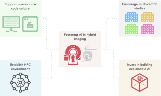
7. Roadmap for fostering AI in hybrid imaging
Commercial AI has grown exponentially in a seemingly short amount of time. This rise is mainly due to the enormous industrial backup, hardware advancements (GPU) and the establishment of strong ML open-source software frameworks (TensorFlow [209], PyTorch [210], Caffe [211], Theano [212], etc.). Most of the commercial AI publications host their code-implementations on GitHub (GitHub Inc. Microsoft, USA) which allows for easy testing and validation. Such an open-source and transparent approach has aided in advancing the commercial AI field significantly. Hybrid imaging community is considerably smaller when compared to the commercial AI world. Despite the disparity in size, there has been a steady rise in AI-based studies. However, most of the hybrid imaging AI approaches are not easily replicable due to two main reasons: (1) limited public repositories and (2) closed nature of their implemented codebase. Creating a centralized database is non- trivial as it has several legal, technical and logistical issues. However, one could open-source their developed codebase associated with their
respective study. It should be noted that mere curation of codes is insufficient. A proper documentation explaining the usage, expected outcomes along with the limitations of the program are extremely important for replicability. Also, one of the most overlooked factors is code maintenance. It is clear that robust maintenance of a codebase is not possible in an academic environment, however a minimum amount of code maintenance should be guaranteed by the research group. Although seemingly exhaustive, going this extra-mile would enhance transparency, improve collaboration, foster trust, promote reproduc-ibility and accelerate development by preventing the reinvention of the wheel.
The radiology community is at the forefront of the medical AI rev-olution. Recently, radiology driven AI studies have started open- sourcing their developed algorithms to promote transparency and reproducibility. Medical imaging AI frameworks are also being estab-lished by industrial and academic institutions to adopt best practices in AI and to support rapid prototyping of AI algorithms [213,214]. Un-fortunately, nuclear medicine studies have been comparatively conser-vative when it comes to open-sourcing its implemented algorithms or executables. To truly embrace AI in hybrid imaging, conscious efforts need to be carried out by both the communities (Fig. 7).
First, sharing code implementations for AI-based studies in medical imaging should be made mandatory by the editors during the peer- review process. Jupyter notebooks [215] offer an excellent way of sharing and explaining codes along with text and images; the native AI and open science community has already adopted such approaches to increase transparency [216]. Many journals offer statistical review with the aid of an extra reviewer, whose sole purpose is to verify the statistics of the reviewed work. Similarly, one could go for a reproducibility re-view, where an IT expert can run the Jupyter notebook to reproduce the results. In short, we would further the field significantly, if we could operate on the principle - ’If you cannot replicate it, then it does not exist!’ [217].
An open-source approach would also minimize the need for centralized data repositories, as the codes could be shared across the private data-silos to facilitate onsite deep learning approaches, such as federated or distributed learning [218]. Multicentric studies should be strongly encouraged compared to single-centre studies, especially in
challenging areas, such as radiomics and deep learning [219], as they can evaluate the robustness of the algorithm better. Federated learning is an exciting approach, since the training process is carried out on data onsite, and only the trained models are exported to a centralised server for multicentric model optimisation, thus, removing the complexity of data-sharing in multicentric studies [218]. Federated learning or deep learning approaches in medical imaging in general mandate a high- performance computing environment. GPUs can significantly accel-erate the training process by 10–30 times in comparison to CPU-based training [220]. Current AI frameworks mainly depend on the CUDA framework from Nvidia for accelerating the training process. And the number of GPUs required depends on the volume and dimensionality of the trained dataset. Medical images are often 3D datasets and can also extend to 4D in case of a dynamic study. And hybrid imaging modalities can significantly increase the dimensionality of the training datasets. Therefore, it is worthwhile to invest in a flexible high-performance computing environment for facilitating deep-learning endeavours.
Finally, deliberate efforts should be focused on building explainable AI for identifying ’Clever Hans’ predictors and also to encourage trust between the clinician and the AI system [202,221]. In the end, the cli-nicians need to explain to the patient why the AI chose a specific therapy and why they think it is a trustworthy option. Development of novel visualisation methods are of utmost importance for making the black box transparent and to identify how the AI algorithm learns (e.g., heat maps) [202,221]. Such visualisation strategies might bring forth unex-pected findings in hybrid imaging datasets.
In summary, medical-AI has the potential to change the clinical landscape of hybrid imaging. Nevertheless, careful and thorough investigation and multi-centric validation of the AI algorithms is necessary prior to clinical deployment, as the algorithms might have a direct or indirect impact in patient management. To support reproduc-ibility, quick development cycles and generally further AI in the hybrid imaging community, collaborative open-source approaches need to be encouraged by the community as a whole, rather than closed private proof-of-concepts.