Structural and biochemical study of the
proteins AmiC, NlpD and FtsW involved in
the bacterial cell division
ByMathieu Rocaboy
Thesis Supervisors: Paulette CHARLIER & Mohammed TERRAK
December 2013 Thesis Committee: Bernard JORIS
Paulette CHARLIER Meriem EL GHACHI Mohammed TERRAK Georges FELLER
Erik GOORMAGHTIGH (Université libre de Bruxelles) Johan WOUTERS (Université de Namur)
Abstract
Cell division in the Gram negative bacterium Escherichia coli is a highly coordinated mechanism involving various physiological functions such as chromosome segregation, cell envelope invagination, peptidoglycan synthesis at the division site and separation of the daughter cells. All these functions require a high level of spatio-temporal regulation in order to preserve the physical integrity of the cell. At least 20 proteins required for a proper cell division are recruited to the division site to form a supramolecular complex called the divisome. This thesis work focused on three major components of the E. coli division machinery: the N-acetylmuramyl L-alanine amidase AmiC, the LytM factor NlpD and the lipid II flippase FtsW. These proteins are recruited at midcell at a late stage of cell division. FtsW is an integral membrane protein crucial for the translocation of the peptidoglycan precursor from the cytoplasm to the periplasm where it will be processed to produce septal peptidoglycan. AmiC acts as a septal peptidoglycan hydrolase that allow the separation of the daughter cells. This enzyme has been shown to be activated by the LytM factor NlpD.
The crystal structure of AmiC from E. coli presented in this work confirms the presence of an inhibitory helix in the active site. The AmiC variant lacking this helix exhibits by itself an activity comparable to that of the wild type AmiC activated by NlpD. Furthermore, the direct interaction between AmiC and NlpD has been detected by microscale thermophoresis with an apparent Kd of about 13 µM. The crystal structure of AmiC also reveals the β-sandwich fold of the AMIN domain, responsible for the septal targeting of AmiC to the division site. The two symmetrical four-stranded β-sheets exhibit highly conserved motifs on the two outer faces. Along with the peptidoglycan binding capacity of the AMIN domain, results obtained so far suggest that the AMIN domain could be involved in the recognition of a specific peptidoglycan architecture or a composition different than the lateral peptidoglycan.
Production screenings of FtsW from different strains were realized and FstW from E. coli was purified. This challenging project will require additional efforts to obtain sufficient amount of protein for structural investigation.
Information gathered in this work confirms the high level of regulation of the hydrolytic activity at the septum and gives a structural basis for a more precise molecular characterization of the division site targeting. Disruption or over-activation of these regulation mechanisms could represent a new strategy in the development of antibacterial compounds.
Résumé
La division cellulaire chez la bactérie à Gram négatif Escherichia coli est un processus cellulaire regroupant de nombreuses fonctions physiologiques telles que la ségrégation chromosomique, l’invagination de la paroi cellulaire, la synthèse de peptidoglycane au site de division et la séparation des cellules filles. Toutes ces fonctions nécessitent un haut niveau de régulation spatio-temporelle afin de préserver l’intégrité physique de la cellule. Au moins 20 protéines impliquées dans la division cellulaire sont recrutées au site de division pour former un complexe multi-protéique appelé divisome. Ce travail de thèse porte sur trois membres de la machinerie de division d’E. coli : la N-acetylmuramyl L-alanine amidase AmiC, le facteur LytM NlpD et FtsW, la flipase du lipide II. Ces protéines sont recrutées au milieu de la cellule à un stade avancé de la division. FtsW est une protéine intégralement membranaire indispensable à la translocation du précurseur du peptidoglycane du cytoplasme vers le périplasme où il sera pris en charge pour produire le peptidoglycane septal. AmiC est une hydrolase du peptidoglycane septal qui permet la séparation des cellules filles. Il a été montré que cette enzyme est activée par NlpD (facteur LytM).
La structure cristallographique d’AmiC d’E. coli présentée dans ce travail confirme la présence d’une hélice inhibitrice dans le site actif. Le variant d’AmiC dépourvu de cette hélice possède une activité comparable à celle d’AmiC sauvage activé par NlpD. De plus, l’interaction directe entre AmiC et NlpD a été détectée par thermophorèse avec un Kd apparent d’environ 13 µM. La structure cristallographique d’AmiC révèle également pour la première fois l’architecture du domaine AMIN avec un repliement de type β-sandwich. Ce domaine, responsable de la localisation au site de division, est composé de deux feuillets symétriques comportant chacun quatre brins β et exposant des motifs conservés sur leurs faces externes. En prenant en compte la capacité du domaine AMIN à lier le peptidoglycane, les résultats obtenus suggèrent que le domaine AMIN pourrait être impliqué dans la reconnaissance d’une architecture ou composition spécifique du peptidoglycane au site de division.
Des criblages de production de protéines FtsW provenant de différentes souches ont été réalisés et la protéine FtsW d’E. coli a été purifiée. Cette partie du projet nécessitera davantage d’efforts pour obtenir des quantités suffisantes de protéines en vue d’une étude structurale.
Les informations récoltées au terme de ce travail confirment le haut niveau de régulation de l’activité hydrolytique au niveau du septum et fournissent une base structurale pour une caractérisation plus précise des déterminants de l’adressage au site de division. La suppression ou la sur-activation de ces systèmes de régulation pourraient représenter une nouvelle stratégie dans le développement de composés antibactériens.
Remerciements
Je souhaite d’abord remercier le Professeur Bernard Joris de m’avoir accueilli au Centre d’Ingénierie des Protéines de l’Université de Liège. Je remercie également le Professeur Moreno Galleni qui lui a succédé au poste de directeur.
Je remercie les Fonds pour la formation à la Recherche dans l’Industrie et dans l’Agriculture (FRIA) pour avoir financé ces années de thèse.
Je tiens à remercier tout particulièrement Paulette Charlier pour m’avoir permis de terminer mon stage de fin de Master au sein de l’équipe de cristallographie. Je la remercie également de m’avoir donné l’opportunité de réaliser ces quatre années de thèse et d’avoir été une promotrice toujours à l’écoute. Merci pour cet encadrement, ce soutien (et ces manons lors des voyages au Synchrotron…) pendant ces quatre années qui resteront une très belle expérience.
Merci Eric et Fred, j’ai énormément appris à vos côtés. Merci pour vos conseils et votre soutien tout au long de cette thèse. Travailler avec vous aura été un vrai plaisir. Merci à Raphael pour m’avoir initié à l’art délicat de la cristallogenèse et pour tous les conseils qu’il m’a donnés.
Un grand merci à Mohammed Terrak pour m’avoir accueilli au labo 1.28 et accepté de m’accompagner en tant que promoteur pendant cette thèse. Merci pour tes innombrables conseils, ta disponibilité et ton écoute qui m’ont grandement facilité la tâche.
Je tiens particulièrement à remercier André Piette et Nicolas Dony avec lesquels j’ai appris énormément de choses durant cette thèse. J’ai vraiment apprécié de travailler avec vous et le fait que cette thèse se soit si bien passée tient beaucoup à votre présence et à votre aide. Merci à Sébastien de Boel pour tous ses conseils et sa disponibilité au début de ma thèse. Merci Adeline pour tes précieux conseils au labo et aussi pendant la phase de rédaction. Isma et Sophie, je vous souhaite le meilleur, bon courage pour la suite ! Samir, j’espère que tu passeras d’aussi bons moments que moi au CIP à condition que tu te fasses aux rudesses du climat… Thank you Badrish, it was a pleasure to work with you (and Mobinius…). Thanks for the invitation in India, it was incredible and I will not forget this amazing trip. !
Merci à Jean-Marie Frère pour la relecture de mon article et à Martine Nguyen-Distèche pour ses conseils et sa précieuse aide bibliographique.
Merci aux réguliers de la salle de pause en biochimie où les leçons de vocabulaire se mêlaient aux concours de chant et aux résumés des aventures de Victor Newman : Fabienne, Florence, Caroline S., Linda, Amandine, Isma, Sophie, Samir, Nicolas, Caroline M., Bryan et Georges.
Merci aux membres des Enzymes Volants, Alain, Philippe, Jean-Marie, Marc, Fred, Dave, Julien et Patrick (a.k.a Michel P.) pour ces quelques années de mini-foot, cette qualification en 8ème de finale restera dans les mémoires.
Un grand merci aux membres du CIP qui m’ont soutenu, aidé et avec qui j’ai énormément appris scientifiquement, le tout dans une excellente ambiance rythmée par les barbecues, trappistes et autres festivités.
Merci à Fred et Mohammed pour leurs patientes relectures de ce manuscrit. Un grand merci à Roya et Marie pour leur précieuse aide et leur soutien dans les derniers moments de rédaction et lors de l’impression de cette thèse.
Abbreviations
A. aeolicus Aquifex aeolicus
ADP Adenosine di-phosphate
Ala Alanine
AMIN Amidase N-terminal domain
ATP Adenosine tri-phosphate
B. henselae Bartonella henselae B. subtilis Bacillus subtilis
C55-P Undecaprenyl-phosphate
C. crescentus Caulobacter crescentus
CABS N-cyclohexyl-3-aminobutanesulfonic acid CAPS N-cyclohexyl-3-aminopropanesulfonic acid CCP4 Collaborative Computational Project No. 4 CSI-Blast Context-Specific Blast
CSS Complexation Significance Score
DAP Diamino-pimelic acid
dcw division cell wall
DDM n-Dodecyl-β-D-maltopyranoside
DNA Deoxyribonucleic acid
DTT Dithiothréitol
E. coli Escherichia coli
EDTA Ethylenediaminetetraacetic acid
EM Electron microscopy
ESRF European Synchrotron Radiation Source Facility
GFP Green fluorescent protein
GlcNAc N-acetyl glucosamine
D-iGlu Iso-glutamate
FIP French beamline for Investigation of Proteins
fts filament-forming temperature-sensitive
G.thermodenitrificans Geobacillus thermodenitrificans
GTP Guanosine triphosphate
LB Luria-Bertani broth
L. lactis Lactococcus lactis
LPS Lipopolysaccharides
LTA Lipoteichoic acid
Lys Lysine
MurNAc N-acetyl muramic acid
NADPH Nicotinamide adénine dinucléotide phosphate
NTA Nitrilotriacetic acid
OD600 Optical density measured at a wavelength of 600 nm
PAGE Polyacrylamide gel electrophoresis
PBP Penicillin-binding protein
PCR Polymerase chain reaction
PDB Protein Data Bank
PEG Polyethylene glycol
PG Peptidoglycan
PEP Phoshpoenolpyruvate
PTS Phosphotransferase system
PISA Proteins, Interfaces, Structures and Assemblies
POTRA Polypeptide transport associated
RBB Remazol Brilliant Blue
RMSD Root-mean-square deviation
S. aureus Staphylococcus aureus
S. pneumoniae Streptococcus pneumoniae S. epidermidis Staphylococcus epidermidis
SDS Sodium dodecyl sulfate
SEDS Shape, elongation, division and sporulation
SPOR Sporulation-related repeat
TB Terrific broth
TEV Tobacco Etch Virus
Tat Twin-arginine transport
UDP Uridine diphosphate
Contents
INTRODUCTION 1
1. Structure of the bacterial cell wall 3
1.1. General points 3 1.2. Peptidoglycan 5 2. Bacterial division 11 2.1. Divisome presentation 11 2.2. Division triggering 12 2.2.1. Min system 13
2.2.2. Nucleoid occlusion system 14
2.3. Early stage of the divisome assembly 15
2.3.1. Constriction ring formation (FtsZ) 15
2.3.2. Membrane anchoring of the Z ring (ZipA, FtsA) 17
2.3.3. Ring stabilization (ZapA-D) 18
2.3.4. Hypotheses for the FtsZ constriction mechanism 19
2.3.5. FtsE/X 20
2.4. Maturation of the divisome 21
2.4.1. FtsK and DNA segregation 21
2.4.2. FtsQLB: the divisome link between cytoplasm and periplasm 22 2.4.3. Translocation of the PG precursor to the periplasm 24 2.4.4. Biosynthesis of glycan chains and incorporation in pre-existent PG 26
2.4.5. Outer membrane invagination 27
2.4.6. Peptidoglycan hydrolysis and bacterial division 29
2.4.6.1. E. coli periplasmic amidases, key players of cell separation 29 2.4.6.2. Regulation of septal amidases: the LytM factors 31 2.4.6.3. Regulation of murein hydrolysis in the context of septum formation 34 2.4.6.4. Coupling murein hydrolase activity and Z ring positioning 35
2.5. Divisome network 35
MATERIAL AND METHODS 41
3. Molecular Biology 43
3.1. Cloning of amiC, nlpD and variants 43
3.2. Cloning of ftsW from different bacteria 44
4. Productions and Purifications 45
4.1. Overexpression and purification of AmiC, NlpD and variants 45 4.2. Overexpression and purification of selenomethionyl-AmiC 45 4.3. Expression tests of FtsW constructs in E. coli C43 (DE3) and membrane preparation 46 4.4. Membrane solubilization and purification of E. coli FtsW 47
4.5. Expression tests of E.coli FtsW in L. lactis NZ9000 47
5. Structural study of AmiC 47
5.1. Crystallization and data collection 47
5.2. Data processing 48
5.3. Structure analysis 48
6. Activity tests 49
6.1. Preparation of peptidoglycan sacculi 49
6.2. SDS detection method (Hayashi) 49
6.3. Labelling of peptidoglycan with Remazol Brilliant Blue 50
6.4. Activity tests with RBB-labelled peptidoglycan 50
6.5. Peptidoglycan-binding assay 50
7. Interaction study 51
RESULTS 53
8. Cloning, Production and Purification 55
8.1. Cloning, production and purification of FtsW 55
8.1.1. ftsW cloning in production vectors 55
8.1.2. Production and purification tests 56
8.1.2.1. Production and purification of E. coli FtsW 56
8.1.2.2. G. thermodenitrificans, A. aeolicus and S. aureus FtsWs 58 8.2. E.coli AmiC & NlpD 60
8.2.1. Cloning of amiC and nlpD 60
9. Crystallogenesis and data collection 66
9.1. Crystallogenesis 66
9.2. Diffraction and data collection 69
10. AmiC crystal structure 70
11. Role for the auto-inhibiting α-helix 78
12. AMIN domain interaction with peptidoglycan sacculi 80
13. The AmiC-NlpD interaction 82
DISCUSSION 85
ANNEXES 99
Annexe 1:
Paper accepted in Molecular Microbiology 101
Annexe 2:
List of protein sequences related to AmiC used for the amino-acid conservation analysis with Consurf 113
1
3
1. Structure of the bacterial cell wall
1.1.
General points
The bacterial cell wall is an important structure of the prokaryote cell. This layer lying above the plasma membrane confers to the bacterium its morphology, allows it to resist internal osmotic pressure, represents a physical protection against toxic substances and can also contribute to the pathogenicity of various micro-organisms. Based on the staining assay developed by Gram in 1884, two major types of cell wall architectures were identified and the corresponding bacteria named Gram positive or Gram negative.
Gram positive bacteria cell wall
The Gram positive cell wall is mainly composed of a layer of peptidoglycan (described in section 1.2) 20 to 80 nanometers thick. Two abundant types of phosphate-rich glycopolymers called teichoic acids, synthesized exclusively by Gram positive bacteria, interact with the peptidoglycan and provide stability to the plasma membrane (Figure 1).
Figure 1. Gram positive enveloppe (Prescott, 2007)
Wall teichoic acids (WTA) are covalently linked to the peptidoglycan via a linkage unit and extend through and beyond the cell wall. Lipoteichoic acids (LTA) are anchored to the
4 cytoplasmic membrane via a glycolipid link. Together with peptidoglycan, lipoteichoic acids influence the elasticity and porosity of the cell wall. They are also involved in pathogenic mechanisms such as resistance against antibiotics and horizontal gene transfer. The existence of a periplasmic space has been suggested by cryo-TEM experiments on Bacillus subtilis (Matias & Beveridge, 2005). Two major parts of the cell wall were discriminated: the inner wall zone with low abundance of substance and designated as the periplasm and the outer wall zone, which contains mature peptidoglycan associated with WTA and cell surface proteins.
Gram negative bacteria cell wall
The cell wall of Gram negative bacteria is characterized by the presence of an outer membrane that determines with the plasma membrane, the limits of the periplasmic space (Figure 2). The latter comprises a thin layer (1 to 3 nm thick) of one or two sheets of peptidoglycan.
Figure 2. Gram negative enveloppe (Prescott, 2007)
The most abundant proteins of the outer membrane, Braun’s lipoproteins, are covalently linked to the peptidoglycan. The outer membrane is a lipid bilayer but unlike the plasmic membrane, phospholipids are only present in the inner leaflet. The outer leaflet is composed of glycolipids, essentially lipopolysaccharides (LPSs) which are known to induce strong inflammatory reactions. The LPS also constitutes a permeability barrier. The high density of LPS molecules
5 at the bacterial surface and the interactions between them restrict the entry of hazardous molecules such as bile salts or antibiotics. However, the outer membrane is not totally hermetic to the external medium. General porins allow a passive transport of compounds not exceeding 700 daltons across the outer membrane. Intake of larger molecules requires specific transporters such as the Phosphotransferase system (PTS) for carbohydrates transport. Export of molecules is also carried out by Gram negative bacteria in various physiological contexts: antibiotics resistance with efflux pumps, synthesis of cell surface organelles with secretion systems or predation with export of bacteriocin in the medium.
1.2.
Peptidoglycan
Introduction
The peptidoglycan (or murein) is a net-like macromolecule that dictates the shape of the bacteria. It is made of glycan chains cross-linked via peptide stems linked to the sugar moieties. This type of linkage confers strength and elasticity to the peptidoglycan and preserves the integrity of the bacterium. The following points will describe the chemical composition, high order structure and biosynthesis of this important bacterial polymer.
General chemical composition and diversity
General composition
The glycan chains of peptidoglycan are made of a repeated basic unit composed of alternating N-acetyl glucosamine (GlcNAc) and N-acetyl muramic acid (MurNAc) linked by a β,1-4 bond (Figure 3). The lactyl group at position 3 on the MurNAc is substituted by a pentapeptide stem of varied amino-acids of L and D series depending on the bacterial species. Most often this peptide stem is made of L-Ala-D-iGlu (iso-glutamate)-meso-DAP (diamino-pimelic acid)-D-Ala-D-Ala in Gram negative bacteria. The meso-DAP is replaced by a L-Lys in some Gram positive bacteria like Staphylococcus aureus. In mature peptidoglycan, the last alanine is lost in cross-linking and maturation reactions catalyzed by transpeptidases and carboxypeptidases respectively (see below).
6
Figure 3. Diversity in peptidoglycan composition. A. Basic unit of E. coli peptidoglycan. B. 4-3 cross-link present in E.coli. C. 4-3 cross-link with peptide bridge (S. aureus). D. Indirect 3-3 cross-link (E. Faecium). E. Deacetylated disaccharide. F. O-acetylation of muramic acid. G. 1,6 anhydroMurNAc disaccharide.
Diversity of peptidoglycan composition among bacteria
The most common cross link (called 4-3) is formed between the D-Alanine at position 4 and the amine function of a DAP (or L-Lysin) at position 3 belonging to an adjacent glycan chain (Schleifer & Kandler, 1972). A 4-2 cross-link has been observed in coryneform bacteria between the α-carboxylic function of D-Glu at position 2 and the carboxylic function of D-Ala at position 4. In both cases, the catalytic reaction linking the two peptide stems is performed by D-D-transpeptidases, members of the penicillin-binding protein family (Sauvage et al., 2008). A third kind of cross-link was observed in Enterococcus faecium (Mainardi et al., 2000): a 3-3 bond connecting two L-Lys at position 3. Interestingly, the formation of this link requires the catalytic action of the L-D transpeptidase Ldtfm, which is insensitive to most β-lactams
antibiotics (Mainardi et al., 2005). L-D transpeptidases have also been found in Escherichia
coli (Magnet et al., 2008) and Mycobacterium tuberculosis (Lavollay et al., 2008). The wide
presence of L-D transpeptidases in the bacterial kingdom along with the potential to bypass the β-lactam sensitive synthesis of peptidoglycan could constitute a threat in the fight against multi-drug resistant bacteria.
7 The cross-link between peptide stems is a direct link in Gram negative bacteria but can be indirect in Gram positive bacteria. In the latter case, the bridge between the lysine of one stem peptide and the D-alanine of the other stem peptide is composed of one to seven residues. For example the S. aureus peptide bridge is composed of five glycine residues (Figure 3C).
Substitutions on the peptidoglycan sugar moieties represent another source of variability in the composition of mature peptidoglycan (Vollmer, 2008). Some of these modifications have important physiological impacts on peptidoglycan integrity and bacterial cell cycle. N-deacetylation of glycan chain and O-acetylation of MurNAc sugar alter the conformation of peptidoglycan and restrict the action of muramidases like lysozyme (Amano et al., 1977, Brumfitt et al., 1958). Recently O-acetylation of both MurNAc and GlcNAc were observed in
Lactobacillus plantarum (Bernard et al., 2011). While the O-acetylation of GlcNAc inhibits the
activity of the major L. plantarum autolysin, the O-acetylation of MurNAc was shown to activate autolysis via the activity of a putative amidase. Thus, the ratio and localization of these glycan chain modifications modulate the peptidoglycan degradation of the bacterium.
The MurNAc moiety can also be modified into 1,6-anhydroMurNAc by lytic transglycosylases. This modification is found at glycan chains termination in Gram negative bacteria like E.coli (Holtje et al., 1975). In the presence of β-lactam antibiotics the peptidoglycan synthesis machinery is inhibited and leads to an uncontrolled hydrolytic activity. Degradation products containing 1,6-anhydroMurNAc are transported to the cytoplasm by the peptidoglycan recycling system. In some Gram negative bacteria like Citrobacter freundii and Enterobacter
cloacae (Holtje et al., 1994, Jacobs et al., 1994), the 1,6 anhydroMurNAc-tripeptide acts as a
signaling molecule to induce the expression of the β-lactam hydrolyzing enzyme AmpC via the transcription factor AmpR.
High order structure
Although the chemical composition of peptidoglycan has been studied for decades, models of its supramolecular arrangement have only been experimentally supported in the last few years. Two main architecture models were proposed: The layered model considers glycan chains running parallel to the cell surface (Ghuysen, 1968) whereas in the scaffold model, they are arranged radially to the cell, perpendicular to the cytoplasmic membrane (Dmitriev et al., 2003) (Figure 4A-B).
8
Figure 4. Peptidoglycan architecture models for Gram negative bacteria. A. Layered model. B. Scaffold model. C. Disorganized layers. Glycan chains and peptide cross links are depicted in green and blue respectively (Gan et al., 2008).
For Gram negative bacteria, experimental data argue in favor of the layered model. Indeed, the observation by fluorescence microscopy of labeled proteins involved in peptidoglycan synthesis reveal a spiral motion (van Teeffelen et al., 2011) in agreement with the proposed mechanism of circumferential insertion of newly synthesized glycan chains (Holtje, 1998). Moreover, direct observation by cryotomography of purified murein from E. coli and
Caulobacter crescentus confirmed that glycan strands are oriented parallel to the cell surface
and in a disorganized hoop-like fashion (Gan et al., 2008) (Figure 4C).
From Gram positive bacteria, which possess a 5- to 10-fold thicker peptidoglycan, three architectural models were proposed, all supported with experimental evidences: Aside from the two aforementioned “layered” and “scaffold” models, a third alternative consists in 50 nm coiled cables of peptidoglycan strands arranged circumferentially around the cell (Hayhurst et
al., 2008) (Figure 5). Recent experiments combining electron tomography on Bacillus subtilis
peptidoglycan and molecular dynamics simulations strongly argue for the layered model and dismiss the two other ones (Beeby et al., 2013). The capacity of B. subtilis to synthesize Gram negative like peptidoglycan during sporulation also argue for a similar synthesis machinery in the bacterial kingdom, leading to a layered murein (Tocheva et al., 2013).
Figure 5. Peptidoglycan architecture models for Gram positive bacteria. A. Layered model. B. Coil-coiled model. C. Scaffold model. The cytoplasmic membrane is depicted in blue. Glycan strands and peptide cross links are shown in green and red respectively (Popham, 2013).
9
Peptidoglycan synthesis
Before being integrated in the nascent glycan chains, disaccharide-pentapeptides precursors have to be synthesized in the cytoplasm and coupled to a lipidic carrier in order to be translocated to the periplasm (Figure 6).
In E.coli, the first cytoplasmic step is the transfer of the enolpyruvyl moiety of phoshpoenol pyruvate (PEP) to uridine diphosphate (UDP); catalyzed by the essential MurA enzyme (Brown
et al., 1995, Zemell & Anwar, 1975). The resulting enolpyruvyl UDP-GlcNAc undergo a
NADPH-dependant reduction by the MurB enzyme to produce the UDP-MurNAc (Taku et al., 1970). The next steps are the stepwise addition of amino-acids to the UDP-MurNAc to obtain the final pentapeptide. The ATP-dependent amino-acid ligases responsible for this sequential synthesis are the following: MurC for the first L-Ala, MurD for D-Glu, MurE for meso DAP and MurF for the terminal dipeptide D-Ala-D-Ala (Bouhss et al., 1999, Bouhss et al., 1997, Bouhss et al., 2008, Eveland et al., 1997). The MurNAc-pentapeptide is then attached to the lipidic carrier undecaprenyl-phosphate (or C55-P) by the MraY tranferase to form the lipid I.
This reaction is followed with the conversion by the enzyme MurG of lipid I into lipid II by the addition of GlcNAc. The lipid II is therefore made of a disaccharide composed of GlcNAc and MurNAc, the latter being substituted at position 3 by a pentapeptide and at position 1 by a C55 isoprenyl diphosphate.
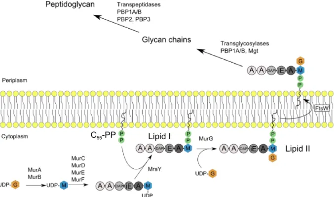
Figure 6. Peptidoglycan synthesis. G: GlcNAc, M: MurNAc. Aminoacids are represented as circles. A (grey circle): L-Ala, iE: iso-glutamate, DAP: diaminopimelic acid, A (white circle): D-Ala.
10 The translocation of lipid II to the periplasm is performed by the flippases FtsW and RodA. The lipid II is then processed by glycosyltransferases that catalyze the β,1-4 bond formation between the GlcNAc of the lipid II precursor and the MurNAc of the growing glycan chain. Peptides protruding on the synthesized glycan strands are cross-linked by transpeptidase reactions to form the peptidoglycan network. During this reaction, the last D-Alanine residue of the donor peptide is lost. Critical proteins involved in these reactions have been identified and belong to the penicillin binding proteins (PBPs) family. PBP1A and PBP1B are bifunctional PBPs that possess both transglycoslyase and transpeptidase activities (Egan & Vollmer, 2013). PBP2 and PBP3 are monofunctional transpeptidases required for elongation and cell division respectively (den Blaauwen et al., 2003, Weiss et al., 1997).
11
2. Bacterial division
2.1.
Divisome presentation
The process responsible for the binary fission of one mother cell into two daughter cells implies the participation of a supramolecular complex called the divisome (Figure 7). This division machinery is composed of, at least, 20 proteins involved in various physiological functions such as chromosome segregation, cell envelope invagination, peptidoglycan synthesis at the division site (called septum) and separation of daughter cells. All these functions require a high level of spatio-temporal regulation in order to preserve the physical integrity of the cell. Mutational experiments have highlighted several genes playing a role in the synthesis of peptidoglycan and division. In E. coli, several are located in the same 2 minutes region of the genome, the dcw cluster (division cell wall) (Ayala et al., 1994). This cluster is conserved in a broad range of bacterial genomes with mainly the same organization, showing its importance in the bacterial kingdom. In the following description, we will focus on the E. coli divisome with few comparisons with known specificities of other bacteria.
The first step of the divisome formation is the polymerization of FtsZ proteins to form a contractile ring (called Z ring). Its stabilization and anchoring to the cytoplasmic membrane is mainly assured by the ZipA, ZapA-D and FtsA proteins. The formation of the Z ring is regulated in time and space by the nucleoid occlusion and the MinCDE systems.
The second step is the maturation of the divisome by the sequential and interdependent recruitment of proteins or protein sub complexes (FtsK, FtQ, FtsL, FtsB, FtsW, PBP3, PBP1B, FtsN, AmiC, etc.) involved in the stabilization of the divisome, septal peptidoglycan synthesis and eventually, separation of the daughter cells.
12
Figure 7. Schematic view of the bacterial divisome of E. coli
2.2.
Division triggering
The polymerization of the Z ring is a key step of the bacterial division that provides the driving force for the cell constriction. Two specialized systems control this event in time and space. The Min system restricts the FtsZ ring polymerization in protofilaments at mid-cell and the Nucleoid Occlusion system allow a proper segregation of the chromosomes between daughter cells (Figure 8). Both mechanisms result in a precise positioning of the contractile ring, with less than 3% relative deviation (den Blaauwen, 2013, Trueba, 1982, Yu & Margolin, 1999).
13 2.2.1. Min system
In rod-shaped bacteria, the Min system allows the positioning at midcell of the Z ring by the creation of a concentration gradient of FtsZ-polymerization inhibitors through the long axis of the cell, the concentration being maximal at the cell poles. In E.coli, in order to achieve this gradient, a pole-to-pole oscillating system carries the FtsZ-polymerization antagonist MinC. This protein is composed of two domains, both interacting with FtsZ. The N-terminal domain inhibits the polymerization of FtsZ and the C-terminal interrupts lateral interactions between FtsZ protofilaments and also interacts with MinD (Hu & Lutkenhaus, 2000, Shiomi & Margolin, 2007). The oscillation is generated by the protein couple MinD/MinE. The MinD is a ParA-like ATPase which binds to the membrane as a symmetrical dimer in an ATP-bound form (Hu et al., 2002). Upon binding of MinE, the ATPase activity of MinD is stimulated. The ADP-form of MinD undergoes conformational changes, disassembling the dimer and provoking its release from the membrane. The iteration of membrane anchoring-release events generates the oscillation phenomenon. MinC also interacts with MinD and thus acts as a passenger of this oscillating system. GFP fusions have revealed this particular pattern for the three proteins involved (Fu et al., 2001, Hu & Lutkenhaus, 1999, Raskin & de Boer, 1999). The concentration gradient of MinD is also proposed to be involved in the segregation of chromosomes. Indeed, MinD possesses a DNA binding capacity that could create a DNA tethering gradient to move duplicated chromosomes from the division site to the cell poles (Di Ventura et al., 2013).
In B. subtilis, the MinE equivalent is absent. The MinCD complex is recruited by DivIVA containing an amphipathic helix. This protein preferentially assembles in negatively curved membranes such as cell poles and division sites (Lenarcic et al., 2009). The most recent model proposes that the membrane curvature at mid-cell allows the recruitment of DivIVa which sequestrates the MinCD complex close to the FtsZ ring and regulates the MinC activity to prevent formation of an aberrantly positioned division septum (Eswaramoorthy et al., 2011). In the vibrioid rod Caulobacter crescentus, the positioning of the FtsZ ring is dictated by the bipolar gradient of the antagonist MipZ ATPase with the low point at midcell. The latter is recruited at the cell poles by ParB which binds to DNA sequences near the segregated chromosome origins (Davis & Austin, 1988, Viollier et al., 2004). ParB and chromosomal DNA are thought to be involved in release-capture events that concentrate MipZ at the cell poles and generate an ATP dependent gradient of this FtsZ antagonist (Kiekebusch et al., 2012).
14
Figure 8. Positioning of the Z-ring with the MinCDE and Nuclear occlusion systems in E.coli (Thanbichler, 2010)
2.2.2. Nucleoid occlusion system
The absence of the Min system leads to improper localization of the contractile ring and generate anucleated minicells (Bi & Lutkenhaus, 1993). However, the Z ring is formed only in DNA-free regions so the nucleoid appears to be involved in a second mechanism to prevent the assembly of the divisome machinery.
In E. coli, screening of mutants lacking the Min system highlighted the SlmA (Synthetically lethal with a defective Min system) antagonist of the FtsZ ring (Bernhardt & de Boer, 2005). This protein binds to specific DNA sites evenly distributed on the chromosome but not in the Ter region known to be the last to partition during the division process (Niki et al., 2000). Upon this required DNA-binding, the SlmA protein oligomerizes to form high order structures able to prevent the polymerization and bundling of FtsZ protofilaments (Tonthat et al., 2011, Tonthat
15 Noc, the SlmA equivalent in B. subtilis, has been shown to target 70 regions of the chromosome also absent in the replication terminus. This protein is a potent division inhibitor involved in the spatial regulation of chromosome segregation but the detailed mechanism is not yet known (Wu & Errington, 2004, Wu et al., 2009).
The existence of the Min system and the nucleoid occlusion mechanism stresses the importance of a properly located polymerization of the FtsZ ring. This step, tightly regulated in time and space, is the starting point for the formation of the macromolecular complex responsible for the bacterial division.
2.3.
Early stage of the divisome assembly
2.3.1. Constriction ring formation (FtsZ)
FtsZ is a protein homologous to tubulin and its GTP-dependant polymerization at midcell is considered as the first step of bacterial division. FtsZ monomers associate in a head to tail manner to form protofilaments with an observed exchange rate of a few seconds between FtsZ molecules present in the protofilaments and the FtsZ monomers or short oligomers. This protein is conserved in most bacteria, all chloroplasts and primitive mitochondria (Beech et al., 2000, Takahara et al., 2000).
FtsZ crystal structures from different organisms (Aquifex aeolicus, B. subtilis, Methanococcus
jannaschi, M. tuberculosis, P. aeruginosa, Thermotoga maritima) show a similar conformation
with two globular domains connected by a central α-helix (Lowe & Amos, 1998, Matsui et al., 2012, Oliva et al., 2007, Szwedziak et al., 2012) (Figure 9A). The N-terminal domain adopts a Rossman fold which binds GTP. The C-terminal domain possesses a chorismate mutase-like fold and a synergy loop that interacts with the N-terminal domain of an adjacent monomer to induce GTP hydrolysis (Lowe & Amos, 1998, Romberg & Levin, 2003) (Figure 9B-C). The ring ultrastructure of the FtsZ polymer was first observed by immunoelectron microscopy in E. coli (Bi & Lutkenhaus, 1991). Its structural arrangement is not fully understood but it has been suggested that this highly dynamic structure is made of overlapping and laterally
16 interacting FtsZ protofilaments of approximately 30 subunits (Chen & Erickson, 2005, Fu et
al., 2010) (Figure 9D). Cryo-EM experiments in Caulobacter support this hypothesis (Li et al.,
2007). Association of protofilaments would not consist in physical and regular interactions expected for protein-protein contacts but Lennard-Jones type interactions involving ions between protofilaments (Horger et al., 2008). An alternative model proposes that FtsZ protofilaments could anneal into much longer filaments exceeding the circumference of the cell. This model is supported by the observation of protofilament annealing in bulk solvent (Chen & Erickson, 2009) and by fluorescence microscopy of the separation of a Z ring into a two turn helix (Erickson et al., 2010). However, this model is not consistent with the high turn-over of subunits in the Z ring that would imply frequent breakage and reannealing events, incompatible with a long and continuous contractile ring.
Figure 9. FtsZ and the Zring. A. FtsZ from M. jannaschii. B. FtsZ dimer. C. Model of a straight FtsZ filament. D. Proposed model for the organization of the Z ring involving short protofilaments (from 80 to 125nm) interacting laterally. Adapted from (Erickson et al., 2010)
17 2.3.2. Membrane anchoring of the Z ring (ZipA, FtsA)
The formation of the Z ring depends on the polymerization of FtsZ but also on its tethering to the membrane by two proteins: FtsA and ZipA (Figure 10). FtsZ can assemble into a ring in the presence of either FtsA or ZipA alone but not in the absence of both (Hale & de Boer, 1999, Pichoff & Lutkenhaus, 2002).
FtsA is a widely conserved ATP binding protein of the actin/Hsp70/hexokinase superfamily that localizes at midcell in a FtsZ-dependent manner (Pichoff & Lutkenhaus, 2002). Tethering of the Z ring to the membrane is mediated by the direct interaction of FtsA with the C-terminal peptide of FtsZ and the presence at the opposite side of FtsA of an amphipathic helix that allows its membrane anchoring (Din et al., 1998, Ma & Margolin, 1999). The FtsZ/FtsA ratio has been demonstrated to be important for a proper division. The cell division is blocked by the overexpression of FtsZ. This effect can be reversed when FtsA is also overexpressed. The normal FtsZ to FtsA ratio is of the order of 100:1 (Dewar et al., 1992). Crystal structure of the
Thermotoga maritima FtsA showed that this protein has an actin-like fold. Moreover,
overexpression in E. coli of FtsAs from T. maritima, E. coli and B. subtilis form filaments. The authors suggest that the Z ring could be tethered to the membrane by an “A-ring” made of FtsA polymers (Szwedziak et al., 2012). FtsA is also involved in the recruitment of the downstream proteins PBP3 and FtsN and a direct interaction with the latter has been demonstrated (Busiek
et al., 2012, Corbin et al., 2004, Rico et al., 2004).
18 ZipA is an essential bitopic membrane protein only present in gammaproteobacteria (de Boer, 2010). This protein bundles FtsZ protofilaments in vitro and modulates their oligomeric state and shape (RayChaudhuri, 1999). Indeed, ZipA binds directly to FtsZ by its C-terminal domain. This interaction has been further characterized by NMR and crystal structures of ZipA in complex with a C-terminal peptide of FtsZ which forms a helix and a beta strand along a hydrophobic groove present in ZipA (Mosyak et al., 2000, Moy et al., 2000). Finally, ZipA is required for the recruitment of downstream proteins such as FtsK, FtsQ, FtsL and FtsN (Hale & de Boer, 2002).
2.3.3. Ring stabilization (ZapA-D)
Stabilization of the Z ring at the early stage is mediated by the four non-essential FtsZ associated proteins ZapA, ZapB, ZapC and ZapD which directly interact with FtsZ (Huang et al., 2013). These four proteins are not present in the whole bacterial kingdom: the gammaproteobacteria possess all the Zap proteins whereas only ZapD is present in betaproteobacteria and ZapA and ZapD in the firmicutes (Natale et al., 2013).
ZapA promotes the Z ring stability by bundling FtsZ protofilaments and in vitro experiments suggest that ZapA is in competition with the FtsZ inhibitor MinC in E. coli (Dajkovic et al., 2008, Gueiros-Filho & Losick, 2002). Besides the direct binding to FtsZ, ZapA also interacts with ZapB, through its coiled-coil domain (Galli & Gerdes, 2012). ZapB is a Z ring stabilizer that localizes at the division site in a ZapA-dependant manner. In vitro experiments have shown that ZapB copellets with FtsZ only in the presence of ZapA, suggesting that ZapA bridges the interaction between ZapB and FtsZ (Galli & Gerdes, 2010). The two other Zap proteins, ZapC and ZapD, also interact with FtsZ but via two different sites, suggesting different molecular mechanisms in the stabilization of the Z ring (Durand-Heredia et al., 2012). As for the FtsA-FtsZ couple, experimental data available so far suggest that a proper stoichiometry of the Zap proteins to FtsZ is required for the formation of the Z ring.
In Gram positive bacteria, the ylmF gene located in a broadly conserved gene cluster codes for the SepF protein that promotes assembly and bundling of FtsZ filaments in B. subtilis (Hamoen
et al., 2006). Unlike Zap proteins alteration, sepF mutants have a cell division defect displaying
19 organization of SepF able to bundle FtsZ protofilaments into tubular structures, suggesting a microtubule-like organization (Gundogdu et al., 2011).
In C. crescentus, FzlA is a critical protein for cell division that was found to mediate the FtsZ polymerization in vitro (Goley et al., 2010). Depletion of FzlA leads to filamentation and cells without constriction. Moreover, FzlA alters the structure of FtsZ filaments into helical bundles and could be involved in the constriction process. The stabilization role of FzlA is consistent with its antagonism towards the FtsZ inhibitor MipZ, involved in the Z ring positioning (cf section 2.2.1). Another FtsZ binding protein, FlzC, is not essential for viability of C. crescentus (Goley et al., 2010).
2.3.4. Hypotheses for the FtsZ constriction mechanism
The FtsZ ring is thought to be the main division component able to provide the driving force leading to membrane constriction. This hypothesis is supported by reconstitution of membrane targeted FtsZ into liposomes where constriction is observed (Osawa et al., 2008). Two main models are proposed to explain the contractile ability of the Z ring (Erickson, 2009).
The sliding model is based on FtsZ protofilaments that can span the circumference of the cell, the two ends interacting via lateral bonds. The increase of lateral bonds is thermodynamically favored and thus induces a condensation of the protofilament providing a constriction force. The proteins involved in the formation and stabilization of the Z ring (FtsA, ZipA, Zap proteins) could also modulate the local environment of the protofilaments and generate a variety of contractile forces. Computational modeling supports the hypothesis that the sliding movement between protofilaments is sufficient to generate a contractile force able to achieve cell division (Lan et al., 2009). GTP hydrolysis would not directly generate contractile force but facilitate the monomer turnover during the condensation events.
The bending model proposes that FtsZ protofilaments can switch from a straight to curved conformation in a GTP hydrolysis-dependent fashion and thus, when tethered to the membrane, impose a bending force to the membrane. Electronic microscopy experiments showed curvature of the Z ring in vitro in E. coli, M. jannaschii and P. aeruginosa (Chen et al., 2012, Huecas & Andreu, 2004, Lu et al., 2000). The recently solved crystal structure of M. tuberculosis FtsZ (MtbFtsZ) in a GTP-bound form gives more insight on the molecular mechanisms involved.
20 Compared to the crystal structure of a FtsZ dimer of S. aureus, a major conformational bend of almost 50° is observed between two adjacent molecules of MtbFtsZ. This observation gives rise to a “hinge opening” mechanism where two consecutive GTP bound-FtsZ molecules in a straight conformation undergo a GTP hydrolysis that induces an opening motion pivoted around a highly conserved interface region. The authors suggest that this mechanism along with the FtsZ tethering by FtsA would constitute a structural scaffold able to provide the driving force for membrane constriction (Li et al., 2013) (Figure 11).
Figure 11. FtsZ is proposed to provide the driving force for cell constriction. A. Superposition of the GDP-bound form of the
M. tuberculosis FtsZ dimer (MtbFtsZ) with the GDP-bound S. aureus FtsZ dimer. B. Proposed mechanism for the cell
constriction with the participation of the membrane anchored FtsA. Adapted from (Li et al., 2013)
2.3.5. FtsE/X
FtsE is an ATPase which forms with the membrane protein FtsX an ATP binding transporter-like complex strongly suggested to be involved in the FtsZ assembly and stability at the division site. In E. coli, GFP-fusion with FtsX showed that the localization of the protein at the division site is dependent on FtsZ, FtsA or ZipA but independent of downstream proteins (Schmidt et
al., 2004). FtsE interaction with FtsZ was assessed by coimmuno-precipitation experiments
(Corbin et al., 2007). In C. crescentus, FtsE was also found to localize at midcell in dividing cells (Goley et al., 2010).
Recently, another role of FtsEX has been established in E. coli. FtsX interacts with the LytM factor EnvC which activates the amidases AmiA and AmiB involved in the cleavage of peptidoglycan during the daughter cells separation (Uehara et al., 2010, Yang et al., 2011). A
21 similar activation pathway is also present in the Gram positive Streptococcus pneumoniae where FtsEX interacts with the putative peptidoglycan hydrolase PcsB (Sham et al., 2011). Mutational studies suggest that the FtsEX-PcsB complex could be involved in the activation of the LytA amidase, known as the major S. pneumoniae autolysin (Sham et al., 2013).
The FtsEX complex in B. subtilis was also found to be required for the hydrolase activity of the endopeptidase CwlO. However, the latter is not involved in the division but in the elongation process (Bisicchia et al., 2007, Hashimoto et al., 2012). Indeed, in a mutant lacking FtsEX, division is not impaired (Garti-Levi et al., 2008).
These results suggest that the FtsEX complex is a broadly conserved hydrolase activator involved in peptidoglycan metabolism in different physiological contexts depending on the bacterial species.
2.4.
Maturation of the divisome
Once the Z ring is formed, a delay is observed in E. coli, B. subtilis and C. crescentus until the complete assembly of the divisome and the beginning of division (Aarsman et al., 2005, Gamba
et al., 2009, Goley et al., 2011). During this period, a set of proteins responsible for the synthesis
of the septal peptidoglycan (at the division site) and eventually the daughter cell separation will be sequentially recruited at midcell in an inter-dependent manner. This second step, termed maturation of the divisome, will lead to a functional division machinery. The transition between the two modes of peptidoglycan synthesis (lateral for the elongation and septal for the division) is progressive and recent results propose that the two machineries interact with each other to promote cell division (van der Ploeg et al., 2013). The proteins part of the maturation phase of the divisome will be described in their recruitment order.
2.4.1. FtsK and DNA segregation
FtsK is a multidomain protein involved in chromosome decatenation and segregation which is essential for cell division (Yu et al., 1998). Its architecture is highly conserved in bacteria. In E
22 and septal localization, followed by a long proline-glutamine linker (Dorazi & Dewar, 2000). The C-terminal domain is a RecA-type ATPase which acts as a DNA translocation machine involved in the chromosome segregation (Dubarry & Barre, 2010). Crystal structure of FtsK from Pseudomonas aeruginosa shows a ring-like hexameric form of FtsK in which the DNA would be translocated in an ATP-dependant manner (Massey et al., 2006). Ftsk also interacts with XerC and XerD that recognize the dif sites (DNA recombination sites) and deconcatenate the chromosomes (Aussel et al., 2002).
Figure 12. Crystal structure of FtsK from P. aeruginosa. A. Monomer of the C-terminal domain (304-811) of FtsK. Two domains are shown: the α domain (blue) is unique to FtsK and the β domain (purple) is RecA related. B. Crystal structure of hexameric FtsK, viewed from the top.
2.4.2. FtsQLB: the divisome link between cytoplasm and periplasm
FtsQ, FtsL and FtsB assemble in a complex (FtsQLB) that is formed independently of the other members of the divisome (Buddelmeijer & Beckwith, 2004). Each of these inner-membrane proteins is bitopic with their major part in the periplasm. Two hybrid experiments have shown that FtsQ interacts with itself, FtsA, FtsX, FtsK, FtsL, FtsB, the recently discovered septal component YmgF (Karimova et al., 2009) and proteins involved in the peptidoglycan synthesis (FtsW, FtsI, FtsN). FtsB also interacts with YmgF whereas FtsL interacts with FtsK, FtsW and
23 YmgF (Akerlund et al., 2002, Di Lallo et al., 2003, Dubarry et al., 2010, D'Ulisse et al., 2007, Grenga et al., 2010, Grenga et al., 2008, Karimova et al., 2005, Karimova et al., 2009).
The crystal structure of the periplasmic part of FtsQ reveals two distinct domains termed α and β (van den Ent et al., 2008). The α domain, adjacent to the cytoplasmic membrane, exhibits a POTRA (polypeptide transport associated) motif in which the second β-strand is essential for midcell localization. The β domain in C-terminal position is essential to the recruitment of FtsL, FtsB and FtsW (Chen et al., 2002, van den Ent et al., 2008).
FtsB and FtsL are involved in the recruitment of late division proteins and are suggested to participate in the Z-ring stabilization (Geissler & Margolin, 2005, Gonzalez & Beckwith, 2009). They are thought to interact with each other through leucine zippers-like motifs located on their periplasmic part (Robichon et al., 2011).
The stoichiometry of the complex is not fully understood. Computational modeling suggest two models: the hexameric 2:2:2 or trimeric 1:1:1, both stable and in agreement with experimental evidences even if the crystal structure of FtsQ as a dimer favors the hexameric model (van den Ent et al., 2008, Villanelo et al., 2011).
Figure 13. FtsQ/FtsL/FtsB complex models produced by dynamics simulation. A. Trimeric 1:1:1 model. FtsB is depicted in blue, FtsL in green and FtsQ in red. Residues involved in interaction regions are represented as sticks B. Hexameric 2:2:2 model. Adapted from (Villanelo et al., 2011)
24 The multiple interactions shared by the FtsQLB complex with components of the divisome along with its requirement for the recruitment of downstream proteins make these three proteins important for the connection between cytoplasmic and periplasmic events of the bacterial division.
2.4.3. Translocation of the PG precursor to the periplasm
Once synthesized by the Mur proteins and MraY, the lipidic precursor of the peptidoglycan (lipid II) has to be translocated into the periplasm in order to provide the disaccharide pentapeptide to the peptidoglycan synthesis machinery. A first candidate for this crucial role, MviN, was identified in E.coli by a bioinformatics approach (Ruiz, 2008). However, the B.
subtilis homologs were found to be not essential for growth, thus discarding a potential lipid II
flippase role in this organism (Fay & Dworkin, 2009). The FtsW protein was identified two years later as the cell division flippase E.coli (Mohammadi et al., 2011). The gene coding for FtsW is located in the dcw cluster including the genes coding for MraY and MurG (Ayala et al., 1994). FtsW shares interactions with multiple actors of the cell division (FtsQ, FtsL, FtsN, PBP3 and PBP1B) and is required for the recruitment of the peptidoglycan synthetase PBP3 (Alexeeva et al., 2010, Di Lallo et al., 2003, Fraipont et al., 2011, Karimova et al., 2005). The latter forms a sub-complex with FtsW independently of the other division proteins (Fraipont et al., 2011).
The topology of FtsW from E. coli shows ten transmembrane (TM) segments linked by cytoplasmic and periplasmic loops (Lara & Ayala, 2002). The large loop between TM 7 and 8 appears to be important in the functioning of FtsW (Pastoret et al., 2004). The loop between TM 9 and 10 is involved in the interaction with PBP3 and PBP1B and could mediate the positioning of the peptidoglycan synthetases (Fraipont et al., 2011). Two other FtsW topologies from S. pneumoniae and M tuberculosis have been detemined so far (Datta et al., 2006, Gérard
et al., 2002). FtsW from M. tuberculosis shows a shorter loop between the TM segments 7-8
and a longer C-terminal periplasmic tail which has been shown to interact with FtsZ and required for a proper cell division in M. smegmatis (Rajagopalan et al., 2005).
25
Figure 14. Membrane topologies of FtsW from E. coli (Lara & Ayala, 2002), S. pneumoniae (Gérard et al., 2002) and
M.tuberculosis (Datta et al., 2006). The numbers indicate residues surrounding transmembrane segments and the C-terminus
of each protein.
FtsW is a member of the SEDS (shape, elongation, division and sporulation) which includes RodA and SpoVE involved in elongation in E.coli and sporulation in B. subtilis respectively (Ikeda et al., 1989). Thus, RodA and SpoVE are candidates for the role of flippase in their respective physiological context. SEDS proteins are typically located in the same operon than their cognate PBP (PBP3 for FtsW, PBP2 for RodA and SpoVD for SpoVE).
26 2.4.4. Biosynthesis of glycan chains and incorporation in pre-existent PG
As mentioned above, the lipid II flippase FtsW interacts with PBP3 and PBP1B in E. coli. These two proteins are responsible for the polymerization of the glycan chains and their incorporation into pre-existent peptidoglycan at the division site (Figure 15).
PBP3 is an essential bitopic protein with a large cytoplasmic part. The latter includes two domains: a N-terminal non-penicillin binding domain suggested to be involved in the correct folding of the protein (Goffin et al., 1996) and a C-terminal which possesses the DD-transpeptidase activity (Pares et al., 1996) allowing peptidoglycan cross-linking. In E. coli, the transmembrane segment is required for the localization of PBP3 at midcell, its dimerization and the interaction with FtsW (Fraipont et al., 2011, Piette et al., 2004, Weiss et al., 1999, Wissel & Weiss, 2004).
Figure 15. Septal peptidoglycan synthesis actors in E. coli. For FtsN, helix and strands are depicted in orange and cyan respectively. For PBP1B, the transglycosylase domain is colored in red, the UB2H domain in yellow, the transpeptidase domain in blue and the transmembrane segment in grey. For PBP3 the transpeptidase domain is colored in blue and the non-penicillin binding domain in grey. Grey cylinders represent transmembrane segments.
27 PBP1B possesses both DD-transpeptidase and transglycosylase domains. In vitro, PBP1B was shown to be more active in conditions favoring its dimerization (Bertsche et al., 2005). The crystal structure of PBP1B suggests that the glycan chain polymerized by the transglycosylase domain could serve as substrate for the transpeptidase domain (Sung et al., 2009). An additional third domain of around 100 residues called UB2H is located between the transpeptidase and transglycosylase domains. Upon interaction with the outer membrane-anchored lipoprotein LpoB, this domain mediates an increase of the transpeptidase activity of PBP1B (Typas et al., 2010) (Figure 15). A model suggests that the porosity and density of peptidoglycan could regulate the activation of PBP1B by LpoB (Paradis-Bleau et al., 2010, Tullman-Ercek et al., 2007, Typas et al., 2010).
FtsN is one of the last proteins recruited at the division site (Figure 15). This essential bitopic membrane protein is thought to improve the assembly and stability of the divisome (Rico et al., 2010). FtsN was shown to interact with PBP1B, PBP3, FtsA and FtsQ (Bertsche et al., 2006, Di Lallo et al., 2003, Karimova et al., 2005, Müller et al., 2007). The periplasmic part of FtsN contains three short α-helices followed by a long linker connected to a C-terminal non-essential SPOR domain (Ursinus et al., 2004, Yang et al., 2004). The latter usually contains around 70 residues with a β-sheet of 4 β-strands flanked by two α-helices on one face. In E. coli, this domain is present in four proteins (FtsN, DamX, DedD and RlpA) that localize at the division site. The SPOR domain of FtsN is known to interact with the peptidoglycan suggesting that this domain recognizes a specific septal peptidoglycan architecture (Arends et al., 2010, Gerding et
al., 2009, Möll & Thanbichler, 2009). Mutational and structural analysis of DamX and FtsN
demonstrate that the β-sheet is involved in the peptidoglycan binding and septal localization of these two proteins (Duncan et al., 2013, Williams et al., 2013). In vitro, FtsN was shown to stimulate the polymerase activity of PBP1B and is suggested to modulate the concerted activities of PBP1B and PBP3 (Müller et al., 2007).
2.4.5. Outer membrane invagination
The Tol-Pal system is often cited for its role in the maintenance of the outer membrane and is composed of two sub-complexes. The first one comprises TolQ, TolR and TolA which are inner membrane proteins forming a complex with a stoichiometry of 4-6:2:1 (Cascales et al., 2001, Zhang et al., 2009) (Figure 16). The second complex includes TolB, a periplasmic protein, and
28 Pal, an outer membrane lipoprotein. Inner and outer membrane can be bridged by specific interactions between the periplasmic part of TolA and Pal or TolB (Carr et al., 2000, Cascales
et al., 2002, Cascales et al., 2000, Lloubes et al., 2001). In vivo cross-linking experiment also
showed interactions between the Pal protein and the outer membrane proteins OmpA and Lpp (Cascales et al., 2002).
Figure 16. Tol-Pal system of E. coli and its interaction network. Interactions are indicated by pink arrows (Godlewska et al., 2009).
Mutational studies showed that the E. coli cells lacking the tolA gene exhibit a chaining phenotype and division defects (Meury & Devilliers, 1999). Moreover, all five proteins of the Tol-Pal system have been shown to accumulate at constriction sites but failed to localize in cells depleted in FtsN. Tol mutants also show a delay in outer membrane invagination and contain outer membrane blebs at constriction sites and cell poles. The authors propose that the Tol-Pal system is a sub-complex of the divisome drawing the outer membrane in the space generated by the separation of the new cell poles during the division process (Gerding et al., 2007). Recently, FtsN and TolQ were shown to interact through their periplasmic parts by a two-hybrid experiment (Teleha et al., 2013).
29 2.4.6. Peptidoglycan hydrolysis and bacterial division
Peptidoglycan hydrolases have been shown to be involved in various physiological contexts such as bacterial growth, peptidoglycan turnover, sporulation and germination events, assembly of secretion systems, pili and flagella (Vollmer et al., 2008). The following section will focus on septal peptidoglycan hydrolases and their respective role and known regulations in the division process of E. coli.
2.4.6.1. E. coli periplasmic amidases, key players of cell separation
The amidases AmiA, AmiB and AmiC belongs to the amidase_3 family and consist of Zn2+ -metallo-enzymes that cleave the amide bond between the lactyl group of the N-acetylmuramic acid and the L-Alanine of the peptide stem. AmiA and AmiC are translocated to the periplasm via the twin-arginine transport (Tat) pathway whereas AmiB transport is mediated by the Sec machinery. Their involvement in the bacterial division have been demonstrated in mutational studies where a triple amidase mutant shows a chaining phenotype in which 90 to 100% of the cells are not able to achieve binary fission (Heidrich et al., 2001). A similar but less severe phenotype is observed for amiA and amiC single mutant with 5-10% and 20-30% of chain-forming cells respectively whereas amiB mutant showed no such division defects (Heidrich et al., 2001). In the absence of AmiC, deletion of the D,D-endopeptidases PBP4 and PBP7 (two low molecular weight PBP) or lytic transglycosylases (e.g. Slt70) exacerbates the chaining phenotype (Priyadarshini et al., 2006). Moreover, deletion of entire families of peptidoglycan hydrolases showed a preponderant role in cell separation for periplasmic amidases followed by lytic transglycosylases and to a lesser extent, D,D-endopeptidases (Heidrich et al., 2002). AmiC and AmiB localize to the division site of constricting cells in a FtsN-dependant fashion thanks to their N-terminal AMIN domain (Bernhardt & de Boer, 2003, Peters et al., 2011). The latter, preponderant in Gram negative bacteria, is mostly present at the N-terminus of periplasmic proteins involved in cell wall metabolism or transport structure such as the type IV pilus (de Souza et al., 2008). Consistent with the septal localization of AmiB and AmiC, AmiA, devoid of AMIN domain, exhibits a dispersed localization throughout the periplasm (Bernhardt & de Boer, 2003) (Figure 17).
30
Figure 17. Cell division amidases of E. coli. (A-C) Localisation studies with GFP variants of AmiA (A-A’), AmiB (B-B’) and AmiC (C-C’). Cell are visualized using GFP (A, B and C) or differential interference contrast (DIC) (A’, B’ and C’). (D) Triple mutant ΔamiA ΔamiB ΔamiC. Cells were stained with the fixable membrane dye FM1-43-FX and visualized by fluorescence. Bars equal 1 µm for (A-B-C) and 8 µm for (D). (E) Topologies of AmiA, AmiB and AmiC.
Catalytic mechanism of amidase_3 family proteins
Despite different folds, the catalytic site and the catalytic mechanism of the amidase_3 family is similar to that of the amidase_2 domain of the major autolysin AtlE from Staphylococcus
epidermidis (Figure 18A and B). Upon approach of the substrate, a water molecule bound to
the active site Zn2+ is shifted toward a conserved glutamic acid which further activates it and favors the nucleophilic attack of the amide bond. This step produces a tetrahedral conformation of the amide carbon and a transient pentameric coordination of the catalytic Zn2+. The tetrahedral intermediate is stabilized by hydrogen interactions involving the carbonyl oxygen of the scissile bond. In the N-acetylmuramoyl-L-alanine amidases, the carbonyl oxygen of the substrate L-Ala and one of the MurNAc oxygen can contribute to the stabilization of the tetrahedral intermediate (Kerff et al., 2010). The proton is transferred to the nitrogen of the scissile bond to form a second transition state characterized by a doubly protonated tetrahedral nitrogen which is potentially stabilized by the MurNAc N-acetyl group (Kerff et al., 2010). In amidase_2 enzyme, a histidine or a lysine is also found across the active site compare to the glutamate (Figure 18A) and is involved in the stabilization of the tetrahedral conformation of the amide carbon. In the amidase_3 family, no residue equivalent to those has been identified.
31
Figure 18. Comparison of amidase_2 and amidase_3 catalytic mechanisms. A. Superposition of the amidase_2 member AmiE (in green) from the major autolysin AtlE (S. epidermidis) and the amidase_3 member CwlV (in orange) from Bacillus polymyxa. Oxygens and nitrogen are depicted in red and blue respectively. B. Proposed catalytic mechanism of AmiE. See details in text. Adapted from (Zoll et al., 2010)
2.4.6.2. Regulation of septal amidases: the LytM factors
Another set of proteins, containing LytM domains (lysostaphin/peptidase M_23), is involved in peptidoglycan hydrolysis. In S. aureus, the LytM protein specifically cleaves the penta-glycine peptide bridge whereas the LytM factor gp13 from the B. subtilis phage Φ29 shows a D,D-endopeptidase activity that cleaves direct cross links in B. subtilis peptidoglycan (Browder
32 In E. coli, four proteins (EnvC, NlpD, YebA and YgeR) contain a LytM domain. Deletion of these proteins leads to a severe chaining phenotype with a preponderant effect of the envC-nlpD double mutation (Figure 20A). Moreover, EnvC and NlpD were shown to localize at the division site, supporting their role in cell separation (Uehara et al., 2009). Unexpectedly, these two proteins do not exhibit hydrolytic activity against peptidoglycan in vitro. Sequence alignment with the active LytM of S. aureus highlighted the lack of zinc-chelating residues in the active sites of EnvC and NlpD. Instead, they specifically activate the three aforementioned amidases: AmiA and AmiB for EnvC and Amic for NlpD (Uehara et al., 2010). More insight in the molecular mechanism of septal amidases activation was gained with the crystal structure of the amidase_3 member AmiB from B. henselae where an α helix is obstructing the active site (Yang et al., 2012) (Figure 19A-B).
Figure 19. A. Alignment amidase_3 members grouped into the following categories: (I) phage endolysins, (II) bacterial amidases involved in mother cell lysis following sporulation and (III) cell separation amidases. The red box highlights a ~ 50 amino acid insertion region found only in the cell separation amidases. B. Crystal structures of amidase_3 members for each aforementioned category: PlyPSA (phage PSA) (I), CwlV (B.polymyxa) (II) and AmiB (B. henselae) (III). The amino acid insertion containing the inhibitory helix is shown by the red arrow. Adapted from (Yang et al., 2012)
Mutational screening of the E. coli AmiB allowed to isolate variants with uncontrolled lytic activity leading to cell lysis when overproduced. These mutations are mainly located in the obstructing α helix, suggesting a conformational switch induced by the LytM factor EnvC to


!["Benjamin Carrión. Correspondencia I. Cartas a Benjamín", Préface de Jorge Enrique Adoum, sélection et notes de Gustavo Salazar [compte-rendu]](data:image/gif;base64,R0lGODlhAQABAIAAAP///wAAACH5BAEAAAAALAAAAAABAAEAAAICRAEAOw==)