Université de Montréal
Elucidation of the biological roles of Wnt5a signaling in follicle
development
Par
Atefeh Abedini Najafabadi
Centre de recherche en reproduction animale (CRRA)
Département de biomédecine vétérinaire
Faculté de médecine vétérinaire
Thèse présentée à la Faculté de médecine vétérinaire
en vue de l’obtention du grade de
philosophiae doctor (Ph.D.)
en sciences vétérinaires
option reproduction
Août, 2015
i
Résumé
La santé folliculaire est déterminée par un nombre de facteurs endocriniens, paracrines et autocrines. Les gonadotrophines hypophysaires sont les principaux moteurs du développement du follicule, mais leurs actions sont modulées localement par les hormones et des facteurs de croissance. Les glycoprotéines de la famille des WNTs représentent une grande famille de molécules impliquées dans différentes voies de signalisation. Ils sont sécrétés dans le but de moduler et coordonner la réponse des follicules aux gonadotrophines, et leurs activités sont indispensables à la fonction ovarienne et à la fertilité féminine. Les WNTs sont généralement classés en fonction de la (des) voie(s) qu’ils activent. Le rôle des membres de la voie canonique WNT et de ses composants tels que CTNNB1, WNT4, WNT2, FZD1 et FZD4 est bien établi au cours du développement du follicule chez les rongeurs. Un rôle similaire des WNTs dans les espèces mono-ovulatoires demeure essentiellement inconnu. De plus, le rôle des WNT non canoniques dans l'ovaire de rongeurs est méconnu.
Les objectifs de cette thèse sont (1) d'élucider la régulation hormonale de l'expression de WNT5A et le rôle physiologique de WNT5A dans les cellules de la granulosa bovine in vitro et (2) d'identifier les rôles physiologiques de WNT5A dans l'ovaire de souris par inactivation génique conditionnelle. Chacun de ces objectifs a mené à la publication d’un article à partir des résultats obtenus au cours de cette thèse.
Dans le premier article, le rôle de WNT5A dans les cellules de la granulosa bovine a été étudié in vitro. Nous avons constaté que WNT5A est un régulateur négatif de la stéroïdogenèse stimulée par la FSH issue des cellules de la granulosa, et qu'il agit en supprimant l'activité de signalisation des WNTs canoniques tout en induisant la voie de signalisation MAPK8/JUN.
ii
Dans le deuxième article, afin d’examiner le rôle de deux WNTs non-canoniques, WNT5A et WNT11, à différents stades de développement folliculaire, nous avons généré des modèles de souris knock-out conditionnels ciblant les cellules de la granulosa pour chacun de ces WNTs. Les résultats obtenus ont permis de mettre en évidence que WNT5A est nécessaire pour assurer la fertilité normale chez la femelle, le développement folliculaire et la stéroïdogenèse ovarienne. Il est aussi un antagoniste de la réponse aux gonadotrophines, agissant par l’intermédiaire de la suppression de la signalisation canonique des WNTs. Chez les souris knock-out pour WNT11, nous ne constatons aucun défaut important dans la fertilité des femelles.
L’ensemble de notre travail met en évidence que WNT5A est essentiel pour le développement normal du follicule et qu’il agit pour inhiber la différenciation des cellules de la granulosa. En résumé, nous avons fourni une étude novatrice et approfondie, utilisant plusieurs modèles et techniques pour déterminer les mécanismes par lesquels WNT5A régule le développement des follicules.
Mots clés: WNT5A, développement folliculaire, cellules de la granulosa, stéroïdogenèse ovarienne, vache, souris knock-out conditionnel.
iii
Abstract
Follicle health is determined by an array of endocrine, paracrine and autocrine factors. Pituitary gonadotropins (LH and FSH) are major drivers of follicle development, but their actions are modulated by local hormones and growth factors. The WNT family of secreted glycoproteins are signaling molecules that act to modulate and coordinate follicular responses to the gonadotropins, and whose activities are indispensable for ovarian function and female fertility. WNTs are normally categorized according to pathway(s) via which they signal. The role of canonical WNT members and components such as CTNNB1, WNT4, WNT2, FZD4 and FZD1 is well established during follicle development in rodents. Whether WNTs play similar roles in monovular species remains essentially unknown. Moreover, the role of non-canonical WNTs in the rodent ovary is unclear.
The objectives of the present thesis were (1) to elucidate the hormonal regulation of WNT5a expression and the physiological role of WNT5a in bovine granulosa cells in vitro and (2) to identify the physiological roles of WNT5a in the mouse ovary by conditional gene inactivation.
The results of this thesis are presented in two articles. In the first article, the role of WNT5a in bovine granulosa cells was investigated in vitro. We found that WNT5A is a negative regulator of FSH-stimulated granulosa cell steroidogenesis, and that it acts by suppressing canonical WNT signaling activity and inducing the noncanonical MAPK8/JUN pathway. In the second article, we generated granulosa-specific knockout mouse models to examine the roles of two non-canonical WNTs, WNT5a and WNT11, in different stages of the follicular development. Our results showed that WNT5a is required for normal female fertility, follicle development and ovarian steroidogenesis, and is an
iv
antagonist of gonadotropin responsiveness that acts via the suppression of canonical WNT signaling. However we did not observe any significant defects in fertility of WNT11 knockout mice.
Together, our work demonstrates that WNT5a is essential for normal follicle development and acts to inhibit differentiation in granulosa cells. In overview, we provided a novel and comprehensive investigation, using multiple models and techniques, to determine the mechanisms by which WNT5a regulates follicle development.
Key words: WNT5a, follicle development, granulosa cells, ovarian steroidogenesis, cow, conditional knockout mouse.
v
Table of contents
Résumé ... i
Abstract ... iii
Table of contents ... v
List of tables ... viii
List of figures ... ix
List of abbreviations ... xii
Dedication ... xvi
Acknowledgment ... xvii
Introduction ... 1
Chapter 1. Literature review ... 3
1. Ovaries ... 4
1.1 Functional and anatomy of ovarian follicle structure ... 4
1.1.1 Oocyte ... 6
1.1.2 Granulosa and cumulus cells ... 9
1.1.2.1 Steroidogenesis ... 11
1.1.2.2 Cumulus expansion ... 14
1.1.3 Theca cells ... 15
1.1.3.1 Steroidogenesis ... 15
1.1.4 Basement membrane ... 18
1.2 Endocrine regulation of follicle growth and development ... 18
1.2.1 Gonadotropins ... 19
1.2.1.1 FSH ... 20
1.2.1.2 LH ... 21
1.2.2 Estradiol and progesterone ... 22
1.2.3 Androstenedione and testosterone... 23
1.2.4 Intraovarian regulators ... 24
1.2.4.1 IGF ... 24
1.2.4.2 TGF-β superfamily ... 25
1.2.4.3 FGFs ... 27
1.2.4.4 EGF ... 29
1.3 Folliculogenesis and the ovarian cycle ... 30
1.3.1 Primordial germ cells ... 31
1.3.2 Transition from oogonia to oocyte ... 32
1.3.3 Formation of primordial follicles ... 34
1.3.4 Transition of primordial follicles to primary follicles ... 35
1.3.5 Follicle growth to pre-antral and antral stages ... 36
1.3.6 Selection process of the dominant follicle ... 38
1.3.7 Atresia ... 42
1.3.8 Ovulation ... 44
vi
2. WNTs and WNT signaling ... 47
2.1 WNT overview ... 47
2.2 WNT genes and proteins ... 47
2.3 WNT receptors ... 48
2.4 WNT inhibitors ... 49
2.5 Intracellular signal transduction ... 51
2.5.1 The canonical pathway ... 52
2.5.2 The non-canonical pathways ... 53
2.6 Role of WNT signaling in the ovary ... 56
2.6.1 CTNNB1 ... 56 2.6.1.1 Steroidogenesis ... 57 2.5.1.2 Follicle development ... 57 2.5.2 WNT4 ... 58 2.5.2.1 Embryonic development ... 58 2.5.2.2 Follicle development ... 59 2.5.3 WNT2 ... 60 2.5.4 FZD1 ... 61 2.5.5 FZD4 ... 62 2.5.6 SFRP4 ... 62
Chapter 2. Hypotheses and objectives ... 64
2. 1 Hypotheses and objectives ... 65
Chapter 3. Article 1 ... 66
3.1 Article 1. ... 67
3.2 Abstract ... 68
3.3 Introduction ... 69
3.4 Material and methods: ... 72
3.5 Results ... 78 3.6 Discussion ... 81 3.7 Acknowledgements ... 84 3.8 References ... 84 3.8 Figures ... 92 3.9 Supplemental figure ... 98 3.10 Tables ... 99 Chapter 4. Articles 2 ... 100 4.1 Article 2 ... 101 4.2 Abstract ... 102 4.3 Introduction ... 103
4.4 Materials and methods ... 107
4.5 Results ... 113
4.6 Discussion ... 120
4.7 Acknowledgments ... 123
vii
4. 9 Figures ... 131
4.10 Supplemental figures ... 145
4.11 Tables ... 150
4.12 Supplemental Tables ... 152
Chapter 5. General discussion and conclusion ... 155
5.1General discussion ... 156
5.2 Conclusion: ... 163
viii
List of tables
Article 1
Table 1. Primer sequences used in real-time PCR. ... 99
Table 2. Effect of WNT5a on the cell cycle, CCND2 mRNA levels and apoptosis in bovine granulosa cells. ... 99
Article 2 Table 1. Mating trials ... 150
Table 2. Ovary weights ... 150
Table 3. Ovulatory rates ... 151
Table 4. Hormone assays ... 151
Table S1. Primer sequences ... 152
Table S2. Mating trials ... 153
Table S3. Mating trials ... 153
Table S4. Mating trials ... 154
ix
List of figures
Literature review
Figure 1. Follicular classification. ... 6
Figure 2. Antral follicle structure with different granulosa cell types surrounding the oocyte. ... 10
Figure 3. Steroidogenesis in mural granulosa cells. ... 13
Figure 4. Enzymatic control of steroidogenesis in the theca cell. ... 17
Figure 5. Regulation of gonadotropins... 20
Figure 6. TGF-β superfamily members and their receptors and binding proteins are expressed in different follicle cells. ... 27
Figure 7. Schematic diagram of the ovary ... 31
Figure 8. Germ cell cluster breakdown and formation of primordial follicles. ... 34
Figure 9. A summary of folliculogenesis in the ewe as an example for mono-ovular species. ... 41
Figure 10. Steroidogenesis in the corpus luteum. ... 46
Figure 11. Regulation of WNT signaling by physiological inhibitors. ... 51
x Article 1.
Figure 1. Regulation of WNT5A in granulosa cells. ... 92 Figure 2. Effect of WNT5A on steroidogenesis and on steady-state levels of mRNA
encoding steroidogenic proteins in bovine granulosa cells. ... 93 Figure 3. WNT5A modulates the activity of multiple signaling pathways in bovine
granulosa cells.. ... 94 Figure 4. WNT5A induced JUN phosphorylation is dependent on JNK and Rac1 activity..
... 95 Figure 5. WNT5A upregulates JUN and FOS mRNA abundance in granulosa cells through
the PCP pathway.. ... 96 Figure 6. Graphical abstract (for review) ... 97 Figure S1. WNT5A inhibited steroid secretion and abundance of mRNA encoding
xi Article 2.
Figure 1. In vivo regulation of Wnt5a and Wnt11 mRNA levels in granulosa cells by gonadotropins. ... 131 Figure 2. Wnt5a and Wnt11 knockdown efficiency in the conditional knockout models 132 Figure 3. Wnt5aflox/-;Amhr2cre/+ mice have smaller ovaries and increased follicular atresia
... 133 Figure 4. Progressive follicle loss and increased follicular atresia in the ovaries of
Wnt5aflox/-;Amhr2cre/+ mice.. ... 135 Figure 5. Quantitative RT-qPCR confirmation of microarray data.. ... 136 Figure 6. Expression of WNT5a target genes is altered in Wnt5aflox/-;Amhr2cre/+ mice. ... 137 Figure 7. Regulation of WNT5a target genes by WNT11. ... 138 Figure 8. WNT5a downregulates canonical WNT signaling and CREB expression in
granulosa cells in vitro. ... 140 Figure 9. WNT5a downregulation of CTNNB1 and CREB is GSK3β-dependent.. ... 141 Figure 10. WNT5a suppresses gonadotropin signaling in granulosa cells. ... 143 Figure 11. Working model of the mechanism of action of WNT5a in granulosa cells. ... 144
Figure S1. Wnt5a and Wnt11 knockdown efficiency in the conditional knockout models ... 145 Figure S2. Expression of WNT5a target genes in Wnt11flox/-;Amhr2cre/+ and Wnt5a
flox/-;Wnt11flox/-;Amhr2cre/+mice. ... 146 Figure S3. WNT5a does not regulate non-canonical signaling effectors in granulosa cells..
... 148 Figure S4. Inhibition of noncanonical signaling does not prevent WNT5a-mediated
xii
List of abbreviations
ADAM: A disintegrin and metalloproteinase domain-containing protein 8 AKAPs: A-kinase anchor proteins
AMH: Anti-Mullerian hormone AREG: Amphiregulin
APC: Adenomatosis polyposis coli Bcl-2: B-cell lymphoma 2
BMP: Bone morphogenetic protein BP: Binding protein
BTC: Betacellulin
CaMK: Calmodulin-dependent protein kinase cAMP: Cyclic adenosine monophosphate CC: Cumulus cells
CEEF: Cumulus expansion enabling factor CK1a: Casein kinase 1a
CL: Corpus luteum CNA: Calcineurin
COC: Cumulus oocyte complex CRD: Cysteine-rich domain CTNNB1: Beta-catenin
CYP11A1: Cytochrome P450 cholesterol side-chain cleavage CYP17A1: Cytochrome P450 17β hydroxylase/C17–20 lyase CYP19A1: Cytochrome P450 aromatase
DAG: Diacylglycerol
Dazla: Deleted in azoospermia Dkk: Dickkopf
xiii ECM: Extracellular matrix
EGF: Epidermal growth factor
EGFR: Epidermal growth factor receptor EREG: Epiregulin
Figla α: Factor in the germline alpha FGF: Fibroblast growth factor Foxl2: Forkhead box protein L2 Foxo3a: Forkhead box O3
FSH: Follicle-stimulating hormone FSHr: FSH receptor
FZD: Frizzled
GDF: Growth and differentiation factor GDNF: Glial cell-derived neurotrophic factor GH: Growth hormone
GnRH: Gonadotropin-releasing hormone GPCRs: G protein coupled receptors GSK3: Glycogen synthase kinase 3 GVBD: Germinal vesicle breakdown
HSD17B1:17β-hydroxysteroid dehydrogenase
HSD3B2:3β-hydroxysteroid dehydrogenase/Δ5-Δ4 isomerase IGF-I: Insulin-like growth factors
IGFBPs: IGF binding protein IαI: Inter-α trypsin inhibitor Int1: Integration 1
IP3: Inositol 1, 4, 5-triphosphate ISH: In situ hybridization IVF: In vitro fertilization JNK: Jun N-terminal kinase
xiv KL: Kit-ligand
LDL: Low-density lipoprotein LEF: Lymphoid enhancer factor LH: Luteinizing hormone
LHR: LH receptor
LIF: Leukemia inhibitory factor
LRPs: Low-density lipoprotein receptor-related protein MI: Metaphase I
MII: Metaphase II
MAPK: Mitogen activated protein kinases MGC: Mural granulosa cells
MIS: Müllerian-inhibitory substance MMTV: Mouse mammary tumor virus MMP: Metalloprotease matrix protein MSH5: Muts homologue 5
NFAT: Nuclear factor of activated T-cells NGF: Nerve growth factor
NLK: Nemo-like kinase
NOBOX: Newborn ovary homeobox protein PCP: Planar cell polarity
PGC: Primordial germ cell PGE: Prostoglandin PI3K: Phosphatidylinositol-3-kinase PKC: Protein kinase C PLC: Phospholipase C PR: Progesterone receptor PRL: Prolactin
xv Ror: Tyrosine kinase-like orphan receptor Ryk: Typical tyrosine kinase receptor SCP2: Sterol carrier protein
SF1: Steroidogenic factor 1
StAR: Steroidogenic acute regulatory protein SPO11: Sporulation protein homology TAK1: TGFβ activated kinase
Sgy: Soggy
sFRP: Secreted FZD-related protein
TCF: T-cell factor TGF: Transforming growth factor TIAR: T-cell intracellular antigen-1-related
TMR: Transmembrane pass receptor
Tsc-1: Tumor suppressor tuberous sclerosis complex 1 TSG-6: Tumor necrosis factor stimulated gene 6 Wg: Wingless
xvi
Dedication
This is dedicated to
my parents and my husband
xvii
Acknowledgment
I would never have been able to finish my thesis without the guidance of my supervisor, my co-supervisor, help from friends, and support from my family and my husband.
First and foremost I want to gratefully acknowledge my supervisor Dr. Derek Boerboom for all his support, kindness, encouragement and confidence that he bestows upon me. I am proud of being his Ph.D student and working under his supervision. I am also thankful for the excellent example he has provided as a successful young leader and researcher. Dr. Boerboom, thank you so much for always being there for your students and listening to us, also for all those English expressions that always make me smile and for all your support not only during tough times of my Ph.D. but also in my personal life. I never forget your fantastic words whenever life was not that kind.
I would also like to thank my co-supervisor, Dr. Christopher Price for his excellent guidance, caring, patience and his sweet smile. For me, you are more than a co-supervisor. I spent more than 2 years of my Ph.D period in your lab which were great moments for me. I am so happy that I could be a part of your lab and you treated me like your students. Dr. Price, thank you so much for permitting us to experience a different relationship between supervisor and students.
I want to express my deeply-felt thanks to Dr. Bruce Murphy for his support and his kindness. Dr. Murphy you are a great person. I really appreciate your personality, your energy and your talents. I am sure that I will miss your jokes.
I would like to thank my advisory committee members, Dr. Younès Chorfi and Dr. Alexandre Boyer for their support, advice, and insight. I would also like to thank my thesis
xviii
committee for their attention, time, and useful suggestions for my research and this dissertation.
It is my pleasure to thank my scientific family, especially the members of Dr. Boerboom and Dr. Price's lab, for being a source of friendship as well as good advice and collaboration. I was so lucky for having you around myself. I am particularly thankful to Dr. Marilène Paquet, Gustavo Zamberlam, Hilda Guerrero, Charlène Rico, Fatiha Sahmi, Jane Fenelon, Mayra Tsoi, Mouhamadou Diaw, Adrien Levasseur and Evelyne Lapointe who were always willing to help and give their best suggestions, and for all the fun we have had in the all four years. In addition, I want to thank Meggie Girard for her technical support.
I must acknowledge the guidance and support from all people in “Centre de Recherche en Reproduction Animale (CRRA)” and “Faculty of Veterinary Medicine”. My sincere gratitude to Eliane Auger, Geneviève Provost and Julie Blouin for their help with administrative concerns throughout the years.
I devote a special thank you to my mother Mehri and my father Mehdi, my elder brother and my younger sisters for all the encouragement, love and supporting me spiritually throughout all my life.
Last but not least, my hearty thanks to my love and my best friend Reza, for all his understanding, support, encouragement and unconditional love. Even though we were miles apart during these 4 years, you were never far from my heart. Reza, thank you so much for believing in me and for supporting me through the good and bad times.
1 Introduction
Follicles are the basic functional units of mammalian ovaries and are composed of germ cells (oocyte) and somatic cells (granulosa, theca and stromal cells) (Hsueh et al., 2015). Follicle development initiates during fetal life (in humans) or after birth (in rodents) when primordial follicles are formed. Once follicles start to grow, the activated primordial follicle composed of a single layer of small granulosa cells surrounding the primordial oocyte develops into a primary, secondary and eventually an antral follicle (Richards and Pangas, 2010). The process of the follicular maturation, known as folliculogenesis, is mainly under control of the gonadotropins (FSH (follicle stimulating hormone) and LH (luteinizing hormone)), which are secreted from the anterior pituitary (Richards et al., 2002). Besides the gonadotropins, ovarian follicle growth is controlled by the production of intraovarian growth regulatory factors that act by autocrine, paracrine and intracrine mechanisms (Richards, 1994).
Members of WNT family of signaling molecules have been shown to impact ovarian cell function and follicle organization. WNTs are secreted signaling molecules that act locally to control different developmental processes including cell proliferation and differentiation (Cadigan and Nusse, 1997). Canonical and non-canonical WNTs transduce their signal via Frizzled (FZD) family receptors to activate diverse signaling cascades (Slusarski et al., 1997), which will be further detailed in the end of chapter 1 of this thesis. Recent reports have suggested roles for canonical WNT signaling in the adult ovary. Indeed, canonical WNTs are required for normal antral follicle development and act by regulating granulosa cell functions including steroidogenesis and proliferation (Boyer et al., 2010a, Boyer et al., 2010b, Wang et al., 2010). Whether WNTs play similar roles in monovular species remains essentially unknown. Likewise, little attention has
2
been paid to the potential roles of non-canonical signaling pathways in the ovary of mono- and polyovular species.
This thesis describes studies that took advantage of both cow and mouse models to investigate the roles of non-canonical WNT signaling in the female fertility. The bovine species is a mono-ovulatory species and serves as a suitable model for studying human reproduction, as the two species have a similar length of gestation and typically release one oocyte at ovulation (Fortune, 1994). The rodent is a widely used model for studying reproduction for several reasons, including cost, ease of maintenance and a short generation interval. Transgenic mouse models have been effective and beneficial in understanding female reproduction (Matzuk and Burns, 2012).
Novel findings in this thesis may provide new insight into the roles and mechanisms of action of the non-canonical WNT pathways in granulosa cells. This thesis may also provide new clues regarding the etiology of various ovarian disorders.
3
4 1. Ovaries
Gametogenesis is the result of a coordinated signaling network between the gonads, pituitary, and hypothalamus. The ovaries are gonads that often found in pairs as part of the vertebrate female reproductive system and are crucial structures for the survival of the species (McDonald and Pineda, 1989). The germinal components (originating from primordial germ cells) start to colonize the gonadal primordia after migration from the yolk sac. The gonadal primordia forms a paired thickening of the coelomic epithelium that lines the ventral–medial surface of the mesonephros (the mid-region of the embryonic kidney), and arises within the intermediate mesoderm between the pronephros (initial kidney) and metanephros (the definitive kidney) on embryonic day 34 in human (Oktem and Oktay, 2008). The ovary is composed of two types of cells; germ cells (oocytes) and somatic cells (granulosa, thecal, and stromal), whose interactions dictate formation of follicles, development of both oocytes and somatic cells as follicles and ovulation (Richards and Pangas, 2010).
The ovary has two fundamental physiological roles. First is the gametogenic function that is responsible for the differentiation and release of a mature oocyte for fertilization. The second is the endocrine function that is essential for follicle development, menstrual/estrous cyclicity, and maintenance of the reproductive tract and its function. These two complementary roles are necessary for successful reproduction (Barnett et al., 2006).
1.1 Functional and anatomy of ovarian follicle structure
Growth and development of the somatic and germ cell compartments of the ovarian follicle occur in a highly coordinated and mutually dependent manner. In
5
mammalian ovaries, the individual graafian=preovulatory follicles consist of layers of theca and granulosa cells surrounding the germ cell (Gilchrist et al., 2004).
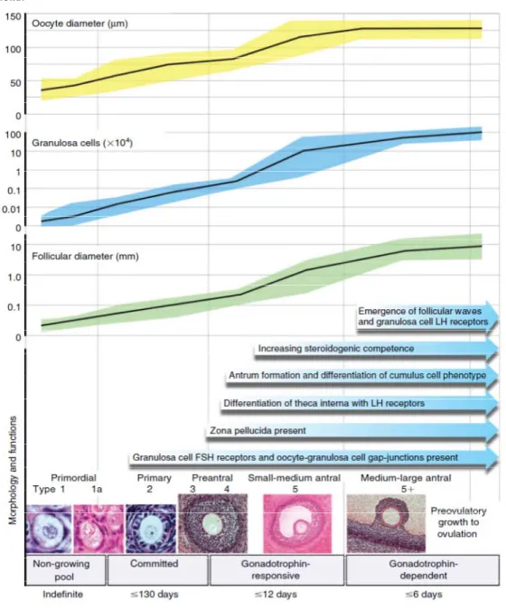
From the morphologic standpoint, the ovarian follicle may be classified into three major groups that are different in their size, complexity, and responsiveness to circulating gonadotropin. Primordial follicles include an oocyte surrounded by a single layer of epithelial, flattened granulosa cells with irregularly shaped nuclei. Theca cells are not present at this stage of folliculogenesis and they lack a distinct vasculature system.
Primary follicle contains a small oocyte with a single layer of cuboidal granulosa cells. Secondary follicles leave the primary stage and begin growth, but not develop a theca layer or antrum cavity. These types of follicles have two or more layer of granulosa cells and zona pellucida surrounding the oocyte. Preovulatory or graafian follicles have a clearly visible antrum. The zona pellucida (ZP) is formed and two layers of theca cells appear in this stage (Figure 1).
The oocyte pool in the mammalian ovary becomes fixed early in life. The number of primordial follicles that undergoing folliculogenesis to reach the mature, graafian=preovulatory stage is very low, and most of the follicles are either under a process of regression (atresia) or remain as primordial follicles without signs of growth. In response to preovulatory gonadotropin surges during each reproductive cycle, the dominant(s) graafian=preovulatory follicle ovulates to release the mature oocyte for fertilization, whereas the remaining theca and granulosa cells undergo transformation to become the corpus luteum (Myers et al., 2004).
6 1.1.1 Oocyte
An oocyte is a female gametocyte or germ cell and is produced in the ovary during gametogenesis. During oogenesis, a primordial germ cell (PGC) undergoes meiosis to
Figure 1. Follicular classification.
(A) Primordial follicles are defined as an oocyte surrounded by a flat layer of granulosa cells. (B) Follicles with cuboidal granulosa cells around the oocyte are classified as primary. (C) Secondary follicles are surrounded by more than one layer of cuboidal granulosa cells, with no visible antrum. (D) Preovulatory follicles are the largest follicles and possess a defined cumulus cell layer. Taken from (Myers et al., 2004).
7
form an oogonium, which becomes a primary oocyte (McDonald and Pineda, 1989). A relatively thick extracellular coat called the zona pellucida (ZP) surrounds the primary oocyte. The width of the ZP increases as the oocyte grows. Far from being a static structure, the ZP plays an important role in facilitating the interaction of sperm and oocyte at the time of fertilization. The ZP first appears as fibrillar material in the space between the oocyte and the cuboidal granulosa cells. As oocyte growth continues, the ZP becomes a denser and thicker network of connected filaments that completely surrounds the oocyte and largely separates it from the granulosa cells. Close contact continues to be maintained between the oocyte and surrounding granulosa cells via gap junction complexes within the zona until the completion of oocyte maturation (Conner et al., 2005).
Meiotic arrest of oocytes and cumulus cell expansion with germinal vesicle breakdown (GVBD) are signs of oocyte maturation. In mammalian follicles, primary oocytes enter meiosis but are arrested at the diplotene stage of prophase I. The oocytes stay in this state for months or years until the preovulatory stage. While the oocyte progresses through the maturation process in response to the preovulatory dose of LH surge, the germinal vesicle of the oocytes in preovulatory follicles undergoes GVBD, which is followed by chromatin condensation and the formation of meiotic spindles. The transition from metaphase I (MI) to metaphase II (MII) includes the extrusion of the first polar body. The maturing oocyte is the site of phosphorylation events that activate or deactivate the proteins involved in the progression of the cell cycle (Chian et al., 2003). Generally, meiotic arrest is controlled by the level of cAMP (cyclic adenosine monophosphate) in the oocyte. Recently, molecular studies have identified G-protein coupled receptors (GPCR) including GPR3 and GPR12, which control cAMP production and inhibit meiotic maturation in the oocytes of antral follicles. In fact, oocytes themselves
8
express a receptor that allows these cells to produce their own cAMP (Hinckley et al., 2005). In addition, AKAPs (PKA anchor proteins) can regulate the cAMP/PKA pathway and consequently meiosis in the mammalian ovary (Burton and McKnight, 2007).
Many of the effects of oocytes on follicular cells can be mimicked by members of the transforming growth factor (TGF) superfamily, including TGFβ1 and growth differentiation factor 9 (GDF-9) (Li et al., 2000). Genetic disruption of Gdf9 in mice causes infertility and abnormal follicle development with arrest at the primary follicle stage. Similar to GDF9, GDF-9B (also called bone-morphogenetic factor-15 (BMP-15)) is an oocyte-derived growth factor that regulates granulosa cell proliferation and differentiation. There is a species-to-species difference in the role of BMP15. Bmp15-null mice are sub-fertile, have defects in cumulus-oocte complex (COC) formation and show a decrease in ovulation rate (Richards and Pangas, 2010). Mutations of GDF9 and BMP15 have been found to cause premature ovarian failure in women (Simpson, 2008).
Oocyte-secreted factors regulate folliculogenesis by modulating a large number of processes associated with granulosa cell growth and differentiation, including cumulus cell mucification and hyaluronic acid production, cellular proliferation, steroid synthesis and luteinization inhibitors (Gilchrist et al., 2004). Experimental ablation of oocytes leads to failure of folliculogenesis. Removing oocytes from preovulatory rabbit follicles results in spontaneous luteinization (el-Fouly et al., 1970). It has been shown that oocytes secrete a potent mitogenic factor(s) that promotes mural granulosa and cumulus cell DNA synthesis and cell proliferation. Importantly, this oocyte mitogen(s) interacts with key known granulosa cell regulators such as FSH, insulin-like growth factors (IGF-I) and androgens, and increase their growth promoting activities (Eppig et al., 1997). Oocytes also promote the primary to secondary and preantral to antral follicle transitions, granulosa
9
cell proliferation, and differentiation before the LH surge, and cumulus expansion and ovulation after the LH surge. It appears that oocyte plays a dominant role in this kind of communication, and the rate of ovarian follicular development is coordinated by the oocyte’s developmental program (Su et al., 2009).
1.1.2 Granulosa and cumulus cells
As follicles grow and the antral cavity forms, granulosa cells separate into two anatomically and functionally distinct subtypes: the cumulus granulosa cells (CC), those surrounding and in contact with the oocyte, and the mural granulosa cells (MGC), which cover the follicle wall forming an epithelium with the basal lamina (Figure 2). Apart from anatomical differences, cumulus cells and MGC are functionally distinct (Albertini et al., 2001, Eppig et al., 1997).
10
In terms of function, CCs have a high rate of proliferation, low steroidogenic capacity, very low LHR (LH receptor) expression and high levels of insulin-like growth factor I compare to MGC, and they have the capacity to secrete hyaluronic acid and undergo mucification and expansion whereas MGC do not (Li et al., 2000). The expression of a variety of growth factors and hormone receptors also differ between MGC and CCs. Cumulus cells play an essential role in the normal growth and development of the oocyte, whereas MGCs serve a primarily endocrine function and support follicle growth. In addition, throughout ovulation, granulosa cells have an endocrine role by undergoing terminal differentiation to luteal cells, whereas cumulus cells mucify and are
Figure 2. Antral follicle structure with different granulosa cell types surrounding the oocyte.
11
ovulated with the oocyte, permitting ovum pickup and the sperm acrosome reaction (Li et al., 2000). Recently, many functional genomics studies have analyzed the importance of different genes by Cre/LoxP-mediated conditional deletion in granulosa cells. Two mouse strains expressing Cre recombinase (Amhr2-Cre and Cyp19-Cre) have been widely used used to target gene inactivation to granulosa cells. Amhr2-Cre is a knock-in of Cre in the anti-Mullerian hormone receptor type II locus, which directs Cre expression at early stages of follicle development. On the other hand Cyp19-Cre is a transgenic strain in which the aromatase promoter drives Cre expression in granulosa cells during antral stages of follicle development (Habenicht and Aitken, 2010).
1.1.2.1 Steroidogenesis
One of the major roles of granulosa cells is the production of sex steroid hormones. Estrogens have a crucial role in controlling fertility and infertility in mammals. Sex steroids play important roles in the development of the ovulatory follicles, the generation of the preovulatory surge of gonadotropins, facilitating sperm transport by changing the consistency of cervical mucus, and preparing the endometrial layer of the uterus for implantation. Compared to cumulus cells, mural granulosa cells are more steroidogenically active, as indicated by higher levels of mRNA expression for steroidogenic enzymes including 17β-hydroxysteroid dehydrogenase (HSD17B1) and aromatase (CYP19A1). The abundance of Cyp19a1 mRNA in ovarian granulosa cells is regulated by factors which stimulate adenylyl cyclase and increase cAMP.
In the ovary, FSH can lead to the activation of second-messenger signaling including cAMP, which drives the expression of CYP19A1 and HSD17B1 and thereby permits MGCs to produce estradiol by converting androgen which comes from the theca
12
cells (Figure 3) (Simpson et al., 2002). The two-cell, two-gonadotropin model describes the role of theca and granulosa cells in the production of steroids, highlighting the cooperation between the two cell types, which is necessary for estrogen production (Li et al., 2000). Other than sex steroids, granulosa cells are capable of producing growth factors including inhibin, activin, bone morphogenetic proteins 2 (BMP-2) and BMP-6. These molecules can communicate between the GCs and the oocyte and theca cells during their development. In addition, after ovulation the luteinized granulosa cells produce progesterone (Gilchrist et al., 2004, Pangas, 2012).
13
Theca cell
Figure 3. Steroidogenesis in mural granulosa cells.
In the growing follicle, androstenedione and testosterone can diffuse through the basement membrane. These androgens are then further transformed into estradiol in granulosa cells. Dotted arrows represent molecule translocations and plain arrows indicate an enzymatic conversion. Reproduced from reference (Lapointe and Boerboom, 2011).
14 1.1.2.2 Cumulus expansion
In most mammals, the LH surge starts an ovulatory cascade by inducing cAMP signaling, which stimulates the expansion of the cumulus cells surrounding the oocytes, the rupture of the follicle wall, and the release of the fertilizable ovum. As mentioned above, one of the distinct differences between MGC and CC is the capacity of cumulus cells to undergo mucification and expansion. CCs secrete proteins creating a large round mass of expanded cumulus cells via an extensive rearrangement of the actin cytoskeleton and induction of hyaluronan synthesis, which transforms the tightly packed cumulus complex into a much larger volume of mucified cells. Hyaluronan is formed by various hyaluronan binding proteins including versican, inter-α trypsin inhibitor (IαI) and tumor necrosis factor stimulated gene 6 (TSG-6). This structure has viscoelastic properties which allow COCs to deform and easily pass through the ruptured follicle wall during ovulation (Nagyova, 2012). In vitro, cumulus expansion is induced by treatment with FSH, which stimulates cAMP signaling to produce a massive mucoid extracellular matrix. COCs expansion varies in different species, for instance mouse COC expansion in culture requires an oocyte secreted factor(s), termed the cumulus expansion enabling factor (CEEF). In comparison, FSH can induce rat, cow, and pig COC expansion in the absence of the oocyte (Gilchrist et al., 2004, Li et al., 2000).
Small molecules such as ions, metabolites and amino acids can be transferred via gap junctions between the oocyte and cumulus cells as well as between cumulus cells to help oocyte growth and development (Gilchrist et al., 2004).
15 1.1.3 Theca cells
Theca cells are vital components of the follicle, providing both structural support and the production of ovarian androgens for the growing follicle as it progresses through the developmental stages to produce a mature and fertilizable oocyte. Androgens are essential substrates for estradiol production in the neighbouring granulosa cells. Theca cells are recruited by factors secreted in activated primary follicles from surrounding stromal tissue (Magoffin, 2005). Theca cells are first observed follicles with two or more layers of granulosa cells. Before this stage, undifferentiated theca cells do not express LH receptors or steoidogenic enzymes. As theca cells are only associated with growing follicles, it is assumed that the follicle itself produces factors that signal to the stroma to recruit cells that form the theca. Theca cells have some necessary roles during folliculogenesis, such as the synthesis of androgens and provide crosstalk with the oocyte and granulosa cells during follicle development. Moreover, they play an important role in the establishment of a vascular system that creates communication with the pituitary gland throughout the reproductive cycle, and delivers essential nutrients to these highly active cells. During atresia, the theca cells are often the last cell type to undergo apoptosis, and after ovulation, theca cells luteinize and form cells of the corpus luteum (Magoffin, 2005, Young and McNeilly, 2010).
1.1.3.1 Steroidogenesis
Theca cells are capable of producing androgen from circulating cholesterol (high and low-density lipoproteins) in response to LH from the pituitary gland, which activates cAMP signaling via a G protein-coupled receptor. Pulsatile release of LH triggers an increase in androstenedione and estradiol secretion by the ovary in many species.
16
Androgens synthesized in theca cells are transported to the granulosa cells where cytochrome P450 aromatase enzyme converts these androgens to estrone and 17β-estradiol (Young and McNeilly, 2010). In theca cells, LH/cAMP induces the expression of the key steroidogenic enzymes cytochrome P450 cholesterol side-chain cleavage (CYP11A1), cytochrome P450 17β-hydroxylase/C17–20 lyase (CYP17A1), 3β-hydroxysteroid dehydrogenase/Δ5-Δ4 isomerase (HSD3B2) and steroidogenic acute regulatory protein (STAR). Cholesterol inside the theca cells is transported by sterol carrier protein 2 (SCP2) to the mitochondria. This internalization of cholesterol by the mitochondria is the rate-limiting step for steroidogenesis. STAR helps cholesterol to enter the inner mitochondrial membrane, where CYP11A1 converts it to pregnenolone. Pregnenolone can then be converted to progesterone by the enzyme 3β-hydroxysteroid dehydrogenase, or to 17α-hydroxypregnenolone by the enzyme CYP17A1 (Figure 4) (Conley and Bird, 1997, Lapointe and Boerboom, 2011).
17
Granulosa
Figure 4. Enzymatic control of steroidogenesis in the theca cell.
LH can stimulate theca cells to convert cholesterol into androstenedione and
testosterone, which can diffuse to the granulosa cell layer. Dashed arrows represent molecule translocations and plain arrows indicate an enzymatic conversion. Reproduced from reference (Lapointe and Boerboom, 2011).
18 1.1.4 Basement membrane
The basement membrane or lamina propria separates the theca and granulosa cell layers. The basal lamina creates a blood–follicle barrier between the two follicular cell compartments, and the presence of this extracellular matrix (ECM) boundary influences the interactions of follicular somatic cells. Some evidence supports the involvement of ECM components in regulating granulosa cell survival, proliferation, and steroidogenesis in the ovarian follicles of several species. A continuous remodelling of the basal lamina occurs during follicle development and its composition is changed during this process (Allegrucci et al., 2003). For instance, the laminin β1 chain was only detectable in the basal lamina of large preantral follicles, and type IV collagens α1 and -6 were reduced considerably in larger antral follicles (Rodgers et al., 2000).
1.2 Endocrine regulation of follicle growth and development
Folliculogenesis describes the progression of a number of small primordial follicles into large preovulatory follicles with the potential to either ovulate or undergo atresia. Regulation of follicle development is a complicated process that is controlled by several factors such as hormones, protein growth factors, and nutrition. Folliculogenesis can be coordinated by these factors via autocrine (affecting the cell type that produces the factor), paracrine (affecting cells adjacent to the one(s) producing the factor) and endocrine (affecting distant cells/organs) mechanisms (Armstrong and Webb, 1997, Webb et al., 2004).
19 1.2.1 Gonadotropins
LH and FSH are called gonadotropins because stimulate the gonads, the testes in males, and the ovaries in females. They are both are essential for steroidogenesis, spermatogenesis, folliculogenesis, and ovulation. They are non-covalently linked heterodimeric glycoproteins include a common α-subunit and specific β subunits. Distinct genes encode the gonadotropin subunits, which are synthesized as precursor proteins and processed and secreted by the pituitary gonadotropes of the anterior pituitary (Burger et al., 2004). Pituitary gonadotropins release is under the acute trophic control of the main reproductive system regulator; the hypothalamic decapeptide gonadotropin-releasing hormone (GnRH). GnRH is released and transported to the anterior pituitary in a pulsatile manner, where it binds to specific receptors and regulates gonadotropin biosynthesis and secretion. The pulse is modulated by a wide variety stimulatory and inhibitory feedback to the both hypothalamus and the pituitary.
Steroidal hormones can regulate gonadotropin levels by adjusting the GnRH pulse frequency at the levels of the in hypothalamus, and by acting directly on the pituitary gonadotrope cells. On the other hand, the members of the transforming growth factor-β superfamily, activin and inhibin, act at the level of the pituitary to control FSH secretion (Figure 5). Both LH and FSH exert their effects on ovarian somatic cells via G protein-coupled receptors that activate several signaling pathways (Burger et al., 2004, Dohler et al., 1977).
20 1.2.1.1 FSH
It is now well accepted that circulating FSH concentrations increase transiently with the emergence of each follicular wave during estrous. FSHR (FSH receptor) mRNA is detected in follicles with only one or two layers of granulosa cells in humans (Oktay et al., 1997).
Suppression of FSH secretion inhibits follicular waves in cattle (Turzillo and Fortune, 1990). One study in cattle shown that a single follicle continues to grow and
Figure 5. Regulation of gonadotropins
FSH and LH plasma concentrations above threshold levels lead to the emergence of greater inhibitory feedback, and thus a reduction in gonadotropin secretion. Estradiol increases GnRH pulse frequency through a positive feedback mechanism, and results in elevated LH secretion. However, progesterone has a negative feedback effect on pulse frequency; therefore LH secretion is suppressed in the presence of a functional corpus luteum. Moreover, activin, opposite to inhibin, can stimulate FSH release.
21
mature because it acquires increased sensitivity to FSH, while further development of other follicles in the same cohort with less sensitivity to FSH is inhibited. Follicles, growing under the FSH support, start producing more estradiol and inhibin, which act on the anterior pituitary to inhibit FSH secretion through a negative feedback mechanism. The decline in FSH concentrations is closely related with the selection of one follicle as dominant and others as its subordinates (Gibbons et al., 1997). Generally, FSH is responsible for oocyte maturation indirectly by inducing Lhcgr in GC and estrogen synthesis. In absence of FSH, follicle development fails and ovulation does not occur. FSH acts through G-protein couple receptors to transduce its signal. Binding of FSH to its receptor results in cAMP synthesis as a second messenger, resulting in stimulation of PKA and the p38 mitogen-activated protein kinase (MAPK) signaling pathway (Levallet et al., 1999).
1.2.1.2 LH
LH acts via G protein coupled receptors in the preovulatory follicle. LH receptors are first expressed on the cells of the theca interna at the tertiary stage of follicle development, and this expression is maintained until the preovulatory stage. In addition, granulosa cells of large estrogenic antral follicles develop LH receptors, as do luteal cells. (Ashkenazi et al., 2005). LH controls granulosa cell differentiation, cumulus expansion and progesterone production. Meiosis in the oocyte, arrested in the first meiotic division during embryogenesis, resumes following the luteinizing hormone surge. LH also up-regulates the expression of proteases that allow the follicle wall to rupture and release the oocyte. Luteinizing hormone increases steroidogenesis by promoting cholesterol transport to mitochondria and the stimulation of the expression of steroidogenic genes.
22
The LH surge induces a large increase in mRNA encoding epiregulin (EREG) and amphiregulin (AREG), which then activate the EGFR (epidermal growth factor receptor) and initiate the ovulatory cascade and induce oocyte maturation (Hsieh and Conti, 2005). The LH surge also results in increased synthesis of prostaglandins in the preovulatory follicle that are required for ovulation (Richards, 1994).
1.2.2 Estradiol and progesterone
Estradiol and progesterone biosynthesis within the ovary is critical for various physiological processes related to normal growth, development, and reproduction (follicle development, regulation of gonadotropin secretion for ovulation and maintenance of pregnancy) (Stormshak and Bishop, 2008).
FSH stimulates the production of estrogens by the granulosa cells of the ovarian follicles. Healthy ruminant follicles contain higher estradiol and lower progesterone concentrations than atretic follicles. It is well established that small follicles contain relatively little estradiol in their follicular fluid, and estradiol concentrations increase with follicle size in healthy growing follicles while decreasing in subordinate follicles. In cattle, once the dominant follicle reaches its maximum diameter, estradiol concentrations dramatically increase in follicular fluid and it decreases when follicle starts regressing. Thus, estradiol concentrations are a key biochemical indicator of health and atresia of ruminant follicles (Price et al., 1995).
Progesterone has a critical role in the regulation of ovulation. The physiological effects of progesterone are mediated by two nuclear receptors (PR-A and PR-B). Studies of progesterone receptor-null (Pr-KO) mice showed that PRs are required for LH-dependent follicle rupture (Conneely, 2010).
23
In general, the action of progesterone on the reproductive tract is to stimulate the proliferation of uterine stromal cells, the epithelial cells of the mammary gland, and to ensure the maintenance of pregnancy. Progesterone, in addition to estradiol, plays an important role in the regulation of LH secretion by exerting negative feedback on the hypothalamus-pituitary axis, thereby reducing the release of GnRH and LH (Murphy, 2000, Niswender et al., 2000).
1.2.3 Androstenedione and testosterone
Androstenedione is a 19 carbon steroid hormone produced in the adrenal glands and the gonads as an intermediate product that can be converted to testosterone or estradiol. Testosterone is the primary mammalian androgen. It is the mainly known as a male sex hormone and an anabolic steroid. Testosterone is primarily secreted in the testicles of males and the ovaries of females, although small amounts are also secreted by the adrenal glands. In theca cells, much cytochrome P450 17α-hydroxylase enzymatic activity is present, and so pregnenolone (the first steroid in the steroidogenesis pathway) is converted to 17α-hydroxypregnenolone. This 17α-hydroxypregnenolone is then converted to androstenedione by P450 17α- hydroxylase (CYP17 or P45017-OH) and 3β-hydroxysteroid dehydrogenase (3β-HSD or HSD3B2). Ruminant theca cells convert limited amounts of androstenedione to testosterone with the enzyme 17β-hydroxysteroid dehydrogenase, and both androstenedione and testosterone can then diffuse to neighbouring granulosa cells for further conversion to estrogens (Gibbons et al., 1997).
24 1.2.4 Intraovarian regulators
Ovarian folliculogenesis is a dynamic process controlled by gonadotropins. However, intraovarian regulators also play key roles in granulosa cell biology, maturation of the oocyte, and neovascularization. These factors can act in an autocrine or paracrine manner through specific cell membrane receptors. The early stages of follicular growth are gonadotropin-independent, and these stages may therefore be controlled by intraovarian signaling (Ben-Ami et al., 2006).
1.2.4.1 IGF
The IGF family members are polypeptides similar to insulin. The IGF system includes two ligands (IGF-I and -II), 6 IGF binding protein (IGFBPs) and two receptors (IGF1R and -2R).
IGFs are regulated by gonadotropins and growth factors such as growth hormone (GH) and oxytocin in ovarian follicles, corpora lutea, and oocytes. In ruminants and rodents, IGF-I plays a key role in reproduction, whereas in human IGF-II appears more important. Whereas I probably has no effects on primordial follicles, I and IGF-II stimulate the growth of secondary follicles in vivo and in vitro. In antral follicles, IGF-I stimulates granulosa cell proliferation and synergizes with gonadotropins to promote differentiation of follicle cells and inhibit apoptosis (Beg and Ginther, 2006). IGFs increase the expression of FSH and LH receptors and the response of granulosa cells, theca cells, and oocytes to gonadotropins. Moreover, IGFs are considered to be one of the main local mediators of many hormones, including gonadotropins and GH (Quirk et al., 2004).
25
IGF-I and IGF-II are produced in large amounts in the corpus luteum to support luteal cell development and function (Beg and Ginther, 2006). Both IGF-I and IGF-II in mice promoted nuclear maturation of oocytes, with stimulation of proliferation and inhibition of apoptosis in the surrounding cumulus oophorus, resulting in increased developmental capacity of the oocytes and blastocyst production after in vitro fertilization (IVF) (Demeestere et al., 2004).
In addition, it is possible that IGF-I and IGF-II mediate effects of nutrients and nutrition-dependent metabolic hormones on reproductive processes (Sirotkin, 2010, Webb et al., 2004). IGF1 seems to stimulate estradiol and progesterone secretion and can stimulate PI3K (phosphoinositide 3-kinase) and PKC (protein kinase C) to increase the expression of CYP19A1 and other steroidogenic enzymes in bovine granulosa cells, and regulate secretion of progesterone and androstenedione in theca cells (Silva and Price, 2002, Stocco, 2008b).
The IGFBPs consist of a family of six non-glycosylated peptides with relatively low molecular weights, which serve to regulate the bioavailability of IGFs within the follicle. IGFBP2, 4 and 5 serve as IGF carriers in the serum, thereby extending their half-lives by preventing their degradation. IGFBPs also regulate the effects of IGFs by preventing their access to cell membrane surface receptors (Ben-Ami et al., 2006).
1.2.4.2 TGF-β superfamily
The TGF-β superfamily of extracellular signaling molecules includes over 35 structurally related but functionally diverse proteins. These proteins are widely distributed throughout the body and acts as extracellular ligands involved in numerous physiological processes during both prenatal and postnatal life. This big superfamily has been divided
26
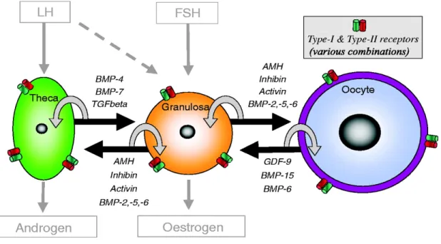
into several subfamilies which include the β (including β1, β2 and TGF-β3), bone morphogenetic protein (BMP, with 20 members), growth and differentiation factor (GDF) (at least nine members), activin/inhibin (including activins A, AB and B and inhibins A and B), and glial cell-derived neurotrophic factor (GDNF) (including GDNF, artemin and neuturin) subfamilies, as well as other members including anti-Mullerian hormone (AMH) and nodal (Knight and Glister, 2006). Several members of this superfamily are expressed by oocytes, granulosa cells and theca cells, and have been implicated in granulosa and theca cell proliferation, atresia, steroidogenesis, oocyte maturation, ovulation and luteinization (Knight and Glister, 2003).
The TGF-β superfamily members GDF-9 and BMP-15 are expressed in the oocyte from an early stage of development, and play key roles in promoting follicle growth to the primary stage. Studies on later stages of follicle development have indicated important roles for activin, BMP-2, 5 and 6, theca cell-derived BMP-2, 4 and 7 and oocyte-derived BMP-6 in promoting granulosa cell proliferation, follicle survival and the prevention of premature luteinization and/or atresia (Figure 6). TGF-β increases LH receptor production in granulosa cells in response to FSH stimulation, whereas it inhibits androgen production in theca cells. In monovular species, dominant follicle selection may depend on the sensitivity of the growing cohort of small antral follicles to FSH. Changes in the intrafollicular concentrations of molecules including activins, AMH, GDF-9 and several BMPs may contribute to this selection process by controlling both FSH- and IGF-dependent signaling pathways in granulosa cells (Knight and Glister, 2003, 2006). Activin may also play a positive role in oocyte maturation and competency, and can increase FSH receptor expression in granulosa cells during the preantral to antral transition. Follicles with greater capacity for activin signaling are more sensitive to FSH, and show better
27
progression to the antral stage (Findlay et al., 2001). One study suggested that inhibin production by large antral follicles is a mechanism for the selection of the dominant follicle and the induction of apoptosis in small subordinate follicles (Vitale et al., 2002).
1.2.4.3 FGFs
The fibroblast growth factor (FGF) superfamily is one of the largest families of growth factors that have been implicated in follicle cell function. There are 22 known
Figure 6. TGF-β superfamily members and their receptors and binding proteins are expressed in different follicle cells.
TGF-β superfamily members including GDF-9, inhibins, activins, TGF-β, BMPs 2, 4, 5, 6 and -7 and AMH are expressed in theca cells, granulosa cells and oocytes. These molecules serve as as intrafollicular autocrine (white arrows) and paracrine (black arrows) signaling molecules. Reproduced from reference (Knight and Glister, 2006).
28
FGFs classed into 7 subfamilies. Most of the members of this family act through single-pass transmembrane tyrosine kinase receptors (FGFR) which are encoded by four separate genes and give rise to four distinct receptor proteins, FGFR1–4 (Schams et al., 2009). Fibroblast growth factors are most famous for their critical roles in organogenesis in the developing embryo (Schams et al., 2009). In the ovary, FGFs are predominantly expressed in oocytes and/or theca cells but are absent from the granulosa layer. FGFs are well known to stimulate granulosa cell proliferation. A consequence of increased proliferation is a decrease in differentiation and estradiol secretion (Buratini et al., 2007). In the reproductive system, FGF2, FGF9, FGF7, and FGF18 play roles in gonadal development and sex differentiation (Portela et al., 2010, Schams et al., 2009). Moreover, FGF7 and FGF10, which belong to the same FGF subfamily, are expressed predominantly in theca cells, and their main receptor (FGFR2) is located on granulosa cells. FGFs are therefore important for paracrine signaling in the ovarian follicle (Portela et al., 2010). FGF2 is one of the members of this family that can regulate angiogenesis in the theca. FGF2 also acts on granulosa cells to promote granulosa cell proliferation, inhibit apoptosis, and decrease steroidogenesis (Jiang et al., 2011). FGF9 is also expressed predominantly in theca cells and stimulates progesterone secretion by granulosa cells. FGF8 cooperates with BMP15 to stimulate glycolysis in cumulus cells (Portela et al., 2010).
FGFs can activate different interacellular pathways including 1) MAPK, widely accepted to be responsible for the mitogenic responses of cells to FGFs, 2) phospholipase C (PLC), which activates PKC and calcium signaling by catalysing the production of diacylglycerol and inositol-1,4,5-triphosphate, and 3) PI3K and AKT (Jiang et al., 2011).
29 1.2.4.4 EGF
Epidermal growth factor (EGF) is a protein with 53 amino acid residues and three intramolecular disulfide bonds, and plays crucial roles in reproduction. Members of the EGF-like family have highly similar structural and functional characteristics. Aside from EGF, other family members include TGF-α, amphiregulin, epiregulin, betacellulin (BTC), epigen, neuregulins and heparin-binding EGF-like growth factor. These proteins can work through four types of transmembrane receptors (Ashkenazi et al., 2005, Ben-Ami et al., 2006).
Because cumulus and oocyte cells do not have LH receptors, it has been proposed that factors released by mural granulosa cells function in an autocrine and paracrine manner to transduce the effects of LH within the follicle. Intrafollicular release of members of the EGF-like family may fulfil this role (Park et al., 2004). It is now known that the EGF-like growth factors AREG, EREG and BTC, rather than EGF, are rapidly and transiently induced in the somatic cells of the preovulatory follicle by LH (Ashkenazi et al., 2005, Hsieh and Conti, 2005, Park et al., 2004). LH/hCG induction of these EGF-like growth factors has been reported in multiple species (Chen et al., 2008, Fru et al., 2007, Hsieh et al., 2007, Wang et al., 2007).
The up-regulation of EGF ligands was initially proposed to occur selectively in mural granulosa cells, from which the ligands would be shed by members of the matrix metalloprotease (MMP) or a disintegrin and metalloprotease (ADAM) families to enable paracrine signaling to the cumulus layer (Conti et al., 2006). The mature growth factors bind to members of the EGFR (also called ERBB1) family, leading to receptor dimerization and autophosphorylation, and activation of downstream signaling cascades
30
including the ERK1/2, AKT, MAPK (via Gγ-c-Src) and PKC pathways to elicit distinct biological effects (Conti et al., 2012).
1.3 Folliculogenesis and the ovarian cycle
The major role of the ovary is the production of mature oocytes for fertilization. Folliculogenesis, the growth and development of ovarian follicles from primordial to preovulatory, is a prolonged process dependent on interactions between the oocyte and the somatic cells including the granulosa and theca cells. The ovarian reserve is determined by the number of primordial follicles in the ovary. Primordial follicles are activated for growth and pass through stages of development until they reach the antral stage. A group of antral follicles is then recruited for further growth, which is followed by the selection of a dominant follicle in monovular species. These processes are under the control of endocrine as well as paracrine factors in the ovary (Fortune et al., 2004, McGee and Hsueh, 2000). In response to preovulatory LH surges during each reproductive cycle, the dominant follicle ovulates and releases the mature oocyte for fertilization, while the remaining theca and granulosa cells will be transformed into corpus luteum through luteinization (Figure 7) (McGee and Hsueh, 2000).
31 1.3.1 Primordial germ cells
The mammalian germ cell lineage is established early during development. Primordial germ cells (PGCs) originate in the proximal region of the epiblast, close to the extra embryonic endoderm, which during gastrulation clusters through the posterior part of the primitive line to become PGCs. These cells move into the endoderm of the yolk sac,
Figure 7. Schematic diagram of the ovary
The ovary is a dynamic organ that undergoes dramatic changes in structure and function. The follicle is the major endocrine and reproductive compartment of the ovary, and undergoes growth, rupture and formation of the corpus luteum during its life span. Taken from (McDonald and Pineda, 1989).
32
then proliferate and migrate via the yolk sac and hindgut endoderm at the caudal end of the embryo, and finally via the dorsal mesentery of hindgut to the genital ridges at the ventral sides of the mesonephros (Barnett et al., 2006). Active oogonia can recruit somatic pregranulosa cells from the surface epithelium at this stage. The oogonia then attach to pregranulosa cells and enter meiosis, during which at least 80% of the germ cells, but not their pregranulosa cells, undergo apoptosis (Scaramuzzi et al., 2011).
Primordial germ cells differentiate under the influence of signals from members of the transforming growth factor β superfamily, including BMP2, BMP4, and BMP8B. BMP2 originates from the endoderm, whereas BMP4 and -8b are from the extra-embryonic ectoderm. Loss of any of these signals prevents the formation of most or all of the primordial germ cells. (Barnett et al., 2006, Ying and Zhao, 2001). Conditional knockout mice have helped to identify genes required for primordial germ cell proliferation and migration. The c-kit receptor tyrosine kinase and its ligand (kit ligand, or stem cell factor) are required for primordial germ cell survival and migration. Expression of integrin β1 on the primordial germ cell surface is also required for successful migration to the genital ridge (Barnett et al., 2006).
1.3.2 Transition from oogonia to oocyte
PGCs are called oogonia once they reach to the gonads. Oogonia are connected to each other via intercellular cytoplasmic bridges. These oogonia are surrounded by mesonephros somatic cells, forming germ cell clusters or nests. These germ cell clusters break down before or after birth (according to species) to allow the formation of primordial follicles (Figure 8). Improper germ cell cluster breakdown may cause polyovular follicles (follicles with more than one oocyte).
33
Oogonia have higher mitotic activity compare to PGCs and undergo several divisions before starting meiosis. This mitotic activity is the major determinant of the size of the oocyte pool. On the other hand, a huge number of oogonia undergo atresia at this stage (approximately 60% in sows, 80% in rodents, 90% in humans and even more in sheep and cows (van den Hurk and Zhao, 2005). The end of reproductive life occurs when the follicle reserve is expended through follicle development and atresia. (Oktem and Urman, 2010).
Several genes are known to play roles in primordial follicle formation, including Dazla (deleted in azoospermia), which is essential for the differentiation of germ cells, and Figla (factor in the germline alpha), which is essential for follicle formation and the interaction between the oocyte and granulosa cells. Nerve growth factor (Ngf) is involved in primordial follicle assembly (Barnett et al., 2006).
34 1.3.3 Formation of primordial follicles
In term of morphology, primordial follicles in most species consist of small diplotene oocytes with a zona pellucida layer, surrounded by a flat layer of granulosa cells (Figure 9) (Rodgers and Irving-Rodgers, 2010). In primordial follicles, the granulosa cells and oocyte have receptors for some growth factors but not for gonadotropins. However, at this stage they do not require FSH and LH for their survival and development (Fortune et al., 2011). Around the initiation of follicle formation, estrogen levels are decreased
Figure 8. Germ cell cluster breakdown and formation of primordial follicles. Germ cells are present in the ovary during embryogenesis. Around birth, germ cell clusters start to break down and form primordial follicles. Several mechanisms including apoptosis are involved in this process. Taken from (Barnett et al., 2006).
35
relative to the period of germ cells clusters, and low amounts of androgens and progestins are detectable. Steroid production could be important for primordial follicle formation, and seems to be independent of gonadotropin secretion (van den Hurk and Zhao, 2005).
A large number of factors including transcription factors, zona proteins, meiosis-specific enzymes and nerve growth factors are important for primordial follicle assembly (Oktem and Urman, 2010). Studies have identified a variety of genes involved in the formation of primordial follicles, which include sporulation protein homology (Spo11), disrupted meiotic cDNA 1 homologue (Dmc1) and muts homologue 5 (Msh5). All three genes are necessary for the initiation of double strand DNA breaks during meiosis and are involved in recombination (Barnett et al., 2006).
1.3.4 Transition of primordial follicles to primary follicles
Primary follicles are similar to primordial follicles in terms of size, but have more cubical-shaped granulosa cells. Primordial follicles first develop into primary follicles prior to or after birth (according to species), and this process continues postnatally until the ovarian reserve is depleted, which takes more than a year in rodents and several decades in women (Oktem and Oktay, 2008).
The transition of a primordial follicle into a primary follicle is more of a slow maturation than a growing process, as the diameter of its oocyte hardly changes (Rodgers and Irving-Rodgers, 2010). Communication among the oocytes, granulosa and certain extra cellular matrix components and autocrine/paracrine growth factors play important roles in this transition and subsequent growth of follicles (Oktem and Urman, 2010).
Inhibitory and stimulatory factors from different cells (oocytes, somatic cells, and stroma) control primary follicle development. Recently, it has been found that inhibitory
36
signals keep primordial follicles in the dormant state. For example, loss of function of some inhibitory molecules for follicular activation, including the tumor suppressor tuberous sclerosis complex 1 (Tsc-1), phosphatase and tensin homolog deleted on chromosome 10 (Pten), Forkhead box O3 (Foxo3a), newborn ovary homeobox protein (Nobox) and forkhead box protein L2 (Foxl2), leads to premature activation of the primordial follicle pool (van den Hurk and Zhao, 2005).
According to studies on transgenic animal models and on the human ovary, several members of the TGF-β super family, such as AMH, activins, BMP-4, BMP-7, and GDF-9, play critical roles in the regulation of primary follicle activation. There are other growth factors and cytokines that act at a paracrine level in the formation of primary follicles, such as kit-ligand, fibroblast growth factor-7 and the leukemia inhibitory factor, which have been shown in vitro to promote follicle growth from the primordial to the primary stage, stimulate oocyte growth and the proliferation of theca cells (Barnett et al., 2006, Oktem and Oktay, 2008).
1.3.5 Follicle growth to pre-antral and antral stages
A follicle with two or more layers of granulosa cells is deemed a secondary or preantral follicle. At this stage, the oocyte enters its growth phase, its diameter increases, granulosa cells become more proliferative, and a theca layer develops around the granulosa cells. Moreover, at this stage the zona pellucida completely forms, providing a protective coat around the oocyte composed of three glycoproteins; ZP1, ZP2 and ZP3 (figure 9) (Eppig, 2001).
During oocyte growth, RNA and protein synthesis increases, and ribosomes, mitochondria and other organelles increase in number. Granulosa cells form gap junctions