Increased platelet reactivity and platelet
–leukocyte aggregation after
elective coronary bypass surgery
Torbjörn Ivert1, Magnus Dalén1, Charlotte Ander2, Ragnhild Stålesen2, Marie Lordkipanidzé 3, & Paul Hjemdahl2
1
Heart and Vascular Theme, Karolinska University Hospital and Department of Molecular Medicine and Surgery, Karolinska Institutet, Stockholm, Sweden,2Department of Clinical Pharmacology, Karolinska University Hospital and Department of Medicine Solna, Karolinska Institutet, Stockholm,
Sweden, and3Faculté de pharmacie, Université de Montréal,and Research center, Montreal Heart Institute, Montréal, Québec, Canada
Abstract
Inflammatory mechanisms are activated, and thrombotic complications occur during the initial months after coronary artery bypass grafting (CABG). Therefore, changes over time of platelet activation and platelet–leukocyte interactions after CABG are of interest. Whole-blood flow cytometry was performed before, and 4–6 days, one month, and three months after elective CABG in 54 men with stable coronary artery disease treated with acetylsalicylic acid (ASA). Single platelets and platelet–leukocyte aggregates (PLAs) among monocytes (P-Mon), neutrophils (P-Neu), and lympho-cytes (P-Lym) were studied without and with stimulation by submaximal concentrations of ADP, thrombin, and the thromboxane analog U46619. White blood cell counts were increased during the initial postoperative course, and platelet counts were increased after one month. Platelet P-selectin expression was significantly enhanced at one month when stimulated by thrombin and U46619 and at three months with ADP and thrombin. All PLAs subtypes were increased at one month without stimulation in vitro. P-Mon and P-Neu stimulated by ADP, thrombin, or U46619 were significantly increased one month after the operation but decreased compared to baseline at three months. Agonist stimulated P-Lyms were increased at one month and remained increased at three months after ADP stimulation. There was significant platelet activation and formation of PLAs unstimulated and after agonist stimulation by ADP, thrombin, and a thromboxane analog after CABG in patients with stable coronary artery disease irrespective of ASA treatment. Changes observed up to three months after CABG support further studies of the clinical implications of protracted increases in platelet activation and platelet–leukocyte interactions.
Keywords
Coronary bypass, platelet activation, platelet aggregates
History
Received 25 June 2018 Revised 14 October 2018 Accepted 22 October 2018
Published online 13 November 2018
Introduction
During recent years, it has become increasingly clear that platelets and platelet–leukocyte interactions are involved in the complex pathophysiology of atherosclerosis and the termi-nology athero-thrombosis is frequently used in this context [1– 8]. Cardiopulmonary bypass activates inflammatory mechan-isms and several aspects of hemostasis [9,10]. We have pre-viously reported on biphasic pro-thrombotic and inflammatory responses after coronary artery bypass grafting (CABG) in a study focusing on the first week after surgery [11]. Thrombo-embolic events may occur during the first month after cardiac surgery and early vein graft failure is related to thrombosis and high platelet reactivity [12,13]. Thus, more intense antiplatelet therapy than acetylsalicylic acid (ASA) alone may be required
to improve the prognosis of patients with stable coronary artery disease undergoing CABG [14,15].
We have previously reported on the efficacy of different ASA dosages following CABG and found that 75 mg twice daily or 160 mg once daily inhibited thromboxane-dependent platelet acti-vation more effectively than 75 mg once daily [16]. The present study describes platelet reactivity and circulating platelet –leuko-cyte aggregates (PLAs) irrespective of ASA dosage following elective CABG in patients without indication for dual antiplatelet therapy. Flow cytometry of single platelets or PLAs is insensitive to ASA treatment as such measurements are performed under static conditions that do not promote thromboxane generation [17–19]. The present aim was to investigate changes in thrombox-ane independent platelet activation and platelet–leukocyte inter-action including responses to agonist stimulation during the recovery phase, at one and three months after elective CABG in patients with stable coronary artery disease.
Patients and Methods Subjects
We included patients with stable angina pectoris scheduled to undergo elective CABG. Exlusion criteria were intake of any other platelet inhibitor than ASA during the last seven days
Color versions of one or more of the figures in the article can be found online atwww.tandfonline.com/iplt.
Correspondence: Torbjörn Ivert, Heart and Vascular Theme, Karolinska University Hospital, Stockholm SE-171 76, Sweden. E-mail: torbjorn. [email protected]
This is an Open Access article distributed under the terms of the Creative Commons Attribution-NonCommercial-NoDerivatives License ( http://creati-vecommons.org/licenses/by-nc-nd/4.0/), which permits non-commercial re-use, distribution, and reproduction in any medium, provided the original work is properly cited, and is not altered, transformed, or built upon in any way.
ISSN: 0953-7104 (print), 1369-1635 (electronic) Platelets, 2019; 30(8): 975–981
prior to surgery, known bleeding disorder or kidney failure, pre-operative platelet count outside of the range 100,000–450,000/µL, or a hemoglobin level below 8 g/L. We screened 75 subjects of whom 54 were included in the final analysis; 20 patients were excluded because of incomplete follow-up with blood samples and one patient with insulin-treated diabetes mellitus and advanced three-vessel disease died suddenly two months after the operation. Results in 42 of the patients have been previously reported regarding the serum thromboxane B2 effect of the ASA doses 75 mg twice daily, 160 mg once daily, and 75 mg once daily [16]. Twelve patients with complete cytometric data and included in the present analyses were excluded in the previous study due to faulty compliance with the prescribed ASA dosage and study procedures.
All patients were treated with ASA (Trombyl®, Pfizer Health AB, Strängnäs, Sweden) 75 mg OD before the operation and they received ASA 75 mg OD or BID or 160 mg OD at discharge from the hospital. The study was approved by the Regional Ethical Review Board in Stockholm (2011/1074–31/1) and by the Swedish Medical Products Agency (EudraCT 2011–002233-19). The study is registered with ClinicalTrials.gov (NCT02482857). A signed consent form was obtained from all participants.
Study Design
All operations were performed through a standard sternotomy with the aid of standard cardiopulmonary bypass using a non-pulsatile centrifugal pump and a membrane oxygenator. The extracorporeal system was primed with Ringer’s acetate. Cold blood cardioplegia was used for myocardial protection at a core temperature of 36°C while the aorta was cross-clamped. Heparin was given before cardiopulmonary bypass (3 mg/kg) to maintain activated clotting time >480 s.
Blood Collection
Blood samples were collected for platelet function testing on admittance before the operation, repeated 4–6 days after surgery before hospital discharge, and at follow-up after one and three to four months. Blood was obtained from an antecubital vein using vacutainer tubes containing EDTA for hematological analyses and sodium citrate for flow cytometry. Complete blood cell counts and mean platelet volume were assessed in samples anticoagu-lated with EDTA, within 20 min of sampling to minimize platelet swelling, using a MICROS 60 cell counter (ABX Diagnostics, Montpellier, France).
Flow Cytometric Analyses
Venous blood was collected by venipuncture without stasis, using siliconized vacutainer tubes containing 1/10 volume of 3.8% trisodium citrate (Becton Dickinson, Meylan, France).
Five µL aliquots of blood were added to 45 µL HEPES-buffered saline (150 mmol/l NaCl, 5 mmol/l KCl, 1 mmol/l MgSO4, 10 mmol/l HEPES, pH 7.4) containing appropriately diluted antibodies as well as agonists or vehicle within 5 min of collection. Samples were incubated at room temperature and static conditions in the dark for 20 min, and then diluted and mildly fixed with 0.5% formaldehyde saline. Agonists (final con-centrations) used to activate platelets and stimulate PLA forma-tion in vitro were as follows: 1 µM adenosine diphosphate (ADP), 0.04 U/mL human alpha-thrombin, or 0.3 µM of the thromboxane analog U46619. We chose these submaximal agonist concentra-tions based on our previous experience to be able to detect bidirectional changes in platelet sensitivity to agonist stimulation,
and to employ several agonists to elucidate possible effects via different activation pathways.
Whole blood flow cytometric measurements of platelet P-selectin expression and PLAs have been described previously [20,21]. Platelets were gated by size and light scatter and verified as CD42b positive in≥99% of events. Total leukocytes were gated as CD45 positive cells using a PE-labeled anti-CD45 MAb (clone J33; Immunotech, Marseille, France) and neutrophils and lymphocytes were determined by size and granularity. Monocytes were further identified using a PC-5 labeled MAb against CD14 (clone RMO52; Beckman Coulter, Miami, FL, USA).
Platelet P-selectin expression data are reported as percentages of P-selectin positive cells in the platelet population. PLAs are presented as percentages of platelet-conjugated leukocytes among leukocyte subtypes, that is, monocytes Mon), neutrophils (P-Neu), and lymphocytes (P-Lym).
Statistics
Descriptive statistics with arithmetic means with standard devia-tions (SD) or 95% confidence intervals (CI) and graphical meth-ods were used to characterize the data. In cases of skewed distributions medians with interquartile ranges (IQR) are pre-sented. A paired t test or in case of skewed distributions the Wilcoxon test were used to analyze changes from baseline to postoperative time points. Repeated measures analysis of variance and the non-parametric Kruskal–Wallis one-way analysis of var-iance were applied to compare multiple measurements. Spearman rank order correlations were used to examine P-selectin expres-sion in relation to PLAs. Two-tailed p-values≤5% were consid-ered statistically significant. STATSTICA 13 (StatSoft, Dell) was used to analyze data.
Results
Preoperative characteristics of the 54 patients are shown in
Table 1. The European System for Cardiac Operative Risk Evaluation (EuroSCORE) II was used to estimate the risk of surgery [22]. All patients received an internal mammary graft,
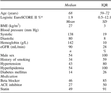
Table I. Characteristics of the 54 patients at recruitment.
Median IQR Age (years) 68 59–72 Logistic EuroSCORE II %* 1.9 0.5–12.1 Mean SD BMI (kg/m2) 27 3 Blood pressure (mm Hg) Systolic 138 19 Diastolic 80 8 Hemoglobin (g/L) 142 10 eGFR (mL/min) 90 28 n % Male sex 54 100 History of smoking 34 59 Hypertension 43 80 Hyperlipidemia 54 100 Diabetes mellitus 14 26 Medication Beta blocker 46 85 ACE inhibitor 27 50 Statin 49 91
SD: standard deviation; IQS = interquartile range; ACE: angiotensin-converting enzyme; eGFR: estimated glomerular filtration rate using the Cockroft and Gault equation [35].
*Mortality risk according to European System for Cardiac Operative Risk Evaluation Score II [22].
and both mammary arteries were used in 19 patients. Mean cardiopulmonary bypass time was 91 min, and an average of 2.9 grafts was inserted. Median blood loss was 580 (interquartile range 450–750) ml. Eight of the 54 included patients (15%) reported minor bleedings such as slight nose bleeds, mouth bleed-ing associated with tooth brushbleed-ing, or bruises at the clinical follow-up after three months. There were no major bleeds or gastrointestinal complications. None of the patients reported recurrent angina during the three months of follow-up.
Hematological Analyses
There was a marked drop in average postoperative hemoglobin level that was normalized at three months after the operation (Figure 1a). White blood cell counts were increased during the initial postoperative course, and platelet counts were slightly reduced after the operation but significantly elevated by 32% after one month without significant changes of mean platelet volumes (Figure 1b-d).
Flow Cytometric Data
The patients were treated with different low dosages of ASA after the operation [16]. Flow cytometric results did not differ between the three treatment groups, and data could thus be pooled for all patients (Supplementary Tables I and II).
Platelet Activation
Single platelet surface P-selectin expression was significantly enhanced at one month when stimulated by thrombin and the thromboxane analog U46619 (by 8% and 23%, respectively), and at three months when stimulated by ADP and thrombin (by 10% and 8%, respectively) (Figure 2). Mean levels without ago-nist stimulation were reduced by 12% from preoperative levels at three months (from 2.5 ± 1.1% to 2.2 ± 1.1%).
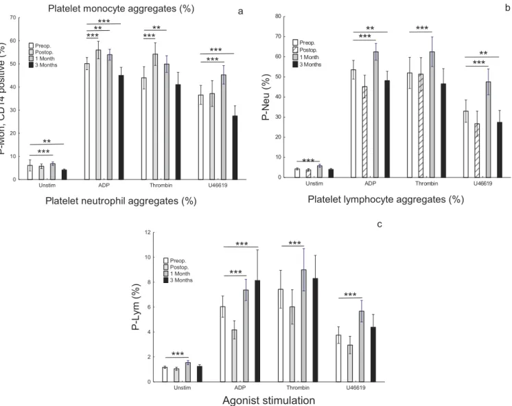
Platelet–Leukocyte Aggregation
The percentages of PLAs separated into subsets are shown in
Figure 3. Baseline unstimulated levels of PLAs were low and
Haemoglobin 0 25 50 75 100 125 150 L/ g ***
a
White blood cell counts
0 2 4 6 8 10 12 0 1 6 L/ ***
b
Platelet counts 0 50 100 150 200 250 300 350 400 0 1 9 L/ ***c
Platelet volume 0 1 2 3 4 5 6 7 8 9 10 Lf 1 Month 3 Months Postop. Preop.d
Figure 1. Mean levels with 95% confidence limits of (a) hemoglobin, (b) white blood cell counts, (c) platelet counts, and (d) platelet volumes in 54 patients before and after CABG. Dashed columns indicate results 4–6 days after surgery, shaded columns at one months and black columns at three months. ***p < 0.001 for difference between postoperative and preoperative levels are indicated.
reflect circulating PLAs when using our methodology [20]. Agonist stimulation increased P-Mon and P-Neu to more than 50% but to less than 10% for P-Lym. In unstimulated samples, all subsets of PLAs were increased one month after CABG. The percentages of CD14 positive P-Mons and P-Neus formed by in vitro stimulation with ADP, thrombin, or U46619 were significantly increased one month after the operation but reduced compared to baseline at three months. Agonist stimu-lated P-Lyms were increased at one month and remained increased at three months after ADP stimulation.
Correlations
There were several significant correlations at one and three months between agonist stimulated levels of platelet P-selectin expression and the corresponding agonist stimulated PLA formation in vitro (Supplementary Table III).
Discussion
In the present study, we found mildly increased platelet respon-siveness to agonist stimulation using whole blood flow cytometry up to three months after elective CABG despite ASA use. Circulating PLAs of all subtypes (platelet-conjugated monocytes, neutrophils, and lymphocytes) were elevated one month after sur-gery, and agonist stimulated PLA formation ex vivo was also enhanced at this time point. Agonist stimulated P-Mon and P-Neu formation was reduced at three months compared to baseline whereas P-Lym formation was not. There was no pattern indicating selectivity for any signaling pathway as platelet and PLA respon-siveness was elevated with stimulation by ADP, thrombin, and a thromboxane analog. We have previously shown that CABG causes early (within one week) activation of circulating platelets and
leukocytes, increased numbers of circulating PLAs, and marked activation of coagulation and inflammatory responses [11]. We presently show that platelet and PLA responses are protracted and in some cases not normalized even after three months.
Only patients with stable coronary artery disease who underwent elective CABG were included in this study as patients treated for acute coronary syndromes require dual antiplatelet treatment after the operation [23]. ASA was not discontinued before the operation, as the associated bleeding risk is small and withdrawal would leave the patient unprotected against acute ischemic events [21,24,25]. Minor bleeding that in some cases might have been associated with ASA treatment was reported by 15% of the patients during the follow-up. Flow cytometric assays under static conditions are independent of thromboxane formation due to platelet aggregation, and can be used to investigate platelets regardless of ASA therapy [17–19]. In agreement with this, we found no differences between groups treated with different ASA dosages and could pool all patients.
CABG performed with the aid of cardiopulmonary bypass caused an early drop in hemoglobin levels reflecting blood loss and hemodilution. Leukocyte counts were the highest 4–6 days after the operation reflecting inflammatory responses to cardio-pulmonary bypass [9]. These changes were normalized three months after the operation. Platelet counts were markedly ele-vated one month after the operation but mean platelet volume that reflects platelet turnover and platelet reactivity (large, newly formed platelets being more reactive) did not change [26].
The postoperative responses to CABG with cardiopulmonary bypass were more pronounced for PLAs than for single platelets. It is of interest that P-Mons may be a more sensitive marker for platelet activation in vivo than P-selectin expression on single platelets [27] presumably since circulating platelets shed the exposed P-selectin in vivo [28]. This may explain why the P-selectin expression of unstimulated (circulating) platelets did
Platelet P-selectin expression
Unstim ADP Thrombin U46619
Agonist stimulation
0 10 20 30 40 50 60 70 80)
%(
st
el
et
al
P
e
vit
i
s
o
p
l
e
S-P
Preop. Postop. 1 Month 3 Months***
***
**
**
**
Figure 2. Mean percentages with 95% confidence limits for platelet P-selectin positive cells in the platelet population in 54 patients before and after CABG. Unstimulated basal levels and levels after agonist stimulation by adenosine diphosphate (ADP), thrombin, and the thromboxane A2analog
U46619 are shown. Dashed columns indicate results 4–6 days after surgery, shaded columns at one month, and black columns at three months. **p < 0.01 and ***p < 0.001 for difference between postoperative and preoperative levels are indicated.
not change, whereas circulating P-Mons were increased at one month. Increased platelet reactivity to agonist stimulation ex vivo was found up to three months after surgery, and larger responses were detected among PLAs than among single plate-lets in the present study. Of interest is that agonist stimulated platelet P-selectin expression correlated with the corresponding formation of PLAs at one and three months, as shown in Supplementary Table III. Thus, platelet sensitization appears to be of importance for the increased reactivity of PLAs after CABG surgery.
Platelets that are adhered to the endothelium and platelets binding to leukocytes are involved in the recruitment of leuko-cytes to the vessel wall and the initiation of vascular inflamma-tion and atherosclerosis [4,6–8,29]. Binding via P-selectin glycoprotein ligand-1 is crucial in the interaction between plate-lets and the microvascular endothelium as well as in interactions with leukocytes and the formation of PLAs [29,30]. Neutrophils can be activated by high mobility group box 1 protein expressed in platelets, and neutrophil extracellular traps play a key role in interaction with platelets [31]. Platelet binding to monocytes increases their adhesiveness and transendothelial migration, and platelet binding to neutrophils promotes transmigration and their
recruitment to inflammatory sites [4,29,30]. Recent work has shown a crucial importance of platelets in guiding neutrophils and inflammatory monocytes to their exit points in the micro-vasculature [32]. The present data showing increased circulating P-Mons and P-Neus, as well as increased responses to agonist stimulation ex vivo one month after the CABG operation, may be interpreted as reflecting inflammatory and prothrombotic chal-lenges to the vasculature. The P-Mon and P-Neu responses were lower than before the operation after three months, indicating wound healing and subsiding inflammation, whereas signs of mildly increased platelet reactivity persisted. Clearance of acti-vated platelets and neutrophils from the circulation and the asso-ciated decrease in platelet–leukocyte aggregates may have contributed to this finding [28,33]. Platelets participate in the regulation of CD4 positive T-cells with implications for immune responses and vascular inflammation [8,34]. Our findings of increased platelet binding to lymphocytes and increased agonist stimulated formation of P-Lyms one month after the CABG procedure are of interest in this context.
Activated platelets and formation of PLAs may increase the risk of thrombosis after CABG. The present data support the need for efficient antiplatelet therapy during the first three months after
Platelet monocyte aggregates (%)
Unstim ADP Thrombin U46619 0 10 20 30 40 50 60 70 ) %( evi ti so p 41 D C, no M-P Preop. Postop. 1 Month 3 Months a *** ** *** *** *** ** ** ******
Platelet neutrophil aggregates (%)
Unstim ADP Thrombin U46619
0 10 20 30 40 50 60 70 80 ) %( ue N-P Preop. Postop. 1 Month 3 Months b
***
***
**
***
***
**
Platelet lymphocyte aggregates (%)
Unstim ADP Thrombin U46619
Agonist stimulation
0 2 4 6 8 10 12 ) %( my L-P Preop. Postop. 1 Month 3 Months c***
***
***
***
***
Figure 3. Mean percentages with 95% confidence limits of (a) CD14 positive platelet–monocyte aggregates (P-Mon), (b) platelet–neutrophil aggregates (P-neu), and (c) platelet–lymphocyte aggregates (P-lym) in 54 patients before and after CABG. Unstimulated levels and levels after agonist stimulation by ADP, thrombin, and the thromboxane A2analog U46619 are shown. Dashed columns indicate results 4–6 days after surgery, shaded columns at one
the operation. Dual antiplatelet therapy may be an option for a limited period of time also among stable patients undergoing elective CABG.
Limitations
We were not able to include all screened patients as we required a follow-up examination of surviving patients three months after the operation, and good-quality blood samples for flow cytometry with minimal in vitro artefacts at all time points. The patients were treated with different low doses of ASA after the operation [16] but we do not believe this has confounded our findings since flow cytometry of single platelets or PLAs is not influenced by the thromboxane path-way of platelet activation [17–19] and the flow cytometric data reported here did not vary between the ASA treatment groups. Conclusions
We conclude that elective CABG in ASA-treated patients with stable coronary artery disease evoked mild signs of platelet acti-vation up to three months after the operation. Increased platelet– leukocyte interactions that may facilitate vascular inflammation and thrombosis were found especially one month after the opera-tion. Our findings support further studies of the clinical implica-tions of protracted platelet activation after CABG.
Supplementary data
Supplementary data for this article can be accessedhere.
Acknowledgment
The authors are grateful for the expert technical assistance of Maud Daleskog.
Funding
The Swedish Research Council/Medicine, the Swedish Heart-Lung Foundation, Karolinska Institutet and the Stockholm County Council.
Conflict of interest
None of the authors have any conflict of interest to report in relation to this work.
ORCID
Marie Lordkipanidzé http://orcid.org/0000-0001-7418-6766 References
1. Ross R. Atherosclerosis is an inflammatory disease. Am Heart J
1999;138:S419–420.
2. Hansson GK. Inflammation, atherosclerosis, and coronary artery dis-ease. N Engl J Med2005;352:1685–1695. doi:10.1056/NEJMra043430. 3. Martins Pda C, Zwaginga JJ. Leukocyte-platelet aggregates: new particles reflecting and effecting cardiovascular disease. Thromb Haemost2005;94:1120–1121.
4. Zarbock A, Polanowska-Grabowska RK, Ley K. Platelet-neutrophil-interactions: linking hemostasis and inflammation. Blood Rev
2007;21:99–111. doi:10.1016/j.blre.2006.06.001.
5. Totani L, Evangelista V. Platelet-leukocyte interactions in cardio-vascular disease and beyond. Arterioscler Thromb Vasc Biol
2010;30:2357–2361. doi:10.1161/ATVBAHA.110.207480. 6. Massberg S, Brand K, Gruner S, Page S, Muller E, Muller I,
Bergmeier W, Richter T, Lorenz M, Konrad I, et al. A critical role of platelet adhesion in the initiation of atherosclerotic lesion forma-tion. J Exp Med2002;196:887–896.
7. Gawaz M, Langer H, May AE. Platelets in inflammation and ather-ogenesis. J Clin Invest2005;115:3378–3384. doi:10.1172/JCI27196.
8. Morrell CN, Aggrey AA, Chapman LM, Modjeski KL. Emerging roles for platelets as immune and inflammatory cells. Blood
2014;123:2759–2767. doi:10.1182/blood-2013-11-462432. 9. Warren OJ, Smith AJ, Alexiou C, Rogers PL, Jawad N, Vincent C,
Darzi AW, Athanasiou T. The inflammatory response to cardiopul-monary bypass: part 1–mechanisms of pathogenesis. J Cardiothorac Vasc Anesth2009;23:223–231. doi:10.1053/j.jvca.2008.08.007. 10. Sniecinski RM, Chandler WL. Activation of the hemostatic system
during cardiopulmonary bypass. Anesth Analg 2011;113:1319– 1333. doi:10.1213/ANE.0b013e3182354b7e.
11. Li N, Astudillo R, Ivert T, Hjemdahl P. Biphasic pro-thrombotic and inflammatory responses after coronary artery bypass surgery. J Thromb Haemost2003;1:470–476.
12. Parang P, Arora R. Coronary vein graft disease: pathogenesis and prevention. Can J Cardiol2009;25:e57–62.
13. Gluckman TJ, McLean RC, Schulman SP, Kickler TS, Shapiro EP, Conte JV, McNicholas KW, Segal JB, Rade JJ. Effects of aspirin responsiveness and platelet reactivity on early vein graft thrombosis after coronary artery bypass graft surgery. J Am Coll Cardiol
2011;57:1069–1077. doi:10.1016/j.jacc.2010.08.650.
14. Verma S, Goodman SG, Mehta SR, Latter DA, Ruel M, Gupta M, Yanagawa B, Al-Omran M, Gupta N, Teoh H, et al. Should dual antiplatelet therapy be used in patients following coronary artery bypass surgery? A meta-analysis of randomized controlled trials. BMC Surg2015;15:112. doi:10.1186/s12893-015-0096-z. 15. van Diepen S, Fuster V, Verma S, Hamza TH, Siami FS, Goodman SG,
Farkouh ME. Dual antiplatelet therapy versus aspirin monotherapy in diabetics with multivessel disease undergoing CABG: FREEDOM insights. J Am Coll Cardiol 2017;69:119–127. doi:10.1016/j. jacc.2016.10.043.
16. Ivert T, Dalen M, Ander C, Stalesen R, Nasman P, Lordkipanidze M, Hjemdahl P. Platelet function one and three months after coronary bypass surgery in relation to once or twice daily dosing of acetylsalicylic acid. Thromb Res2017;149:64–69. doi:10.1016/j.thromres.2016.11.018. 17. Rinder CS, Student LA, Bonan JL, Rinder HM, Smith BR. Aspirin
does not inhibit adenosine diphosphate-induced platelet alpha-gran-ule release. Blood1993;82:505–512.
18. Chronos NA, Wilson DJ, Janes SL, Hutton RA, Buller NP, Goodall AH. Aspirin does not affect the flow cytometric detection of fibri-nogen binding to, or release of alpha-granules or lysosomes from, human platelets. Clin Sci (Lond)1994;87:575–580.
19. Li N, Hu H, Hjemdahl P. Aspirin treatment does not attenuate platelet or leukocyte activation as monitored by whole blood flow cytometry. Thromb Res2003;111:165–170.
20. Li N, Goodall AH, Hjemdahl P. Efficient flow cytometric assay for platelet-leukocyte aggregates in whole blood using fluorescence signal triggering. Cytometry1999;35:154–161.
21. Li N, Wallen NH, Hjemdahl P. Evidence for prothrombotic effects of exercise and limited protection by aspirin. Circulation
1999;100:1374–1379.
22. Nashef SA, Sharples LD, Roques F, Lockowandt U. EuroSCORE II and the art and science of risk modelling. Eur J Cardiothorac Surg
2013;43:695–696. doi:10.1093/ejcts/ezs468.
23. m. Authors/Task Force, Windecker S, Kolh P, Alfonso F, Collet JP, Cremer J, Falk V, Filippatos G, Hamm C, Head SJ, Juni P, et al. 2014 ESC/EACTS guidelines on myocardial revascularization: the task force on myocardial revascularization of the european society of cardiology (ESC) and the European association for cardio-thoracic surgery (EACTS)developed with the special contribution of the European association of percutaneous cardiovascular interventions (EAPCI). Eur Heart J2014;35:2541–2619. doi:10.1093/eurheartj/ehu278. 24. Lordkipanidze M, Diodati JG, Pharand C. Possibility of a rebound
phenomenon following antiplatelet therapy withdrawal: a look at the clinical and pharmacological evidence. Pharmacol Ther
2009;123:178–186. doi:10.1016/j.pharmthera.2009.03.019. 25. Beving H, Zhao C, Albage A, Ivert T. Abnormally high platelet
activity after discontinuation of acetylsalicylic acid treatment. Blood Coagul Fibrinolysis1996;7:80–84.
26. Lordkipanidze M. Platelet turnover in atherothrombotic disease. Curr Pharm Des2012;18:5328–5343.
27. Michelson AD, Barnard MR, Krueger LA, Valeri CR, Furman MI. Circulating monocyte-platelet aggregates are a more sensitive mar-ker of in vivo platelet activation than platelet surface P-selectin: studies in baboons, human coronary intervention, and human acute myocardial infarction. Circulation2001;104:1533–1537.
28. Maugeri N, Rovere-Querini P, Evangelista V, Covino C, Capobianco A, Bertilaccio MT, Piccoli A, Totani L, Cianflone D, Maseri A, et al. Neutrophils phagocytose activated platelets in vivo: a phosphatidylserine, P-selectin, and {beta}2 integrin-depen-dent cell clearance program. Blood 2009;113:5254–5265. doi:10.1182/blood-2008-09-180794.
29. Da Costa Martins PA, van Gils JM, Mol A, Hordijk PL, Zwaginga JJ. Platelet binding to monocytes increases the adhesive properties of monocytes by up-regulating the expression and functionality of beta1 and beta2 integrins. J Leukoc Biol 2006;79:499–507. doi:10.1189/ jlb.0605318.
30. Lam FW, Burns AR, Smith CW, Rumbaut RE. Platelets enhance neutrophil transendothelial migration via P-selectin glycoprotein ligand-1. Am J Physiol Heart Circ Physiol 2011;300:H468–475. doi:10.1152/ajpheart.00491.2010.
31. Maugeri N, Campana L, Gavina M, Covino C, De Metrio M, Panciroli C, Maiuri L, Maseri A, D’Angelo A, Bianchi ME, et al. Activated
platelets present high mobility group box 1 to neutrophils, inducing autophagy and promoting the extrusion of neutrophil extracellular traps. J Thromb Haemost2014;12:2074–2088. doi:10.1111/jth.12710. 32. Zuchtriegel G, Uhl B, Puhr-Westerheide D, Pornbacher M, Lauber K, Krombach F, Reichel CA. Platelets guide leukocytes to their sites of extravasation. PLoS Biol 2016;14:e1002459. doi:10.1371/journal.pbio.1002459.
33. Ma R, Xie R, Yu C, Si Y, Wu X, Zhao L, Yao Z, Fang S, Chen H, Novakovic V, et al. Phosphatidylserine-mediated platelet clearance by endothelium decreases platelet aggregates and pro-coagulant activity in sepsis. Sci Rep 2017;7:4978. doi:10.1038/ s41598-017-04773-8.
34. Li N. CD4+ T cells in atherosclerosis: regulation by platelets. Thromb Haemost2013;109:980–990. doi:10.1160/TH12-11-0819. 35. Cockcroft DW, Gault MH. Prediction of creatinine clearance from serum creatinine. Nephron 1976;16:31–41. doi:10.1159/ 000180580.