Université de Montréal
A hypoxia response element in the Vegfa promoter is required for basal Vegfa expression in skin and for optimal granulation tissue formation during wound healing in mice
par Domenic Ciarlillo
Département de biomédecine vétérinaire Faculté de médecine vétérinaire
Mémoire présenté à la Faculté de médecine vétérinaire en vue de l’obtention du grade de maître ès sciences (M. Sc.)
en sciences vétérinaires option biomédecine
Décembre 2016
ii RÉSUMÉ
L’hypoxie contribue à la guérison cutanée via l’induction de HIF-1 (hypoxia-inducible factor-1). HIF-1 gère l’expression de VEGFA (vascular endothelial growth factor A) en se liant au HRE (hypoxia response element) présent au niveau de son promoteur. Par contre, dans le contexte de la guérison cutanée il est incertain si l’hypoxie et HIF-1 contribuent à l’augmentation de l’expression de VEGFA. Pour vérifier cette hypothèse, la guérison cutanée et l’expression de VEGFA ont été étudiées dans un modèle murine, Vegfa/, possédant un HRE non-fonctionnel dans le promoteur de Vegfa. De manière inattendue, le niveau d’ARNm de Vegfa présent dans la peau intacte des souris mutantes était diminué. Par contre, le niveau d’ARNm de Vegfa dans le tissu de granulation n’était pas altéré par rapport à celui de souris normales. Similairement, le niveau d’ARNm des gènes ciblés par Vegfa, Pdgfb et Sdf-1, étaient aussi comparativement diminués dans la peau intacte des souris mutantes mais aucune différence significative ne fut observée dans le tissu de granulation de plaies. L’analyse histologique des plaies en guérison chez les souris mutantes a démontré un tissu de granulation altéré en quantité et qualité (densité de capillaires). Par contre, la différence dans l’épithélialisation et le taux de guérison cutanée entre les deux populations de souris était non-significative. Les résultats démontrent qu’HIF-1 n’est pas un facteur majeur dans la régulation de Vegfa dans le contexte de la guérison cutanée. Cependant, HIF-1 est nécessaire pour maintenir un niveau d’expression basale (Vegfa et ses gènes cibles) adéquate et est aussi nécessaire pour la formation d’un tissu de granulation de qualité optimal suivant une blessure cutanée.
iii ABSTRACT
Hypoxia in skin wounds is thought to contribute to healing through the induction of hypoxia-inducible factor-1 (HIF-1), a key transcriptional regulator of genes associated with healing. HIF-1 regulates the expression of vascular endothelial growth factor A (VEGFA) by binding a hypoxia response element (HRE) in its promoter. Although VEGFA is a key regulator of angiogenesis during healing, whether hypoxia and HIF-1 serve to induce its expression in this context is unknown. To test this hypothesis, we studied VEGFA expression and wound healing in the Vegfa/ mouse model, which lacks a functional HRE in the Vegfa promoter. Unexpectedly, decreased levels of Vegfa mRNA were found in intact skin in Vegfa/ mice, whereas levels in granulation tissue during wound healing were not altered. Likewise, mRNA levels of VEGFA target genes Pdgfb and Sdf-1 were decreased in skin but unchanged in granulation tissue in the Vegfa/ mice. Decreased Vegfa levels in skin could not be attributed to decreased HIF-1 protein expression in the skin of Vegfa/ mice, and were therefore the consequence of a loss of HIF-1 responsiveness of the Vegfa promoter. Histologic analyses of healing wounds in Vegfa/ mice revealed significant defects in granulation tissue, both in terms of quantity and capillary density, although epithelialization and healing rates were unaltered. It can be concluded that HIF-1 is not a major regulator of Vegfa during wound healing. Rather, HIF-1 serves to maintain basal levels of Vegfa and VEGFA target genes in intact skin, required for optimal granulation tissue formation in response to wounding.
iv TABLE OF CONTENTS
Résumé ………. ii
Abstract ………. iii
Table of Contents ……… iv
List of Tables ……….. vii
List of Figures ………. viii
Abbreviations ……… x
Appendix ……….. xiv
CHAPTER 1: Introduction ……….. 1
Physiology of Wound Healing ……….. 1
Hemostasis ……….. 1
Inflammation ……….. 2
Early Inflammatory Phase ………. 2
Late Inflammatory Phase ……….. 3
Proliferation ………. 3
Angiogenesis ………. 4
Cell-Cell Junctional Adhesion ……… 5
Endothelial Cell Sprouting ……….. 5
Tip Cell Guidance……… 7
Tip Cell Anastomosis and Tubulogenesis ………. 7
VEGF……… 8 VEGF Family ……… 8 VEGFA ……… 9 VEGF165………. 10 VEGF Receptors………. 10 VEGFR1……….. 10 VEGFR2……….. 11 NRP……… 12
v
VEGFA Function………. 12
VEGFA Expression………. 14
Remodelling and Scar Formation……….. 15
Oxygen and Wound Healing……… 16
Oxygen and Cell Function……… 16
Energy Production……….. 16
Defence against Pathogens and ROS Production…….... 16
Oxygen Levels in Healing Wounds………... 17
Hypoxia-Inducible Factor-1……….. 18
Structure……….. 18
HIF-1α……… 19
HIF-1β……… 21
HIF-1 Complex Stability……….. 21
HIF-α Residues………. 24
Prolyl Hydroxylase Domain……… 24
VCB Cul2 E3 ligase complex……….…. 24
Arrest-Defective-1……… 25
Factor inhibiting HIF-1……….. 25
HIF-1 Complex Activity………. 26
MAP Kinase Pathway……….. 26
ERK1/ERK2 Pathway ……….. 26
Wound Healing Models and Measurements……….. 26
Research Hypotheses……… 28
Research Objectives……….. 29
CHAPTER 2: Article………. 30
Abstract………. 31
Introduction……….. 32
Materials and Methods……….... 34
vi
Part I: Molecular Profile……… 34
Real-time RT-PCR………. 35
Western Blot……….. 36
Part II: Wound Healing Assay……… 37
Histological Analyses………. 38
Statistical Analyses………... 38
Results ……… 39
Part I: Molecular Profile……… 39
Part II: Wound Healing Assay……… 42
Discussion………... 45
Acknowledgements……….. 46
References……….. 47
CHAPTER 3: Discussion and Future Experiments ……….... 49
vii LIST OF TABLES
Chapter 1: Introduction
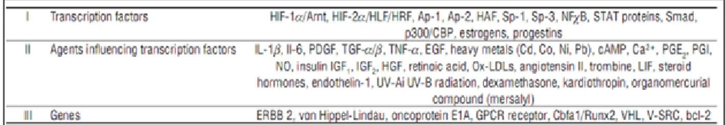
Table I Factors capable of influencing Vegf gene expression………. 8
Table II Target genes of the HIF-1 complex……… 19
Chapter 2: Article – Materials and Methods
viii LIST OF FIGURES
Appendix
Figure S1………. xiv
MAP kinase cascades in mammalian cells Figure S2……….. xv
Hif-1α mRNA levels in intact skin and in granulation tissue in wild type and mutant (Vegfaδ/δ) mice Chapter 1: Introduction Figure 1 Phases of Wound Healing………... 1
Figure 2 Endothelial Cell Sprouting……… 6
Figure 3 VEGFA Isoform Structures……… 9
Figure 4 HIF-α Isoforms and Protein Domains……… 20
Figure 5 HIF-1α Stability under Normoxic Conditions……….. 22
Figure 6 HIF-1α Stability under Hypoxic Conditions……….. 23
Chapter 2: Article – Results Figure 7……… 39
ix
Vegfa mRNA levels in intact skin and in granulation tissue in wild type and mutant (Vegfaδ/δ)
mice
Figure 8……… 40
Pdgfb and Sdf-1 mRNA levels in intact skin and in granulation tissue in wild type and Vegfaδ/δ
mice
Figure 9……… 41
HIF-1α and VEGFA expression in intact skin of wild type and Vegfaδ/δ mice
Figure 10………. 42 Healing kinetics and macroscopic appearance of wounds in wild type and Vegfaδ/δ mice
Figure 11………. 43
Epithelialization of healing wounds in wild type and Vegfaδ/δ mice
Figure 12………. 44
x ABBREVIATIONS
2-Oxoglutarate 2-OG
Activator protein AP
Adenosine triphosphate ATP
Angiopoietin ANGPT
Arrest-defective-1 ARD1
Aryl hydrocarbon nuclear translocator ARNT
Asparagine 803 N803
Basic fibroblast growth factor bFGF Basix-helix-loop-helix-Per-ARNT-Sim bHLH-PAS
Beta-actin ACTB
c-Jun NH2-terminal kinase JNK
C-terminal transactivation domain C-TAD
Carbon dioxide CO2
Cluster of differentiation 31 CD31
Cobalt chloride CoCl2
Delta-like 4 Dll4
Early inflammatory phase EIP
Egg-laying nine EGLN
Epidermal growth factor EGF
Epidermal growth factor receptor EGFR
Erythropoietin EPO
Extracellular matrix ECM
Extracellular signal-regulated kinase ERK
Factor inhibiting HIF-1 FIH-1
xi
Fibroblast growth factor FGF
fms-like tyrosine kinase-1 Flt-1
Glucose transporter-1 GLUT-1
Hematopoietic stem cell HSC
Hematoxylin-eosin-phloxine-saffron HEPS
Hepatocyte growth factor HGF
HIF-prolyl hydroxylase HPH
Hydrogen peroxide H2O2
Hydroxyl ion OH
-Hypoxia-inducible factor HIF
Hypoxia response element HRE
Insulin-like growth factor IGF Interferon-γ INF-γ
Interleukin IL
Internal ribosome entry site IRES
Jagged-1 Jag1
Keratinocyte growth factor KGF
Kinase domain region KDR
Late inflammatory phase LIP
Lysine 532 K532
Macrophage colony stimulating factor M-CSF
Matrix metalloproteinase MMP
Membrane type-1 matrix metalloproteinase MT1-MMP
Mitogen-activated protein MAP
N-terminal transactivation domain N-TAD
Neuropilin NRP
xii
Nitric oxide synthase NOS
Nuclear factor-1 NF-1
Nuclear factor kappa B NF-κB
Optimal cutting temperature OCT
Oxygen-dependent degradation domain ODDD
Peroxide anion HO2
-Phospholipase C PLC
Placental growth factor PlGF
Platelet derived growth factor PDGF
Platelet factor-IV PF4
Proline 402 P402
Proline 564 P564
Prolyl hydroxylase domain PHD
Quantitative polymerase chain reaction qPCR Quantitative reverse transcription polymerase chain reaction RT-qPCR
Reactive oxygen species ROS
Receptor tyrosine kinase RTK
Ribosomal protein L19 RPL19
Specificity protein 1 Sp1
Stress-activated protein kinase SAPK
Stromal cell-derived factor-1 SDF-1
Superoxide anion O
2-Tissue inhibitors of metalloproteinases TIMPs Tissue-type plasminogen activator tPA
Transforming growth factor TGF
Tumor necrosis factor TNF
xiii
Vascular endothelial cadherin VE-cadherin
Vascular endothelial growth factor VEGF Vascular endothelial growth factor receptor VEGFR
Vesiculo-vacuolar organelles VVO
xiv APPENDIX
FIGURE S1| MAP kinase cascades in mammalian cells. Overview of the various branches involved in MAP kinase cell signalling. Image source Zhang and Liu, 2002 figure 1.
xv
A B
FIGURE S2|Hif-1α mRNA levels in intact skin and in granulation tissue in wild type and mutant (Vegfaδ/δ) mice: Quantitative
PCR analysis was used to measure mRNA levels of (A) Hif-1α in intact skin ***P = 0.0010, WT: 1.3 ± 0.2, Vegfaδ/δ: 0.49 ± 0.06 and
(B) Hif-1α in granulation tissue. No significant difference was observed when comparing granulation tissue Hif-1α mRNA levels between both populations of mice. Messenger RNA levels are relative to that of Rpl19. Values represent mean ± SEM (n = 6). A two-tailed unpaired t-test was utilized for the statistical analysis of the intact skin data set. A Sidak-corrected repeated measures 2-way ANOVA was utilized for the statistical analysis of the granulation tissue data set.
CHAPTER 1
INTRODUCTION
Physiology of Wound Healing
The complex series of events involved in the healing of a wound can be divided into 4 phases: hemostasis, inflammation, proliferation and remodelling/scar formation (Figure 1). An alteration of either phase may lead to the development of a chronic, non-healing wound [1-5].
Hemostasis
Tissue injury immediately triggers a transient vasoconstriction (lasting roughly 5-10 minutes) and initiates the activation of the extrinsic coagulation cascade [2, 6-8]. Through
2
platelet degranulation various growth factors and cell signalling molecules, platelet derived growth factor (PDGF), fibroblast growth factor (FGF), β-thromboglobulin, serotonin, bradykinin, prostaglandins, prostacyclins, thromboxane, histamine, insulin-like growth factor (IGF)-1, epidermal growth factor (EGF), transforming growth factor (TGF)-β and platelet factor-IV (PF4), among others, are released into the wound environment [2, 6, 8]. Approximately 20 minutes post-wounding, through the release of vasoactive substances such as histamine, vasodilation and an increased vascular permeability are favored [8]. The response initiated by these factors favors a healthy inflammatory response by attracting and activating certain cells such as polymorphonuclear leukocytes, macrophages, fibroblasts and endothelial cells, and contributes to hemostasis via the activation of the complement and kinin cascades [2, 6, 8]. Ultimately, platelet aggregation within the fibrin blood clot at the site of injury serves the purpose of diminishing blood loss [2, 6, 7].
Inflammation
The inflammatory phase quickly follows hemostasis and is characterized by the 5 cardinal signs of inflammation; redness, warmth, swelling, pain and loss of function [7]. The inflammatory phase can be further divided into an early inflammatory phase (EIP) (days 1-2) and a late inflammatory phase (LIP) (days 2-3) [2].
Early Inflammatory Phase
The classical and alternative pathways of the complement cascade are activated upon initiation of the EIP. The activation of these pathways ultimately results in the attraction of neutrophils to the wound site via chemoattractants such as TGF-β and/or complement fragments C3a, C5a [2, 9]. Therefore, the first inflammatory cells to reach the wound are neutrophils, which do so within 24-36h after wounding [2, 9]. Neutrophils then begin to eliminate pathogens and foreign matter either by phagocytosis, enzymatic destruction (via degranulation products such as collagenases and elastases) or through the production of free
3
radicals [2, 6, 8, 9]. Neutrophil activity usually ceases roughly 72 hours post-wounding, once the affected region has been cleared of bacteria and debris [2, 10].
Late Inflammatory Phase
The cell type that predominates this portion of the inflammatory phase and that is essential for normal wound progression is the monocyte/macrophage [2, 8]. Similarly to neutrophils, blood monocytes are attracted to the wound by several chemoattractants (e.g. PDGF, TGF-β) but make a tardier appearance, arriving at the wound site 48-72 hours after injury [2, 9]. Once they have arrived at the wound site, blood monocytes become tissue macrophages. The primary role of macrophages is that of repair. Although they possess phagocytic and enzyme degrading capabilities to control and neutralize potential pathogens, they are the main producers of various growth factors (PDGF, TGF-β, and FGF) that will promote the proliferation of smooth muscle cells, the proliferation of endothelial cells, the initial migration of fibroblasts, keratinocytes and endothelial cells within the wound, as well as the production of the extracellular matrix (ECM) [2, 6-9].
Proliferation
Approximately 3 days after wounding and lasting until 2-4 weeks post-wounding, the proliferative phase follows the inflammatory phase [2, 7, 9]. Fibroblast migration, deposition of ECM and the formation of granulation tissue are all associated with the proliferative phase [2, 7, 9, 11]. Similarly to neutrophils and monocytes, fibroblasts are attracted to the wound by various factors [e.g. PDGF, TGF-β and basic fibroblast growth factor (bFGF)] [2, 7]. The proliferation of fibroblasts results in the production of components (fibronectin, hyaluronan, collagen and proteoglycans) essential for the formation of the new ECM [2].
The newly produced ECM allows for the regulation of differentiation, growth, expansion, and migration of cells [2, 3]. The ECM is able to modify cytokine activity, which is necessary for the regulation of its own components [12]. Overall, the ECM behaves as a
4
cellular scaffold, permitting the regeneration of wounded skin [3]. The main components of the ECM are collagen, adhesive glycoproteins (fibronectin, laminin, thrombospondin and integrins), and proteoglycans (glycosaminoglycans such as dermatan sulphate, heparan sulphate and hyaluronic acid) [2].
Re-epithelialization is another essential process stimulated during the proliferative phase [9, 11]. Two molecules are primarily responsible for the restoration of an epidermal lining: EGF, whose production is attributed to keratinocytes and platelets, and TGF-α, which is produced mainly by activated macrophages [9].
The proliferative phase also encompasses the production of another key component in the healing of the wound: granulation tissue. Granulation tissue is mainly composed of a network of capillaries and collagen and it serves as a transient dermal substitute until the dermis can be fully repaired [6]. The quality of the granulation tissue within a wound may serve as an indication of the wound health [2]. By 3-5 days after wounding, a healthy granulation tissue bed should appear hyperaemic, moist, shiny and granular [2]. Conversely, the granulation tissue of unhealthy wounds can be described as friable, soft, and of a beefy-red color [2, 13]. Microscopically, granulation tissue is primarily characterized by macrophages, proliferating fibroblasts and capillaries [2, 14].
As the proliferative phase ends, the ratio of hyaluronic acid to chondroitin sulfate is altered (the amount of chondroitin sulfate increases), consequently reducing the migratory/proliferative capabilities of fibroblasts. This favors fibroblast differentiation, thereby denoting the initiation of the remodelling phase [7].
Angiogenesis
Angiogenesis is an essential component of the wound healing process occurring within the proliferative phase and ensures the development of a network of capillaries [2, 6, 11]. Specific growth factors responsible for the creation of these blood vessels are vascular endothelial growth factor (VEGF), bFGF and TGF-β, which can originate from epidermal cells, fibroblasts, macrophages and endothelial cells [1, 9]. Angiogenesis is a process whose
5
function is directly proportional to oxygen tension levels and that consists of the formation of new blood vessels within an organism [15-18]. Angiogenesis can occur due to endothelial cell splitting, known as intussusception, or by endothelial cell sprouting [19-21]. Angiogenesis differs from vasculogenesis, the latter being responsible for the formation of the very first vascular networks within the viable embryo where mesodermal cells differentiate into angioblasts, the precursors to endothelial cells [22, 23]. Angiogenesis mediated by VEGF is an essential component of many physiological processes such as embryonic development, the ovarian cycle and bone remodelling and it also contributes to pathological conditions such as wound healing, retinopathy and tumor growth and development [17, 21, 24-28]. Many factors are involved in the process of angiogenesis; these include but are not limited to PDGF, TGF-β, tumor necrosis factor (TNF)-α, TNF-β, bFGF, FGF, TGF-α, hepatocyte growth factor (HGF), interleukin (IL)-8, and angiopoietins, as well as one of the most important factors, VEGF [15, 27, 29].
Cell-Cell Junctional Adhesion
Protein complexes present within the endothelium are responsible for stabilizing the vascular endothelial cell lining and ensuring that a proper endothelial cell-cell adhesion exists. These protein complexes are structurally altered throughout angiogenesis in order for this process to successfully reach completion [22]. The major player regulating endothelial cell-cell adhesion is a member of the cadherin family, vascular endothelial cadherin (VE-cadherin) [19, 22].
Endothelial Cell Sprouting
Before endothelial tip cell sprouting can occur, specific steps are required for vascular endothelial cells to become permissive to phenotypic changes. Matrix metalloproteinases (MMP) such as membrane type-1 matrix metalloproteinase (MT1-MMP) and disintegrin as well as VEGF-activated endothelial cell protease secretions are required for the
6
FIGURE 2| Endothelial cell sprouting: Figure demonstrating the influence of VEGF in the specialization of either a tip or stalk endothelial cell phenotype. This decision is mediated through the regulation of Notch signalling between neighbouring endothelial cells. Image source
Eilken and Adams, 2010, figure 2
remodelling/degradation of the sub-endothelial basement membrane surrounding blood vessels [19, 22]. Endothelial cell motility also must be enhanced through the modulation of VE-cadherin. It has been postulated that exposure to VEGF promotes VE-cadherin endocytosis, thereby leading to an increase in endothelial cell motility. Finally, endothelial cell phenotypic specialization, tip cell or stalk cell, is required for angiogenesis to proceed (Figure 2) [22]. This phenotypic specialization is regulated via the Notch pathway [20, 22]. In the absence of VEGF, a balance exists regarding Notch cell signalling between adjacent endothelial cells. A factor named Delta-like 4 (Dll4) is produced by adjacent endothelial cells and activates the neighbouring cell’s Notch signalling pathway. In the presence of a VEGF gradient, VEGF will stimulate one of the neighbouring endothelial cells to a greater extent resulting in a higher degree of Dll4 production and therefore will induce a more robust Notch pathway activation. Increased Notch signalling results in decreased expression of vascular
7
endothelial factor receptor (VEGFR)-2 and VEGFR3, increased expression of VEGFR1 and will result in the undertaking of a stalk cell phenotype, whereas the adjacent Dll4-expressing cell will undertake a tip cell phenotype [20, 22]. Stalk cells also express a factor named Jagged-1 (Jag1), which inhibits the characteristics of a motile, sprouting tip cell phenotype [22].
Tip Cell Guidance
Once endothelial cells have undertaken a tip cell phenotype, the sprouting cells’ motile filopodia respond to environmental cues which guide the path of new vessel growth [22]. An essential component required for vessel support during its growth is the sheathing of blood vessels by ECM components, primarily mature collagen [18]. The presence of an ECM lacking mature collagen results in newly branched blood vessels that are delicate and poorly assembled [18]. Tip cell guidance is another process in which the importance of heparin-binding domains present in select VEGF isoforms can be observed. VEGF binds to heparin sulfate proteoglycans present in the ECM and through this binding establishes a gradient that aids in attracting tip cell direction [22].
Tip Cell Anastomosis and Tubulogenesis
VE-cadherin is also present on the filopodial tips of sprouting endothelial cells. In this scenario, this cell-cell junctional adhesion complex may aid in the anastomosis of two sprouting endothelial cells. The interaction of both VE-cadherin complexes on their respective tip cells may initiate junction assembly, which will reduce tip cell motility. A phenotypic cell conversion occurs, tip cell to stalk cell, as the process of tubulogenesis commences. The tip cells are introduced into the endothelial cell lining where they become stalk cells and the lumen of the future complete blood vessel is formed [22]. Finally a new round of cell sprouting occurs as other endothelial cells are exposed to and modulated by pro-angiogenic factors [22].
8
VEGF
Among the growth factors required for the development of a blood vessel network, VEGF was the first to be identified, due to its ability to increase vascular permeability [15, 25, 30]. VEGF is essential for the viability of the developing embryo as well as being an important factor for the proper maintenance and health of the adult vasculature [31, 32]. Embryonic mice genetically engineered to lack one functional Vegf allele, Vegf+/-, die early in embryonic
life, at E9.5-E10.5, mainly due to severe vascular abnormalities that are predominantly cardiovascular related [19, 21, 23, 25, 29, 31, 33-35]. On the other hand, overexpression of
Vegf (as little as twice the normal amount) leads to death of the organism [25, 31]. Many cell
types such as macrophages, glial cells, keratinocytes, endothelial cells, dermal fibroblasts and tumor cells, are capable of secreting VEGF through various pathways [26, 30] (Table I lists factors capable of influencing Vegf gene expression). However, keratinocytes remain the major source of VEGF production within cutaneous wounds [30].
VEGF Family
The VEGF family is composed of several members [VEGFA, VEGFB, VEGFC, VEGFD, VEGFE, placental growth factor (PlGF) and VEGFFs (snake venom-derived VEGF related proteins)], but the regulation of endothelial cells seems to be predominantly controlled via VEGFA [19, 21, 31, 35-37]. The action and function of VEGFC and VEGFD predominantly lies in the formation of new lymphatic vessels, (lymphangiogenesis), however, they also behave as influential lymphatic endothelial cell regulatory proteins [23].
TABLE I| Factors capable of influencing Vegf gene expression: Table depicting various factors affecting Vegf gene expression, whether they are transcription factors, agents influencing transcription factors, or genes. Image source Jośko and Mazurek, 2004, table 1
9
FIGURE 3| VEGFA isoform structures: Major VEGFA isoforms found within humans (mice isoforms possess one less amino acid). Exon 1 encodes for the signal sequence [15]. Exon 3 confers the ability to dimerize as well as interact with VEGFR1 [15, 38]. VEGFR2-binding is enabled by the presence of exon 4 within the fully formed protein structure [15, 38]. Exon 5 ensures that VEGFA isoforms are vulnerable to plasmin cleavage [15, 38]. Exon 6 and 7 are heparin-binding domains. The presence/absence of these two domains explains why some VEGFA isoforms, such as VEGF189,are mainly sequestered in the ECM via binding to various heparan-containing ECM components, as opposed to VEGF121 which is a fully diffusible VEGFA isoform [38]. Furthermore, a portion of exon 7 and all of exon 8 are responsible for the ability to bind to coreceptors NRP1/NRP2. This interaction is required for enhanced VEGFR2 cell signalling via the binding of VEGFA to its receptor [38]. Image source Raimondi and Ruhrberg, 2013, figure 1.
VEGFA
The gene for VEGFA consists of 8 exons and 7 introns which, when subjected to RNA splicing, gives rise to various isoforms [15, 38] (Figure 3). The currently known VEGFA isoforms include VEGF206, VEGF189, VEGF183, VEGF165, VEGF148, VEGF145, VEGF121, VEGF189b, and VEGF165b, where VEGF165 was found to be the predominantly active isoform in terms of VEGF activity [15, 31, 36]. Consequently, in the following text describing functional activities related to this growth factor, the use of the term VEGFA will refer to the most biologically active isoform, VEGF165. The majority of VEGFA isoforms can exist in either a soluble or ECM bound form, where each form plays a role in dictating the final functional and structural outcome of the vasculature. Soluble VEGFA forms promote vascular hyperplasia, decreased vascular density and poor vascular branching, whereas ECM bound forms favor highly branched vessels and a vasculature characterized by thin walls [31].
10 VEGF165
VEGF165 possesses intermediary, exon inclusions allows this isoform to be bound to the ECM as well as being secreted, and therefore optimal structural characteristics compared to other VEGFA isoforms, thereby making this isoform the predominant mediator in carrying out the various functions of VEGFA [27]. To further enforce that VEGF165 is the predominant functionally active VEGFA isoform, mice solely expressing VEGF164 (mouse VEGFA isoforms possess one less amino acid) do not possess any morphological vascular differences when compared to wild type mice [21, 27, 31]. VEGF165 possesses a heparin-binding domain and therefore is mitogenically active. A portion of VEGF165 is secreted, however a large portion of VEGF165 remains bound to the cell surface and ECM [27, 39]. Similarly to VEGF189, VEGF165 bound to the ECM can be released as a secreted form via the action of plasmin [27].
VEGF Receptors
There exists 4 different VEGF receptors; VEGFR1, VEGFR2, VEGFR3 and Neuropilin- 1/Neuropilin-2 (NRP1/NRP2) [27]. VEGFR3 is not a suitable receptor for VEGFA, but rather behaves as a receptor for VEGFC and VEGFD and therefore is the only receptor that will not be outlined [21, 27, 33].
VEGFR1
VEGFR1, also known as fms-like tyrosine kinase-1 (Flt-1), is a receptor tyrosine kinase (RTK) [27, 33, 40]. Ligands capable of interacting with this receptor include VEGFA, PlGF and VEGFB [27]. Flt-1 expression is influenced by oxygen tension levels because it is a hypoxia-inducible gene [33, 34, 41, 42]. Alternative splicing of Flt-1 can result in the expression of a soluble form of the receptor that acts as a negative regulator of VEGFA activity through its ability to bind and sequester the ligand VEGFA [19-22, 27, 31]. Therefore VEGFR1 has been deemed a “decoy receptor” [19, 27, 31, 43]. Although this receptor primarily functions as a
11
“decoy receptor” it still plays an important role in monocyte chemotaxis, inflammation, increasing vascular permeability, cell migration, hematopoietic stem cell (HSC) repopulation and the induction of matrix metalloproteinase-9 (MMP-9), urokinase-type plasminogen activator (uPA) and tissue-type plasminogen activator (tPA) [21, 23, 25, 27, 31, 33, 37]. Plasminogen activators (uPA and tPA) are responsible for the transformation of plasminogen to plasmin, a predominant fibrinolytic agent, necessary for fibrin dissolution and consequently permitting wound re-epithelialization [44]. MMP-9 on the other hand results in the excision of collagenous anchors tethering keratinocytes to the basal lamina [44].
VEGFR2
VEGFR2, also known as kinase domain region (KDR) or fetal liver kinase-1 (Flk-1), is structurally identical to VEGFR1, however the affinity of VEGFR2 for its ligand is 10 times lower than is that of VEGFR1 [21, 27]. Known ligands for this receptor include VEGFA, VEGFC and VEGFD. VEGR2 signalling is the main route through which VEGFA can carry out its principal functions [22, 25, 27, 31, 36, 37, 43]. Flk-1-/- embryonic mice suffer the same fate as Flt-1-/- mutants, which is embryonic death at E8.5-9.5 however vascularization does not occur
in the former [21, 25-27, 33, 34]. Oxygen tension levels alter the expression of Flk-1, which is down-regulated under hypoxic conditions [23]. However, receptor phosphorylation is increased under low oxygen tension levels [31]. Upon ligand binding, six tyrosine residues, 951, 996, 1054, 1059, 1175 (of particular importance), and 1214, located within VEGFR2, are phosphorylated thereby permitting the subsequent activation of various signalling pathways, such as extracellular-regulated kinase (ERK) 1/2 [19, 31]. Activation of this receptor has been associated with many functions such as endothelial cell migration, maturation, proliferation, survival, differentiation and vasculogenesis [19, 33].
12 NRP
NRP1 is a non-tyrosine kinase receptor that influences neuronal activity and whose normal ligand belongs to the collapsin-semaphorin family [21, 27, 31, 35, 36]. NRP1 influences the effects of angiogenesis, explaining the specific interactions between NRP1 and select VEGF family members (VEGFA, VEGFB, VEGFE and PLGF), whereas NRP2 regulates lymphangiogenesis [21, 22, 36, 37]. Neuropilin receptors can act as powerful VEGFA signalling enhancers when co-expressed on cells with VEGFR2. All VEGFA isoforms are capable of interacting with NRP1 via the inclusion of exon 8 in their protein structure [21, 31]. Exon 7, a heparin-binding domain within the VEGFA protein structure, along with the heparin-binding domain found within the NRPs, leads to the formation of a surface whereby VEGFA/NRP can interact with VEGFRs via the mediation of heparan [21]. This interaction is capable of promoting an altered presentation of the VEGFA ligand to VEGFR2 in a manner that enhances the signal pathway induced by receptor stimulation. Like Flt-1 and Flk-1, Nrp-1-/- embryonic
mice are not viable and are characterized by vascular abnormalities, demonstrating the necessity of the enhanced VEGFA signal transduction induced by this coreceptor [27, 33, 36].
VEGFA Function
VEGFA possesses two main functions. The first is to increase the nutrient supply to tissues and the second is playing a role in waste management by increasing the removal rate of waste products [15, 45]. VEGFA accomplishes these fundamental functions via three processes: vasodilation, angiogenesis and increased vascular permeability [15, 21, 45].
Vasodilation directly supports both of VEGFA’s main functions. VEGFA is a potent vasodilator capable of dilating blood vessels very rapidly and to a large degree [15, 27]. The increase in blood vessel diameter will result in increased blood flow, thereby allowing more oxygen and glucose to reach the target tissue as well as increasing the removal rate of waste products such as carbon dioxide (CO2), H+ and lactate [15]. The mechanism underlying the VEGFA-induced vasodilation is based upon the action of nitric oxide (NO) [15, 27, 33]. VEGFA
13
binds to its receptor, VEGFR2, which will then activate the phospholipase C (PLC) pathway. The end result of the activation of the PLC pathway is an increase in intracellular Ca2+ that interacts with calmodulin to stimulate nitric oxide synthase (NOS) and therefore NO production. NO is then capable of freely diffusing through the endothelial cells and acting on the smooth muscle cells surrounding the blood vessels and relaxing them, leading to vasodilation [15]. NO is also part of a positive feedback loop because NO is capable of stimulating VEGFA expression [33].
VEGFA is one of the most potent angiogenic factors since it is capable of driving all the important processes required for angiogenesis, including endothelial cell proliferation and migration, basement membrane degradation, tube and lumen formation, increased vascular permeability, and new vessel formation [15, 22, 26, 46-48]. This growth factor is able to stimulate the growth of new vessels, whether they are arteries, veins or lymphatics [27]. In the context of wound healing, angiogenesis allows essential nutrients and oxygen to be delivered to mitotically active cells, thereby increasing healing rates [30]. VEGFA is also capable of upregulating other factors, such as PDGFB and stromal cell-derived factor-1 (SDF-1), which play an important role in angiogenesis [49-51]
VEGFA is capable of increasing the hydraulic conductivity of fluid as well as increasing the diffusive permeability of certain macromolecules by altering the number or length of pores present in the vasculature [15, 27]. The increase in vascular permeability allows for the extravasation of certain proteins, fibrinogen and plasminogen, within the area surrounding the wound. The presence and deposition of these factors within the ECM directly aids subsequent tissue healing by creating a suitable ECM substrate for tissue growth as well as by creating an environment that is pro-angiogenic [15]. The mechanism of VEGFA-induced increased vascular permeability is similar to that of vasodilation. VEGFA will bind to its receptor, VEGFR2, thereby leading to the activation of PLC and, ultimately, to an increase in intracellular Ca2+. However, the increase in Ca2+ does not stimulate downstream factors but, rather, it directly affects endothelial cells [15, 27]. Endothelial cell exposure to increased Ca2+ levels will result in contraction of the cells, clustering of vesicles inside the cells, and the
14
formation of vesiculo-vacuolar organelles (VVO) leading to an increased permeability to fluid and macromolecules [15, 25].
VEGFA Expression
Oxygen tension levels play a very important role in the ability of the HIF-1 complex to mediate VEGFA gene expression [27]. However, hypoxia-regulated VEGFA expression is not the only pathway capable of stimulating this gene, which is expressed within nearly all tissues in the adult [25, 52] (Table I). Hypoglycemia has been found to stimulate VEGFA expression [33, 53, 54]. Furthermore, acidosis is also documented to lead to increased VEGFA levels [55]. Similarly to HIF-1α, many growth factors are able to stimulate VEGFA gene expression; these include EGF, TGF-α, TGF-β, TNF-α, keratinocyte growth factor (KGF), IGF-1, FGF, bFGF and PDGF [18, 25, 27, 33, 34, 56, 57]. Cytokines, IL-1α, IL-6 and IL-8, also play a role in the up-regulation of this gene [25, 27, 33, 56]. Chemical induction of VEGFA expression is not the only manner in which VEGFA expression is stimulated. Physical forces such as a simple stretch can increase VEGFA gene expression [25].
Reactive oxygen species (ROS) are also mediators able to increase VEGFA expression [15, 57, 58]; fibroblasts, endothelial cells and macrophages are all stimulated to express VEGFA due to ROS exposure [58]. Furthermore, hydrogen peroxide (H2O2) is capable of directly stimulating the VEGFA promoter resulting in increased expression and secretion levels of this growth factor by keratinocytes and macrophages [18, 59]. This HIF-1 independent H202-induced VEGFA expression is mediated via the interaction of Sp1 with the VEGFA promoter [57, 59].
Located within the Vegfa promoter are multiple binding sites for different transcription factors such as HIF-1, activator protein (AP)-1, AP-2, Sp1, nuclear factor (NF)-1, nuclear factor kappa B (NF-Κb) and Sp1-related factors, signifying a control of VEGFA expression other than hypoxia via the activation of different signalling cascades [16, 34, 46, 60]. The MAP kinase signalling pathway documented to induce Vegfa expression is that of ERK1/ERK2 pathway [34, 61]. The ERK1/ERK2 pathway has been shown to increase Vegfa
15
gene expression via the binding of transcription factors AP-2 and Sp1 to their appropriate recognition DNA sequences within the Vegfa promoter [29, 56]. Moreover, the active combinatory effect of Sp1 alongside AP-2 has been documented to increase Vegfa expression [46, 52]. Macrophage colony stimulating factor (M-CSF) secreted by various cell types including endothelial cells, fibroblasts, tumor cells, and monocytes, has been found to upregulate Vegfa expression via the activation of the ERK pathways [46, 62].
Remodelling and Scar Formation
The process of remodelling has already begun as the first signs of granulation tissue formation appear within the wound bed. Therefore the fourth and final wound healing phase begins around 1 week post-wounding and can last ≥1-2 years [2, 9]. The ECM is continuously being remodelled, due to constant remodelling of collagen. This dynamic state is present until an equilibrium is reached at roughly 21 days post-wounding [2, 7]. MMPs are responsible for proteolysis and for controlling cell migration through the ECM [2, 63]. Initially MMPs are beneficial to the wound environment as they promote angiogenesis, cleanse the wound via debridement, and aid in epithelialization [5]. MMPs vary considerably as several different types exist, each with their own target. Interstitial collagenases alter collagen I, II, and III, gelatinases degrade amorphous collagen and fibronectin and stromelysins alter components of the ECM such as laminin [2]. Uncontrolled MMP activity impairs wound healing since MMPs degrade collagen, essential for proper healing [2, 63]. Therefore MMPs are usually found in the form of zymogens requiring an activating catalyst (proteases such as plasmin) found only in regions of injured tissue [2].
As the wound progresses in its remodelling, certain changes can be observed. There is an increased activity of MMP inhibitors [tissue inhibitors of metalloproteinases (TIMPs)] and reduced MMP activity, mainly due to the activity of TGF-β [2, 9]. A reduction in the number of macrophages and fibroblasts can be observed as can a reduction in cellular metabolic activity [2, 9]. Blood perfusion to the wound site is reduced as well [2, 9]. Therefore, the granulation tissue is slowly replaced by avascular scar tissue and, as the scar matures,
16
collagen fiber diameter increases and type III collagen is replaced by type I until a ratio (collagen I : collagen III) of 4 : 1 is achieved [2, 6, 7, 9]. In addition to the superior organizational state of the collagen fibers, cross-linking of these fibers also occurs, thereby increasing the tensile strength of the scar tissue as the wound heals [6, 9]. The wound tensile strength within the first week of healing is minimal. In humans, within 4-6 weeks post-wounding the wound tensile strength increases to 30-50% of the strength of intact skin and 60% wound tensile strength is achieved by 6 months [4]. The maximum tensile strength reached in a healed wound approximately 1 year after wounding is 80% of the strength of healthy intact skin [2, 4, 7].
Oxygen and Wound Healing
Oxygen and Cell Function
Energy Production
Essential intracellular processes such as biosynthesis, movement, transport and proliferation depend on an energy source in the form of adenosine triphosphate (ATP) [18, 59, 64]. ATP is synthesized via oxidative phosphorylation, requiring oxygen for its completion, in amounts that are necessary to satisfy each cell’s purpose [18, 41, 58, 59].
Defence against Pathogens and ROS Production
Respiratory burst refers to the production of ROS such as peroxide anion (H02-), hydroxyl ion (OH-) and superoxide anion (O2-), which are produced by various leukocytes and serve to destroy invading pathogen and control against infection [18, 41, 58, 59]. This process is highly dependent on the presence of oxygen in the environment [41, 57-59]. The active enzyme responsible for this process, NADPH-linked oxygenase, requires a high concentration of oxygen in order to be fully active since the production rate of the toxic radicals is directly
17
proportional to the degree of enzyme activity (50% activity at 45-80 mmHg and maximal activity may require oxygen levels >300 mmHg) [18, 41, 59].
PDGF, EGF, TNF-α, and IL-1β are capable of increasing the synthesis of ROS through fibroblast and leukocyte stimulation via Rac1 [57, 59]. ROS production alters the behaviour and function of cells/processes via its influence on increasing cytokine release, stimulating angiogenesis and increasing cell motility [18, 41, 58].
Oxygen Levels in Healing Wounds
Within the first few days after wounding, oxygen levels are drastically reduced giving rise to a hypoxic environment [59]. A significant decrease in oxygen levels is present approximately 48 hours following injury and maximal hypoxia (≤ 10 mmHg) is attained close to 4 days post-wounding [65]. Oxygen tension levels vary within the wound itself with levels as low as 10 mmHg recorded at the center of the wound and levels of 60 mmHg at the wound periphery [18, 41]. Hypoxia is primarily the consequence of the traumatic destruction of the vasculature as well as the augmentation of oxygen consumption rates due to increased cellular density and metabolic activity [18, 41, 53, 58, 59, 66]. Acute hypoxia favors wound healing because the low oxygen tension environment promotes fibroblast proliferation, keratinocyte motility, procollagen synthesis, angiogenesis, ROS production and an increase in the expression of various growth factors such as TGF-β1, PDGF and VEGFA [41, 58, 59, 64, 67, 68]. Furthermore, low oxygen tension levels have been documented as the predominant inducer of VEGFA expression [23]. However, hypoxic conditions that persist (chronic hypoxia) negatively impact wound healing since minimum oxygen tension levels of 30-40 mmHg are required for efficient wound healing mechanisms [41, 58, 66]. Therefore angiogenesis, re-epithelialization, fibroblast collagen deposition and the ability of leukocytes to control the presence of pathogens, are all impaired under hypoxic conditions [18, 41, 58, 66]. Moreover, fibroblast proliferation rates decrease, procollagen synthesis decreases, and TGF-β1 expression is downregulated [41, 58, 59]. Hence, the continued presence of hypoxia has been linked to delayed wound healing [68]. In a normally healing wound, oxygen tension levels
18
return to normal and stabilize over time as a result of angiogenesis due to the influence of VEGFA [41, 59]. Hypoxic conditions have been shown to favor the stability of Vegfa mRNA due to the prolongation of the gene transcript’s half-life, normally approximately 30 minutes, which is extended by 3-8-fold [32, 53, 69, 70]. Similarly to HIF-1α, VEGFA possesses an internal ribosome entry site (IRES) that enables this growth factor to be adequately expressed at the protein level in spite of hypoxic/stressful conditions [26, 33, 42, 53, 60]. Therefore under low oxygen tension levels, VEGFA levels ultimately increase due to increased mRNA stability, increased transcription rates, and increased translation rates [16, 32, 33, 42, 56].
Hypoxia-Inducible Factor-1
Structure
Hypoxia-inducible factor-1 (HIF-1) is a heterodimeric transcription factor that induces the expression of a plethora of genes important for cell survival, erythropoiesis, metabolic regulation, cell proliferation and vascular biology [41, 59, 71, 72] (Table II). HIF-1 was first discovered in the nuclear extracts of liver cells, Hep3B cells, exposed to hypoxic conditions. It was discovered that the HIF-1 complex interacted with a specific DNA sequence, 5’-TACGTGCT-3’, located within the enhancer region of the erythropoietin (EPO) gene [73]. Furthermore, the consensus sequence, 5’-RCGTG-3’, was not only found to be present at the level of the EPO enhancer region but was also located within the DNA of various genes such as VEGFA, angiopoietin (ANGPT)-1, ANGPT2, PDGFB, glucose transporter-1 (GLUT-1) and SDF-1 [SDF-18, 7SDF-1, 74, 75]. The protein subunits (α and β) constituting the HIF-SDF-1 complex are members of the basic-helix-loop-helix-Per-ARNT-Sim (bHLH-PAS) protein family that utilize the bHLH and PAS motifs to dimerize and consequently become functionally active [17, 18, 72, 73, 76]. The bHLH is not only necessary for proper heterodimerization between HIF-1 subunits but also possesses a “basic” region responsible for HRE binding, subsequently resulting in the promotion of target gene expression [71, 72, 76, 77]. HIF-α subunits contain three possible
19
TABLE II| Target genes of the HIF-1 complex: Genes are categorized according to their cell function contribution upon activation. The gene of particular importance for all the necessary steps of angiogenesis is VEGFA. Modified image source Ke and Costa, 2006, table 2.
isoforms, whereas only one possible configuration for the HIF-β subunit exists [72]. Furthermore, knockdown experiments have demonstrated that the HIF-1α isoform is of predominant importance for target gene expression when exposed to hypoxic conditions [77]. Figure 4 depicts the essential domains required for proper protein function as well as the residues that are post-translationally modified in order to modulate the activity of this transcription factor.
HIF-1α
The importance of HIF-1α is unequivocal since embryonic lethality arising at E10.5 accompanied by the development of a faulty vasculature characterizes Hif-1α-/- mice [17, 71,
20
FIGURE 4| HIF-α isoforms and protein domains: Graphical representation of the protein structures of the various components of the HIF-α subunits (HIF-1α/HIF-2α/HIF-3α) and HIF-1β subunit. The important functional domains are color coded between different subunits and the HIF-1α residues of importance are depicted in the image. These important proline residues that affect HIF-1α stability are conserved within the other HIF-α subunits as depicted in the image. The structure of the alternatively spliced HIF-3α subunit, IPAS, is also shown. Image source Ke and Costa, 2006,
figure 1.
named N-terminal transactivation domain (N-TAD) and C-terminal transactivation domain (C-TAD) [72, 77]. The transactivation domains permit the recruitment of essential transcription enhancing co-activators such as p300/CBP and Ref-1 [72, 77]. Oxygen levels do not seem to alter the transcription rate of this gene [72, 77]. HIF-1α expression levels are influenced by the activity of various transcription factors such as AP-1, NF-1, NF-κB and constitutive expression is predominantly maintained via Sp1 [77]. HIF-1α is ubiquitously expressed in all murine tissues and its expression is upregulated in the presence of various growth factors such as IGF-1, IGF-2, EGF and bFGF [71, 72, 77, 79]. Furthermore, due to the presence of an IRES located within the 5’ untranslated region of Hif-1α, translation rates are not affected by variable oxygen tension levels, as compared to a generalized reduction in protein synthesis under hypoxic conditions [18, 32, 53, 72, 77]. Moreover, oxygen tension levels alter the stability of HIF-1α/HIF-2α/HIF-3α, via their oxygen-dependent degradation domain (ODDD) [18, 72, 77]. As a result, under normoxic conditions at the cellular level, minimal HIF-1α protein levels are observed [34].
21
HIF-1β
HIF-1β, also known as the aryl hydrocarbon nuclear translocator (ARNT), is constitutively expressed and is therefore present at stable levels under hypoxic/normoxic conditions [18, 72, 76, 79]. HIF-1β also possesses a transactivation domain however this domain has not been shown to have a significant effect on the positive modulation of HIF-1 gene target induction [77]. Similarly to null Hif-1α mutants, embryonic Hif-1β-/- mice are
non-viable and develop an abnormal vasculature [71].
HIF-1 Complex Stability
The stability of HIF-1α is strongly influenced by ambient oxygen tension levels. Under normoxic conditions enzymes capable of hydroxylating specific HIF-1α amino acid residues results in the ability of a polyubiquitinylating complex, VCB-Cul2-E3 ligase complex, to bind and mark the protein for proteasomal degradation [72]. However, once oxygen is removed from the environment (hypoxia), HIF-1α is no longer marked for proteasomal degradation due to the absence of hydroxylated HIF-1α amino acid residues [72]. This therefore allows HIF-1α to migrate to the nucleus where dimerization with HIF-1β occurs and target gene expression is promoted via the recruitment of co-activators [72]. A detailed explanation of all enzymes and factors influencing HIF-1α stability is found in the figure captions of figures 5 and 6.
24
HIF-α Residues
HIF-1α stability and activity is influenced by specific post-translational modifications of certain residues encompassed within its ODDD: proline 402 (P402), proline 564 (P564) and lysine 532 (K532) [72, 77]. Modifications of asparagine residue (N803), within its C-TAD, also result in variable HIF-1α activity [72].
Prolyl Hydroxylase Domain
Prolyl Hydroxylase Domain (PHD), also known as HIF-prolyl hydroxylase (HPH) or Egg-laying Nine (EGLN) is a member of the 2-oxoglutarate (2-OG) dependent dioxygenases, enzymes capable of hydroxylating certain residues via molecular oxygen [18, 72]. Three isoforms of this enzyme have been discovered: PHD1/HPH3/EGLN2, PHD2/HPH2/EGLN1 and PHD3/HPH1/EGLN3 where PHD2 is the most potent in terms of its ability to hydroxylate HIF-α residues [72]. The proper function of PHD requires two cofactors, ascorbate and iron (Fe2+). Iron chelators and metal irons such as, Co2+, Ni2+ and Mn2+ are able to significantly reduce PHD enzyme activity by altering the availability and stability of the ferrous ion [72, 76]. PHD enzymatic function relies on splitting molecular oxygen and consequently hydroxylating HIF-1α-containing P402 and P564 residues, thus promoting the interaction between HIF-1α and the von Hippel-Lindau (pVHL) ubiquitin E3 ligase complex [18, 72, 80].
VCB-Cul2 E3 Ligase Complex
PHD-mediated hydroxylation of P402 and P564 found in HIF-1α allows for specific binding of HIF-1α to pVHL. Once pVHL complexes with HIF-1α, other factors necessary for the complete formation of the pVHL ubiquitin E3 ligase complex (VCB-Cul2 E3 ligase complex) can be recruited. Elongin-C, elongin-B, cullin-2 and Rbx1 are recruited to form the VCB-Cul2 E3 ligase complex [72, 76]. HIF-1α is consequently polyubiquitinylated and therefore marked for proteasomal degradation [18, 72, 76, 77, 80]. The phenotype for a null mutation of the
25
gene encoding for pVHL, a tumor suppressor gene, has been linked with contributing to the progression of certain tumors [33, 71]. An inactive pVHL would increase HIF-1α stability and therefore increase HIF-1 complex formation, thereby leading to the activation of genes favoring tumor progression, such as the angiogenic factor VEGFA [71].
Arrest-Defective-1
Arrest-defective-1 (ARD1) is an acetyltransferase capable of post-translationally modifying K532 found on HIF-1α [72, 76]. Acetylation of this residue favors the interaction of HIF-1α with pVHL [72, 76]. Gene expression and translation of this protein are both downregulated under hypoxic conditions, which may increase HIF-1α stability; however the activity of this enzyme is not influenced by oxygen tension levels [72].
Factor inhibiting HIF-1
Factor inhibiting HIF-1 (FIH-1) is a 2-OG-dependent dioxygenase, like PHD [18, 72]. Asparagine residues found within the C-TAD (N803 in HIF-1α and N851 in HIF-2α) can be hydroxylated via FIH-1, thereby preventing p300/CBP coactivator recruitment necessary for successful gene target expression via the HIF-1 complex [18, 72, 76, 77, 80]. Oxygen tension levels do not affect expression levels of FIH-1 and FIH-1’s function does not alter HIF-1α/HIF-2α’s stability [72, 76]. However, the formation of a ternary complex between FIH-1, pVHL and HIF-1α leading to the recruitment of a histone deacetylase via pVHL facilitates FIH-1’s transcriptional repression of HIF-1 induced gene expression [72].
26
HIF-1 complex activity
MAP Kinase Pathway
Serine/threonine kinases are proteins that make up the mitogen-activated protein (MAP) kinase signalling cascade family [34, 81]. There exists an abundance of factors capable of triggering the various branches of the MAP kinase signalling cascade such as growth and stress factors, hormones, and ECM components [34]. The p42/p44 (also known as extracellular signal-regulated kinase: ERK2/ERK1) MAP kinase pathway as well as the stress-activated protein kinase (SAPK) MAP kinase pathways, p38 and c-Jun NH2-terminal kinase (JNK), are all components categorized under MAP kinase signalling (Figure S1) [34, 81, 82].
ERK1/ERK2 Pathway of the MAP Kinase Family
Various components are capable of activating the ERK pathway, such as ROS, Ca2+, VEGFA, PDGF and TGF-β [59]. Activation of the ERK pathway leads to the phosphorylation of HIF-1α and increases the transactivational capacity of HIF-1 [46, 59, 77]. The phosphorylation of 1α residues does not seem to affect the stability of this subunit but rather entices HIF-1β to interact with its counterpart, thereby allowing dimerization to further carry out its target gene expression activation [59, 76]. Furthermore, direct phosphorylation of the coactivator p300/CBP via the ERK pathway has been shown to positively influence the transcription-inducing activity of the HIF-1 complex [77].
Wound Healing Models and Measurements
Various animal wound models exist in the literature ranging from incisional, excisional, burn and granulation tissue models [83]. For this study an excisional wound model was selected for the same reasons outlined by Galiano et al [83]. The wounds are easily
27
harvested and specimens can be analyzed via immunohistochemistry or molecular profiling [83]. Furthermore, an excisional mouse wound model allows for a simpler analysis of wound healing processes such as epithelialization, granulation tissue formation, scar formation, contraction and angiogenesis which was of particular importance for this study [83]. Wound location selection, dorsum of mice, was based upon the area most unlikely to be affected by post-operative auto-inflicted trauma. Auto-inflicted trauma to experimentally created wounds would skew future data analyses regarding wound healing rates as well as gene expression profiles.
A macroscopic as well as a histological approach was utilized in this study in order to quantify and evaluate wound healing in the murine model. Macroscopically, surface area was measured, using Image J software, from digital photographs taken from a standardized distance from the wound, on pre-determined post-operative days, in order to determine the rate of wound healing. With regards to the histological analysis of wound healing, specific parameters (epidermal tongue length, epidermal gap, wound width) were used to evaluate wound healing, reflecting measurements found within the literature [84]. As the wound heals new epithelium originating from the wound margins will proliferate towards the center of the wound until a complete new epithelial cover is formed. The new epidermis originating from the wound margins is denoted as the epidermal tongue and therefore each wound analyzed histologically will have a left and right epidermal tongue. The distance in between both epidermal tongues, still uncovered by new epidermis, is denoted the epidermal gap. Finally, the distance between wound margins (thereby including the left epidermal tongue, epidermal gap and right epidermal tongue) represents the wound width. In this study these histological parameters were used to calculate the percentage of neo-epidermal coverage. This calculation resulted in the addition of the left and right epidermal tongues divided by the wound width. It was decided to present the data in this manner as it depicted the information in a more accurate and logical manner.
28 RESEARCH HYPOTHESES
It is well documented that a hypoxic environment allows for the formation of the active HIF-1 complex, which stimulates the induction of certain genes necessary for wound healing such as Vegfa, a potent angiogenic agent. However, whether HIF-1 acts to induce Vegfa expression in the context of cutaneous wound healing has not been established. This study aimed to compare cutaneous wound healing between wild type and mutant mice incapable of inducing Vegfa expression via 1, in order to better understand the contribution of HIF-1-induced Vegfa expression with regards to the healing process. The following hypotheses were formulated:
1) A hypoxia response element (HRE) at the level of the Vegfa promoter is required for normal Vegfa expression in cutaneous wounds.
2) HIF-1 induced Vegfa expression is required for optimal Vegfa downstream target expression within a cutaneous wound healing setting.
3) HIF-1 induced Vegfa expression is required for optimal cutaneous wound healing progression, including wound healing rates and granulation tissue formation.
29 RESEARCH OBJECTIVES
The objectives of this study were to determine the effect of a lack of HIF-1-induced Vegfa expression on cutaneous wound healing within Vegfaδ/δ mice when compared to their wild
type counterparts.
Objective 1: To compare mRNA and protein expression levels of various pro-angiogenic factors (Vegfa, Hif-1α, Sdf-1, Pdgfb) between wild type (WT) and mutant (Vegfaδ/δ) mice
throughout the course of cutaneous wound healing
• Real-time qPCR of Vegfa, Sdf-1, Pdgfb and Hif-1α throughout the course of wound healing
• Western blot of HIF-1α and VEGFA within intact skin
Objective 2: To compare the rate of cutaneous wound healing between WT and mutant mice • Measure the surface area of healing wounds on days 0, 3, 7, 10 and 14
Objective 3: To examine the differences in wound healing progression between WT and mutant mice, at a histological level
• Measure various histological features (epidermal tongue length, epidermal gap, wound width) to estimate progression of epidermal coverage;
• Measure the surface area of granulation tissue in cross sections of wound samples; • Calculate the capillary density within the granulation tissue of healing wounds
CHAPTER 2 - Article in preparation for submission to PLoS One
A Hypoxia Response Element in the Vegfa Promoter is Required for Basal Vegfa Expression in Skin and for Optimal Granulation Tissue Formation During Wound Healing in Mice
Domenic Ciarlillo1, Christophe Céleste2, Peter Carmeliet3, Derek Boerboom4, Christine Theoret5
1,2,4,5Département de biomédecine vétérinaire, Faculté de médecine vétérinaire, Université de Montréal, St-Hyacinthe, Canada.
3Laboratory of Angiogenesis and Vascular Metabolism, Vesalius Research Center, University of Leuven, Leuven, Belgium.
Corresponding author: christine.theoret@umontreal.ca (CT)
1 Conceptualization, formal analysis, investigation, methodology, validation, visualization, writing – original draft preparation
2 Conceptualization, funding acquisition, investigation, methodology, supervision, writing - review and editing
3 Resources, writing - review and editing
4 Conceptualization, formal analysis, supervision, resources, validation, writing - original draft preparation
5 Conceptualization, funding acquisition, investigation, methodology, project administration, resources, supervision, validation, writing – original draft preparation
Role of HIF-1 in Basal VEGFA Expression in Skin
31 ABSTRACT
Hypoxia in skin wounds is thought to contribute to healing through the induction of hypoxia inducible factor-1, a key transcriptional regulator of genes associated with healing. Hypoxia inducible factor-1 regulates the expression of vascular endothelial growth factor A (VEGFA) by binding a hypoxia response element in its promoter. Although VEGFA is a key regulator of angiogenesis during healing, whether hypoxia and hypoxia inducible factor-1 serve to induce its expression in this context is unknown. To test this hypothesis, we studied VEGFA expression and wound healing in mutant mice that lack a functional hypoxia response element in the VEGFA promoter. Decreased levels of VEGFA mRNA were found in intact skin of mutant mice whereas levels in granulation tissue during wound healing were not altered. Likewise, mRNA levels of VEGFA target genes, platelet-derived growth factor B and stromal cell-derived factor-1, were decreased in skin but unchanged in granulation tissue of mutant mice. Decreased VEGFA mRNA levels in skin of mutant mice could not be attributed to decreased hypoxia inducible factor-1 protein expression, and were therefore a consequence of the loss of hypoxia inducible factor-1 responsiveness of the VEGFA promoter. Histologic analyses of healing wounds in mutant mice revealed significant defects in granulation tissue, both in terms of quantity and capillary density, although epithelialization and healing rates were unaltered. We conclude that hypoxia inducible factor-1 is not a major regulator of VEGFA during wound healing; rather, it serves to maintain basal levels of VEGFA and its target genes in intact skin, required for optimal granulation tissue formation in response to wounding.
32 INTRODUCTION
Hypoxia within skin wounds arises primarily in response to traumatic destruction of the cutaneous vasculature and augmentation of cellular oxygen consumption rates due to temporary increases in cellular density and metabolic activity [1-4]. Acute hypoxia favors wound healing because low oxygen tension levels promote angiogenesis, fibroplasia, epithelialization, and extracellular matrix (ECM) synthesis [1, 2, 4-6]. These effects are regulated, at least in part, by hypoxia-induced increases in the expression of various growth factors including transforming growth factor (TGF)-β1, platelet-derived growth factor (PDGF) and vascular endothelial growth factor (VEGFA) [1, 2, 4, 6].
Hypoxia triggers the accumulation of hypoxia-inducible factor (HIF)-1, the cellular hypoxia sensor, which is the key element in the process of oxygen homeostasis and in the re-establishment of blood vessels in hypoxic areas [1, 4]. HIF-1 is a heterodimeric transcription factor that induces the expression of a plethora of genes that mediate adaptive responses to hypoxia such as angiogenesis and cell proliferation/survival [1, 4, 7]. It exerts its
transcriptional regulatory effects by binding a site named the hypoxia-response element (HRE) found within the promoters of various genes including VEGFA, angiopoietin (ANGPT)1,
ANGPT2, PDGFB, glucose transporter (GLUT)-1 and stromal cell-derived factor (SDF)-1 [3,
7-9].
Angiogenesis is a physiological process whereby new blood vessels form from pre-existing vessels [10, 11]. In the wound environment, these new vessels are required to deliver essential nutrients and oxygen to mitotically active cells, thereby ensuring that healing progresses normally [12]. VEGFA is a key regulator of angiogenesis in a variety of developmental and physiological processes including wound healing [13, 14]. VEGFA is also capable of upregulating other factors, such as PDGFB and SDF-1, which play an important role in angiogenesis [15-17]. HIF-1 is thought to be a major regulator of Vegfa expression in several cell types, and acts via a well-characterized HRE in the Vegfa promoter [7, 18]. However, whether HIF-1 acts to induce Vegfa expression in the context of cutaneous wound healing has not been established. In the current study, we tested this idea in vivo using the
33
Vegfa/ mouse model, which lacks a functional HRE in the Vegfa promoter, and therefore cannot respond to hypoxia/HIF-1 with an increase in Vegfa transcriptional activity [19]. We predicted that Vegfa upregulation in response to wounding would fail to occur in mutant mice, and that wound healing would be compromised due to poor angiogenesis and the presence of granulation tissue of inferior quantity and/or quality. While our experimental goal was achieved, the outcome of our study demonstrated that, contrary to prediction, HIF-1 is required for basal expression of Vegfa in intact skin, rather than in granulation tissue during wound healing.