O
pen
A
rchive
T
OULOUSE
A
rchive
O
uverte (
OATAO
)
OATAO is an open access repository that collects the work of Toulouse researchers and
makes it freely available over the web where possible.
This is an author-deposited version published in :
http://oatao.univ-toulouse.fr/
Eprints ID : 18542
To link to this article : DOI:10.1016/j.otsr.2015.12.010
URL :
https://doi.org/10.1016/j.otsr.2015.12.010
To cite this version : Delclaux, Stéphanie and Trang Pham, Thi Thuy
and Bonnevialle, Nicolas and Aprédoaei, Costel and Rongières,
Michel and Bonnevialle, Paul and Mansat, Pierre Distal radius
fracture malunion: Importance of managing injuries of the distal
radio-ulnar joint. (2016) Orthopaedics & Traumatology: Surgery &
Research, vol. 102 (n° 3). pp. 327-332. ISSN 1877-0568
Any correspondence concerning this service should be sent to the repository
administrator:
staff-oatao@listes-diff.inp-toulouse.fr
Distal
radius fracture malunion: Importance of managing injuries
of
the distal radio-ulnar joint
S. Delclaux
∗, T.T. Trang Pham , N. Bonnevialle , C. Aprédoaei , M. Rongières ,
P.
Bonnevialle , P. Mansat
Département de chirurgie orthopédique et traumatologique, hôpital Pierre-Paul-Riquet, CHU de Toulouse-Purpan, place du Dr-Baylac, 31059 Toulouse cedex 9, France Keywords: Distal radius Osteotomy Radio-ulnar joint Malunion
a b s t r a c t
Background: Distal radius malunion is a major complication of distal radius fractures, reported in 0 to 33% of cases. Corrective osteotomy to restore normal anatomy usually provides improved function and significant pain relief. We report the outcomes in a case-series with special attention to the potential influence of the initial management.
Material and methods: This single-centre retrospective study included 12 patients with a mean age of 35 years (range, 14–60 years) who were managed by different surgeons. There were 8 extra-articular fractures, including 3 with volar angulation, 2 anterior marginal fractures, and 2 intra-articular T-shaped fractures; the dominant side was involved in 7/12 patients. Initial fracture management was with an anterior plate in 2 patients, Kapandji intra-focal pinning in 5 patients, plate and pin fixation in 2 patients, and non-operative reduction in 3 patients. The malunion was anterior in 10 patients, including 2 with intra-articular malunion, and posterior in 2 patients. Corrective osteotomy of the radius was performed in all 12 patients between 2005 and 2012. In 11/12 patients, mean time from fracture to osteotomy was 168 days (range, 45–180 days). The defect was filled using an iliac bone graft in 7 patients and a bone substitute in 4 patients. No procedures on the distal radio-ulnar joint were performed.
Results: All 12 patients were evaluated 24 months after the corrective osteotomy. They showed gains in ranges not only of flexion/extension, but also of pronation/supination. All patients reported improved wrist function. The flexion/extension arc increased by 40◦(+21◦of flexion and +19◦of extension) and the
pronation/supination arc by 46◦(+13◦of pronation and +15◦of supination). Mean visual analogue scale score for pain was 1.7 (range, 0–3). Complications recorded within 2 years after corrective osteotomy were complex regional pain syndrome type I (n = 1), radio-carpal osteoarthritis (n = 3), and restricted supination due to incongruity of the distal radio-ulnar joint surfaces (n = 3). This last abnormality should therefore receive careful attention during the management of distal radius malunion.
Discussion: In our case-series study, 3 (25%) patients required revision surgery for persistent loss of supination. The main error in these patients was failure to perform a complementary procedure on the distal radio-ulnar joint despite postoperative joint incongruity. This finding and data from a literature review warrant a high level of awareness that distal radio-ulnar joint congruity governs the outcome of corrective osteotomy for distal radius malunion.
1. Introduction
Distal radius fractures account for 75% of all forearm fractures
[1,2]. Malunion is a major complication of fractures involving the
∗ Corresponding author. Département de chirurgie orthopédique et traumatologique–urgences mains, hôpital Pierre-Paul-Riquet, CHU de Toulouse-Purpan, place du Dr-Baylac, 31059 Toulouse cedex 9, France.
Tel.: +33 6 25 77 02 81.
E-mail address:stephanie.delclaux@laposte.net(S. Delclaux).
distal radius and ulna [3], seen in up to 33% of cases[4].
Mal-union causes pain, deformity, motion range limitation, and loss of strength. Malunion can be extra-articular or intra-articular and can cause severe functional impairments. A clinical assessment of the functional impact combined with a detailed imaging study workup allows classification of the malunion and provides an evaluation of its severity and of any alterations in joint congruity. Arthrography combined with computed tomography (CT) and, in selected cases, arthroscopy contributes additional information on the condition of the cartilage and ligaments (radio-carpal, radio-ulnar, and intra-carpal), as well as on joint relationships. Corrective osteotomy of
the radius, which is usually performed using the opening-wedge technique, restores normal anatomy to optimise wrist function. Intra-articular malunion is particularly challenging to treat, and its outcomes are less predictable. Finally, in every case, the relation-ships with the distal radio-ulnar joint (DRUJ) should be evaluated after correction of the malunion, to determine whether an addi-tional surgical procedure on the distal ulna is appropriate.
The objective of this study was to evaluate the outcomes of surgical treatment of distal radius malunion in our surgical centre.
2. Material and methods
A single-centre retrospective study was conducted in patients managed by multiple surgeons. The centre was a department of orthopaedic and trauma surgery with a level 1 emergency centre in a university hospital. A single investigator, who was indepen-dent from the surgeons, recorded preoperative and postoperative clinical and radiological data from the medical charts. The diagno-sis of malunion was based on excessive anterior or posterior tilt combined with shortening of the radius. Surgery was indicated if the patient reported functional impairment and had radiological evidence of malunion.
Consecutive patients who underwent corrective osteotomy of the radius to treat distal radius malunion between 2005 and 2012 were included. Exclusion criteria were distal radius malunion that was not corrected or that was treated only by a surgical procedure on the DRUJ.
Before corrective osteotomy, antero-posterior radiographs, true lateral radiographs, and CT of the wrist were obtained in all patients. Radial inclination and ulnar variance were determined on the antero-posterior radiographs and radial joint surface
obliq-uity on the lateral radiograph (Table 1). None of the imaging
studies obtained before corrective osteotomy showed evidence of osteoarthritis.
2.1. Operative technique
The procedures were performed by seven different surgeons, under regional and local anaesthesia.
2.1.1. Volarly angulated malunion (n = 10)
The Henry volar approach was used in all 10 patients. The flexor tendons and median nerve were reflected. The pronator quadratus muscle was approached and, if still present, opened longitudinally and separated from the volar aspect of the radius. The fracture site was identified by inserting two pins parallel to the joint space. A saw was then used to perform a transverse bone cut parallel to the two pins, while leaving an intact posterior hinge. The cut was opened by placing a Meary spreader and the correction was checked by
fluo-roscopy. The goal was to obtain 10◦of forward tilt of the radial joint
surface. The defect created by the osteotomy was filled with a rect-angular graft of iliac cortical and cancellous bone in 5 patients and a rectangular hydroxyapatite block (Biosorb, SBM, Lourdes, France) in 4 patients. No graft material was used in the remaining patient. The osteotomy site and graft were then stabilised by implanting
a volar plate (Aptus Radius®, Medartis, Basel, Switzerland) with
four epiphyseal screws and three diaphyseal screws. In 2 patients,
the osteotomy extended to the intra-articular compartment. No procedures were performed on the head of the ulna.
2.1.2. Dorsally angulated malunion (n = 2)
In both patients, a dorsal longitudinal incision was performed between the third and fourth extensor compartments. The dorsal retinaculum was opened and the terminal branch of the posterior inter-osseous nerve was routinely isolated and removed. Two par-allel pins were implanted to identify the fracture site and to guide the transverse osteotomy performed using a saw. The osteotomy extended to the intra-articular compartment in 1 patient. A Meary
spreader was used to correct the deformity by inducing 10◦ of
forward tilt of the radial joint surface. A graft composed of iliac cortical and cancellous bone was implanted to fill the defect.
Fix-ation was with a dedicated dorsal plate (Aptus Radius®, Medartis,
Basel, Switzerland) after resection of Lister’s tubercle. The extensor retinaculum was placed beneath the extensor tendons to protect them from the underlying plate. No procedure on the head of the ulna was required in either patient.
2.2. Postoperative management
Immobilisation was with a forearm orthosis for 45 days in all patients. Radiographs were then taken to check that the osteotomy site had healed. The orthosis was then removed and a rehabilitation programme followed for 2 months on average (range, 1–4 months). 2.3. Assessment methods
A single investigator who was independent from the surgeons retrospectively assessed the medical charts. The clinical evalu-ation relied on the visual analogue scale (VAS) pain score and goniometer measurements of wrist flexion/extension and prona-tion/supination. We were able to contact 10 of the 12 patients,
who completed the Quick-DASH questionnaire[5] after a mean
follow-up of 4.5 years (range, 2–7 years).
Antero-posterior and lateral radiographs of the treated wrist in the neutral position were obtained immediately after surgery then 24 months later. Radial inclination, ulnar variance, and radial joint surface obliquity were measured on the radiographs. The radiographs were examined for signs of osteoarthritis, which was
classified according to Knirk and Jupiter[6].
Mean preoperative and postoperative values of continuous wrist motion variables were compared using the non-parametric Mann–Whitney test.
3. Results
Of the 12 patients included in the study, 11 underwent cor-rective osteotomy 45 to 180 days after the fracture, i.e., after a mean interval of 168 days. The time from fracture to corrective osteotomy was 15 years in the remaining patient. Follow-up was at least 24 months in all patients.
Mean patient age at the time of the fracture was 35 years (range, 14–60 years). The dominant side was involved in 7 patients. Of the 12 fractures, 8 were extra-articular, 2 anterior marginal, and 2 intra-articular and T-shaped. Distal radial joint surface tilt was dorsal in 5 patients and volar in 7 patients. Other fractures, found in 7 patients,
Table 1
Computed tomography measurements on the 12 distal radius fracture malunions (mean values).
Number Ulnar variance (mm) Radial inclination (degree) Distal radial joint surface obliquity (degree) Volarly angulated malunion 10 +6 (+7 to +3) 25◦(23◦to 26◦) +31◦(+29◦to +33◦)
Table 2
Main characteristics of the fractures and initial treatment in the 12 study patients. 8 extra-articular
fractures
3 volarly angulated 5 dorsally angulated Initial treatment Kapandji pinning, n = 2
Non-operative treatment, n = 1 Kapandji pinning, n = 3 Non-operative treatment, n = 2 2 anterior marginal fractures 2 volarly angulated – Initial treatment Plate + pin fixation,
n = 1 Non-operative treatment, n = 1 – 2 intra-articular T-shaped fractures 2 volarly angulated – Initial treatment Plate + pin fixation,
n = 1 Non-operative treatment, n = 1
–
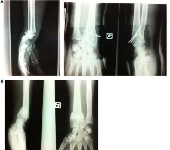
involved the styloid process of the ulna (n = 7), olecranon (n = 1), and trapezium (n = 1). The initial treatment consisted of Kapandji intra-focal pinning in 5 patients, volar plate and pin fixation in 2 patients, and non-operative management in 5 patients.Table 2lists the main characteristics of the fractures and treatment methods. The malunion was volarly angulated in 10 patients, including 2 with intra-articular involvement, and dorsally angulated in 2 patients (Fig. 1).
The main patient complaint was restricted pronation and supination, followed by pain. With the elbow flexed, mean motion ranges measured by goniometry before osteotomy were as follows:
flexion, 37.5◦(10◦to 50◦); extension, 41.6◦(20◦to 60◦); pronation,
Table 3
Clinical outcomes after 24 months in 12 patients (mean values).
Motion Flexion/extension Pronation/supination Before osteotomy 37◦(10◦to 50◦) 42◦(20◦to 60◦) 54◦(30◦to 80◦) 30◦(0◦to 60◦) After osteotomy 59◦(15◦to 70◦) 60◦(35◦to 75◦) 67◦(35◦to 82◦) 45◦(15◦to 60◦)
Pain VAS score – Before osteotomy 2 (2–4) – After osteotomy 2 (0–3)
Functional score Quick-DASH – After osteotomy 20 (10–42) –
54.1◦(30◦to 80◦); and supination, 30◦(0◦to 60◦). The mean VAS pain score was 2.4 (range, 1–4).
3.1. Clinical outcomes
Patients consistently reported improved wrist motion and func-tion 24 months after corrective osteotomy. Of the 12 patients, 10 were satisfied or very satisfied with the procedure. The mean VAS pain score was 1.7 (range, 0–3) and the mean Quick-DASH score was 20.4 (range, 10–42).
Table 3compares pain during wrist motion and wrist motion ranges before and after corrective osteotomy. The flexion/extension
arc increased by 40◦ (+21◦of flexion and +19◦of extension) and
the pronation/supination arc by 28◦(+13◦of pronation and +15◦of
supination).
Fig. 1. A. Example of an extra-articular fracture with dorsal angulation managed with Kapandji intra-focal pinning. B. Anterior malunion of the distal radius in the same
Table 4
Radiological outcomes after 24 months in the 12 study patients (mean values).
Ulnar variance (mm) Radial inclination (degree)
Distal radial joint surface obliquity (degree)
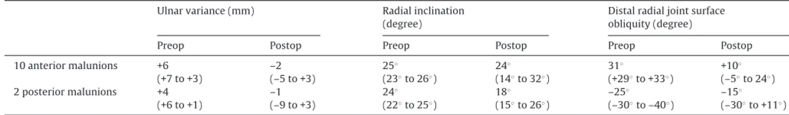
Preop Postop Preop Postop Preop Postop 10 anterior malunions +6 (+7 to +3) –2 (–5 to +3) 25◦ (23◦to 26◦) 24◦ (14◦to 32◦) 31◦ (+29◦to +33◦) +10◦ (–5◦to 24◦) 2 posterior malunions +4 (+6 to +1) –1 (–9 to +3) 24◦ (22◦to 25◦) 18◦ (15◦to 26◦) –25◦ (–30◦to –40◦) –15◦ (–30◦to +11◦) 3.2. Radiological outcomes
Progressive radio-carpal osteoarthritis developed in 3 patients. The osteoarthritis stage according to the Knirk and Jupiter classifi-cation was 1 in 1 patient and 2 in 2 patients[6].
Mean ulnar variance was –1.3 (range, –9 to +3), mean radial
inclination was 21.25◦(range, 14◦ to 32◦), and mean radial joint
surface obliquity was –2.5◦(range, –30◦to 24◦).Table 4compares
preoperative and postoperative radiographic values. Persistent posterior tilt was noted in the patients with posterior malunion.
All grafts showed good osteo-integration with no difference between iliac bone grafts and bone substitute.
3.3. Complications and revisions
Complex regional pain syndrome type I developed in 1 patient during the 24 months following corrective osteotomy. In 3 patients,
the range of supination was limited (mean, 6◦). Dorsal subluxation
of the ulnar head was noted in 1 of these patients (Fig. 2). All 3
patients required revision surgery with resection-stabilisation of
the distal ulna[7]within 2 years after corrective osteotomy.Table 5
shows the preoperative and postoperative radiological data of these 3 patients.
Osteoarthritis of the radio-scaphoid joint developed after cor-rective osteotomy in 3 patients. Among them, 1 required revision surgery for complete wrist denervation 18 months after the osteotomy. In 2 of these 3 patients, the malunion was intra-articular.
No cases of infection or non-union were recorded. No compli-cations related to iliac graft harvesting were observed. Neither was there any complications related to plate fixation, and none of the plates were removed within 2 years after corrective osteotomy.
3.4. Statistical results
We found no statistically significant differences between the preoperative and postoperative motion range values.
Fig. 2. Radiographs before and after osteotomy: persistent subluxation of the distal
radio-ulnar joint.
4. Discussion
In 1973, Kapandji[8]described an internal fixation technique
involving pinning of the fracture site. Kapandji pinning became the standard of treatment for extra-articular fractures of the
Table 5
Preoperative and postoperative data in the 3 patients who underwent revision surgery with distal radio-ulnar joint resection-stabilisation within 2 years after corrective osteotomy.
Type of fracture Angles before osteotomy
Type of osteotomy Angles after osteotomy Patient #1 Dorsal angulation UV +2
RI 24◦ Anteversion 18◦ Anterior, bone substitute RU +3 RI 32◦ Retroversion 12◦ Patient #2 T-shaped intra-articular fracture with volar angulation
RU 0 RI 24
Retroversion 28◦
Posterior, iliac bone graft
RU +2 Retroversion 30◦
Patient #3 Volar angulation RU +2 RI 24◦ Anteversion 30◦ Anterior, bone substitute RU –9 RI 30◦ Anteversion 10◦
distal radius. However, in patients with posterior and/or ante-rior comminution, this technique can be followed by secondary displacement with excessive reduction, resulting in malunion
[9]. Thus, a study by Herzberg and Dumontier[10] reported at
the 2000 SOFCOT symposium showed secondary displacement in 30% of cases despite immobilisation for 6 to 8 weeks. In 2012,
Obert et al.[11]reported a decrease in the frequency of secondary
displacement since the introduction of volar plate fixation. Of our 12 patients, the 5 patients managed with Kapandji pinning had volarly angulated malunion, although 4 of them initially had a dorsally displaced fracture. These 5 patients had posterior comminution and 3 of them exhibited excessive anterior reduction on the radiograph taken 15 days after initial surgery.
The quality of anatomic restoration of the distal radius and DRUJ governs the functional outcomes. In a 1997 cadaver study,
Bron-stein et al.[12]showed that 5 mm of ulnar translation of the radial
epiphysis resulted in a mean pronation loss of 23%. Shortening of the radius by 10 mm decreased pronation by 47% and supination by 29%.
Most studies included dorsally and volarly angulated malunions.
However, Saffar et al.[13]described specific features of anterior
malunions: limitation of volar angulation by the strong anterior capsular plane with minimal shortening of the radius and prona-tion of the distal fragment responsible for incongruity of the DRUJ with a marked decrease in the range of supination. Prommersberger
et al.[14]noted this rotational deformity in 23 of 37 malunions.
Rotational deformity tended to be more common in volar malu-nions, which seemed more poorly tolerated compared to dorsal malunions. The need to correct not only all volar or dorsal deformi-ties, but also the rotational deformities substantially increases the complexity of the procedure.
Saffar et al.[13]reviewed the various treatment options:
iso-lated osteotomy of the radius, isoiso-lated procedure on the ulna, or both. In their study, optimal effectiveness was obtained by combin-ing corrective osteotomy of the distal radius with a procedure on the head of the ulna, the main goal being to restore normal anatomy. All 12 patients in our study underwent isolated osteotomy of the distal radius and 3 of them required revision surgery for persis-tent loss of pronation/supination. This 25% revision rate within 2 years after corrective osteotomy is substantial. The postopera-tive radiographs in these 3 patients showed failure to correct the ulnar variance compared to the pre-osteotomy radiographs. In these 3 patients, detailed preoperative imaging studies combined with an evaluation of motion range would have identified the need for an additional procedure in combination with the radial osteotomy.
Based on our findings, we agree with Saffar et al.[13]and De
Smet et al.[15]that the objective of corrective osteotomy should
consist of restoring DRUJ congruity. A procedure on the DRUJ should be added when the intra-operative evaluation shows persistent limitation of the pronation/supination arc and/or failure to restore DRUJ congruity.
Coulet et al.[16] pointed out that DRUJ congruity cannot be
restored by radial osteotomy alone if ulnar variance exceeds 10 mm preoperatively. Preoperative computed arthro-tomography is cru-cial in this situation to choose between conservative and radical treatment. When the joint surfaces are intact, conservative treat-ment consisting in stabilisation and/or shortening of the ulna can be considered. In contrast, radical treatment is in order if the lesions involve the joint. Resection of the distal ulna, first described by
Darrach[17]in 1913, was performed in 3 of our patients using the
modified technique developed by Mansat et al.[7]With this
tech-nique, nearly 95% of patients are satisfied and a marked increase
in the pronation/supination arc is obtained (+43◦of supination and
+18◦of pronation), thus meeting the main demand of the patients
in our study.
Although the radiological angles were returned to their normal
values, the mean supination increase was only 6◦in the 10 patients
with anterior malunions, after exclusion of the 2 patients who had revision surgery. These fair outcomes are at variance with the
excel-lent results reported by Sato et al.[18], who obtained a nearly 70◦
increase in supination 25 months after isolated radial osteotomy. The limited improvement in supination is probably ascribable to failure to correct the rotational component of the radial malunion. Simply restoring ulnar variance failed to ensure good DRUJ con-gruity.
Saffar et al.[13]used a lateral approach posterior to the first
extensor compartment in 4 patients. The distal attachment of the brachioradialis muscle was reflected in the posterior-to-anterior direction. They reported that this approach allowed better con-trol of the distal fragment and therefore improved correction of the displacement in pronation. They advocated a cylindrical osteotomy in the sagittal plane. This osteotomy allows rotation without shortening, thereby restoring radial length without graft
implantation. In the study by Saffar et al.[13], a graft was needed
in only 3 patients, compared to 9 of 10 patients with volarly angulated malunion in our study. This decrease in the need for iliac graft harvesting is of interest in view of the 31% donor-site
complication rate reported by Gupta et al. [19] Mathew et al.
[20] described an antero-lateral approach involving
anterior-to-posterior detachment of the radial artery, brachioradialis muscle, and first extensor compartment. This approach may have the dual advantage of improving epiphyseal reduction control and allowing graft harvesting from the operative site, eliminating
the morbidity associated with iliac graft harvesting[19]. These
new surgical approaches may result in better correction of the rotational component of the malunion, thereby improving DRUJ congruity.
Our 2 patients with posterior malunions were managed with a dorsal approach and opening-wedge osteotomy filled with an iliac graft. Biplanar closing-wedge osteotomy, a technique described
by Posner et al.[21], has the singular advantage of avoiding the
morbidity associated with bone graft harvesting. This technique must be combined with DRUJ resection-stabilisation as described
by Darrach, because it increases the loss of radial length[21,22].
Closing-wedge osteotomy can be performed either alone or com-bined with translation to centre the distal fragment on the radial shaft.
More generally, radial malunions are responsible for carpal bone malalignment, which correlates directly with the functional
out-comes[23]. Gupta et al.[24] reported that measuring the angle
of radiolunate flexion could distinguish between two patterns of malalignment, namely mid-carpal and radio-carpal. In our study,
the radiolunate angle [24] was greater than 25◦ in 8 patients,
indicating mid-carpal instability. The remaining 4 patients had a
radiolunate angle equal to or lower than 25◦, indicating
radio-carpal instability. As reported by Gupta et al.[24], radial osteotomy
corrected the radiolunate angle in 9 patients, leaving only 3 patients
with a radiolunate angle greater than 25◦.
5. Conclusion
The management of distal radius malunions is complex. The pri-mary objective of surgery is restoration of the normal anatomy, with the goal of improving wrist range of motion and decreasing pain. In our study, isolated transverse osteotomy of the distal radius failed to restore sufficient supination. To obtain optimal clin-ical outcomes, distal radius osteotomy must correct the rotational component of the malunion. Surgery on the ulna to restore DRUJ anatomy is needed in some cases to optimise the postoperative functional outcomes.
Disclosure of interest
The authors declare that they have no competing interest.
References
[1]Jupiter JB. Current concepts review: fractures of the distal end of the radius. J Bone Joint Surg Am 1991;73:461–9.
[2]Mansat P. Traitement des fractures anciennes de l’extrémité distale des 2 os de l’avant-bras. Encyclopédie Médico-ChirurgicaleTechniques Chirurgicales–Orthopédie-Traumatologie 2006:44–346 [Éditions Scientifiques et Médicales Elsevier SAS, Paris, tous droits réservés].
[3]Judet T, Piriou P, De Thomasson E. Traitement orthopédique des fractures de Pouteau-Colles selon R. Judet. In: Allieu Y, editor. Fractures du radius distal de l’adulte. Paris: Expansion Scientifique Publications; 1998. p. 58–66.
[4]Cole JM, Obletz BE. Comminuted fractures of the distal end of the radius treated by skeletal transfixation in plaster cast: an end-result study of 33 cases. J Bone Joint Surg Am 1966;48:931–45.
[5]Dubert T, Voche P, Dumontier C, Dinh A. [The DASH questionnaire. French translation of a trans-cultural adaptation]. Chir Main 2001;20:294–302.
[6]Knirk JL, Jupiter JB:. Intra-articular fractures of the distal end of the radius in young adults. J Bone Joint Surg Am 1986;68:647–59.
[7]Mansat P, Ayel JE, Bonenvialle N, Rongières M, Mansat M, Bonnevialle P. Long-term outcome of distal ulna resection-stabilisation procedures in post-traumatic radio-ulnar joint disorders. Orthop Traum Surg Res 2010;36(3):216–21.
[8]Kanpandji AI. Ostéosynthèse des fractures récentes de l’extrémité inférieure du radius chez l’adulte. In: Duparc J, editor. Conférence d’enseignement de la Sofcot. Paris: Elsevier; 1994. p. 19–39.
[9]Fritz T, Wersching D, Klavora R, Krieglstein C, Friedl W. Combined Kirshner wire fixation in the treatment of Colles fracture. A prospective, controlled trial. Arch Orthop Trauma Surg 1999;119:171–8.
[10]Herzberg G, Dumontier C. Symposium : les fractures fraiches du radius distal chez l’adulte. Rev Chir Orthop 2000;86(Suppl. 1):1585–8.
[11]Obert L, Rey PB, Uhring J, Gasse N, Rochet S, Lepage D, et al. Fixation of distal radius fractures in adults: a review. Orthop Traum Surg Res 2013;99:216–34.
[12]Bronstein AJ, Trumble TE, Tencer AF. The effects of distal radius fracture malalignment on forearm rotation: a cadaveric study. J Hand Surg Am 1997;22:258–62.
[13]Saffar P, Tafnkji Y. Cal vicieux du radius en flexion. Chir Main 2005;24:299–304.
[14]Prommersberger KJ, Froehner SC, Schmitt RR, Lanz UB. Rotation deformity in malunited fractures of the distal radius. J Hand Surg Am 2004;29:110–5.
[15]Van Cauwelaert J, De Smet L. Corrective osteotomy for malunion of the dis-tal radius in young and middle-aged patients: an outcome study. Chir Main 2003;22:84–9.
[16]Coulet B, Id el Ouali M, Boretto J, Lazerges C, Chammas M. Is distal ulna resection influential on outcomes of distal radius malunion corrective osteotomies? Orthop Traum Surg Res 2011;97:479–88.
[17]Darrach W. Partial excision of lower shaft of ulna for deformity Colles fracture. Clin Orthop 1992;275:3–4.
[18]Sato K, Nakamura T, Iwamoto T, Toyama Y, Ikegami H, Takayama S. Corrective osteotomy for volarly malunited distal radius. J Hand Surg Am 2009;34:27–33.
[19]Gupta AR. Perioperative and long-term complications of iliac crest bone graft harvesting for spinal surgery: a quantitative review of literature. Int Med J 2001;8:163–6.
[20]Mathew P, Garcia-Elias M. Anterolateral surgical approach to the malunited distal radius fracture for corrective osteotomy and bone-graft harvest. Tech Hand Up Ext Surg 2013;1:17.
[21]Posner MA, Ambrose L. Malunited Colles’fractures: correction with a biplanar closing wedge osteotomy. J Hand Surg Am 1991;16:1017–26.
[22]Sennwald G, Fischer W, Stähelin A. Le cal vicieux du radius distal et son traite-ment : à propos de 122 radius. Int Orthop 1992;16:45–51.
[23]Bickerstaff DR, Bell MJ. Carpal malalignment in Colles’ fractures. J Hand Surg Br 1989;14(2):155–60.
[24]Gupta A, Batra S, Jain P, Sharma SK. Carpal alignment in distal radial fractures. BMC Musculoskelet Disord 2002;3(3):14.