1
Risk factors associated with bovine tuberculosis and molecular characterization of
1
Mycobacterium bovis strains in urban settings in Niger 2
3
Boukary A.R.1,2,6,7, Thys E.2, Rigouts L.2,7, Matthys F.3†, Berkvens D.2, Mahamadou I.4, 4
Yenikoye A.5, Saegerman C.6* 5
6
1 Department Animal Health and Livestock Promotion, ONG Karkara, Niamey, Niger BP 2045, Niamey, Niger
2 Department of Biomedical Sciences, Institute of Tropical Medicine, Nationalestraat 155, B-2000, Antwerp, Belgium
3 Department of Public Health, Institute of Tropical Medicine, Nationalestraat 155, B-2000, Antwerp, Belgium
4 Ministry of Livestock, Direction of Animal Health, Niamey, Niger 5 Faculty of Agronomy, University of Niamey, BP 10895, Niamey, Niger
6 Department Infectious and Parasitic Diseases, Faculty of Veterinary Medicine, University of Liege, Boulevard de Colonster 20, B42, B-4000 Liege, Belgium
7 Department of Veterinary, Pharmaceutical and Biomedical Sciences, University of Antwerp, Universiteitsplein 1, B-2610 Wilrijk, Belgium
7
† This paper is dedicated to the memory of Francine Matthys who was the former medical 8
director of MSF Belgium and a prominent public health researcher in the control of human 9
tuberculosis in developing countries. 10
* Corresponding author: Prof. Claude Saegerman, Department of Infectious and Parasitic 11
Diseases, Epidemiology and Risk Analysis Applied to Veterinary Sciences (UREAR), Faculty 12
of Veterinary Medicine, University of Liege, Boulevard de Colonster, 20, B42, B-4000, 13
2 Liege, Belgium. Tel: + 32 4 366 45 79; Fax: + 32 4 366 42 61; E-mail: 14 claude.saegerman@ulg.ac.be 15 16 17 Abstract 18
A retrospective and a longitudinal survey were carried out at the abattoir of Niamey. Results 19
showed a highly significant difference of suspected tuberculosis (TB) gross lesions among 20
different animals species (P <0.0001). The proportion of carcasses with TB-like lesions was 21
0.19% among cattle, 0.11% among camels, 0.001% among sheep and 0.0006% among goats. 22
In cattle, cows are significantly more affected than the other categories (P<0.001). Also in 23
cattle, TB-like lesions are mostly localized in the lungs (92.77%) followed by the lymph 24
nodes (50.87%) and the liver (32.40%). The prevalence of gross lesions compatible with 25
bovine TB (BTB) is strongly influenced by the season (P<0.0001), closely correlated with the 26
origin of the animals (P<0.001), and has a negative impact on the weight of affected animals 27
(P <0.0001). Sixty two samples of suspected TB gross lesions were subject to microbiological 28
analysis and molecular typing of strains. Mycobacterium bovis was identified in 18 animals 29
showing 5 different spoligotypes, belonging to type "African 1" (Af1) previously identified in 30
Central and West Africa. In addition, a profile (SB1982) not previously reported distinguished 31
by the absence of spacers 3, 4, 9, 16, 22, 30 and 39-43 has been characterized in this study. To 32
assess risk factors for BTB transmission, a questionnaire on animal husbandry practices, food 33
habits, and clinical signs of TB in animals and humans was submitted to the heads of 1,131 34
randomly selected households. The main risk factors identified are: consumption of 35
unpasteurized milk (91%) and lack of hygiene within households (32 to 74%). Clinical signs 36
that could be attributed to TB were also reported both in humans and in animals of the 37
households. 38
3 39
Key words: Mycobacterium bovis, bovine tuberculosis, risk factors, livestock, urban settings
40 41 42
1. INTRODUCTION
43 44Tuberculosis due to Mycobacterium bovis is often neglected as zoonotic disease in 45
developing countries (Acha and Szyfres, 2005). In sub-Saharan Africa (SSA), bovine 46
tuberculosis (BTB) is a serious threat for the economy but also for public health and animal 47
health (Cosivi et al., 1998; Michel et al., 2006; Cleaveland et al., 2007; Humblet et al., 2009). 48
BTB is widely distributed in SSA, where 85% of the herds and 82% of the human population 49
live in areas where the disease has been reported (Cosivi et al., 1998). In most African 50
countries, BTB control measures are not applied (OIE, 2007). Additionally, there are very 51
complex interactions between the rural pastoral livestock systems and the semi-intensive 52
system practiced in and around urban settings (Thys et al., 2006; Boukary et al., 2007). These 53
interactions and inadequate sanitation measures are important risk factors favouring 54
endemicity of zoonotic tuberculosis (Sidibé et al., 2003; Mfinanga et al., 2003; Cleaveland et 55
al., 2007). The nutritional habits of the population consuming unpasteurized milk also endorse 56
infection with M. bovis (Kang'ethe et al., 2007). However, TB due to M. bovis has hardly been 57
studied in the sub-Saharan context where epidemiologic aspects of the disease remain largely 58
unknown (Cosivi et al., 1998). Whether in animals or humans, the pathogen itself, M. bovis, is 59
rarely studied (Thoen and Bloom, 1995). The Interafrican Bureau for Animal Resources 60
(AU/IBAR, 2006) indicates that in 2006 only 9 of 53 African countries reported cases with a 61
total of 176 outbreaks of BTB. Mostly cattle are affected representing 98.9% of reported 62
animal cases. 63
4 Use of in vitro culture and subsequent molecular typing enabled progress in the 64
characterization of M. bovis strains in some African countries (Rigouts et al., 1996; Portaels et 65
al., 2001; Haddad et al., 2004). Molecular typing of M. bovis isolates from Tanzania (Daborn 66
et al., 1997) has shown two lineages of M. bovis: an aboriginal lineage with atypical 67
properties and a lineage imported from Europe displaying the classical spoligotyping profile. 68
Subsequent work in Central and West Africa performed by Njanpop-Lafourcade et al. (2001), 69
Diguimbaye et al. (2006), Schelling et al. (2005), Cadmus et al. (2006) and Müller et al. 70
(2008; 2009) revealed a predominant characteristic M. bovis spoligotype in Central Africa and 71
West Africa, called Af1 type. 72
Data on the importance of BTB in Niger are particularly scarce and not updated. 73
Investigations of Alambedji (1984) and Bloch and Diallo (1991) by single intradermal skin 74
test gave low prevalence rates varying from 1.56 to 3.20% among cattle. 75
The present work aims to determine the prevalence of BTB suspected gross lesions at the 76
abattoir of Niamey, to contribute to the knowledge of current circulating M. bovis strains in 77
Niger, and to identify risk factors for transmission of BTB from animal to human. 78
79 80
2. MATERIALS AND METHODS
8182
2.1. Survey site and animal husbandry systems
83
The survey was carried out at the abattoir of Niamey located in the industrial area of the city 84
on the banks of the Niger River. The ―Abattoir de Niamey‖ is a semi-autonomous company 85
which was founded in 1967. It’s an old structureand the entire infrastructureisaging. At the 86
moment of the study, the staff wascomposedof 60techniciansincluding 10 permanentstaff 87
inspectorsworking under the supervisionof asworn veterinarian. Thisstaffisassistedbymore 88
5 than 300 temporary workers and butchers. Slaughter and carcass inspection is doneat night 89
between 10PM and5 AM. 90
The Niamey abattoir is in charge of animal slaughtering and refrigeration, and has a capacity 91
of 10,000 tons per year. It is supplied by animals from the city livestock market (called 92
Tourakou) and from rural markets, mainly those located in the areas of Torodi, Tera, Ayorou, 93
Balleyara, Kollo and Boubon in the surrounding of Niamey (Figure 1). In rural areas (Ru), 94
animals are managed under a traditional husbandry system (extensive/transhumant)) 95
depending entirely on natural pastures and farm by-products without extra feed supplements 96
or adequate health services. Cattle and small ruminants are usually driven to pasture together. 97
Camels are generally led in separate herds and their feeding is mainly based on tree fodder. In 98
the urban (Ur) and periurban (Pu) areas of Niamey city the livestock system is directed 99
towards dairy production and animals are more fed with supplements, including agro-100
industrial by-products and kitchen waste. 101
102
2.2. Characterization of suspected BTB infection at the abattoir of Niamey
103
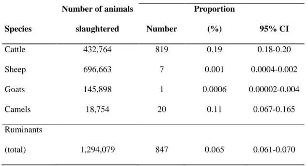
Determination of the prevalence of TB-like lesions by retrospective data collection: Data on 104
432,764 cattle, 696,663 sheep, 145,898 goats and 18,754 camels slaughtered at the abattoir of 105
Niamey from January 2003 to December 2008 were collected from the official records and 106
analysed (Table 1). Carcasses underwent a standard meat inspection including the 107
examination of the following lymph nodes: parotid, retropharyngeal, mediastinal, 108
tracheobronchial, mesenteric, submaxillary, iliac, precrural, prescapular, supra-mammary, 109
inguinal, apical, ischiatic and portal nodes. Organs/tissues including lungs, liver, kidneys, 110
mammary glands, intestines, heart, abdominal and thoracic cavities, cerebral membranes and 111
bones (ribs and vertebrae) were also thoroughly examined. Organs showing gross visible 112
lesions compatible with BTB were confiscated. According to the regulation at the abattoir of 113
6 Niamey, suspected cases of active or stabilized tuberculosis that are confirmed by the 114
veterinary inspector are subject to total or partial condemnation and destruction. 115
116
Characterization of suspected tuberculosis gross lesions by a longitudinal survey: An 117
additional one-year longitudinal survey was implemented independently during standard meat 118
inspection at the abattoir from July 2007 to June 2008. For all the cases of condemnation due 119
to suspected TB data related to the animals (age, breed, sex, geographic origin and clinical 120
history), the production and marketing system (including the various stakeholders), the 121
affected organs and the description of the observed lesions were collected and analysed. 122
123
Bacteriology and molecular characterization of mycobacteria 124
A total of 147 samples were collected from 140 carcasses of animals (130 cattle, 3 sheep 125
and 7 camels) with suspected BTB lesions. The frequent power cuts and difficulties in 126
maintaining the cold chain during the survey period did not allow a good preservation of the 127
specimens. At the end, only 62 samples from 60 animals were available for analysis; these 128
samples covered the entire period studied and were subjected to laboratory-based analyses. 129
They were packed in 1.2 mL Eppendorf® tubes containing semi-solid transport medium 130
(Portaels et al., 2001) and sent at room temperature to the Mycobacteriology Unit of the 131
Institute of Tropical Medicine, Antwerp, Belgium. Acid fast bacilli (AFB) were detected by 132
microscopy using the Ziehl-Neelsen (ZN) technique applying the ATS scale to quantify the 133
bacterial load (American Thoracic Society, 1999). In vitro culture on Löwenstein-Jensen and 134
Stonebrink medium was carried out after decontamination using the inverted Petroff method 135
(Durnez et al., 2008). Spoligotyping was carried out to identify M. bovis in all isolates 136
(Kamerbeek et al., 1997). In addition, PCR targeting the 16S rRNA gene to detect M. 137
tuberculosis-complex (Durnez et al., 2008) was used for 30 specimens of animals that yielded
7 contaminated or negative (sub-) cultures, but yet positive smears in order to maximize 139
recovery of probable M. bovis. Subsequently, PCR-positive samples were subjected directly 140
to spoligotyping. 141
All spoligotypes were identified using the database on www.Mbovis.org (Smith and 142
Upton, 2011). Presence or absence of the RDAf1 region was determined for all identified M. 143
bovis cases as described (Müller et al., 2009).
144 145
2.3. Determination of risk factors for BTB transmission
146 147
From July 2007 to March 2008, a cross sectional household survey was conducted in the 148
Pu zone from July to September 2007, in the Ru zone from October to December 2007 as well 149
as in the Ur zone of Niamey city from January to March 2008. In total, 1,131 households (399 150
in Ur, 400 in Pu and 332 in Ru) were randomly selected from an up-to-date census database. 151
62% of household heads surveyed are Fulani ethnic, 20% are Zarma and 11% are Tuareg. The 152
Fulani are traditionally nomadic herders and their presence in Ur and Pu areas is quite recent 153
and due to climate changes observed in recent decades. 154
The questionnaire used in the face-to-face interview of the household heads included 155
questions related to animal husbandry practices, food habits and the presence of clinical signs 156
of TB both in animals and humans. 157
158
2.4. Statistical analysis
159 160
Descriptive statistics and comparisons were carried out using Intercooled Stata 9.2 for 161
Windows (StataCorp LP, USA). Generalised linear models (Poisson regression and logistic 162
regression) were employed to examine the effects of different risk factors: Poisson regressions 163
8 yielded incidence rate ratios (IRR), logistic regressions resulted in odds ratios. Welch's test 164
was used to appreciate the weight difference between the carcasses carrying TB-like lesions 165
and healthy carcasses. 166 167 168
3. RESULTS
169 1703.1. Determination of the prevalence of suspected TB gross lesions by retrospective
171
data collection
172
Based on data recorded over the 6 years, it appears that the demand for meat consumption 173
had remained fairly constant. The average number of animals slaughtered per month at the 174
Niamey abattoir during this period was: 6011 (315) cattle, 9676 (970) sheep and 2026 175
(361) goats. The survey conducted between July 2007 and June 2008 showed that the daily 176
workload for meat inspectors does not seem to be a factor affecting their performance. 177
However, they complain of poor equipment and poor working conditions. 178
At the Niamey abattoir, seizures of carcasses for suspicion of TB-like lesions were rare. 179
Indeed, out of a 1,294,079 animals slaughtered during the 6-years period, condemnations 180
based on suspected TB gross visible lesions included 847 carcasses (Table 1). During the 181
entire period, the maximum daily record of suspected carcasses in cattle was 4. The average 182
apparent prevalence of TB-like lesions was 0.19% (95% CI: 0.18 - 0.20) for cattle and 0.11% 183
(95% CI: 0.07 - 0.17) for camels. Regarding small ruminants only 7 cases were reported in 184
sheep and one in goats. 185
Based on the observation of TB-like lesions there is a highly significant difference in 186
susceptibility to infection among different species (P<0.001). Camels and cattle are species at 187
9 significantly high risk compared to goats and to sheep. There is no significant difference in 188
susceptibility to infection between sheep and goats. 189
190
Insert Table 1
191 192
Condemnations due to suspected TB lesions mostly concerned cattle with 819 cases on 193
a total of 847 for all included species. Of these 819 cases, only 747 cases (91.2 %) could be 194
described, whereas for the remaining detailed descriptions were lacking in the registers. Data 195
collected were related to the affected organs, types of lesions observed and the weight of 196
carcasses. 197
In cattle, the majority of the TB-like lesions were detected in the lungs (92.77%). Lung 198
lesions were in most cases accompanied by reactions in the lymph nodes (50.87%) and 199
lesions in the liver (32.40%). Other affected organs were: the heart (0.80%), the kidneys 200
(0.54%) and the spleen (0.27%). About one tenth (9.77%) of the cases were suspected of 201
generalized miliary TB. 202
The apparent prevalence of TB-like lesions in cattle at the abattoir in Niamey is strongly 203
influenced by the season (P<0.0001). The Incidence Rate Ratio (IRR), of suspected BTB 204
shows two peaks (Figure 2). The first slight peak is observed during the hot dry season in 205
April (P<0.05) and two strong peaks are observed during the first half of the rainy season in 206
July (P<0.001) and in August (P<0.01). 207 208 Insert Figure 2 209 210 211
3.2. Characterization of suspected TB gross lesions by a longitudinal survey
10 213
A total of 71,373 cattle, 124,759 sheep, 19,731 goats and 2,604 camels were slaughtered 214
at the abattoir of Niamey during the one-year survey. The 71,373 cattle included 4,086 oxen, 215
14,630 bulls, 31,190 cows and 21,467 calves (animals younger than 4 years are considered as 216
calves at the abattoir of Niamey). A total of 130 cattle, 3 sheep and 7 camels presented gross 217
visible lesions compatible with BTB. 218
Experts of the abattoir consider that 50% of animals slaughtered are from the Tourakou 219
market in Niamey, 30% come from the Torodi area and 20% from other rural markets. 220
Data collected by this independent tracking confirms the picture observed in cattle 221
during the six-year period. Out of the 130 BTB suspected cases in cattle, organs and tissues 222
most affected were lungs, lymph nodes, liver and to a lesser extent kidneys and heart (Table 223
2).
224
The presence of macroscopic lesions is closely correlated with the origin of the animals 225
(P<0.001). Over half (56.2%) of the cases were from the rural zone of Torodi, 22.3% from the 226
urban community of Niamey and 20.5% from the other rural areas. Compared to urban 227
markets, the risk of having animals suspected of TB is statistically higher in Torodi (OR=4.2; 228
95% CI: 2.7 - 6.5) and in other rural markets (OR=2.4; 95% CI: 1.4 - 4.1). 229
In cattle, there was a significant difference in sensitivity to infection between the different 230
categories (bulls, oxen, cows and calves) (df=3; Chi2=84 P<0.001). Cows are significantly 231
more sensitive to infection than the other categories (OR=9.4; 95% CI: 4.4 - 20.2). 232
A significant negative relationship was found between the presence of TB-like lesions on the 233
carcasses and the weight of affected animals. Overall, we found that the carcasses of cattle 234
suspected for BTB are significantly lighter than those of healthy animals (Welch's test 235
P<0.0001). The difference of weight between healthy carcass and those with gross visible 236
lesions is on average 14 kg (95% CI: 8 - 20). 237
11 238 Insert Table 2 239 240 241 242
3.3. Molecular characterisation of mycobacteria
243
Acid-fast bacilli were detected by smear microscopy in 31 (50.0%) of the 62 post-mortem 244
samples. Overall, we observed a high contamination rate (25.8%) for culture, most probably 245
due to suboptimal storage conditions. Thirteen samples were positive for culture among 246
which 7 were clearly identified as M. bovis type Af1 by the non-appearance of spacer 30 and 247
the Af1-specific deletion (Table 3). Five isolates were identified as non-tuberculous 248
mycobacteria (NTM); the latter were not further identified to the species level. The remaining 249
culture (BK 090046) yielded a weak spoligotype result and failed in subculture; most 250
probably it was M. bovis as spacers 39-43 were clearly missing, and the Af1 specific deletion. 251
However, the spoligotyping reactions were too weak to determine the exact M. bovis 252
spoligotype (SB type). In addition, in 11 animals that yielded negative or contaminated 253
culture results but a MTB-complex positive PCR, M. bovis type Af1 was clearly identified by 254
spoligotyping and the RDAf1 PCR directly in the specimen. For 6 more specimens we noted a 255
weak spoligotype reaction even though the MTB-complex-specific PCR turned positive; 256
again most probably the latter are M. bovis as spacers 39-43 were clearly missing for all of 257
them; five showed the Af1-specific deletion, whereas one failed in this assay, probably due to 258
the low DNA content. Spoligotypes were too weak to determine the exact SB type in the 259
seven specimens. 260
So, M. bovis type Af1 was clearly identified in 18 animals (16 bovines, 1 camel and 1 sheep), 261
and 5 different spoligotypes were observed (Table 3). SB0944 profile was the most abundant 262
being identified in 1 sheep (Bali-Bali race) and 12 bovines (11 belonging to the Djeli and 1 to 263
12 the Mbororo breed). SB0300 was found in two Djeli cows, SB1440 in one Djeli cow, SB1433 264
in one camel. Furthermore a new strain not yet reported to the M. bovis database prior to this 265
study was found in a Djeli cow. Hence, it has now been reported to the database, and 266 designated as SB1982. 267 268 Insert Table 3 269 270 271
3.4. Determination of risk factors for BTB transmission
272
Table 4 shows that, in most cases, mating was not controlled, especially in urban areas.
273
Eighty-five to 94% of the households consumed fresh milk or products derived from 274
unpasteurized milk. In Pu and Ru zones, poor hygiene was scored in 74% of the households 275
and in 32% in Ur zones. On average, only 1% of households employed disinfectants for 276
cleaning kitchen tools used to prepare food from animal products. 277
Large proportions of livestock keepers observed severe weight loss in their animals (48.6% in 278
Ur, 58.8% in Pu and 51.8% in Ru) despite provision of additional feed. 18%, 27% and 25% of 279
household’s heads interviewed respectively in Ur, Pu and Ru zones reported animal death 280
casualties due to chronic cough. 281
Regarding the disease in humans, 43.1% of the interviewed household’s heads in Ru stated to 282
have observed in their vicinity clinical signs that could be related to TB. Some stated to suffer 283
themselves from persistent cough or knew people with similar symptoms. Chronic cough in 284
humans was also reported to have been observed by respondents in Ur (22.8%) and in Pu 285
(30.5%). In the rural zone of Torodi, a significant relationship (OR=5; 95% CI: 3.0 - 26.5) 286
was found between the presence of people with severe chronic cough in households and the 287
presence of animals suffering from chronic cough within the same household. 288
13 289 Insert Table 4 290 291
4. DISCUSSION
292 2934.1. Determination of the prevalence of suspected tuberculosis gross lesions by
294
retrospective data collection
295
In SSA, research on animal TB mainly focused on cattle. Data on other domestic species 296
are particularly scarce (Cosivi et al., 1998; Razanamparany et al., 2006; Diguimbaye et al., 297
2006) and should be further investigated in the future. 298
Our results showed the presence of TB-like lesions in all animal species slaughtered at the 299
Niamey abattoir, but the risk of suspected BTB infection is significantly higher in cattle and 300
camels compared with goats and sheep that are less susceptible to M. bovis. Nevertheless, the 301
apparent prevalence of TB-like lesions (0.19%) observed in cattle slaughtered at the abattoir 302
of Niamey is among the lowest recorded on the African continent. It varies in West African 303
abattoirs from 1 to 8.8% (Dao, 2005, Diguimbaye et al., 2006, Njanpop-Lafourcade et al., 304
2001; Cadmus et al., 2006; Müller et al., 2008). Our finding is also lower than the 7.9 to 305
19.8% prevalence record in EasternEastern Africa (Regassa et al. 2008; Cleaveland et al. 306
2007; Biffa et al., 2011). 307
The low prevalence observed in small ruminants compared to cattle and camels can be 308
explained partly by the difference in sensitivity of a physiopathological point of view between 309
these species. Indeed, sheep and goats are inherently more resistant to contracting the disease, 310
i.e. they require a much higher infective dose than cattle before infection can become 311
established (Allen, 1988; WHO, 2001). According to some authors, BTB infection in sheep 312
and goats is a sign of infection in other in-contact species (Allen, 1988; Ayele et al. 2004). 313
14 The disease does not spread easily between small ruminants (Allen, 1988). When exposure to 314
infection is high, there is no doubt that small ruminants can become infected, and display 315
lesion morphology and distribution in the body similar to cattle (Fischer et al., 2009). In Niger 316
cattle and small ruminants normally graze together, and this practice could constitute a higher 317
risk for transmission of bovine TB among these animals. 318
The probability of lesions to be detected at slaughter is difficult to assess (Müller et al., 2008). 319
The very low figures at Niamey could be due to underestimation, which in turn can be 320
attributed i.a. to the lack of rigor in the veterinary inspection. According to Assaged et al. 321
(2004), meat inspection at the abattoir can detect only 55% of infected animals with 322
confirmed visible lesions. The low number of carcasses condemned for BTB reasons may also 323
be linked to the high incidence of illegal slaughtering. Indeed, at the abattoir in Niamey, any 324
animal found with BTB lesions is subjected to total or partial condemnation. This urges 325
butchers to clandestine slaughtering. As a result, there may be TB-infected meat on the 326
market and an increased risk of transmission to people (Asiimwe et al., 2009). 327
Analysis of data recorded from the abattoir during the 6-years period shows a strong 328
seasonal variability of condemnation due to BTB (P<0.001). The frequency of gross lesions is 329
higher in the beginning of the rainy season (July-August). This may be explained by the 330
massive destocking of animals by farmers. Indeed, this period concurs with the return of 331
animals from transhumance. Sick animals and those with poor general condition are usually 332
sold or culled. 333
Lung lesions are the major cause of condemnation due to BTB in SSA (Dao, 2005, 334
Diguimbaye et al. 2006; Njanpop-Lafourcade et al., 2001; Cadmus et al., 2006; Müller et al., 335
2008; Asiimwe et al., 2009). Our results show that 92.77% of the 747 detected gross lesions 336
are in the lungs with 28.65% of animals presenting lung lesions only, whereas the remaining 337
presented multiple lesions involving the lungs and other organs. This suggests that in this 338
15 setting the lungs are the entry port of M. bovis infection. This is in agreement with the 339
findings of Müller et al. (2008) showing that the presence of pulmonary lesions was closely 340
associated with M. bovis infection. Lymph node reactions (50.87%) are also characteristic for 341
BTB. They are the tissues that are investigated primarily by meat inspectors. Lesions in the 342
liver are frequent too (32.40%). This organ contains usually caseous nodules which can 343
reflect chronic infection (Thorel, 2003). 344
345
4.2. Characterization of suspected tuberculosis gross lesions by longitudinal survey
346 347
It should be noted that so far, no serious investigation has been conducted on this topic in 348
Niger and control of TB is limited to meat inspections in abattoirs. Cows are significantly 349
more at risk to BTB infection than bulls, oxes and calves. 350
The high prevalence of TB-like lesions observed in cows may be explained by the fact that 351
they remain longer in the herds for milk production while the other categories are slaughtered 352
or sold earlier. It has been shown that the prevalence of bovine tuberculosis increases with age 353
of animals especially in regions where infection is endemic (Blancou et al., 1971; Sidibé et 354
al., 2003, Cleaveland et al., 2007; Humblet et al., 2009). Indeed, in endemic situations, older 355
animals are more likely to be exposed and to develop the disease. Moreover, under the effect 356
of stress or age, latent infections are reactivated resulting in a higher prevalence in older 357
animals (Pollock and Neill, 2002). 358
In this survey, the presence of TB-like lesions in cattle is closely related to the origin 359
of the animals (P<0.001). Rural areas that supply Niamey in cattle are the most affected. 360
Compared to urban markets, the risk of having animals with gross visible lesions is 361
statistically higher in Torodi and other rural markets. Indeed, 70.77% of animals with gross 362
lesions were from Ru with 56.15% of them originating from the Torodi area, which is located 363
16 in the far west of Niger at the border with Mali, Burkina Faso and Benin. Our results 364
corroborate those found by Alambedji (1984) who reported that 46% of cattle suspected of 365
BTB in the Niamey abattoir are from Torodi. 366
Due to its geographical position, Torodi constitutes a crossroad for trade and transit of 367
cattle. The complex interactions between transhumant livestock system, the semi-intensive 368
system practiced by farmers and the presence of an important livestock market in this area 369
promote contact between animals from different regions with a significant risk of spread of 370
BTB. In addition, it should be noted that Torodi is located at the edge of the natural park that 371
is shared by Niger, Benin and Burkina Faso. The proximity of this natural reserve leads 372
necessarily to close contact between domestic animals and wildlife which can be a source of 373
transmission of M. bovis (Zieger et al., 1998). The role of wildlife in transmission or 374
recurrence of BTB in domestic animals has been well documented (Michel et al., 2006; 375
2008). In South Africa, for example, BTB is now a serious problem in the Kruger National 376
Park where the disease was first diagnosed in buffalo in 1996 (Bengis et al. 1996). Even in 377
countries where BTB is eradicated, wildlife remains a risk of transmission to domestic 378
animals through the parks. Woodford (1982) found M. bovis in warthogs (Phacochoerus 379
aethiopicus) and buffaloes Syncerus caffer (Sparrman, 1779) in the Ruwenzori National Park
380
in Uganda. Keet et al. (1996) also reported bovine TB in other wildlife at the same park. 381
Bovine tuberculosis can be a serious threat for the economy in sub-Saharan Africa (Cosivi 382
et al., 1998). Indeed BTB causes a decrease in financial capital and an increase of production 383
costs for the farmers. The disease also causes indirect losses in agricultural productivity, due 384
to the loss of animal traction and manure. We observed significant differences in body weight 385
(P <0.0001) between healthy carcasses and those with gross visible lesions; a decrease of 14 386
kg in average weight in cattle presenting TB-like lesions was seen. Similarly, Blancou and 387
Cheneau (1974) found weight losses ranging from 3.1 to 9.7 kg in Malagasy zebu carcasses 388
17 with gross visible lesions. The loss of weight due to suspected BTB varies depending on the 389
disease status and the farming system and is higher as the lesions caused by the pathogen are 390
more severe (Blancou and Cheneau, 1974). In contrast, Biffa et al. (2011) found no significant 391
correlation between body condition and infection with bovine tuberculosis in cattle in 392
Ethiopia and suggested that body condition may not be considered a reliable predictor of TB 393
under Ethiopian conditions. 394
395 396
4.3. Molecular characterisation of mycobacteria
397 398
As far as we know, this is the first study conducted on molecular characterization of M. 399
bovis isolates from slaughter animals in Niger. However, the low number of samples analyzed
400
(62/147) due to frequent power cuts hampers the study. Freeze-thaw cycles and frequent 401
transfers of samples from one location to another due to load shedding of electricity were the 402
basis for the loss of a number of samples. Müller et al. (2008) were confronted to the same 403
situation in a similar study conducted at the abattoir of Bamako in Mali. 404
Several mycobacterium were isolated from 50% (n=62 samples) of the 60 carcasses with 405
characteristic TB-like lesions. This is relatively higher than the 35.0% (n=60) and the 31.2% 406
(n=105) respectively from cattle carcasses in Mali and in Ethiopia (Müller et al., 2008; Biffa 407
et al., 2011), but still lower than expected. As suggested by some authors (Cleaveland et al., 408
2007; Müller et al., 2008; Biffa et al., 2011), we agree in our case that the relatively low 409
isolation frequency could be due to reduced sensitivity of culture arising from prolonged 410
storage of specimens. The low recovery of bacteria could also be explained by the high 411
amount of completely calcified lesions without viable tubercle bacilli (Müller et al. 2008). 412
18 We clearly identified M. bovis in 18 animals (n=60) and NTM were found in 7 animals. It’s 413
well established that in sub-Saharan Africa, TB-like lesions may be caused by an array of 414
pathogens amongst which NTM could play a crucial role (Müller et al. 2009). This suggestion 415
is strengthen by the findings of a previous study in some African countries where NTM such 416
as M. fortuitum, M. kansasii, M. aquae and M. smegmatis were cited as causative agents of 417
TB-like lesions in domestic animals (Diguimbaye et al., 2006; Müller et al., 2008; Sahraoui et 418
al., 2009 ; Biffa et al., 2010; Mamo et al., 2011) 419
Among the spoligotypes identified in Niger, SB0944 is present in 13 (72%) of the positive 420
samples, which confirms the predominance of this strain in Central and West African regions. 421
Indeed, this strain represents a significant proportion of spoligotypes identified in Nigeria 422
(46.1%), in Cameroon (62.7%), in Chad (40%) and in Mali (9%) (Njanpop-Lafourcade et al., 423
2001; Cadmus et al., 2006; Diguimbaye et al., 2006; Müller et al., 2008). Based on the 424
similarity of the SB0944 pattern with the BCG-like profile commonly seen in strains from 425
France, an influence of the French colonial history on the West African M. bovis population 426
has been suggested (Njanpop-Lafourcade et al., 2001). Our discovery of this strain in Niger 427
confirms its regional coverage. 428
429
Up to now, The SB1433 and SB1440 strains have been shown in Nigeria and the SB0300 430
strain in Mali. The presence of these three strains in Niger suggests that this country should 431
play a central role through its particular geographical position. Indeed, Niger is a country of 432
important transboundary transhumance and a major exporter of cattle to other countries 433
(MRA, 2001). A feature common to all these strains identified in Cameroon, Chad, Nigeria, 434
Mali and now in Niger is the lack of the spacer 30 typical for the Af1 M. bovis type (Müller et 435
al., 2009). This suggests a close relationship between strains from Niger and those other 436
countries. In addition to the absence of spacer 30, SB0300 also lacks spacer 6, a characteristic 437
19 not seen in the strains from Central African countries (Njanpop-Lafourcade et al., 2001; 438
Diguimbaye et al., 2006). Müller et al. (2008) suggest that spoligotype pattern SB0300 may 439
have evolved from strains with spoligotype pattern SB0944 either by drift or a selective 440
sweep. Similarly, the new strain (SB1982) might have been imported from neighbouring 441
countries, where it remained undetected due to non-systematic isolation and/or typing of M. 442
bovis, or it might be the result of ongoing evolution cattle and/or other animal species (e.g.,
443
wildlife animals), reflecting the long-term presence of M. bovis in the country. Indeed, this 444
strain lacks spacer 6 and 22 in addition. 445
The fact that the strain SB1433 was only found in camel can be explained by the separate 446
husbandry of camels in Niger making direct contact with other species rare. However, this 447
hypothesis has to be verified as the animal in which this strain was isolated is certainly not 448
representative of the entire camel husbandry system. We also know that this animal came 449
from the livestock market of Tourakou, but we had no information on its exact origin. Some 450
authors (Wernery et al. 2007; Mamo et al., 2011) reported that BTB occurs more frequently in 451
camels when they are kept close to other camels or in close contact with cattle. Cases of 452
contamination of camels by wildlife (gazelles) have also been reported in the Arabian 453
Peninsula (Ostrowski et al., 1998). Further investigations with a larger number of animals are 454
therefore needed to better understand the epidemiology of tuberculosis in camels in Niger. 455
456
4.4. Determination of risk factors for BTB transmission
457 458
One of the characteristics of animal husbandry in SSA, whatever the livestock system 459
considered, is the close proximity between humans and animals (Cosivi et al. 1998). In 460
pastoral areas, humans and animals share the same microenvironment and water sources 461
especially during the hot and dry seasons, which is a high potential risk of transmission of M. 462
20
bovis (Cosivi et al., 1998; Mfinanga et al., 2003; Ameni et al., 2006). In urban and suburban
463
areas, the closeness is such that humans and animal reservoirs live confined in unsanitary and 464
poorly ventilated places (Sidibé et al., 2003; Boukary et al., 2007; Regassa et al., 2008). Our 465
results show the existence of BTB in Niger in all animal husbandry systems, since M. bovis 466
has been identified in animals from urban, periurban and rural areas. During the survey in 467
those three areas, some livestock keepers declared to have observed clinical signs 468
characteristic for BTB in their animals. 469
Poor farming practices, consumption of unpasteurized milk and poor food hygiene 470
conditions are important risk factors for transmission of M. bovis from animals to humans 471
(Kazwala et al., 1998; Sidibé et al., 2003; Ameni et al., 2006; Cleaveland et al., 2007; 472
Kang'ethe et al., 2007; Humblet et al., 2009). M. bovis is usually transmitted by ingestion of 473
unpasteurized milk and causes extrapulmonary tuberculosis especially in children (Kleeberg, 474
1984; Dankner and Davis, 2000). The proportion of tuberculosis due to M. bovis is currently 475
not known with precision in developing countries (Cosivi et al., 1998) however it would be, 476
according to some authors (Collins and Grange, 1983; Dankner and Davis, 2000), comparable 477
to that existing in 1945 in Great Britain, where 30% of cases of tuberculosis among children 478
under 5 years were due to M. bovis. Our study revealed that the potential risk of 479
contamination of humans with TB is high, as respectively 22.8, 30.5 and 43.1% of the 480
respondents in Ur, Pu and the Ru said they knew people with symptoms that can be related to 481
tuberculosis. Risk factors for transmission of M. bovis to humans was higher in the rural zone 482
of Torodi where a significant interaction (P=0.01) was found between the records of people 483
suffering of severe chronic cough and the presence of animals suffering also from chronic 484
cough within the same household. This results corroborate those found by Adamou (2005) 485
which estimated the prevalence of human TB pulmonary smear positive cases to 144 per 486
100,000 inhabitants in the rural area of Gaya, Niger. This prevalence is higher than the 487
21 national average of Niger, which is 77 TB pulmonary smear positive cases per 100,000 488
inhabitants in 2007 (WHO, 2011). More investigations in the rural area should be performed 489
in order to confirm its probable role as an epidemiological outbreak of BTB in Niger. 490
It should be noted that in Niger, as in other countries of SSA, cultures and customs play 491
an important role in the persistence and spread of TB (Mfinanga al., 2003; Ayele et al., 2004). 492
In many African cultures TB is stigmatised which generally pushing patients to hide their 493
illness for fear of the discrimination they can suffer from that (Edginton et al., 2002). 494 495 496
5. CONCLUSION
497 498Our results corroborate the work of several authors who have shown the existence of BTB 499
in sub-Saharan Africa. Indeed, we isolated five different M. bovis strains with one new strain 500
designated as SB1982 from samples coming from the abattoir of Niamey. The existence of 501
strains common to several countries in West and Central Africa suggests that transboundary 502
movements of livestock is a major risk factor for transmission of BTB between animals in this 503
geographical area. It also highlighted factors that expose humans to infection with M. bovis: 504
eating habits, husbandry practices, and lack of hygiene and sanitation. 505
For a better control and eventual eradication of BTB in Niger and SSA, a better 506
understanding of the epidemiology and dynamics of circulating M. bovis strains is imperative. 507
Our recommendations are to: 508
Evaluate the actual prevalence and economic impact of BTB in different geographical 509
areas and different farming systems, especially in areas of high suspicion, like the region 510
of Torodi; 511
22 Further investigate on husbandry practices in relation to the forest environment for a 512
better understanding of the interaction between domestic animals and wildlife and the 513
potential role of wildlife in the maintenance and transmission of BTB; 514
Implement measures to control risk factors related to the transmission of the disease from 515
animal to human in different husbandry systems, especially in urban and suburban 516
livestock systems; 517
Implement coordinated actions stimulating a synergy among researchers and institutions 518
in human medicine and animal sciences (Zinsstag et al., 2005). 519
520 521
23
TABLES AND FIGURES
522 523
Table 1: Different animal species slaughtered at the abattoir of Niamey and cases of
524
condemnation due to BTB in 2003-2008 (retrospective survey) 525
526
Table 2: Characterization of BTB macroscopic lesions observed in cattle slaughtered at the
527
abattoir of Niamey from July 2007 to June 2008 528
Legend: * Livestock market of the urban community of Niamey
529 530
Table 3: Samples yielding positive culture and/or PCR-based results for the
531
detection/identification of mycobacteria 532
Legend: F= female; M= male; Pos = culture positive without culture number; Neg = remained
533
negative in culture; Cont = contaminated culture; NT = Not tested; M.sp = mycobacterial 534
species different from M. tuberculosis-complex; Spoligotypes were obtained from isolates in 535
case of successful culture and from the decontaminated biopsies in case culture failed. 536
537
Table 4: Exploratory variables in the three strata (%)
538 539
Figure 1: View of the study zone and location of livestock markets (including that of Torodi)
540
witch supply Niamey abattoir 541
542
Figure 2: Monthly incidence rate ratio (IRR) for bovine carcasses in the abattoir of Niamey,
543
2003-2008 544
545 546
24
References
547 548
Acha P., and B. Szyfres, 2005: Zoonoses et maladies transmissibles à l'homme et aux 549
animaux. 3th Edn. OIE, Paris. 550
Adamou M., 2005: Problématique du sous dépistage des cas de tuberculose pulmonaire frottis 551
positifs dans le district sanitaire de Gaya/Niger : analyse causale et perspectives 552
d’amélioration. Master thesis in Public Health. Institute of Tropical Medicine, Antwerp, 553
Belgium. 554
Alambedji A.I., 1984: Contribution à l’étude de la tuberculose bovine au Niger. PhD thesis, 555
EISMV, Dakar, Senegal. 556
Allen G.M. 1988: Tuberculosis in sheep - a very rare disease. Surveillance. 15, 8-9. 557
Ameni G., A. Aseffa, H. Engers, D. Young, G. Hewinson, and M. Vordermeier, 2006: Cattle 558
Husbandry in Ethiopia Is a Predominant Factor Affecting the Pathology of Bovine 559
Tuberculosis and Gamma Interferon Responses to Mycobacterial Antigens. Clin. Vaccine 560
Immunol. 13, 1030–1036. 561
Asseged B., Z. Woldesenbet, E. Yimer, and E. Lemma, 2004: Evaluation of abattoir 562
inspection for the diagnosis of M. bovis infection in cattle in Addis Ababa abattoir. Trop. 563
anim. Health Prod. 36, 537-546. 564
Asiimwe B.B., J. Asiimwe, G. Kallenius, F.K. Ashaba, S. Ghebremichael, M. Joloba, and T. 565
Koivula, 2009: Molecular characterisation of Mycobacterium bovis isolates from cattle 566
carcases at a city abattoir in Uganda. Veterinary Record. 164, 655-658. 567
American Thoracic Society, 1999: Diagnostic standards and classification of tuberculosis in 568
adults and children. Official statement of the American Thoracic Society and the Centers for 569
Disease Control and Prevention. 570
25 AU/IBAR. 2006: Pan Africain Animal Health Yearbook. African Union, Interafrican bureau 571
for animal resources, Nairobi, Kenya. 572
Ayele W.Y., S.D. Neill, J. Zinsstag, M.G. Weiss, and I. Pavlik, 2004: Bovine tuberculosis: an 573
old disease but a new threat to Africa. Int. J. Tuberc. Lung. Dis. 8, 924-937. 574
Biffa D., A. Bogale, and E.Skjerve, 2010: Diagnostic efficiency of abattoir meat inspection 575
service in Ethiopia to detect carcasses infected with Mycobacterium bovis: Implications for 576
public health. BMC Public Health. 10:462. DOI 10.1007/s11250-010-9729-5. 577
Biffa D., F. Inangolet, A. Bogale, J. Oloya, B. Djønne, and E. Skjerve, 2011: E. Risk factors 578
associated with prevalence of tuberculosis-like lesions and associated mycobacteria in cattle 579
slaughtered at public and export abattoirs in Ethiopia. Trop Anim Health Prod. 43, 529–538. 580
Blancou J., C. Rorhibach, A. Perdrix, A. Chogel, and G. Rosner, 1971: La tuberculose bovine 581
à Madagascar. Revue Elev. Méd. vét. Pays trop. 24, 505-517. 582
Blancou J.M., and Y. Cheneau, 1974 : Influence de la tuberculose sur le gain de poids de 583
zébus à l’engrais. Revue Elev. Méd. vét. Pays trop. 45, 75-80. 584
Bloch N., and I. Diallo, 1991 : Enquête sérologique et allergologique sur les bovins au Niger. 585
Revue Elev. Méd. vét. Pays trop. 44, 117- 122. 586
Boukary A.R., M. Chaïbou, H. Marichatou, and G.Vias, 2007: Caractérisation des systèmes 587
de production laitière et analyse des stratégies de valorisation du lait en milieu rural et 588
périurbain au Niger : cas de la communauté urbaine de Niamey et de la commune rurale de 589
Filingué. Revue Élev. Méd. vét. Pays trop. 60, 113-120. 590
Cadmus S., S. Palmer, O. Melissa, D. James, G. Karen, S. Noel., J. Keith, H.R. Glyn, and V. 591
Stephen, 2006: Molecular Analysis of Human and Bovine Tubercle Bacilli from a Local 592
Setting in Nigeria. Journal of Clinical Microbiology. 44, 29–34. 593
26 Cleaveland S., D.J. Shaw, S.G. Mfinanga, G. Shirima, R.R. Kazwala, E. Eblate, and M.Sharp, 594
2007: Mycobacterium bovis in rural Tanzania: Risk factors for infection in human and cattle 595
populations. Tuberculosis. 87, 30-43. 596
Collins C.H., and J.M. Grange, 1983: The bovine tubercle bacilli: a review. J. App. Bacteriol. 597
55, 13-29. 598
Cosivi O., J.M. Grange, C.J. Daborn, M.C. Raviglione, T. Fujikura, D. Cousins, R.A. 599
Robinson, H.F. Huchzermeyer, and F.X. Meslin, 1998: Zoonotic tuberculosis due to 600
Mycobacterium bovis in developing countries. Emerg. Infect. Dis. 4, 59-70.
601
Daborn C.J., R.R. Kazwala, and D.M. Kambarage, 1997: Bovine tuberculosis research 602
programme in Tanzania: interim results. , In Berrada, J. , N. Bouchriti, and M. Bouslikhane 603
(eds), Animal tuberculosis in Africa and Middle East. pp. 151–198. Actes Editions, Rabat, 604
Morocco. 605
Dankner W.M., and C.E. Davis, 2000: Mycobacterium bovis as a significant cause of 606
tuberculosis in children residing along the United States-Mexico border in the Baja California 607
region. Pediatrics. 105:79. DOI: 10.1542/peds.105.6.e79 608
Dao M., 2005: Contribution à l’étude de la tuberculose bovine au Mali: Enquête aux abattoirs 609
de Bamako et de Mopti ; isolement de 10 souches de Mycobacterium bovis. PhD thesis, 610
EISMV, Dakar, Senegal. 611
Diguimbaye C., M. Hilty, R. Ngandolo, H.M. Hassane, E.P. Gaby, F. Baggi, M. Tanner, E. 612
Schelling, and J. Zinsstag, 2006: Molecular Characterization and Drug Resistance Testing of 613
Mycobacterium tuberculosis Isolates from Chad. J. Clin. Microbiol. 44, 1575–1577.
614
Durnez L ., M. Eddyani, G.F. Mgode, A. Katakweba, C.R. Katholi, R.R. Kazwala, F. 615
Portaels, and H. Leirs, 2008: First Detection of Mycobacteria in African Rodents and 616
Insectivores, Using Stratified Pool Screening. Appl. Environm. Microbiol.74, 768-773.
27 Edginton M.E., C.S. Sekatane, S.J. Goldstein, 2002: Patients’ beliefs: do they affect 618
tuberculosis control? A study in a rural district of South Africa. Int J Tuberc Lung Dis. 6, 619
1075–1082. 620
Fischer R.J., T.L. Johnson, S.J. Raffel, and Schwan T.G., 2009: Mycobacterium bovis and M. 621
tuberculosis in Goats, Nigeria. Emerging Infectious Diseases. 15, 2066–2067. 622
Haddad N, M. Masselot, and B. Durand, 2004: Molecular differentiation of Mycobacterium 623
bovis isolates. Review of main techniques and applications. Research in Veterinary Science.
624
76, 1–18. 625
Humblet M.F., M.L. Boschiroli, and C. Saegerman, 2009: Classification of worldwide bovine 626
tuberculosis risk factors in cattle: a stratified approach. Vet. Res. 40:50. DOI: 627
10.1051/vetres/2009033. 628
Kamerbeek J., L. Schouls, A. Kolk, M. Van Agterveld, D. Van Soolingen, S. Kuijper, A. 629
Bunschoten, H. Molhuizen, R. Shaw, M. Goyal, and J. Van Embden, 1997: Simultaneous 630
detection and strain differentiation of Mycobacterium tuberculosis for diagnosis and 631
epidemiology. Journal of Clinical Microbiology. 35, 907-914. 632
Kang’Ethe E.K., C.E. Ekuttan, V.N. Kimani, and M.W. Kiragu, 2007: Investigations into the 633
prevalence of bovine brucellosis and the risk factors that predispose humans to infection 634
amaong urban dairy and non-dairy farming households in Dagoretti division, Nairobi,Kenya. 635
East African Medical Journal. 84, 96-99. 636
Kazwala R.R., D.M. Kambarage, C.J. Daborn, J. Nyange, S.F.H. Jiwa, and J.M. Sharp, 2001: 637
Risk factors associated with the occurrence of bovine tuberculosis in cattle in the Southern 638
Highlands of Tanzania. Vet. Res. Commun. 25, 609–614. 639
Kleeberg H.H., 1984: Human tuberculosis of bovine origin in relation to public health. Rev. 640
sci. tech. Off. int. Epiz. 3, 11-32. 641
28 Kudi A.C., D.J.U. Kalla, Y. Alkali, S.M. Ladan, M.C. Kudi, and H. Mai, 1997: Abattoir 642
survey of small ruminant diseases in Bauchi, Nigeria. Revue Élev. Méd. vét. Pays trop. 50, 643
281-284. 644
Mamo G., G. Bayleyegn, T.S. Tessema, M. Legesse, G. Medhin, G. Bjune, F. Abebe, and G. 645
Ameni, 2011: Pathology of Camel Tuberculosis and Molecular Characterization of its 646
Causative Agents in Pastoral Regions of Ethiopia. PloS ONE. 6, 1-8. 647
Mfinanga S.G., O. Mørkve, R.R. Kazwala, S. Cleaveland, J.M. Sharp, G. Shirima, and R. 648
Nilsen, 2003: Tribal differences in perception of tuberculosis: a possible role in tuberculosis 649
control in Arusha, Tanzania. Int. J Tuberc Lung Dis. 7, 933–941. 650
Michel A.L., R.G. Bengis, D.F. Keet, M. Hofmeyr, L.M. de Klerk, P.C. Cross, A.E. Jolles, D. 651
Cooper, I.J. Whyte, P. Buss, and J.Godfroid, 2006: Wildlife tuberculosis in South African 652
conservation areas: Implications and challenges. Veterinary Microbiol. 112, 91–100. 653
Michel A.L., M.L. Coetzee, D.F. Keet, L. Mare, R. Warren, D. Cooper, R.G. Bengis, K. 654
Kremer, and P. van Helden, 2008: Molecular epidemiology of Mycobacterium bovis isolates 655
from free ranging wildlife in South African game reserves. Veterinary Microbiol. 1 :9. 656
DOI:10.1016/j.vetmic.2008.07.023 657
MRA (Ministère de Ressources Animales/Ministry of Animal Resources), 2001 : Document 658
cadre pour la relance du secteur de l’élevage au Niger. Niamey. 659
Müller B., B. Steiner, B. Bonfoh, A. Fané, N. Smith, and J. Zinsstag, 2008: Molecular 660
characterisation of Mycobacterium bovis isolated from cattle slaughtered at the Bamako 661
abattoir in Mali. BMC Veterinary Research. 4, 1-6. 662
Müller B., M. Hilty, S. Berg, M.C. Garcia-Pelayo, J. Dale, M.L. Boschiroli, S. Cadmus, B.N. 663
Richard Ngandolo, S. Godreuil, C. Diguimbaye-Djaibe, R. Kazwala, B. Bonfoh, M.B. 664
Njanpop-Lafourcade, N. Sahraoui, D. Guetarni, A. Aseffa, M.H. Mekonnen, V.R. 665
29 Razanamparany, H. Ramarokoto, B. Djønne, J. Oloya, A. Machado, C. Mucavele, E. Skjerve, 666
F. Portaels, L. Rigouts, A. Michel, A. Müller, G. Källenius, P.D. van Helden, R.G. Hewinson, 667
J. Zinsstag, S.V .Gordon, and N.H. Smith, 2009: African 1, an Epidemiologically Important 668
Clonal Complex of Mycobacterium bovis Dominant in Mali, Nigeria, Cameroon, and Chad. J 669
Bacteriol. 191, 1951-1960. 670
Njanpop-Lafourcade B.M., J. Inwald, A. Ostyn, B. Durand, S. Hughes, M. Thorel, G. 671
Hewinson, and N. Haddad, 2001: Molecular Typing of Mycobacterium bovis Isolates from 672
Cameroon. Journal of Clinical Microbiology. 39, 222–227. 673
OIE. 2007: Santé animale mondiale en 2007. Organisation mondiale de la santé animale 674
(OIE), Paris, France. 675
Ostrowski, S., E. Bedin, D.N. Lenain, and A.H. Abuzinada, 1998: Ten years of Arabian Oryx 676
conservation breeding in Saudi Arabia— achievements and regional perspectives. Oryx. 32, 677
209. 678
Pollock J., and S. Neill, 2002: Mycobacterium bovis infection and tuberculosis in cattle. Vet J. 679
163, 115–127. 680
Portaels F., R.C. Johnson, and W.M. Meyers, 2001: Buruli Ulcer. Diagnosis of 681
Mycobacterium ulcerans. A Manual for Health Care Providers. World Health Organization,
682
Geneva. 683
Razanamparany R.V., R. Quirin, A. Rapaoliarijaona, H. Rakotoaritahina, E.J. Vololonirina, T. 684
Rasolonavalona, S. Ferdinand, C. Sola, N. Rastogi, H. Ramarokoto, and S. Chanteau, 2006: 685
Usefulness of restriction fragment length polymorphism and spoligotyping for 686
epidemiological studies of Mycobacterium bovis in Madagascar: description of new 687
genotypes. Vet. Microbiol. 114, 115-122. 688
30 Regassa A., G. Medhin, and G. Ameni, 2008: Bovine tuberculosis is more prevalent in cattle 689
owned by farmers with active tuberculosis in central Ethiopia. Vet J. 178, 119-25. 690
Rigouts L., B. Maregeya, H. Traore, J.P. Collart, K. Issette, and F. Portaels, 1996: Use of 691
DNA restriction fragment typing in the differentiation of Mycobacterium bovis complex 692
isolates from animals and humans in Burundi. Tubercle Lung Dis. 77, 264–268. 693
Sahraoui N., B. Müller, D. Guetarni, F. Boulahbal, D. Yala, R. Ouzrout, S. Berg, N.H. Smith, 694
and J. Zinsstag, 2009: Molecular characterization of Mycobacterium bovis strains isolated 695
from cattle slaughtered at two abattoirs in Algeria. BMC Veterinary Research. 5:4 696
doi:10.1186/1746-6148-5-4 697
Schelling E., C. Diguimbaye, M. Hilty, F. Baggi, R. Ngandolo, and J. Zinsstag, 2005: 698
Epidémiologie moléculaire des premiers isolements de mycobactéries chez l’animal du 699
Tchad. Epidémiol. et santé anim. 48, 81-91. 700
Sidibé S.S., N.A. Dicko, A. Fané, R.M. Doumbia, C.K. Sidibé, S. Kanté, O. Mangané, B. 701
Konaté, A.Z. Koné, M.S. Maïga, and M. Fofana, 2003: Tuberculose bovine au Mali : résultats 702
d’une enquête épidémiologique dans les élevages laitiers de la zone périurbaine du district de 703
Bamako. Revue Élev. Méd. vét. Pays trop. 56, 115-120. 704
Smith N.H. and P. Upton, 2011: Naming spoligotype patterns for the RD9-deleted lineage of 705
the Mycobacterium tuberculosis complex; www.Mbovis.org. Infection, Genetics and 706
Evolution. In press. 707
Thoen C.O., and B.R. Bloom, 1995: Pathogenesis of Mycobacterium bovis. In: Thoen, C. O., 708
and J.H. Steele, (eds), Mycobacterium bovis infection in animals and humans. pp. 3-14. 709
AMES, Iowa. 710
31 Thorel M.F., 2003: Tuberculose. In: Lefèvre P. J. Blancou, and R. Chermette (eds), 711
Principales maladies infectieuses et parasitaires du bétail. pp. 927-949. Editions Médicales 712
Internationales, Paris. 713
Thys E., H. Schiere, G. Van Huylenbroeck, A. Mfoukou-Ntsakala, M. Oueadraogo, and S. 714
Geerts, 2006: Three approaches for the integrated assessment of urban household livestock 715
production systems: Cases from Sub-Saharan Africa. Outlook on Agriculture. 35, 7–18. 716
Wernery U., J. Kinne, K.L. Jahans, H.M. Vordermeier, J. Esfandiari, R. Greenwald, B. 717
Johnson, A. Ul-Haq, and K.P. Lyashchenko, 2007: Tuberculosis outbreak in a dromedary 718
racing herd and rapid serological detection of infected camels. Veterinary Microbiology. 122, 719
108–115. 720
WHO (World Health Organization), 2001: Zoonoses and communicable diseases common to 721
man and animals. 3td edn. Edition, Volume I., Washington, D.C. U.S.A. 722
WHO (World Health Organization), 2011: Stratégie de Coopération, un aperçu. 723
http://www.who.int/countryfocus/cooperation_strategy/ccsbrief_ner_fr.pdf. 724
Zinsstag J., E. Schelling, K. Wyss, and M.B. Mahamat, 2005: Potential of cooperation 725
between human and animal health to strengthen health systems. Lancet. 366, 2142–2145. 726
32 728
Table 1: Different animal species slaughtered at the abattoir of Niamey and cases of
729
condemnation due to BTB in 2003-2008 (retrospective survey) 730 731 732 Species Number of animals slaughtered
Carcasses confiscated for TB lesions
Number Proportion (%) 95% CI Cattle 432,764 819 0.19 0.18-0.20 Sheep 696,663 7 0.001 0.0004-0.002 Goats 145,898 1 0.0006 0.00002-0.004 Camels 18,754 20 0.11 0.067-0.165 Ruminants (total) 1,294,079 847 0.065 0.061-0.070 733 734 735 736
33
Table 2: Characterization of BTB macroscopic lesions observed in cattle slaughtered at the
737
abattoir of Niamey from July 2007 to June 2008 738
739
Breed Organ/tissue affected Sex
Location Zone Azawak Mbororo Djeli Lung Nodes Liver Kidney Heart Female Male
Balleyara Ru 1 1 2 - - - - 1 1 Kollo Ru - 2 7 6 1 - - - 6 3 Say Ru - 4 4 8 5 - - - 3 5 Tera Ru 1 4 4 9 4 1 - - 4 5 Torodi Ru - 11 62 73 40 7 1 1 34 39 Tourakou* Ur 3 14 12 29 16 4 - - 16 13 Total 5 36 89 127 66 12 1 1 64 66 740 Legend table 2: 741
* Livestock market of the urban community of Niamey. 742
743 744
34
Table 3: Samples yielding positive culture and/or PCR-based results for the detection/identification of mycobacteria
745 746 ITM specimen number Animal Microscopy (ATS scale) Culture MTBc-specific PCR Identification by spolygotyping RDAf1
Species Breed Sex Age Origin Organ Type of lesion
BK091163 Sheep Bali-bali F 5 Torodi Lung Caseous tubercles 1+ Pos NT M. bovis SB0944 Deletion
BK090032 Bovine Djeli M 7 Torodi Lung Caseous tubercles 4+ Pos NT M. bovis SB0944 Deletion
BK090037 Bovine Djeli M 6 Torodi Lung Gray nodules 2+ Pos NT M. bovis SB0944 Deletion
BK091153 Bovine Djeli F 6 Torodi Prescapular node Caseous-calcified tubercles 1+ Pos NT M. bovis SB0944 Deletion
BK091154 Bovine Djeli F 15 Torodi Lung Caseous tubercles 1+ Pos NT M. bovis SB0944 Deletion
BK091155 Bovine Djeli F 7 Torodi Liver Caseous-calcified tubercles 2+ Pos NT M. bovis SB0944 Deletion BK090048 Bovine Mbororo M 6 Balleyara Liver Caseous tubercles 1+ Pos NT M. bovis SB0944 Deletion
BK093747 Bovine Djeli F 5 Tera Lung Caseous tubercles 1+ Cont Pos M. bovis SB0944 Deletion
BK097348 Bovine Djeli F 4 Tourakou Lung Miliary tubercles 4+ Cont Pos M. bovis SB0944 Deletion
BK097352 Bovine Djeli F 11 Torodi Lung Caseous tubercles 4+ Neg Pos M. bovis SB0944 Deletion
BK090030 Bovine Djeli F 15 N'dounga Lung Lesoins 2+ Neg Pos M. bovis SB0944 Deletion
BK090035 Bovine Djeli F 4 Torodi Lymph node Lesoins 4+ Neg Pos M. bovis SB0944 Deletion
BK091156 Bovine Djeli F 6 Torodi Lung Caseous tubercles 4+ Neg Pos M. bovis SB0944 Deletion
BK097355 Bovine Djeli F 8 Torodi Lung Caseous tubercles 4+ Neg Pos M. bovis SB0300 Deletion
BK097356 Bovine Djeli F 7 Tourakou Prescapular node Caseous tubercles 4+ Neg Pos M. bovis SB0300 Deletion
BK097346 Camel Dromedary M - Tourakou Lung Gray nodules 1+ Cont Pos M. bovis SB1433 Deletion
BK091158 Bovine Djeli F 9 Tourakou Lung Miliary tubercles 2+ Neg Pos M. bovis SB1440 Deletion
BK097342 Bovine Djeli F 10 Tourakou Lung Caseous -calcified tubercles 4+ Cont Pos M. bovis SB1982 Deletion
BK090046 Bovine Mbororo F 10 Tourakou Lung Caseous tubercles 1+ Pos Pos M. bovis? Deletion
BK090034 Bovine Djeli F 4 Torodi Diaphragm Lesoins 1+ Neg Pos M. bovis? Deletion
BK090036 Bovine Djeli F 4 Torodi Lung Caseous tubercles 1+ Neg Pos M. bovis? Deletion
BK097339 Bovine Djeli F 7 Torodi Lung Miliary tubercles 4+ Cont Pos M. bovis? Deletion
BK097350 Bovine Djeli F 6 Tourakou Liver Caseous tubercles 1+ Cont Pos M. bovis? Deletion
BK097357 Bovine Mbororo F 8 Tourakou Lung Gray nodules 1+ Neg Pos M. bovis? PCR
negative BK097341 Bovine Crossbred F 8 Tourakou Prescapular node Caseous -calcified tubercles 1+ Cont Pos M. bovis? PCR
35
BK097344 Bovine Crossbred F 7 Tourakou Lung Caseous tubercles 1+ Cont Neg NTM NT
BK090044 Bovine Djeli F 7 Tourakou Liver Caseous tubercles 1+ Pos Neg NTM NT
BK090053 Bovine Djeli M 8 Torodi Lung Gray nodules 4+ Pos NT NTM NT
BK091158 Bovine Djeli F 9 Tourakou Lung Miliary tubercles 2+ Pos Neg NTM NT
BK090042 Bovine Mbororo M 6 Tera Lung Caseous tubercles 2+ Pos Neg NTM NT
BK091152 Bovine Mbororo F 5 Tourakou Prescapular node Caseous -calcified tubercles 1+ Pos Neg NTM NT 747
Legend table 3: F = female; M = male; Pos = culture positive without culture number; Neg = remained negative in culture; Cont = contaminated
748
culture; NT = Not tested; NTM (non-tuberculous mycobacteria) = mycobacterial species different from M. tuberculosis-complex; microscopy 749
scaling according to American Thoracic Society (ATS), 1999; RDAf1 = PCR to detect region of difference specific for M. bovis Af1 type; 750
spoligotyping and RDAf1 results were obtained from isolates in case of successful culture and from the decontaminated biopsies in case culture 751
failed BUT 16S-based PCR was positive for M. tuberculosis-complex. 752
36
Table 4: Exploratory variables in the three strata (%)
753 754
Factor Ur PU Ru
Uncontrolled mating on animals 92.1 75.3 65.7
Consumption of unpasteurized milk 85.5 93.7 90.9
Households using disinfectants 1.4 0.5 0.7
Presence of animals with severe states of weight loss despite a good diet
48.6 58.8 51.8
Herds with animals died of persistent cough 18.0 27.0 25.0 People suffering from persistent cough 22.8 30.5 43.1 755
756 757
37
Fig. 1.
758