To cite this version :
Liu, Yanping and Tourbin, Mallorie
and Lachaize, Sébastien and
Guiraud, Pascal Nanoparticles in wastewaters: hazards, fate and
remediation. (2014) Powder Technology, vol. 255. pp. 249-156.
ISSN 0032-5910
O
pen
A
rchive
T
OULOUSE
A
rchive
O
uverte (
OATAO
)
OATAO is an open access repository that collects the work of some Toulouse researchers
and makes it freely available over the web where possible.
This is an author’s version published in :
http://oatao.univ-toulouse.fr/9755
Official URL :
https://dx.doi.org/10.1016/j.powtec.2013.08.025
Any correspondence concerning this service should be sent to the repository administrator :
Nanoparticles in wastewaters: Hazards, fate and remediation
Y. Liu
a,b,c,
M. Tourbin
r,S.
Lachaize
d,e,P. Guiraud
a,b,c,*a Université de Toulouse, INSA, UPS, /NP, LISBP, 135 Avenue de Rangueil, F-31077 Toulouse, France b INRA, UMR792 Ingénierie des Systèmes Biologiques et des Procédés, F-31400 Toulouse, France ' CNRS, UMR5504,F-31400 Toulouse, France
d Université de Toulouse, JNSA, UPS, LPCNO, 135 avenue de Rangueil, F-31077 Toulouse, France e CNRS, LPCNO, F-31077 Toulouse, France
r Laboratoire de Génie Chimique, 4 allée Emile Manso, F-31432 Toulouse, France
ARTICLE INFO ABSTRACT
Keywords: Nanopartides Hazards Removal Water Fate
The increasing use of nanoparticles will inevitably result in their release into the aquatic environment and there
by cause the exposure ofliving organisms. Due to their larger surface area, high ratio of particle number to mass,
enhanced chemical reactivity, and potential for easier penetration of cells, nanoparticles may be more taxie
than larger particles of the same substance. Sorne researchers have been showing some relations between
nanoparticles and certain diseases. However, the doses, surface shapes, material toxicity and persistence of
nanoparticles may ail be factors in determining harmful biological effects. In order to better evaluate their
risks, potential exposure route of nanoparticles has to be taken into consideration as well. Finally, a brief summa
ry of techniques for nanoparticle removal in waters and wastewaters is presented, but it seems that no treatment
can absolutely protect the public from exposure to a large-scale dissemination of nanomaterials.
1. Introduction
Particles in the nano-sized range, for example soot and organic colloids, have been present on earth for millions of years. Recently, how ever, nanoparticles have attracted a lot of attention because of our increasing ability to synthesize and manipulate such materials [ 1 ]. The Woodrow Wilson Database Iisted an inventory of 1317 consumer products containing engineered nanoparticles (March 2011) currently on the market, which has grown by nearly 521% since March 2006. The largest product category is health and fitness (including cosmetics and sunscreens), with a total of 738 products, followed by home and garden (209), automotive (126), food and beverage (105).
Commercially important nanoparticles include metal oxide nanopowders, such as silica (Si02), titania (Ti02), alumina (Ah03) or
iron oxides (Fe304, Fe203), and other nanoparticle materials like
semiconductors metals or alloys. Besides these, molecules of special interest that fall within the range of nanotechnology are fullerenes and dendrimers (tree-like molecules with defined cavities), which may find application for example as drug carriers in medicine [2].
Nanowires, nanotubes or nanorods as Iinear nanostructures can be generated from different material classes. As one of the most promising * Corresponding author at: Université de Toulouse, INSA, UPS, !NP, USBP, 135 Avenue de Rangueil, F-31077 Toulouse, France. Tel.: +33 561559686; fax: +33 561559760.
E-mail addresses: daniellelyp@gmail.com (Y. Liu), mallorie.tourbin@ensiacet.fr (M. Tourbin), sebastien.lachaize@insa-toulouse.fr (S. Lachaize),
pascal.guiraud@insa-toulouse.fr (P. Guiraud). http :/ /dx.doi.org/10.1016/j.powtec.2013.08.025
linear nanostructures, carbon nanotubes can be expected to find a broad field of application in nanoelectronics, catalysis, design of nove) gas sen sors, enzymatic biosensors, immunosensors and DNA probesand, and also as fillers for nanocomposite materials with special properties [3-5]. Nanolayers are another important topic within the range of nanotech nology. Through nanoscale engineering of surfaces and layers, a vast range of functionalities and new physical effects ( e.g. magnetoelectronic or optical) can be achieved. Moreover, the surfaces and layers of nano scale are often needed to optimize the interfaces between different material classes (e.g. semiconductors on silicon wafers), and to have the desired special properties [3]. In addition, nanolayers can also be coated to fight erosion, corrosion in metals.
Nanoporous ( usually called mesoporous) materials with the pore size in the nanometer range have a broad range of industrial applica tions due to their outstanding properties [2]. Their large surface area which generally certifies large quantities of active centers, as well as their narrow pore size distribution makes mesoporous materials widely used in shape-selective catalysis, membrane filtration and energy storage [6-9].
The reasons that make these nanomaterials so different and so in triguing may be as follows. The extremely small feature size at the nanoscale is of the same scale as the critical size for physical phe nomena. Fundamental electronic, magnetic, optical, chemical, and biological processes are different at this Ievel. Surfaces and interfaces are also important in explaining nanomaterial behavior. In bulk mate rials, only a relatively small percentage of atoms will be at or near a surface or interface (like a crystal grain boundary). In nanomaterials,
the small feature size ensures that many atoms, sometimes half or more in some cases ( size < 5 nm ), will be near interfaces. Surface proper ties such as energy levels, electronic structure, and reactivity can be quite different from bulk ones, and then give rise to quite different material properties [10].
Such exceptional properties of nanomaterials might not only favor their applications, but also cause their nove! toxicity. In fact, the reactive surface of ultra-small particles can result in the direct generation of harmful oxyradicals (ROS): these can cause cell injury by attacking DNA, proteins and membranes [11-13]. Furthermore, the ability of these particles to penetrate the body and cells ( e.g., via fluid-phase endocytosis and caveolae) provides potential routes for the delivery of nanoparticle-associated toxic pollutants to sites where they would not normally go [ 13 ]. Nanoparticles can then behave like a vector on which hazardous compounds are concentrated. It is worth knowing that medicinal applications of nanoparticles benefit the same property to deliver drugs to diseased cells in order to improve the bioavailability of a drug; but biodistribution of some nanoparticles may not be known exactly, so they may accumulate in the body over time, leading to po tential dangers.
Summarily, as the nanotechnology industries start to corne on line with larger scale production, it is inevitable that nanoscale products and by-products will enter the aquatic environment [13,14], since in dustrial products and wastes tend to end up in waterways ( e.g., drainage ditches, rivers, lakes, estuaries and coastal waters) despite safeguards. Accidentai spillages or permitted release of industrial effiuents in aquatic systems could lead to direct exposure to nanoparticles for humans via inhalation of water aerosols, skin contact and direct inges tion of contaminated drinking water or particles adsorbed on vegeta bles or other foodstuffs [14]. More indirect exposure could arise from ingestion of organisms such as fish and shellfish (i.e. mollusks and crustaceans) as a part of the human diet.
2. The potential hazards of nanoparticles
Although the risks of nano- or ultrafine particles seem to be plausi ble, hazards relevant to humans and other mammals have been studied. Much of this research has been done with experimental mammals, but animal experiments cannot be the only basis for precise quantitative estimates regarding actual risk for humans because of the differences between experimental animais and humans that make extrapolations uncertain [15,16].
2.1. Risks of inhaled nanoparticles
The high deposition efficiency of inhaled nanoparticles in the pul monary region increases in people with asthma or chronic obstructive Jung disease [17]. Inflammation of the Jung is often seen as a response to the inhalation of nanoparticles as well. In addition, exposure to car bon nanotubes can give rise to the formation of interstitial granulomas in animal experiments [15].
2.1.1. Meta! oxide nanoparticles (Ti02 and Si02)
In vivo pulmonary toxicity studies in rats, Warheit et al. [18] demon strated that ultrafine Ti02 had low inflammatory potential and Jung
tissue toxicity. Studying the effect of ultrafine carbon and Ti02 particles
ranging from 12 to 220 nm, Môller et al. [19] saw evidence for impaired defense ability in the rat Jung. Renwick et al. [20] showed that ultra fine Ti02 and carbon black particles impaired phagocytosis by alveolar mac
rophages more strongly than fine particles of the same materials. There was also evidence that nanoparticles might act as an adjuvant for aller gie sensitization [ 15 ].
Moreover, submicron and nanoscale amorphous silica spheres and rods as mode! materials were synthesized by Brown et al. [21] for shape-driven toxicological experimentation. Their results showed that
shape-driven agglomeration may be a factor in the pathogenesis of particle-induced Jung disease.
2.1.2. Carbon nanotubes and carbon nanoparticles
In the studies of Môller et al. [19] and Renwick et al. [20] in 2.1.1, ultrafine carbon particles have been proved to be taxie to some extent. Deckers et al. [22] further compared the toxic effect of aluminum oxide, titanium oxide nanoparticles to multi-walled carbon nanotubes. Carbon nanotubes were more taxie than metal oxide nanoparticles. They also demonstrated significant difference in biological response as different functions of nanomaterials. Prevailing theories suggested that acicular or fiber-like particles induce enhanced toxicity over iso tropie materials through hindrance of phagocyte-mediated clearance mechanisms and through the aggravation of proximal cells via me chanical interactions. Moreover, Lundborg et al. [23,24] found that rat and human alveolar macrophages had impaired function due to aggregates of ultrafine carbon particles, which may be linked to in creased infection risk and decreased protection of sensitive Jung cells.
2.1.3. Quantum dots (QDs)
Recently, researchers are more and more focused on the influence of size, crystalline structure, and chemical composition of nanoparticles in the investigation of their toxicities. Clift et al. [25] studied the uptake, kinetics and cellular distribution of different surface coated QDs and demonstrated that surface coating has a significant influence on the mode of nanoparticle interaction with cells, as well as the subsequent consequences of the interaction.
22. Risks of contacted nanoparticles
22.1. Meta! oxide nanoparticles (Ti02 and ZnO)
Apart from exposure by inhalation, dermal penetration of nano particles is a matter of interest for humans. The application of Ti02
and ZnO nanoparticles in sunscreens, currently the most important use of ultrafine metal oxide particles in persona! care products high lights the dermal penetration of nanoparticles. Ti02 and ZnO particles
sized -15 to 50 nm can be photocatalytically active on exposure to sun light [15]. According to a study of Dunford et al. [26], this also holds for coated particles that are actually applied in sunscreens.
Menzel et al. [27] demonstrated in experiments that Ti02 nano
particles can penetrate pig skin through the stratum comeum into the underlying stratum granulosum within the first 8 h after applica tion. And studies with ZnO suggest that ZnO nanoparticles may penetrate deep into the rat and rabbit skin [26]. ZnO and Ti02
nanoparticles may also become involved in damaging nucleic acids and other cell components by photocatalytic reactions on exposure to sunlight due to penetration into the stratum granulosum [26]. Ti02
nanoparticles may furthermore become involved in causing allergie reactions [15,27].
Wu et al. [28] evaluated the potential toxicity of Ti02 nanoparticles
in hairless mice and porcine skin. Their results indicate that Ti02
nanoparticles can penetrate through the skin, reach different tissues and induce diverse pathological lesions in several major organs. Topical application of nano-Ti02 for a prolonged period can induce dermal
toxicity, most likely associated with free radical generation, oxidative stress, and collagen depletion that can lead to skin aging.
22.2. Carbon nanotube
Dermal exposure of humans may also be important in handling nanoparticles in laboratories or industries. Glove deposits of single walled carbon nanotube during handling were estimated by Maynard et al. [29]. They showed that substantial deposits on skin or gloves could originate in handling carbon nanotubes, and presented evidence that ( unrefined) carbon nanotubes may cause dermal toxicity due to oxidative stress [ 15 ].
2.3. Risks of nanoparticles in the aquatic environment
2.3.1. C50-fullerene
For aquatic animais, other routes of entry such as the passage across gill and other externat suiface epithelia act. Studies with fish by Oberdôrster [30] indicated that C60-fullerene may be internalized
by these routes and induce lipid peroxidation in brain of juvenile largemouth bass. Zhu et al. [31] investigated different toxicities to daph nia magna between tetrahydrofuran (lHF)-solubilized nC60 and water
stirred-n�0. There were 100% mortality in the 1HF-n�0-exposed fish
between 6 and 18 h, white the water-stirred-n�0-exposed fish showed
no obvious physical effects after 48 h. I<ovochich et al. [32] found the same results that THF-solubilized n�0 but not fullerol or aqueous
n�0 generated cellular toxicity in a mouse macrophage cell. These sug
gest that the toxicity of nanoparticles can be greatly affected by various factors.
2.3.2. Meta/ oxide nanoparticles (Ti02, Ce02, Si02 and Fe203)
Zhu et al. [33] found that nTiO2 exerted minimal toxicity to daphnia
within the traditional 48 h exposure time, but caused high toxicity when the exposure time was extended to 72 h. This demonstrated that exposure duration may be a contributing factor in nanoparticle mediated toxicity. Moreover, upon chronic exposure to nîiO2 for
21 days, daphnia displayed severe growth retardation and mortality, as well as reproductive defects. A significant amount of nTiO2 was
also found to be accumulated in daphnia, and these daphnia displayed difficulty in eliminating nîiO2 from their body, presenting increased
bioconcentration factor values. This high level of bioaccumulation may interfere with food intake and ultimately affect growth and reproduction. Genotoxic and ecotoxic assessments of widely used nanoparticles, CeO2, SiO2 and TiO2, have been conducted on two aquatic sentine! spe
cies, the freshwater crustacean daphnia magna and the larva of the aquatic midge chironomus riparius [34]. A statistically significant correlation was observed between DNA damage and mortality in the CeOrexposed chironomus riparius, which suggests that CeOrinduced DNA damage might provoke higher-level consequences. Auffan et al. [35]observed a reduction of 21 ± 4% of the Ce4+ atoms localized at
the surface of CeO2 nanoparticles due to the interactions with organic
molecules present in biological media. These particles induced strong DNA lesions and chromosome damage related to an oxidative stress. SiO2 nanoparticles did not seem to affect the DNA integrity; whereas,
the mortality of both the SiOrexposed daphnia magna and chironomus riparius increased. In addition, the TiO2 nanoparticle did not lead to sig
nificant alterations in geno- or ecotoxic parameters of both species in the work of Lee et al. [34].
Furthermore, Zhu et al. [36] used early life stages of the zebrafish to
examine the ecological effects of iron oxide nanoparticles on aquatic species. These nFe2O3 nanoparticles aggregated rapidly and settled out
of the water column where they contacted directly with zebrafish em bryos. These aggregation and sedimentation phenomenon of nFe2O3
is similar to that of other nanoparticles, including Cu, Ag, TiO2, nZnO,
nAl2O3, fullerene, and single-walled carbon nanotube [37-44]. Their
studies also demonstrated that nFe2O3 aggregates caused a serious
delay in embryo hatching, malformation in some zebrafish embryos and larvae, and eventually mortality. It is worth noting that the condi tions in the present study were those of an "ideal" experimental situa tion, using standard zebrafish culture medium as a simulated aquatic environment. However, in a real environment such as lakes and rivers, nFe2O3 may behave differently.
2.3.3. Other nanoparticles ( Ag, Si, CdSe and carbon nanotube)
Bilberg et al. [45] investigated acute toxicity of nanosivler to zebrafish (Danio rerio) in a 48 h static renewal study and compared with the toxicity of silver ions (AgNO3). The study demonstrated that
silver nanoparticles were lethal to zebrafish. Their observations also re vealed increased rate of operculum movement and surface respiration
after nanosilver exposure, suggesting respiratory toxicity. Moreover, the silver ions were approximately 3.4 times more toxic than the silver nanoparticles by mass of silver added to the tanks, indicating that nanoparticles form of silver are less toxic than their soluble forms by mass added.
Ong et al. [46] used silicon (nSi), cadmium selenide (nCdSe), silver
(nAg) and single-walled carbon nanotubes to assess nanoparticle effects on zebrafish hatch. Exposure of 10 mg/L nAg and nCdSe delayed zebrafish hatch, and 100 mg/L of nCdSe inhibited hatch and the embryos died within the chorion. Both the morphology and the movement of the embryos were not affected. It was determined that the main mechanism of hatch inhibition by nanoparticles is likely through the interaction of nanoparticles with the zebrafish hatching enzyme. lt was concluded that the observed effects arose from the nanoparticles themselves and not their dissolved metal components.
In conclusion, recent studies have shown some relevancy between nanoparticles and pathologies by animal experiments. Although more precise data and the mechanism of their risks have to be further studied, it is important to take into account that many nanostructures may cause potential risks for human health.
2.4. Physico-chemical features relevant to particle toxicology
As mentioned above, toxicities of nanoparticles can be influenced by many factors. It is recognized that biologically available suiface area is probably the most critical parameter for the effects of the nanomaterials. Additionally, particle surface chemistry, biodegradability, concentration, shape, solubility, particle size and suiface charges are ail found to be significant factors in determining harmful biological effects.
Generally, for the same mass of particles with the same chemical composition and crystalline structure, a greater toxicity was found from nanoparticles than from their larger counterparts. The higher sur face area of nanoparticles causes a dose dependent increase in oxidation and DNA damage, much higher than larger particles with the same mass dose [47,48].
For concentration-dependent toxicity of nanoparticles, there are many contradictory results related to their toxic effects at different con centrations. Comparing these results of different studies, one must take into account that there are differences in the aggregation properties of nanoparticles in air and water, causing inherent discrepancies be tween inhalation studies and instillation or in vitro experiments. The aggregation of nanoparticles may reduce their toxicity, due to a more effective macrophage clearance for larger particles compared to smaller ones [49,50]. Thus, experiments performed with high concentrations of nanoparticles may not be as toxic as lower concentrations of the same nanoparticles, because most aggregates may be formed for a high con centration of nanoparticles [51,52].
Particle chemistry is another critical in determining nanoparticle toxicity. lt is especially relevant from the point of view of cell molecular chemistry and oxidative stress. Other words, depending on their chem istry, nanoparticles can show different cellular uptake, subcellular local ization, and ability to catalyze the production ofROS [53]. For example, rutile TiO2 nanoparticles (200 nm) were found to induce oxidative DNA
damage in the absence of light, but anatase TiO2 nanoparticles of the
same size did not [51].
Furthermore, particle surface plays a critical role in toxicity as makes contact with cells and biological materials. Suifactants can drastically change the physicochemical properties of nanoparticles, affecting their cytotoxicity. Functional groups are usually attached either covalently or non-covalently onto the nanoparticles by chemical processes [54]. For example, place-exchange reaction is the most versatile and widely used method for introducing functional groups to Au nanoparticles. Although some findings showed that functionalized Au nanoparticles are not cytotoxic, a slight reduction in the reactive oxygen and nitrite species can be caused by them [ 55 ]. Additional, nano-TiO2 has been
Cr (III), Mn (Il), Ni (Il), Zn (Il), Cd (Il), Mo (VI) [56]. However, when an aqueous suspension of bacteria and other micro-organisms is in the presence ofTiO2 in the dark, a slight reduction for nano-TiO2 to adsorb
metals in the concentration of colonies was found due to the possible agglomeration ofîiO2 with the bacterial cells and subsequent sedimen
tation [57].
2.4.1. The challenge to relate the physicochemical properties of colloidal
nanoparticles to their cytotoxidty
Until now, researchers cannot agree with each other on the dose at which nanoparticles cause a biological response. Sorne of them mea sured the dose of toxicity by total weight, some others by the number of particles per volume. Beckman 1 found that the best way to pin point how toxic the particles are to cells was to calculate the dose based on the total surface area of the nanomaterial. Brown et al. [21] investigated the shape-driven toxicity of amorphous silica and showed that this may be the main reason for the pathogenesis of Jung disease. Zhu et al. [ 33] proposed that exposure duration has to be considered in nanoparticle mediated toxicity. And Auffan et al. [35] pointed out that chemically sta ble metallic nanoparticles have no significant cellular toxicity, whereas nanoparticles able to be oxidized reduced or dissolved are cytotoxic and even genotoxic for cellular organisms.
It seems that different parameters play major roles under different conditions, complicating the toxicity evaluation of nanoparticles. Conse quently, there would be little pressure to defense or treat wastewaters containing such particles that may present nove! toxicity.
2.4.2. Predicting nanoparticle interactions in human bodies
The impact of nanomaterials on living cells can be broken down into the interactions between the nanomaterial and the individual cell com ponents. The membrane interface is the first interactional medium be tween a material and a cell [58]. Foley et al. [59] demonstrated that a fullerene derivative could cross the external cellular membrane and it localizes preferentially to the mitochondria. Yang et al. [60] pointed to possible negative impacts of nanomaterials in daphnia magna. They concluded that long-term exposure (21 days) of low-level C60 caused
significant cellular damage, leading to cell dysfunction and cell lysis or necrosis in daphnia magna.
Nanomaterials that interact with proteins may alter protein structure as well. Highly selective protein adsorption on nanoparticles, added to the fact that particles can reach subcellular locations, results in signifi cant new potential impacts for nanoparticles on protein interactions and cellular behavior. The Joss of secondary structure and consequent changes in the activity of proteins upon binding to nanoparticles could be seen as a drawback or a potential source of nanoparticle toxicity [ 61 ]. Other literature focuses on the detrimental effects of nanomaterial DNA interactions [58]. For example, hydroxyl radicals (·OH) associated with TiO2 nanoparticle induced cytotoxicity and oxidative DNA damage
in fish cells [62]. A nove! fullerene-lysine conjugate has been synthe sized and was found to cleave the supercoiled DNA; superoxide radical generated on photoirradiation seems to be the ROS behind the DNA cleavage, which may give negative effects [63].
Finally, besides the potential hazards of nanoparticles, their occur rence and fate in aqueous system are also important in determining their final toxicity to public.
3. Occurrence and fate of nanoparticles in water
Nanoparticles are expected to be present in water environments with very low concentration from mg/1 to µg/L according to modeling studies [64-67]; but it still lacks of data reporting the realistic concen tration ranges of nanoparticles in natural aqueous system, due to accel erating introduction of nanoparticles into various applications.
1 M. Beckman, Nanoparticle toxidty doesn't get wacky at the smallest sizes, Pacifie Northwest
National Laboratory, 2009.
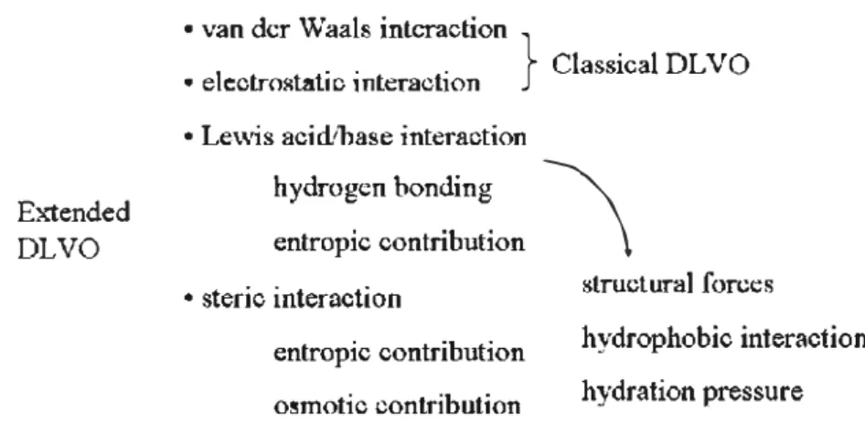
The aggregation and sedimentation of nanoparticles may occur naturally in the presence of suspended or dissolved substances in water (e.g. nature organic matter), which would favor the removal of nanoparticles from suspensions [68]. Fig. 1 gives the general schema of interactions in governing environmental colloidal processes and stability.
However, the combined effect of pH, ionic strength, electrolyte species and concentrations, and other characteristics of water would either make nanoparticles aggregated by charge neutralization, bridging, electrical double layer compression and by other mechanisms [70], or may cause nanoparticles to be more stable [71,72]. Thus, uptake of nanoparticles by animais and biomagnification in the food-chain is always possible.
Recently, silver nanoparticles (AgNPs) are used increasingly in con sumer products such as water treatment for their antimicrobial proper ties. This increased use raises ecological concern because of the release of AgNPs into the environment Once released, zero-valent silver may be oxidized to Ag+ and the cation liberated, or it may persist as AgNPs [73]. Aggregation of silver nanoparticles appears to strongly influence other processes, including precipitation, adsorption and dissolution, and thus is particularly important in determining the transport of the particles in aquatic systems. However, studies of aggregation stability of silver nanoparticles showed that aggregation is confined or limited to very slow rate and the particles in the system stay suspended for a relatively long term [74].
Furthermore, the dispersion stability of nanoparticles is also impor tant in determining their potential exposure to organisms, which will depend on their intrinsic properties, including sizes, shape, core com position, surface properties and concentrations [75]. Since most of the engineered nanoparticles are surface-modified, functionalized, or coated during manufacturing, their stability against aggregation and deposition may increase [76]. For example, in China and Taiwan, silica nanoparticles are commonly used as abrasive in chemical mechanical polishing (CMP) industries for the manufacture of integrated circuits and electronic chips. The effiuents of these factories are mainly com posed of silica nanoparticles (1.3-8.5 g/1 which correspond to 0.05%-0.36% in volume ratio) with an average size around 100 nm. These particles present a negative charge at the surface, and the suspensions are very stable [77-83].
Other fates of aquatic nanoparticles include adsorption/desorption, degradation, reaction and so on; but it is likely for nanoparticles (maybe even bind with toxic pollutants during their exposure) to enter into aquatic animais, and then accumulate into human body be fore their removal. Therefore, it is reasonable to begin considering how to remove nanoparticles from water, especially from wastewater such as the CMP effluents that has an important total solid content in the nano-range and has been produced in large quantities.
4. Technologies of nanoparticle separation
The conventional wastewater treatments including chemical, physi cal and biological methods may not be adapted to the recovery of nano scaled particles, first of ail due to their size, but also due to their original properties [13,15]. In the last few years, some researchers [84-88] have begun to study how to separate nanoparticles from waters, but com pared to their plentiful applications and toxicities explorations, separa tion researches of nanoparticles are still limited.
4.1. Coagulation and electrocoagulation (EC) processes
Chang et al. [89] studied the treatment of nanoparticles by chemical coagulation as it exists in the unity of wastewater treatment plant. Their structural researches on the nanoparticle agglomeration due to coagula tion have demonstrated the interest of coagulant addition for a good recovery, but its effect on nanoparticle removal ( <9%) was still not satisfying. Chuang et al. [90] investigated colloidal silica removal in
• van der Waals interaction
} ClassicalDLVO
• e)ectrostatic interaction • Lewis acid/base interaction
Extended
DLVO
hydrogen bonding entropie contribution
• steric interaction
\
structural forceshydrophobie interaction hydration pressure entropie contribution
osmotic contribution
Fig. 1. General schema of interactions in governing environmental colloidal processes and stability (adapted from Grasso et al. [691). coagulation processes for wastewater reuse in another high-tech indus
trial park. Experimental results illustrated two coagulants (PACI and Al2(OH)0Cl6_0) demonstrated the same capacity on silica nanoparticle
removal (approximately 80%). Chemical coagulation and flocculation processes have the advantages to benefit a fast increase of particle size and an easy removal of particles by sedimentation, but the high quanti ties of coagulant and sludge are their shortcomings.
EC was then tested as an alternative to chemical coagulation because it does not require the direct addition of a chemical coagulant [71,80]. Lai and Lin [77] investigated the treatment of copper CMP wastewater from a semiconductor plant by EC. The test results indicated that EC with Al/Fe electrode pair was very efficient and able to achieve 99% cop per ion and 96.5% turbidity removal in Jess than 30 min. Another group [78] used electro-coagulation-flotation (ECF) process with surfactant to treat CMP wastewater. The addition of surfactant CTAB that could both increase the particle size and produce positive-charged bubbles (particles are negative-charged) effectively increased the removal of the turbidity. The sludge volume and settling time were significantly Jess in the solution containing CTAB than in the one without CTAB. Com paring with chemical coagulation process, EC process has Jess sludge and no preliminary pH adjustments. Main disadvantage ofEC is the dis solution of electrodes into wastewater streams as a result of oxidation. Thus electrodes need to be regularly replaced. Other disadvantages contain the relatively high usage of electricity, and high conductivity of the water suspension required.
At the present time, excellent coagulation/flocculation performances have been reached using organic reagents such as polyamines and polyDADMACs authorized by the Directorate General for Health of several countries. After optimization of the aggregating process with cationic organic polymers, the size distributions of SiO2 nanoparticles
(about 80 nm) can reach between 10 µm and 1 mm, with fractal di mensions (compactness) ranging from 2.3 to 2.5. The performances obtained show that the use of these organic reagents is a promising potential route to treat other types of nanoparticles. The treatment also proposed to reach a ratio of average diameters dp10JdNP of 3500,
and therefore it facilitates the elimination of these nanoparticles ag glomerated by filtration [91 ].
42. Flotation process
Lien and Liu [81] tested the treatment of CMP effiuents by dissolved air flotation (DAF). Very effective removal of particles (turbidity decrease > 98%) from wastewater was found when CTAB was used as a collector. It is proposed that both the change of interface properties and the aggregation of nanoparticles contributed to flotation removal ofparticles.
Tsai et al. [83] studied nano-bubble flotation with coagulation by PACl/sodium oleate (NaOI) as a combination of activator/collector for the cost-effective treatment of CMP wastewater. Their work increased
the wastewater clarification efficiency by 40% as compared with tradi tional coagulation/flocculation process. More than 95% of the turbidity, total solids and total silica removal efficiendes were observed.
Furthermore, Liu et al. [92] tested the flotation ofnanosilica suspen sions in a new designed continuous DAF system. They showed that flotation had an interesting separation efficiency (removal of 99% of nanoparticle concentration) only when additives were added to over come the hydrophilic behaviors of silica. In the conditions tested in their work, the quantities of additives were a Iittle more than the ones necessary to perform aggregation and settling separation, but the flota tion combined with coagulation process needed much Jess time to achieve the separation purpose.
4.3. Filtration process
Due to the small size, the separation of nanoparticles from liquid medium by conventional filtration without pre-coagulation could be difficult [93]. In the Iast few years, coagulation or electrocoagulation with filtration was then explored by many researchers [94-95].
Zhong et al. [96] used the tubular ceramic membranes to remove nickel catalysts from slurry, indicating a separation efficiency of 100%. Springer et al. [97] evaluated the feasibility of membrane ultrafiltration for the removal of SiO2 nanoparticles (78 nm mean hydrodynamic
diameter), the 10 kDa membrane showed the highest expectable reten tion rates (>99.6%). Additionally, it was demonstrated that approxi mately 98% of fullerol could be removed by a polyamide nanofiltration membrane [98].
Based on several studies, the removal efficiendes for nanoparticles could be greatly improved by using membrane filtration as the final process. However, membrane filtration process is mainly hindered by membrane fouling, leading to flux decline and the increase of operating costs.
4.4. Biological process
During conventional wastewater treatment process, the nanoparticles could be incorporated into the sewage sludge through aggregation and sorption and further accumulated in sewage sludge over time. Once attached to biomass, the removal of nanoparticles is assodated with the setting and removal of the biomass [99]. A recent full-scale wastewater treatment test indicated that TiO2 nanomaterials were trapped in bio
mass, which could then be settled and removed by secondary sedimenta tion or membrane filtration process, and more than 96% of the TiO2 could
be finally removed [100]. Adsorption to activated sludge seems to be the major removal mechanism for nanoparticles in this study.
However, Limbach et al. [87] investigated the capture of nanoparticles by clearing sludge. Their study demonstrated a significant influence of the nanoparticle surface charge and the addition of dispersion stabi lizing surfactants as routinely used in the preparation of nanoparticle
derived products. A detailed investigation on the agglomeration of oxide nanoparticles in wastewater streams revealed a high stabilization of the particles against clearance ( adsorption on the bacteria from the sludge). Their present results indicate a limited capability of the biological treatrnent step to completely remove oxide nanoparticles from wastewater.
Another study suggested that the predominant mechanisms of engineered nanoparticle removal (95% of nano-copper were removed) were aggregation and settling rather than biosorption in activated sludge biomass [101 ]. The different conclusion may be attributed to the different types of nanoparticles investigated in these studies. It should be noted that some nanoparticles entrapped within the sludge materials may still have an effect on the activity of the essential bacteria or may re-enter the environment through application of sewage sludge to fields, indneration or landfilling [102]. More experiments will be required for in depth studies on the detailed mechanism of nanoparticle adsorption to sludge.
4.5. Other processes for nanoparticle separation
Zarutskaya and Shapiro [84] applied magnetic filters to capture nanoparticles with permanent magnetic moments. The influence of filtration operating conditions and particle diameters on their behavior and magnetic capture efficiency was investigated numerically, but this work was limited to the separation of magnetic nanoparticles.
Chin et al. [79] used synthesized magnetite nanoparticles to aggre gate target nanoparticles by the electrostatic attraction between the two oppositely charged particles. By optimizing experimental condi tions, the residual turbidity could be removed until less than 1 NID, but large amount of magnetic nanoparticles had to be used.
Michael and Armstrong [103] summarized separation and analysis of nanoparticles by capillary electrophoresis. The advantage of this method is that the separation of different colloids is possible. However, ail of the above technologies have obvious disadvantage for a large scale wastewater ( containing nanoparticles) treatment.
5. Conclusion
As we ail know, the potential advantages of nanomaterials are im mense, but so are the potential dangers. More attention on the risk assessment and waste management associated with nanomaterials are required. Nevertheless, the knowledge about occurrence, fate and toxicity of nanoparticles are still lacking. The pathways of nanoparticle penetration into bodies by food (fish and shellfish) are still uncertain. Consequently, characterization and quantification of nanoparticles may be firstly crucial to evaluate their potential risks and to compare different research results.
Until now, a wide number of techniques including microscopy tech niques ( transmission electron microscopy, confocal laser scanning mi croscopy, scanning electron microscopy and atomic force microscopy), static and dynamic light scattering (SLS, OLS), X-ray diffraction (XRD), inductively coupled plasma (ICP-OES and ICP-AES), X-ray photoelec tron spectroscopy (XPS) have been developed for detection and charac terization of nanomaterials. However, it is still a great challenge to directly detect engineered nanoparticles in natural water due to their low concentrations.
In order to promote the feasibility of these analytical techniques, several key issues are needed to be addressed: (i) minimizing the alter ation of samples and Joss of components from sampling to analysis; (ii) minimizing artifacts in estimation of size distribution during frac tionation process; (iii) ensuring the representativeness of samples; (iv) distinguishing between engineered nanomaterials and their natu rally occurring counterparts; (v) validating detected results by several methods.
On the other hand, the development of wastewater treatment contain ing nanoparticles is urgent for hazard reduction in aquatic environment. It
may be difficult for one type of method to treat the complex matrix con taining nanoparticles, different techniques are usually required to comple ment one another for achieving better removal efficiency. Although some methods are effective in removing spedfic types of nanoparticles at labo ratory scale, their effidendes for difl"erent nanoparticles in full scale are still unknown. It might be a good news that nanoparticles could be sta bilized by sewage sludge in nature, even if the interaction between nanoparticles and sewage sludge has not been clear. More researches and funds are required to fill these knowledge gaps even though the reg ulations have not yet been imposed.
Acknowledgments
The authors are grateful to CNRS (Projet PEPS), Région Midi Pyrénées and French National Research Agency (NANOSEP, ANR-08-ECOT-009) for financial support. We acknowledge China scholarship council (CSC) for the scholarship.
References
[1] B. Nowack, T.D. Bucheli, Occurrence, behavior and effects of nanoparticles in the environment, Environ. Pollut 150 (2007) 5-22.
[2] W. Luther, Classification of nanomaterials for commercial purposes including nanoparticles, fullerenes, dendrimers, nanowires, nanotubes, nanolayers and nanopores, Technologies Division of VDI (Verein Deutscher lngenieure) Report: Industrial Application of Nanomaterials - Chances and Risks, Tech. Anal. (2004).
[3] Y.B. Vasudeo, R. Rangaprasad, Nanomaterials, nanotechnology and their relevance to polymers, Technical Aarticles & Reports on Plastic lndustry, 2004. 1-1 O.
[4] M. Trojanowicz, Analytical application of carbon nanotubes: a review, Trends Anal. Chem. 25 (2006) 480-487.
[5] D. Vairavapandian, P. Vichchulada, M.D. Lay, Preparation and modification of carbon nanotubes: review of recent advances and applications in catalysis and sensing, Anal. Chim. Acta 626 (2008) 119-129.
[6] P. Wang, Z. Wang,]. Li, Y. Bai, Preparation, characterization, and catalytic characteris tics of Pd nanoparticles encapsulated in mesoporous silica, Microporous Mesoporous Mater. 116 (2008) 400-405.
[7] Y. Shao, X. Wang, M. Engelhard, C. Wang, S. Dai, J. Liu, Z. Yang, Y. Lin, Nitrogen doped mesoporous carbon for energy storage in vanadium redox flow batteries, J. Power Sources 195 (2010) 4375-4379.
[8] P. Kumar, V.V. Guliants, Periodic mesoporous organic-inorganic hybrid materials: application in membrane separations and adsorption, Microporous Mesoporous Mater. 132 ( 2010) 1-14.
[9] C. Liu, L Wang, W. Ren, Z. Rang, X. Wang,J. Wang, Synthesis and characterization of a mesoporous silica tube (MCM-48) membrane on a large-pore Œ-Al203 ceramic,
Microporous Mesoporous Mater. 106 (2007) 35-39.
[10] Y. Liu, Elimination de nanoparticules d'effluents liquides, Université de Toulouse, 2010. (phD thesis).
[11] D.M. Brown, M.R. Wilson, W. MacNee, V. Stone, K. Donaldson, Size-dependent proinflammatory effects of ultrfine polystyrene particles: a rote for surface area and oxidative stress in the enhanced activity of ultrafines, Toxicol. Appl. Pharrnacol. 175 (2001) 191-199.
[12] X.K. Cheng, A.T. Kan, M.B. Tomosom, Naphthalene adsorption and desorption from aqueous C60 fullerene, J. Chem. Eng. Data 49 (2004) 675-683.
[13] M.N. Moore, Do nanoparticles present ecotoxicological risks for the health of the aquatic environment? Environ. !nt. 32 (2006) 967-976.
[14] C.G. Daughton, Non-regulated water contaminants: emerging research, Environ. Impact Asses 24 (2004) 711-732.
[15] L Reijnders, Cleaner nanotechnology and hazard reduction of manufactured nanoparticles,J. Clean Prad. 14 (2006) 124-133.
[16] G. Oberdôrster, Toxicology of ultrafine particles; in vivo studies, Philos. Trans. R. Soc. Land. A 358 (2000) 2719-2740.
[ 17] D.C. Chalupa, P.E. Morrow, G. Oberdôrster, M.]. Utell, M.W. Frampton, Ultrafine particle deposition in subjects with asthma, Environ. Health Perspect. 112 (2004) 679-682.
[18] D.B. Warheit RA Hoke, C. Finlay, E.M. Donner, K.L Reed, C.M. Sayes, Development of a base set of toxicity tests using ultrafine Ti02 partiel es as a component of nano particle risk management, Toxicol. Lett. 171 (2007) 99-110.
[19] W. Môller, T. Hofer, A. Ziesenis, E. Karg, J. Heyder, Ultrafine particles cause cy toskeletal dysfunctions in macrophages, Toxicol. Appl. Pharmacol. 182 (2002) 197-202.
[20] LC. Renwick, K. Donaldson, A. Clouter, lmpairrnent of alveolar macrophage phago cytosis by ultrafine particles, Toxicol. Appl. Pharrnacol. 172 (2001) 119-127.
[21] S.C. Brown, M. Kama!, N. Nasreen, A. Baumuratov, P. Sharrna, V.B. Antony, B.M. Moudgil, Influence of shape, adhesion and simulated lung mechanics on amor phous silica nanoparticle toxidty, Adv. Powder Technol. 18 (2007) 69-79.
[22] A.S. Deckers, B. Gouget, M.M. L'Hermite, N.H. Boime, C. Reynaud, M. Carrière, In vitro investigation of oxide nanoparticle and carbon nanotube toxicity and intracellular accumulation in A549 human pneumocytes, Toxicol. 253 (2008) 137-146.
[23] M. Lundborg, A.Johansson, L Lastbom, P. Camner, Ingested aggregates ofultrafine carbon particles and interferon impair rat alveolar macrophage function, Environ. Res. Sect A 81 (1999) 309-315.
[24] M. Lundborg, U. Johard, L Lastbom, P. Gerde, P. Camner, Human alveolar macro phage phagocytotic function is impaired by aggregates of ultrafine carbon parti cles, Environ. Res. 86 (2001) 244-253.
[25] M,J.D. Clift, B.R. Rutishauser, D.M. Brown, R. Duffin, K. Donaldson, L Proudfoot, K. Guy, V. Stone, The impact of different nanoparticle surface chemistry and size on uptake and toxicity in a murine macrophage cell line, Toxicol. Appl. Pharmacol. 232 {2008) 418-427.
[26] R. Dunford, A. Salinaro, L Cai, N. Serpone, S. Horikoshi, H. Hidaka, Chemical oxida tion and DNA damage catalysed by inorganic sunscreen ingredients, FEBS Lett 418
(1997) 87-90.
[27] F. Menzel, T. Reinert, J. Vogt, T. Butz, Investigations of percutaneous uptake of ultrafine Ti02 particles at the high energy ion nanoprobe LIPSION, Nue!. Instr.
Meth. B 219-220 (2004) 82-86.
[28] J. Wu, W. Liu, C. Xue, S. Zhou, F. Lan, L Bi, H. Xu, X. Yang, F.D. Zeng, Toxicity and penetration of Ti02 nanoparticles in hairless mice and porcine skin alter
subchronic dermal exposure, Toxicol. Lett 191 {2009) 1-8.
[29] AD. Maynard, PA Baron, M. Foley, AS. Shvedova, E.R. Kisin, V. Castranova, Exposure to carbon nanotube material: aerosol release during the handling of unrefined single walled carbon nanotube material, J. Toxicol. Environ. Health A 67 (2004) 87-107.
[30] E. Oberdôrster, Manufactured nanomaterials (fullerenes, C,,0) induce oxidative
stress in the brain of juvenile largemouth bass, Environ. Health Perspect 112 (2004) 1058-1062.
[31] S. Zhu, E. Oberdôrster, M.L Haasch, Toxicity ofan engineered nanoparticle (fullerene, Cso in two aquatic species, daphnia and fathead minnow, Mar.Environ. Res. 62 (2006) S5-S9.
[32] M. Kovochich, B. Espinasse, M. Auffan, E.M. Hotze, L Wessel, T. Xia, A.E. Nel, M.R. Wiesner, Comparative toxicity of C60 aggregates toward mammalian cells: role of
tetrahydrofuran (lHF) decomposition, Environ. Sei. Technol. 43 (2009) 6378-6384. [33] X. Zhu, Y. Chang, Y. Chen, Toxicity and bioaccumulation ofTi02 nanoparticle aggre
gates in daphnia magna, Chemosphere 78 (2010) 209-215.
[34] S.W. Lee, S.M. Kim, J. Choi, Genotoxicity and ecotoxicity assays using the freshwa ter crustacean daphnia magna and the larva of the aquatic midge chironomus riparius to screen the ecological risks of nanoparticle exposure, Environ. Toxicol. Pharmacol. 28 (2009) 86-91.
[35] M. Auffan, J. Rose, T. Orisere, M.D. Meo, A. Thill, O. Zeyons, O. Proux, A. Masion, P. Chaurand, O. Spalla, A. Botta, M.R. Wiesner, J. Bottera, Ce02 nanoparticles induce
DNA damage towards human dermal fibroblasts in vitro, Nanotoxicology 3 (2009) 161-171.
[36] X. Zhu, S. Tian, Z. Cai, Toxicity assessment of iron oxide nanoparticles in zebrafish (danio retrio) early life stages, PLoS One 7 (2012) e46286.
[37] Y. Zhang, Y. Chen, P. Westerhoff, K. Hristovski,J.C. Crittenden, Stability of commer cial metal oxide nanoparticles in water, Water Res. 42 (2008) 2204-2212. [38] J. Cheng, E. Flahaut, S.H. Cheng, Effect of carbon nanotubes on developing zebrafish
(danio rerio) embryos, Environ. Toxicol. Chem 26 (2007) 708-716.
[39] X. Zhu, L Zhu, Y. Chen, S. Tian, Acute toxicities of six manufactured nanomaterial suspensions to Daphnia magna, J. Nanopart. Res. 11 (2009) 67-75.
[40] R.J. Griffitt, R. Weil, KA Hyndman, N.D. Denslow, K. Powers, D. Taylor, D.S. Barber, Exposure to copper nanoparticles causes gill injury and acute lethality in zebrafish (danio rerio), Environ. Sei. Technol. 41 (2007) 8178-8186.
[41] X. Zhu, L Zhu, Y. Li, Z. Duan, W. Chen, P.J. Alvarez, Developmental toxicity in zebrafish embryos alter exposure to manufactured nanomaterials: buckminsterful Ierene aggregates (nC60) and fullerol, Environ. Toxicol. Chem. 26 (2007) 976-979.
[42] X. Zhu, L Zhu, Y. Lang, Y. Chen, Oxidative stress and growth inhibition in the fresh water fish carassius auratus induced by chronic exposure to sub-lethal fullerene aggregates, Environ. Toxicol. Chem. 27 (2008) 1979-1985.
[43] X. Zhu,J. Wang, X. Zhang, Y. Chang, Y. Chen, The impact ofZnO nanoparticle aggre gates on the embryonic development of zebrafish (danio rerio), Nanotechnology 20 (2009) 195103.
[ 44] P.V. Asharani, Y.W. Lian, Z. Gong, S. Valiyaveettil, Toxicity of silver nanoparticles in zebrafish models, Nanotechnology 19 (2008) 255102.
[45] K. Bilberg, M.B. Hovgaard, F. Besenbacher, E. Baatrup, In vivo toxicity of silver nanoparticles and silver ions in zebrafish (danio rerio),J. Toxicol. 9 (2012), http://dx.doi.org/10.1155/2012/293784 {Article ID 293784).
[46] K.J. Ong, X. Zhao, M.E. Thistle, T,J. MacCormack, R,J. Clark, G. Ma, Y. Martinez-Rubi, B. Simard, J.S.C. Loo, J.G.C. Veinot, G.G. Goss, Mechanistic insights into the effect of nanoparticles on zebrafish hatch, Nanotoxicology, http://dx.doi.org/10.3109/ 17435390.2013.778345.
[ 47] L Risorn, P. Mailer, S. Loft, Oxidative stress-induced DNA damage by particulate air pollution, Mutat Res. 592 (2005) 119-137.
[48] K. Donaldson, V. Stone, Current hypotheses on the mechanisms of toxicity of ultrafine particles, Ann. 1st Super. Sanita 39 (2003) 405-410.
[49] G. Oberdôrster, E. Oberdôrster,J. Oberdôrster, Nanotoxicology: an emerging disci pline evolving from studies of ultrafine particles, Environ. Health Perspect 113 (2005) 823-839.
[ 50] S. Takenaka, E. Karg, C. Roth, H. Schulz, A. Ziesenis, U. Heinzmann, P. Schramel, J. Heyder, Pulmonary and systemic distribution of inhaled ultrafine silver particles in rats, Environ. Health Perspect. 109 (2001) 547-551.
[51] J.R. Gurr, A.S.S. Wang, C.H. Chen, K.Y. Jan, Ultrafine titanium dioxide particles in the absence of photoactivation can induce oxidative damage to human branchial epithelial cells, Toxicology 213 (2005) 66-73.
[52] C. Buzea, 1.1.P. Blandina, K. Rabbie, Nanomaterials and nanoparticles: sources and toxicity, Biointerphases 2 (2007) MRl 7-MRl 72.
[53] T. Xia, M. Kovochich,J. Brant, M. Hotze,J. Sempf, T. Oberley, C. Sioutas,J.1. Yeh, M.R. Wiesner, A.E. Nel, Comparison of the abilities of ambient and manufactured nanoparticles to induce cellular toxicity according to an oxidative stress paradigrn, Nano Lett. 6 (2006) 1794-1807.
[ 54] A. Ahmad, S. Senapati, M.I. Khan, R. Kumar, M. Sastry, Extra-/intracellular biosynthe sis of gold nanoparticles by an alkalotolerant fungus, Trichothecium sp, J. Biomed. Technol. 1 (2005) 47-53.
[55] C.S. Yah, The toxicity of gold nanoparticles in relation to their physiochemical properties, Biomagn. Res. 24 (2013) 400-413.
[ 56] A. Kaur, U. Gupta, A review on applications of nanoparticles for the pre concentra tion ofenvironmental pollutants,j. Mater. Chem 19 (2009) 8279-8289. [ 57] Z. Clemente, V. Castro, C. Jonsson, L F. Fraceto, Ecotoxicology of nano-TiO, - an
evaluation of its toxicity to organisms of aquatic ecosystems, Int. J. Environ. Res. 6 (2012) 33-50.
[58] M.R. Wiesner,J. Bottera, Environmental nanotechnology: applications and impacts of nanomaterials, first ed. McGraw-Hill Companies, New York, 2007.
[59] S. Foley, A.D.M. Curtis, A. Hirsch, M. Brettreich, A. Pelegrin, P. Seta, C. Larroque, Interaction of a water soluble fullerene derivative with reactive oxygen species and mode! enzymatic systems, Fuller. Nanotub. Car. N. 10 (2002) 49-67. [60] X.Y. Yang, R.E. Edelmann, J.T. Oris, Suspended Cso nanoparticles protect against
short-term UV and uoranthene photo-induced toxicity, but cause longterm cellular damage in daphnia magna, Aquat Toxicol. 100 (2010) 202-210.
[61] 1. Lynch, KA Dawson, Protein-nanoparticle interactions, Nanotoday 3 (12) (2008) 40-48.
[62] J.F. Reeves, SJ. Davies, NJ.F. Dodd, A.N.Jha, Hydroxyl radicals ('OH) are associated with titanium dioxide (Ti02) nanoparticle-induced cytotoxicity and oxidative DNA
damage in fish cells, Mutat Res. 640 (2008) 113-122.
[63] A. Kumar, M.V. Rao, S.K. Menon, Photoinduced DNA cleavage by fullerene-lysine conjugate, Tetrahedron Lett. 50 {2009) 6526-6530.
[64] A. Boxall, Q, Chaudhry, C. Sinclair, A.Jones, R. Aitken, B.Jefferson, C. Watts, Current and future predicted environmental exposure to engineered nanoparticles, Central Science Laboratory, Departrnent of the Environment and Rural Aff airs, London, UK, 2007.
[ 65] F. Gottschalk, T. Sonderer, R. W. Scholz, B. Nowack, Modeled environmental concen trations of engineered nanomaterials (Ti02, ZnO, Ag, CNT, Fullerenes) for different
regions, Environ. Sei. Technol. 43 (2009) 9216-9222.
[66] N.C. Mueller, B. Nowack, Exposure modeling of engineered nanoparticles in the environment, Environ. Sd. Technol. 42 (2008) 4447-4453.
[ 67] C. Neal, H.Jarvie, P. Rowland, Titanium in UK rural, agricultural and urban/industrial rivers: geogenic and anthropogenic colloidal/sub-colloidal sources and the signifi cance of within-river retenti on, Sd. Total. Environ. 409 (2011) 1843-1853. [68] H. Weinberg, A. Galyean, M. Leopold, Evaluating engineered nanoparticles in natu
ral waters, TrAC Trends Anal. Chem. 30 (2011) 72-83.
[69] D. Grassa, K. Subramaniarn, M. Butkus, K. Strevett, J. Bergendahl, A review of non-DLVO interactions in environmental colloidal systems, Rev. Env. Sd. Biotechnol. 1 (2002) 17-38.
[70] Y. Zhang, Y. Chen, P. Westerhoff, J. Crittenden, Impact of natural organic matter and divalent cations on the stability of aqueous nanoparticles, Water Res. 43 (2009) 4249-4257.
[71] H. Hyung,J.D. Fortner,J.B. Hughes,J. Kim, Natural organic matter stabilizes carbon nanotubes in the aqueous phase, Environ. Sd. Technol. 41 (2006) 179-184. [72] A.A. Keller, Stability and aggregation of metal oxide nanoparticles in natural aqueous
matrices, Environ. Sei. Technol. 44 (2010) 1962-1967.
[73] J. Dobias, R. Bernier-Latrnani, Silver release from silver nanoparticles in natural waters, Environ. Sei. Technol. 47 (2013) 4140-4146.
[74] L Kvitek, M. Vanickova, A. Panacek, J. Soukupova, M. Dittrich, E. Valentova, R. Prucek, M. Bandrova, D. Milde, R. Zboril, Initial study on the toxicity of silver nanoparticles (NPs) against paramecium caudatum, J. Phys. Chem. C 113 (2009) 4296-4300.
[75] J. Labille,J. Brant, Stability ofnanoparticles in water, Nanomedicine 5 (2010) 985-998. [76] J. Labille, J. Feng, C. Botta, D. Borschneck, M. Sammut, M. Cable, M. Auffan, J. Rose, J. Bottera, Aging of Ti02 nanocomposites used in sunscreen. Dispersion and fate
of the degradation products in aqueous environment, Environ. Pollut 158 (2010) 3482-3489.
[77] C.L Lai, S.H. Lin, Electrocoagulation of chemical mechanical polishing (CMP) waste water from semiconductor fabrication, Chem. Eng.J. 95 (2003) 205-211. [78] C.Y. Hu, S.L Lo, C.M. Li, W.H. Kuan, Treating chemical mechanical polishing (CMP)
wastewater by electro-coagulation-flotation, J. Hazard. Mater. 120 (2005) 15-20. [79] CJ. Chin, P.W. Chen, LJ. Wang, Removal ofnanoparticles from CMP wastewater by
magnetic seeding aggregation, Chemosphere 63 (2006) 1809-1813.
[80] K.T. Kin, H.S. Tang, S.F. Chan, S. Raghavan, S. Martinez, Treatment of chemical mechanical planarization wastes by electrocoagulation/electro-fenton method, IEEE Trans. Semicond. Manuf. 19 (2006) 208-215.
[81] C.Y. Lien, J.C. Liu, Treatment of polishing wastewater from semiconductor manu facturer by dispersed air flotation,J. Environ. Eng. 132 (2006) 51-57.
[82] G.C.C. Yang, C.M.T. Tsai, Performance evaluation of a simultaneous electrocoagulation and electrofiltration module for the treatrnent of Cu-CMP and oxide-CMP wastewa ters, J. Membr. Sei. 286 (2006) 36-44.
[83] J.C. Tsai, M. Kumar, S.Y. Chen, J.G. Lin, Nano-bubble flotation technology with coag ulation process for the cost-effective treatment of chemical mechanical polishing wastewater, Sep. Purif. Technol. 58 (2007) 61-67.
[84] T. Zarutskaya, M. Shapiro, Capture of nanoparticles by magnetic filters, J. Aerosol Sei. 31 (2000) 907-921.
[85] SA Lee, K.H. Choo, C.H. Lee, H.I. Lee, T. Hyeon, W. Choi, H.H. Kwon, Use of ultrafil tration membranes for the separation of Ti02 photocatalysts in drinking water
[86] M.A Rodriguez, D.W. Armstrong, Separation and analysis of colloidal/nano-partides induding microorganisms by capillary electrophoresis: a fundamental review, j. Chromatogr. B 800 (2004) 7-25.
[87] LK Limbach, R. Bereiter, E. Müller, R. Krebs, R. Galli, W.J. Stark, Removal of oxide nanopartides in a mode! wastewater treatrnent plant: influence of agglomera tion and surfactants on clearing efficiency, Environ. Sei. Technol. 42 (2008) 5828-5833.
[88] Y. Liu, M. Tourbin, S. Lachaize, P. Guiraud, Silica nanoparticle separation from water by aggregation with AICls, Ind. Eng. Chem. Res. 51 (2012) 1853-1863.
[89] M.R. Chang, D.J. Lee, J.Y. Lai, Nanopartides in wastewater from a sdence-based in dustrial park - coagulation using polyalurninium chloride, j. Environ. Manage. 85 (2007) 1009-1014.
[90] S.H. Chuang, T.C. Chang, C.F. Ouyang, J.M. Leu, Colloidal silica removal in coagula tion processes for wastewater reuse in a high-tech industrial pari� Water Sd. Technol. 55 (2007) 187-195.
[91] M. Bizi, Stability and flocculation of nanosilica by conventional organic polymer, Nat Sd. 4 (2012) 372-385.
[92] Y. Liu, M. Tourbin, S. Lachaize, P. Guiraud, Flotation separation ofSiO2 nanopartides
from wastewaters, STP2012 - 7ème colloque Science et Technologie des Poudres, Toulouse, 2012.
[93] Y.T. Lin, M. Sung, P.F. Sanders, A Marinucci, C.P. Huang, Separation of nano-sized colloidal particles using cross-flow electro-filtration, Sep. Purif. Technol. 58 (2007) 138-147.
[94] C. Huang, W. Jiang, C. Chen, Nano silica removal from lC wastewater by pre coagulation and microfiltration, Water Sei. Technol. 50 (2004) 133-138.
[95] G.C.C. Yang, q. Li, Electrofiltration of silica nanoparticle - containing waste water using tubular ceramic membranes, Sep. Purif. Technol. 58 (2007) 159-165.
[96] Z.X. Zhong, W.X. Li, W.H. Xing, N.P. Xu, Crossflow filtration of nanosized catalysts suspension using ceramic membranes, Sep. Purif. Technol. 76 (2011) 223-230.
[97] F. Springer, S. Laborie, C. Guigui, Removal of SiO2 nanoparticles from industry
wastewaters and subsurface waters by ultrafiltration: investigation of process effi dency, deposit properties and fouling mechanism, Sep. Purif. Technol. 108 (2013) 6-14.
[98] KD. Pickering, Photochemistry and environmental applications of water-soluble fullerene compounds, Rice University, 2005. (PhD thesis).
[99] B. Kim, C.S. Park, M. Murayama, M.F. Hochella, Discovery and characterization of silver sulfide nanoparticles in final sewage sludge products, Environ. Sd. Technol. 44 (2010) 7509-7514.
[100] P. Westerhoff, G.X. Song, K. Hristovski, M.A Kiser, Occurrence and removal oftita
nium at full scale wastewater treatment plants: implications forTiO2 nanomaterials,
J. Environ. Monit. 13 (2011) 1195-1203.
[101] R. Ganesh, J. Smeraldi, T. Hosseini, L Khatib, B.H. Oison, D. Rosso, Evaluation of nanocopper removal and toxidty in municipal wastewaters, Environ. Sei. Technol. 44 (2010) 7808-7813.
[102] N. Wu, Y. Wyart, Y. Liu, j. Rose, P. Moulin, An overview of solid/liquid separation methods and size fractionation techniques for engineered nanomaterials in aquatic environment, Environ. Technol. (2013) 1-17.
[103]AR. Michael, D.W. Armstrong, Separation and analysis of colloidal/nanoparticles including microorganisms by capillary electrophoresis: a fundamental review, j. Chromatogr. B 800 (2004) 7-25.