A. Palla S. Hegemann U. Widmer D. Straumann
Vestibular and auditory deficits
in Fabry disease and their response
to enzyme replacement therapy
Introduction
In Fabry disease, an X-linked lysosomal storage disor-der, deficient activity of the enzyme α-galactosidase A (α-Gal A) [6, 22] leads to intracellular accumulation of globotriaosylceramide predominantly in the vascular
tissue, eye, skin, kidney, heart, and nervous system [7, 10]. Disease prevalence is estimated between 1:40,000 and 1:117,000 live births [27]. If later-onset variant phe-notypes are included, incidence ranges from 1:3100 to 1:4600 [39]. The clinical onset in childhood is character-ized by painful acroparaesthesias, hypohydrosis, typical angiokeratoma, gastrointestinal symptoms, such as
ab-JON 2575
Received: 31 December 2006 Accepted: 24 January 2007 Published online: 15 October 2007
A. Palla, MD (쾷) · D. Straumann, MD Neurology Department
Zurich University Hospital Frauenklinikstrasse 26 8091 Zurich, Switzerland Tel.: +41-1/255-5500 Fax: +41-1/255-4507 E-Mail: [email protected] S. Hegemann, MD
Dept. of Otolaryngology, Head & Neck Surgery
Zurich University Hospital, Switzerland U. Widmer, MD
Dept. of Medicine
Zurich University Hospital, Switzerland Grant/financial support:
Swiss National Science Foundation (#3200B0–105434); Betty and David Koetser Foundation for Brain Research, Zurich, Switzerland.
■ Abstract Progressive hearing
(pHL) and vestibular (pVL) loss are frequent deficits in Fabry dis-ease (FD). Recently, enzyme re-placement therapy (ERT) with
hu-man α-galactosidase A has become
available. Here, we investigate the association between pHL and pVL in FD and their ERT responses. Pure tone audiometry (PTA) and head impulse testing (HIT) were administered at baseline in 47 pa-tients (25 male, 18–60 y; 22 female, 17–74 y), of whom 24 also received caloric irrigation (CI). Of the 47 patients, 38 (24 male) were tested both before and during ERT (fol-low-up ≤ 60 months). ERT con-sisted of agalsidase alfa infusions. At baseline, pHL was present in 88 % of males and 86 % of females. Over all tested frequencies (range: 0.5–6 kHz), pHL was significantly (two-way ANOVA: p < 0.05) greater at higher age and in males, with largest deficits at high frequencies. When assessed with HIT, 80 % of males and 77 % of females had pVL. pVL was significantly greater at higher age and in males. Tested
with CI, 21 % of males and 0 % of females had pVL. No associations among individual semicircular canal (SCC) deficits, as tested by HIT, and hearing was observed in individual ears. After ≥ 18 months of ERT, pVL was significantly smaller than at baseline (ANOVA for HIT: p < 0.01). In contrast, pHL remained unchanged by ERT over 60 months (p > 0.05). We conclude that pHL and pVL prevalences are similar in FD. To detect pVL, HIT is more sensitive than CI. We specu-late that pHL and pVL emerge from lesions within the vestibulo-cochlear labyrinth, because no spe-cific patterns of vestibulo-cochlear deficits were observed, as expected if lesions were more proximal along the inferior or superior branch of the vestibulo-cochlear nerve or labyrinthine artery. Fi-nally, ERT stabilizes auditory and even improves vestibular function.
■ Key words lysosomal disease ·
Fabry disease · head impulse test · caloric irrigation · pure tone audiogram
dominal pain and diarrhea, and corneal dystrophy [7, 10, 26]. Renal failure, cardiomyopathy, and cerebrovas-cular disease cause premature death, on average about two decades earlier than in the general population [24–26]. Although inheritance is X-chromosomal linked, females can be affected to a mild or severe degree due to random X-chromosomal inactivation [42]. Re-cently enzyme replacement therapy (ERT) with
recom-binant or gene-activated human α-Gal A provided
evi-dence of reversibility of several clinical manifestations, notably of cardiac, renal, and peripheral neuropathic symptoms [5, 11, 19, 36, 40, 43].
Hearing loss has often been linked with Fabry dis-ease, but only recent studies have ascertained its high prevalence [9, 18, 26]. Predominantly, hearing loss in Fabry disease is of sensorineural type [14, 15, 18, 34]. Likewise, vestibular function seems to be commonly af-fected in Fabry disease, but so far only small case series are available [9, 29, 32]. When cochlear and vestibular impairments in patients with Fabry disease are com-pared, hearing loss is more frequently reported than vestibular loss and an independent involvement be-tween the cochlea and the vestibular labyrinth has been suggested [9, 29]. While studies agree on the prevalence of hearing loss, the prevalence of vestibular impairment varies considerably, i. e. ranges from 30 % [9] to 50 % [29] when tested with caloric irrigation, and up to 70 % when tested with search-coil head impulses [32].
Caloric irrigation and the search-coil head impulse test are both validated assessments of peripheral vestibular function. The advantage of the search-coil head impulse test over caloric irrigation consists in a more physiological stimulation of the high-frequency range of the vestibular system and the possibility to de-termine the function of individual semicircular canals [3, 4, 37].
The present study was prospectively conducted in Fabry patients with the following goals: 1) to determine the prevalence of auditory and vestibular impairments in male and female patients; 2) to compare vestibular re-sponses as tested by head impulses and caloric irriga-tion; 3) to correlate vestibular function to hearing; 4) to determine the effect of ERT on vestibular and auditory function.
Subjects and methods
■Subjects
Forty-seven patients (25 male, 18–60 y, mean 40 y; 22 female, 17–74 y, mean 34 y) diagnosed with Fabry disease (FD) were vestibularly and audiologically tested at baseline examination. The diagnosis of FD was confirmed in all patients by enzyme assay or DNA analysis. Pa-tients were consecutively recruited starting February 2001.Vestibular assessments at baseline included head impulse testing in all 47 pa-tients, of whom 24 patients (16 male, 18–60 y, mean 41 y; 8 female, 17–74 y, mean 44) also received caloric irrigation. Enzyme
replace-ment therapy (ERT) was given to 38 (24 male) of the 47 patients. All FD patients receiving ERT were tested at relatively regular intervals during a period of up to 60 months. ERT consisted of intravenous in-fusions of gene-activated or recombinant human α-galactosidase A. Agalsidase alfa (Replagal®; TKT Europe – SS, Danderyd, Sweden) was given at a dose of 0.2 mg/kg every two weeks. The comparison group for vestibular head impulse testing comprised 28 healthy subjects (13 male; aged 18–75 y, mean 44), while normal values for audiometric testing were taken from ISO 70291, an internationally accepted
age-and gender-matched control data set. Informed consent was obtained from patients and healthy human subjects after a full explanation of the experimental procedure. The protocol was approved by a local ethics committee and was in accordance with the ethical standards laid down in the 1964 Declaration of Helsinki for research involving human subjects.
■Auditory testing
Routine pure tone audiometry was performed for auditory assess-ment. The audiometric type, amount, and configuration of hearing loss were further analyzed. The type of hearing loss was classified as sensorineural (average air-bone gap of less than 15 dB for 0.5, 1 and 2 kHz), conductive (normal bone conduction thresholds and average air-bone gap of 15 dB or more for 0.5, 1, and 2 kHz), or mixed (bone conduction threshold greater than 20 dB HL in combination with av-eraged air-bone gap 15 dB or more for 0.5, 1, and 2 kHz) according to the European Working Group on Genetics and Hearing Impairment.2
Hearing loss was categorized according to pure-tone air conduction thresholds at 0.5, 1, 2, 4, and 6 kHz relative to 90thpercentiles of the
age- and gender-matched ISO 7029 control data. To determine the shape of audiometric configurations, we computed the following thresholds (T) for each of the two ears in every patient:
low frequency threshold: T(low) = [T(0.5 kHz) + T(1 kHz)]/2 mid frequency threshold: T(mid) = T(2 kHz)
high frequency threshold: T(high) = [T(4 kHz) + T(6 kHz)]/2 Using these three thresholds, audiometric configurations were de-fined as follows (modified definitions from European Working Group on Genetics and Hearing Impairment2and [9]):
앫 flat: |T(low) – T(mid)| ≤ 15 dB HL AND |T(mid) – T(high)| ≤ 15 dB HL
앫 mid frequency U-shaped: |T(mid) – T(low)| > 15 dB HL AND |T(mid) – T(high)| > 15 dB HL
앫 high frequency: |T(high) – T(low)| > 15 dB HL AND |T(mid) – T(low)| < [T(high) – T(low)]/3
앫 sloping: |T(high) – T(low)| > 15 dB HL AND |T(mid) – T(low)| > [T(high) – T(low)]/3
앫 low frequency: |T(low) – T(high)| > 15 dB HL AND |T(mid) – T(high)| < [T(low) – T(high)]/3
앫 rising: |T(low) – T(high)| > 15 dB HL AND |T(mid) – T(high)| > [T(low) – T(high)]/3
1International Organization for Standardization (2000). Acoustics –
Statistical distribution of hearing thresholds as a function of age, ISO 7029:2000
2http://www.gendeaf.org. Recommendations for description of
ge-netic and audiological data for families with nonsyndromic heredi-tary hearing impairment; Composed by the GENDEAF study group on genotype phenotype associations
■Vestibular testing
Caloric irrigation was performed according to the Fitzgerald-Hallpike testing protocol [13]. In sequence, unilateral 30 °C-cold and 44 °C-warm water irrigations during 20 s with 20 mL of water were performed on either side and eye movement responses were video-oculographically recorded for 180 s [37]. The asymmetry of periph-eral vestibular function in percent, i. e. the canal paresis factor, was de-termined by Jongkees formula [21]. For quantitative head impulse testing, three-dimensional eye and head movements were measured in a magnetic frame (Remmel type system, modified by A. Lasker, Bal-timore, MA, USA) using dual search-coils. Search-coils were cali-brated before each session [41]. After anesthetizing the conjunctiva with oxybuprocaine 0.4 %, one search-coil was placed on the right eye around the cornea and the other was tightly fixed on the forehead with adhesive tape. Voltages were sampled at 16 bits at a frequency of 1000 Hz and stored on the hard disk of a computer. During experi-ments, subjects were seated inside the magnetic coil frame (side length: 1.4 m). Care was taken to position the center of the inter-pupillary line in the center of the magnetic frame. Rotational head thrusts (amplitude: 20–40°; duration: 150–200 ms; peak velocity: ~ 300 °/s; peak acceleration: ~ 10000 °/s2) were applied approximately
along the planes of the horizontal, left anterior and right posterior (LARP), and right anterior and left posterior (RALP) semicircular canals by an investigator standing behind the subject. The directions of the head impulses were pseudorandomly intermingled and four to six impulses were performed in each direction. For each semicircular canal in the appropriate stimulus plane, the gain of the vestibulo-oc-ular reflex (VOR) was determined by computing the coefficient ‘eye-in-space displacement divided by head-‘eye-in-space displacement’ with head-in-space moved from 3° to 7° eccentricity from straight-ahead [31]. Note that a decreased gain value in the direction of an individ-ual semicircular canal does not necessarily imply that the pathology is within this canal; the pathology could as well be somewhere along the primary vestibular afferents from this semicircular canal. ■Statistical analysis
To analyze the interference between the patterns of auditory and vestibular damage, we used chi-square testing. Wilcoxon rank sum test for equal medians was performed to determine the effect of age on the high frequency vestibulo-ocular reflex as tested by head im-pulses in healthy controls. The effects of age and gender on vestibular and audiometric data at baseline data were analyzed with two-way analysis of variance (ANOVA). Two-way ANOVA was also used to de-termine whether semicircular canals for vestibular function or fre-quencies for audiometric functions were differentially affected by FD. If the outcome of the two-way ANOVA was statistically significant, we performed multiple comparisons testing to discern the grouping variables that differed significantly. To compare baseline data be-tween different groups (e. g. males vs. females), we applied unpaired t-tests. Effects of ERT at different time intervals were evaluated by one-way ANOVA. If the outcome of ANOVA testing was statistically significant, we performed multiple comparison testing to determine, between which periods of ERT significant differences occurred.
Results
■ Vestibular and auditory function at baseline
Fig. 1 depicts measures of vestibular (head impulse test-ing) and auditory (pure tone audiography) function in a typical patient with Fabry disease (U. H.; age: 44 years). For head impulses along the horizontal plane, gains of
the vestibulo-ocular reflex (VOR) were bilaterally re-duced (Fig. 1A), reflecting hypofunction of the horizon-tal semicircular canals (SCC) on both sides. Along the RALP (= right anterior, left posterior) plane, VOR gains were reduced for upward head impulses and normal for downward head impulses (Fig. 1B), corresponding to re-duced left posterior SCC and normal right anterior SCC function. VOR gains along the LARP (= left anterior, right posterior) plane were reduced in both directions (Fig. 1C), i. e. both left anterior SCC and right posterior SCC functions were reduced. In this patient, caloric irri-gation revealed a normal vestibular function (canal paresis factor: 16 %; data not shown). Pure-tone au-diometry demonstrated bilateral high-frequency hear-ing loss with a mean high-frequency loss (air conduc-tion at 4 and 6 kHz) of 35 dB for the right and 67.5 dB for the left ear (Fig. 1D). However, when these audiometric data were corrected for age (ISO 7029, see Methods), the high-frequency hearing loss was restricted to the left ear (with 18.5 dB above the mean of the 90thpercentiles for 4 and 6 kHz). Thus, in this Fabry patient, vestibular deficits were found on both sides, while hearing func-tion was impaired on one side only.
Fig. 2 shows the percentages of male (left bars) and female (right bars) patients (N = 47; male = 25) with vestibulo-auditory impairment at baseline examination assessed by head impulse testing along different semi-circular canal planes (HSCC: horizontal semisemi-circular canals; ASCC: anterior semicircular canals; PSCC: poste-rior semicircular canals), caloric irrigation (CP: canal paresis factor), and pure tone audiometry (Audio). Bars indicating the percentage of bilateral (gray area) and unilateral (white area) deficits are piled. For example, the horizontal semicircular canals (Fig. 2, leftmost bar) were affected bilaterally in 20 % and unilaterally in 48 % of male patients. For caloric testing, only the percentage of patients with a pathological canal paresis factor – a measure of peripheral vestibular asymmetry (see Meth-ods) – is depicted (white area), since no generally ac-cepted parameter for bilateral impairment assessed by caloric irrigation is available. Recall that caloric irriga-tion was only performed in 24 patients (16 male), while head impulses and pure tone audiometry were com-pleted in all 47 patients.
Altogether, in 80 % of male and 77 % of female pa-tients, head impulse tests in at least one of six semicir-cular canal directions were abnormal. Pathological asymmetries of caloric responses, i. e. with a canal pare-sis factor > 25 %, were found in 21 % of male and in none of female patients. Thus, vestibular deficits were more frequently identified with search-coil head impulse test-ing than with caloric irrigation. In pure tone audiome-try, 88 % of male and 86 % of female patients showed im-paired auditory function compared to age- and gender-matched ISO control data. Hearing impairment was sensorineural in all affected patients, i. e. no
con-ductive or mixed hearing impairment was observed. Configurations of audiometric thresholds were high-frequency in 40 % of males and 9 % of females, flat in 52 % of males and 86 % of females, and sloping in 8 % of males and 5 % of females (see Methods for definitions).
■ Relation between vestibular and auditory deficits at
baseline
We asked whether there was an association between vestibular and hearing impairments in Fabry patients at baseline. Table 1 compares vestibular (as assessed with head-impulse testing) and auditory function in the pooled population of male and female patients. Clearly, vestibular function did not always parallel auditory function, i. e. a vestibular deficit in a patient did not im-ply a hearing impairment in the same patient, and vice versa (chi square: p > 0.05). Likewise, no association be-tween vestibular and auditory function was found when analyzing data from males and females separately (data not shown).
As demonstrated in Table 2, there was not even an
as-Fig. 1 Example of head impulses along the horizontal (A), RALP (= right-ante-rior & left-posteright-ante-rior semicircular canals) (B) and LARP (= left-anterior & right-posterior semicircular canals) (C) planes as well as pure tone audiometry (D) in a typical patient with Fabry disease (U. H.; age 44 y) at baseline examination. A, B, C If the vestibulo-ocular reflex (VOR) were perfectly compensatory, traces would be parallel to the abscissa (head-in-space axis); if the VOR were absent, traces would move on a 45° slope. Traces are clipped beyond 10° eccentricity of head-space. Dashed vertical lines in-dicate intervals used to determine the gains (see Methods). g: median gain value during head impulses. D Pure tone audiogram. Circles: hearing thresholds of the right ear; crosses: hearing thresh-olds of the left ear
-15 -10 -5 0 5 10 15 head-in-space [˚] -15 -10 -5 0 5 10 15 ey e -in-spac e [ °] head-in-space [°] ey e -in-spac e [ °] g = 0.65 g = 0.64 a b d c -15 -10 -5 0 5 10 15 -15 -10 -5 0 15 left right RALP down head-in-space [°] g = 0.55 g = 0.42 -15 -10 -5 0 5 10 15 -15 -10 -5 LARP down LARP up ey e -in-spac e [ °] 0 5 10 15 g = 0.59 RALP up 5 10 g = 0.82 -10 0 10 20 30 40 50 60 70 80 90 100 110 120 130 125 250 500 1k 2k 4k 8k 12k Hz dB HL
Fig. 2 Percentages of male (left columns; N = 25) and female (right columns; N = 22) patients at baseline examination with unilateral (white area) and bilateral (gray area) vestibular and auditory impairments. Horizontal (HSCC), anterior (ASCC), and posterior (PSCC) semicircular canal function assessed with search-coil head impulse testing. CP canal paresis factor, i. e. asymmetry of the vestibular func-tion tested by caloric testing. Audio pure tone audiometry (hearing loss relative to ISO) 0 10 20 30 40 50 60 70 80 90 100 HSCC ASCC PSCC CP Audio P atients with v estibulo -audit or y impairment [%] m : males f : females : unilateral : bilateral m f m f m f m f m f
sociation between vestibular and auditory function at the level of ipsilateral labyrinths or their afferents. Whether or not a Fabry patient had a vestibular deficit on one side (as assessed with head-impulse testing) was independent of the presence of an auditory deficit on the same side, and vice versa (chi square: p > 0.05).Also sep-arate analyses among male and female patients found no association between vestibular and auditory function on the same side (data not shown).
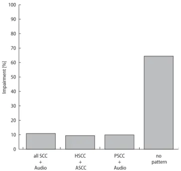
Fig. 3 compares patterns of ipsilateral vestibular and auditory deficits of Fabry patients (results from right and left sides are pooled). 32 % of patients showed typi-cal patterns of vestibular and auditory deficits that are expected if lesions were situated along the branches of the vestibulo-cochlear nerve or labyrinthine artery [12, 33]. These patterns include 1) horizontal and anterior SCC deficits, 2) posterior SCC and hearing deficits, and 3) deficits of all SCC and hearing. In the majority of pa-tients (68 %), however, no pattern was identifiable.
■ Factors determining vestibular and auditory function
at baseline and its characteristics during ERT
To statistically analyze the influence of age and gender on vestibular function, we first computed the average VOR gain of head impulse testing along the three ipsi-lateral semicircular canals of each ear in every patient. This average VOR gain was then normalized by
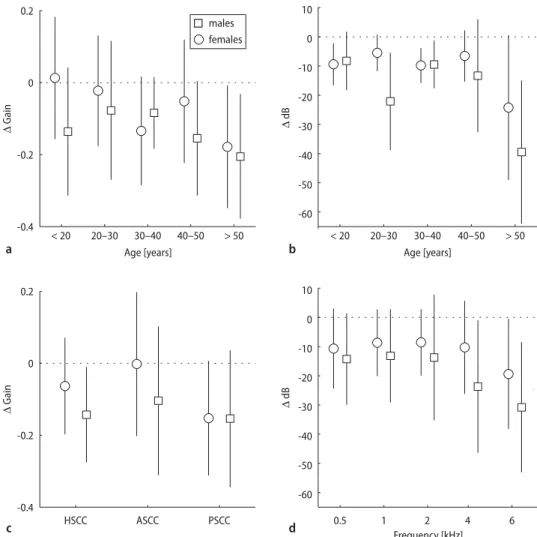
sub-tracting the grand average of unilateral VOR gains in the healthy control group. Since average unilateral VOR gains in our group of healthy subjects did not signifi-cantly decline with age (Wilcoxon rank sum test for equal median p > 0.05), gain values of Fabry patients were not age-corrected. Two-way analysis of variance (ANOVA) demonstrated a significant (p < 0.01) influ-ence of age and gender on vestibular function in the Fabry patients. Fig. 4A illustrates that vestibular func-tion in Fabry patients declines with age, i. e. the durafunc-tion of the disease, and is more reduced in male (squares) than in female (circles) patients.
A similar analysis was performed to investigate the influence of age and gender on auditory function. First, differences of pure-tone air conduction thresholds rela-tive to the 50 % percentile of age- and gender-matched ISO 7029 control data were computed for the five tested frequencies (0.5, 1, 2, 4, 6 kHz). These values were then averaged separately for each ear to obtain a unilateral measure of auditory function.As for vestibular function, age and gender significantly (two-way ANOVA: p < 0.01) influenced auditory function, with a decline of auditory function with age (or duration of disease) and more re-duced function in male patients, as shown in Fig. 4B. While all semicircular canals in Fabry patients showed similar (p > 0.05) impairment (Fig. 4C), auditory deficits
Table 1 Vestibular and auditory function of either ear in Fabry patients assessed by search-coil head impulse testing and pure tone audiometry
Auditory function Vestibular function Total (no. of patients) (no. of patients)
normal affected
normal 2 4 6
affected 8 33 41
total 10 37 47
Number of Fabry patients with normal or abnormal auditory or vestibular function. Significance of chi square: p > 0.05
Table 2 Right- and left-ear vestibular and auditory function in Fabry patients as-sessed by search-coil head impulse testing and pure tone audiometry
Auditory function Vestibular function Total (no. of ears) (no. of ears)
normal affected
normal 9 12 21
affected 28 45 73
total 37 57 94
Number of ears in Fabry patients with normal or abnormal auditory or vestibular function. Significance of chi square: p > 0.05
Fig. 3 Percentages for patterns of impaired semicircular canal (SCC) and auditory function within the same ear, as expected by different topographical locations of lesions due to impairments of labyrinthine nerve or blood supply or lesions within the labyrinth. All: impairment of all three SCC and hearing within the same ear; N = 11. HSCC + ASCC deficit of the horizontal and anterior SCC as expected from a lesion along the superior branch of the vestibular nerve or labyrinthine artery; N = 9. PSCC + Audio deficit of the posterior SCC and auditory function as expected from a lesion along the inferior branch of the vestibular nerve or labyrinthine artery; N = 10. No pattern no association of the three SCC and auditory function; N = 64
0 10 20 30 40 50 60 70 80 90 100 all SCC + Audio HSCC + ASCC PSCC + Audio no pattern Im pairment [%]
were significantly (p < 0.01) different among tested fre-quencies with largest differences at high frefre-quencies (Fig. 4D).
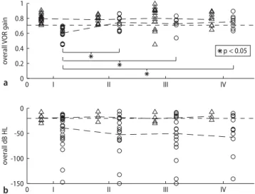
Fig. 5 illustrates the effect of ERT on overall vestibu-lar and auditory function. As for Fig. 4, only results from head impulse testing were considered for vestibular function. Overall vestibular function of a patient was de-fined as the average of VOR gains obtained by head im-pulses along the six semicircular canal directions (Fig. 5A). Likewise, the overall auditory function of a subject was defined as the average of hearing thresholds at 0.5, 1, 2, 4, and 6 kHz of both ears, whereby age- and gender-correction of hearing thresholds were computed
by subtracting the age- and gender-matched 90th
per-centiles at each frequency before averaging (Fig. 5B). Patients were divided into two groups depending on whether the overall VOR gain (Fig. 5A) or the overall hearing threshold (Fig. 5B), respectively, was within the normal range (group_N, triangles) or reduced (group_R, circles) relative to healthy subjects. At base-line examination, i. e. prior to ERT administration, aver-age overall VOR gains of group_R were not significantly
different between males and females although overall VOR gains of females were less reduced than those of males (average overall gains for male 0.59 ± 0.09, for fe-male 0.61 ± 0.08; unpaired t-tests: p > 0.05; data not shown). In contrast, for pure tone audiometry, the aver-age overall hearing thresholds for group_R at baseline were significantly different between male and female patients with hearing thresholds in females less im-paired than in males (average overall hearing thresholds for male 47.3 dB ± 35.6, for female 21.7 dB ± 10.9; un-paired t-tests: p < 0.05; data not shown). Overall VOR gains and hearing thresholds at baseline in group_N were not significantly different between male and fe-male patients (unpaired t-tests: p > 0.05; data not shown).
Because number of female patients receiving ERT (N = 14) was small, data of male and female patients were pooled for further analysis. Pooled data of group_N (triangles) and group_R (circles) were then partitioned into baseline examination (period I, i. e. prior to ERT administration) and according to the time passed from the beginning of the ERT to the date of
ex-Fig. 4 Differences of VOR gains ob-tained by search-coil head impulse test-ing along the three ipsilateral SCC of each ear in every patient relative to grand average of unilateral gains in healthy controls (A, C) and differences of pure-tone air conduction thresholds rel-ative to ISO 7029 (B, D) in males (squares) and females (circles) patients at baseline examination. A Average VOR gain deficits (error bars: ± 1SD) plotted at different age intervals; C as A but plot-ted for individual SCC. B Average pure-tone air conduction thresholds relative to 50thpercentile of ISO 7029 (error bars:
± 1SD) plotted at different age intervals; D as B but plotted for individual fre-quencies. HSCC horizontal SCC; ASCC an-terior SCC; PSCC posan-terior SCC. Note that impairment of auditory function was significantly greater at higher frequency and at higher age, whereas vestibular function was significantly more reduced at higher age, but with no preference for a specific SCC -60 -50 -40 -30 -20 -10 0 10 Δ dB < 20 20–30 30–40 40–50 > 50 Age [years] -0.4 -0.2 0 0.2 Δ G ain ASCC PSCC HSCC -0.4 -0.2 0 0.2 Δ G ain Age [years] < 20 20–30 30–40 40–50 > 50 -60 -50 -40 -30 -20 -10 0 10 Frequency [kHz] Δ dB 0.5 1 2 4 6 males females a b c d
amination (period II: 1 day – 1.5 years; period III: 1.5 years – 3 years; period IV: 3 years – maximal 5 years; dashed lines connect average of corresponding groups; dashed horizontal line: lower limit of data from healthy subjects). For group_N, no significant change in both av-erage overall VOR gain (Fig. 5A) and avav-erage overall hearing threshold (Fig. 5B) was noted during 60 months (ANOVA: p > 0.05). In contrast, average overall VOR gain differed significantly among the periods in group_R (ANOVA: p < 0.01). Specifically, multiple comparison re-vealed a significant (p < 0.05) vestibular improvement during the first 1.5 years of ERT (Fig. 5A). Thereafter, overall average VOR gain did not significantly change.
For auditory function, no significant changes of aver-age hearing thresholds in group_R were observed dur-ing 60 months, although a slight tendency for further de-terioration was visible (Fig. 5B). The same analysis as for overall hearing thresholds (see Fig. 5B) was repeated for single frequencies (0.5, 1, 2, 4, 6 kHz), average high fre-quency (4 and 6 kHz), and average low frefre-quency (0.5 and 1 kHz). In these frequency-specific analyses, we also did not observe any significant changes in both group_N and group_R during ERT (data not shown). Fi-nally, since in baseline examination, hearing thresholds of female patients differed significantly from male pa-tients, all above analyses were repeated for male patients only. Again, no significant changes of auditory function in group_ N and group_R were observed during ERT (data not shown).
In summary, ERT in Fabry patients led to a significant
improvement of vestibular function, which took place within the first year of treatment, while auditory func-tion did not significantly change within the first five years of ERT.
Discussion
Our study confirms the high prevalence of both pro-gressive hearing and vestibular loss in patients with Fabry disease and presents detailed analysis on the as-sociation between auditory and vestibular deficits be-fore and during treatment with enzyme replacement therapy (ERT). Sensorineural hearing loss was observed in 88 % of males and 86 % of females and comprised all frequency ranges. Hearing loss was significantly influ-enced by age and gender. In particular, age-corrected au-ditory function was more impaired at higher age and deficits were greater in male than in female patients. In addition, auditory function differed significantly among tested frequencies with largest deficits at high frequen-cies. Vestibular deficits were present in 80 % of males and 77 % of females. As in auditory function, vestibular function was significantly more impaired in males and at higher age.
The high prevalence of vestibular deficits in our Fabry patients clearly differs from reports of previous studies [9, 29]. We believe that our finding is most likely due to the higher sensitivity of the search-coil head im-pulse test in detecting vestibular hypofunction com-pared to caloric irrigation [37]. In fact, only about 21 % of our patients showed vestibular abnormalities when assessed by caloric irrigation. This relatively low per-centage of caloric abnormality agrees with results from the previous studies [9, 29]. Two factors may explain why patients with Fabry disease show vestibular hypofunc-tion more frequently when evaluated with search-coil head impulse testing compared to caloric irrigation: 1) The canal paresis factor, which is used to quantify caloric irrigation becomes pathological only if vestibu-lar damage is asymmetric; in other words, the canal paresis factor is insensitive to roughly bilateral vestibu-lar hypofunction. Head impulses, however, show that bi-lateral vestibular damage is not uncommon in Fabry pa-tients (see Fig. 2). 2) Typically, the recovery of the vestibulo-ocular reflex (VOR) is frequency-dependent and incomplete at higher frequencies and accelerations [28, 30, 31]. While in patients after vestibular neuritis or vestibular neurectomy the low and medium frequency VOR, as assessed by turntable testing or caloric irriga-tion, is centrally compensated within several weeks, the high-frequency VOR, as assessed by head impulse test-ing, remains deficient even after many years [1–3, 8, 17, 20, 23, 37, 38]. As an additional factor we cannot exclude the possibility that high vestibular frequencies are more vulnerable to the pathological processes of Fabry
dis-Fig. 5 Average of overall VOR gains (error bars: ± 1SD) for search-coil head im-pulses (A) and overall hearing thresholds (error bars: ± 1SD) for pure tone audiom-etry (B) at baseline and during enzyme replacement therapy (I: baseline examina-tion, i. e. prior to ERT; II: 1 day – 1.5 year; III: 1.5 year – 3 years; IV: 3 years – maximal 5 years). Data from males and females are pooled. Triangles: patients with normal values (group_N). Circles: patients with reduced values (group_R). Asterisks: sig-nificant (p < 0.05) differences of average VOR gains in group_R between periods I and II, I and III as well as I and IV. Note that here only patients receiving ERT were included in baseline examination (N = 38; males: N = 24)
0 I II III IV -150 -100 -50 0 0 I II III IV 0 0.2 0.4 0.6 0.8 1 : p < 0.05 o verall dB HL o verall VOR gain a b
ease than low vestibular frequencies. The fact that, in our study, all Fabry patients with pathological caloric irriga-tion had reduced head impulses as well and were more than 50 years old might support this hypothesis.
The location of vestibulo-cochlear damage in Fabry disease is not known. Possible sites are the vestibulo-cochlear labyrinth, the eighth cranial nerve or the root entry zone of the eighth cranial nerve. We speculate that the impairment is most likely located within the vestibulo-cochlear labyrinth, since the majority of Fabry patients showed no specific lesion pattern, as one would expect if lesions were proximal of the labyrinth along the superior (i. e. concomitant anterior SCC and lateral SCC deficits) or the inferior (i. e. concomitant posterior SCC and auditory deficit) branch of the vestibular nerve or labyrinthine artery [12]. We emphasize, however, that with our functional methods, we are not able to deter-mine whether neural (including hair cells) or vascular structures within the labyrinth are affected in Fabry dis-ease. Histological findings by Schachern et al. in tempo-ral bones of two patients with Fabry disease suggest, on the basis of glycosphingolipid accumulation in the stria vascularis and in the spiral ganglion cells, that the etiol-ogy of cochlear lesions at the level of the hair cells is pri-marily of vascular origin [35]. A recent audiometric study by Ries et al. also comes to the conclusion that the auditory deficit is due to a cochlear impairment [34].
The effect of enzyme replacement therapy (ERT) was analyzed for vestibular and auditory function at follow-up examinations over five years. Previous studies in pa-tients after vestibular neuritis and neurectomy have shown that unilateral vestibular deficits also influence the vestibulo-ocular reflex towards the contralateral healthy side by central mechanisms [23, 31]. Therefore, to investigate vestibular function during ERT, we aver-aged the vestibulo-ocular responses along the six semi-circular canals for each patient. For comparison, the same binaural averaging was done for auditory func-tion. During ERT over 60 months, auditory function did not significantly change in our patients, i. e. there was no indication for recovery, but also no indication for dete-rioration. So far, three other studies have reported on au-ditory function under ERT. While Conti et al. did not see an improvement during the first 12 months [9], Hajioff et al. found a significant decrease of sensorineural
hear-ing loss in the high frequency range after 18 months [15]. Hajioff et al. three years later found in the Fabry Outcome Survey a significant decrease of sensorineural hearing loss over all frequencies after 12 months [16]. A comparison between our and the other studies should only be done with caution because of different analyti-cal methods. Conti et al. and the Fabry Outcome Survey study excluded extreme audiometric frequencies when calculating the amount of hearing impairment, while Hajioff et al. focused on the high frequency hearing im-pairment and averaged the hearing thresholds at 4 and 8 kHz. Our study provides evidence that the overall au-ditory function (average over all hearing thresholds in both ears) as well as the high-frequency auditory func-tion (average of hearing thresholds at 4 and 6 kHz in both ears) remains stable during ERT. Thus, to date whether or not auditory function improves under ERT is still not conclusively answered.
In contrast to auditory function, the vestibular func-tion in its high-frequency range (assessed by head im-pulse testing) significantly improved during the first one and a half years of ERT and remained unchanged there-after. In a preliminary study, we reported on the effect of ERT on vestibular function as tested by head impulses in a much smaller population of Fabry patients. There we found a tendency of vestibular recovery within the first 12 months; these results, however, were not statistically significant [32]. With about twice the number of Fabry patients in the present study the vestibular improve-ment turned out to be significant.
In conclusion, we have demonstrated that in patients with Fabry disease vestibular deficits occur as fre-quently as auditory deficits. Functional tests support the hypothesis that the vestibular and auditory systems are mainly affected within the labyrinth, probably at the level of the hair cells. For the detection of vestibular deficits the search-coil head impulse test is more sensi-tive than caloric irrigation. Finally, ERT significantly im-proves the vestibular function in its high-frequency range, while the auditory function is at least stabilized.
■Acknowledgements The authors thank Dr. S. Marti and Dr. K. We-ber for performing some of the measurements as well as A. Züger, T. Schmückle, and E. Schafflützel for technical assistance.
References
1. Allum JH, Ledin T (1999) Recovery of vestibulo-ocular reflex-function in subjects with an acute unilateral peripheral vestibular deficit. J Vestib Res 9:135–144
2. Aw ST, Fetter M, Cremer PD, Karlberg M, Halmagyi GM (2001) Individual semicircular canal function in supe-rior and infesupe-rior vestibular neuritis. Neurology 57:768–774
3. Aw ST, Halmagyi GM, Haslwanter T, Curthoys IS, Yavor RA, Todd MJ (1996) Three-dimensional vector analysis of the human vestibuloocular reflex in response to high-acceleration head rotations. II responses in subjects with unilateral vestibular loss and selective semicircular canal occlusion. J Neuro-physiol 76:4021–4030
4. Aw ST, Haslwanter T, Halmagyi GM, Curthoys IS, Yavor RA, Todd MJ (1996) Three-dimensional vector analysis of the human vestibuloocular reflex in response to high-acceleration head rotations. I. Responses in normal sub-jects. J Neurophysiol 76:4009–4020 5. Beck M, Ricci R, Widmer U, Dehout F,
de Lorenzo AG, Kampmann C, Linhart A, Sunder-Plassmann G, Houge G, Ramaswami U, Gal A, Mehta A (2004) Fabry disease: overall effects of agalsi-dase alfa treatment. Eur J Clin Invest 34:838–844
6. Brady RO, Gal AE, Bradley RM, Martensson E, Warshaw AL, Laster L (1967) Enzymatic defect in Fabry’s disease. Ceramidetrihexosidase defi-ciency. N Engl J Med 276:1163–1167 7. Brady RO, Schiffmann R (2000)
Clini-cal features of and recent advances in therapy for Fabry disease. JAMA 284: 2771–2775
8. Brantberg K, Magnusson M (1990) The dynamics of the vestibulo-ocular re-flex in patients with vestibular neuri-tis. Am J Otolaryngol 11:345–351 9. Conti G, Sergi B (2003) Auditory and
vestibular findings in Fabry disease: a study of hemizygous males and het-erozygous females. Acta Paediatr Suppl 92:33–37
10. Desnick, R, Ioannou, Y, Eng,C (2001)
α-Galactosidase A deficiency: Fabry disease. In: Scriver CR, Beaudet AL, Sly WS, Valle D (eds) The metabolic and molecular bases of inherited disease. New York: McGraw-Hill, pp 3733–3774 11. Eng CM, Guffon N, Wilcox WR,
Ger-main DP, Lee P, Waldek S, Caplan L, Linthorst GE, Desnick RJ (2001) Safety and efficacy of recombinant human alpha-galactosidase A-replacement therapy in Fabry’s disease. N Engl J Med 345:9–16
12. Fetter M, Dichgans J (1996) Vestibular neuritis spares the inferior division of the vestibular nerve. Brain 119: 755–763
13. Fitzgerald G, Hallpike C (1942) Studies in human vestibular function. I. Obser-vations of the directional preponder-ance of caloric nystagmus resulting from cerebral lesions. Brain 65: 115–137
14. Germain DP, Avan P, Chassaing A, Bonfils P (2002) Patients affected with Fabry disease have an increased inci-dence of progressive hearing loss and sudden deafness: an investigation of twenty-two hemizygous male patients. BMC Med Genet 3:10
15. Hajioff D, Goodwin S, Quiney R, Zuckerman J, MacDermot KD, Mehta A (2003) Hearing improvement in pa-tients with Fabry disease treated with agalsidase alfa. Acta Paediatr Suppl 92:28–30
16. Hajioff D, Hegemann S, Conti G, Beck M, Sunder-Plassmann G, Widmer U, Mehta A, Keilmann A (2006) Agalsi-dase alpha and hearing in Fabry disease: data from the Fabry Outcome Survey. Eur J Clin Invest 36:663–667 17. Halmagyi GM, Curthoys IS, Cremer PD, Henderson CJ, Todd MJ, Staples MJ, D’Cruz DM (1990) The human horizontal vestibulo-ocular reflex in response to high-acceleration stimula-tion before and after unilateral vestibular neurectomy. Exp Brain Res 81:479–490
18. Hegemann S, Hajioff D, Conti G, Beck M, Sunder-Plassmann G, Widmer U, Mehta A, Keilmann A (2006) Hearing loss in Fabry disease: data from the Fabry Outcome Survey. Eur J Clin Invest 36:654–662
19. Hilz MJ, Brys M, Marthol H, Stemper B, Dutsch M (2004) Enzyme replacement therapy improves function of C-, Adelta-, and Abeta-nerve fibers in Fabry neuropathy. Neurology 62: 1066–1072
20. Imate Y, Sekitani T (1993) Vestibular compensation in vestibular neuronitis. Long-term follow-up evaluation. Acta Otolaryngol 113:463–465
21. Jongkees LBW (1966) The evaluation of the vestibular caloric test. In: Wolf-son RJ (ed) The vestibular system and its diseases. University of Pennsylvania Press Philadelphia, pp 323
22. Kint JA (1970) Fabry’s disease: alpha-galactosidase deficiency. Science 167: 1268–1269
23. Lasker DM, Hullar TE, Minor LB (2000) Horizontal vestibuloocular reflex evoked by high-acceleration rotations in the squirrel monkey. III. Responses after labyrinthectomy. J Neurophysiol 83:2482–2496
24. MacDermot KD, Holmes A, Miners AH (2001) Anderson-Fabry disease: clini-cal manifestations and impact of dis-ease in a cohort of 60 obligate carrier females. J Med Genet 38:769–775 25. MacDermot KD, Holmes A, Miners AH
(2001) Anderson-Fabry disease: clini-cal manifestations and impact of dis-ease in a cohort of 98 hemizygous males. J Med Genet 38:750–760 26. Mehta A, Ricci R, Widmer U, Dehout F,
Garcia de LA, Kampmann C, Linhart A, Sunder-Plassmann G, Ries M, Beck M (2004) Fabry disease defined: baseline clinical manifestations of 366 patients in the Fabry Outcome Survey. Eur J Clin Invest 34:236–242
27. Meikle PJ, Hopwood JJ, Clague AE, Carey WF (1999) Prevalence of lysosomal storage disorders. JAMA 281:249–254
28. Minor LB, Lasker DM, Backous DD, Hullar TE (1999) Horizontal vestibu-loocular reflex evoked by high-acceler-ation rothigh-acceler-ations in the squirrel monkey. I. Normal responses. J Neurophysiol 82:1254–1270
29. Morgan SH, Rudge P, Smith SJ, Bron-stein AM, Kendall BE, Holly E, Young EP, Crawfurd MD, Bannister R (1990) The neurological complications of Anderson-Fabry disease (alpha-galac-tosidase A deficiency)-investigation of symptomatic and presymptomatic patients. Q J Med 75:491–507 30. Paige GD (1989) Nonlinearity and
asymmetry in the human vestibulo-ocular reflex. Acta Otolaryngol 108:1–8 31. Palla A, Straumann D (2004) Recovery
of the high-acceleration vestibulo-ocular reflex after vestibular neuritis. J Assoc Res Otolaryngol 5:427–435 32. Palla A, Widmer U, Straumann D
(2003) Head-impulse testing in Fabry disease-vestibular function in male and female patients. Acta Paediatr Suppl 92:38–42
33. Rambold H, Boenki J, Stritzke G, Wisst F, Neppert B, Helmchen C (2005) Dif-ferential vestibular dysfunction in sudden unilateral hearing loss. Neurol-ogy 64:148–151
34. Ries M, Kim HJ, Zalewski CK, Mas-troianni MA, Moore DF, Brady RO, Dambrosia JM, Schiffmann R, Brewer CC (2006) Neuropathic and cere-brovascular correlates of hearing loss in Fabry disease. Brain 130:143–150 35. Schachern PA, Shea DA, Paparella MM,
Yoon TH (1989) Otologic histopathol-ogy of Fabry’s disease. Ann Otol Rhi-nol Laryngol 98:359–363
36. Schiffmann R, Kopp JB, Austin HA III, Sabnis S, Moore DF, Weibel T, Balow JE, Brady RO (2001) Enzyme replacement therapy in Fabry disease: a random-ized controlled trial. JAMA 285: 2743–2749
37. Schmid-Priscoveanu A, Bohmer A, Obzina H, Straumann D (2001) Caloric and search-coil head-impulse testing in patients after vestibular neuritis. J Assoc Res Otolaryngol 2:72–78 38. Schmid-Priscoveanu A, Straumann D,
Bohmer A, Obzina H (1999) Vestibulo-ocular responses during static head roll and three-dimensional head im-pulses after vestibular neuritis. Acta Otolaryngol 119:750–757
39. Spada M, Pagliardini S, Yasuda M, Tukel T, Thiagarajan G, Sakuraba H, Ponzone A, Desnick RJ (2006) High incidence of later-onset Fabry disease revealed by newborn screening. Am J Hum Genet 79:31–40
40. Spinelli L, Pisani A, Sabbatini M, Petretta M, Andreucci MV, Procaccini D, Lo SN, Federico S, Cianciaruso B (2004) Enzyme replacement therapy with agalsidase beta improves cardiac involvement in Fabry’s disease. Clin Genet 66:158–165
41. Straumann D, Zee DS, Solomon D, Lasker AG, Roberts DC (1995) Transient torsion during and after saccades. Vision Res 35:3321–3334
42. Whybra C, Kampmann C, Willers I, Davies J, Winchester B, Kriegsmann J, Bruhl K, Gal A, Bunge S, Beck M (2001) Anderson-Fabry disease: clinical manifestations of disease in female heterozygotes. J Inherit Metab Dis 24:715–724
43. Wilcox WR, Banikazemi M, Guffon N, Waldek S, Lee P, Linthorst GE, Desnick RJ, Germain DP (2004) Long-term safety and efficacy of enzyme replace-ment therapy for Fabry disease. Am J Hum Genet 75:65–74