Review
The surprising diversity of clostridial hydrogenases:
a comparative genomic perspective
Magdalena Calusinska,
1Thomas Happe,
2Bernard Joris
1and Annick Wilmotte
1Correspondence Annick Wilmotte [email protected]
1Center for Protein Engineering, University of Lie`ge, Alle´e de la Chimie 3, B4000 Lie`ge, Belgium 2Lehrstuhl fu¨r Biochemie der Pflanzen, AG, Photobiotechnologie, Ruhr – Universita¨t Bochum,
44780 Bochum, Germany
Among the large variety of micro-organisms capable of fermentative hydrogen production, strict anaerobes such as members of the genus Clostridium are the most widely studied. They can produce hydrogen by a reversible reduction of protons accumulated during fermentation to dihydrogen, a reaction which is catalysed by hydrogenases. Sequenced genomes provide completely new insights into the diversity of clostridial hydrogenases. Building on previous reports, we found that[FeFe] hydrogenases are not a homogeneous group of enzymes, but exist in multiple forms with different modular structures and are especially abundant in members of the genus Clostridium. This unusual diversity seems to support the central role of hydrogenases in cell metabolism. In particular, the presence of multiple putative operons encoding multisubunit [FeFe] hydrogenases highlights the fact that hydrogen metabolism is very complex in this genus. In contrast with[FeFe] hydrogenases, their [NiFe] hydrogenase counterparts, widely represented in other bacteria and archaea, are found in only a few clostridial species. Surprisingly, a
heteromultimeric Ech hydrogenase, known to be an energy-converting[NiFe] hydrogenase and previously described only in methanogenic archaea and some sulfur-reducing bacteria, was found to be encoded by the genomes of four cellulolytic strains: Clostridum cellulolyticum, Clostridum papyrosolvens, Clostridum thermocellum and Clostridum phytofermentans.
Introduction
Molecular hydrogen is a key intermediate in the metabolic interactions of a wide range of micro-organisms. The main routes for biohydrogen production are photoproduction and dark fermentation with the latter providing higher rates of gas evolution without external energy requirements and the possibility of converting a wide range of biomass-based substrates into hydrogen. Among a large variety of micro-organisms capable of fermentative hydrogen pro-duction, strict anaerobes such as members of the genus Clostridium are the most widely studied (Levin et al., 2004). Clostridia are the dominant micro-organisms in mixed microaerophilic communities capable of hydrogen pro-duction from biomass waste treatment. They can produce hydrogen by butyric and mixed acid fermentations at optimal pH values ranging from 4.5 to 5.5 (Fang & Liu, 2002). While fermentative conditions, such as substrate
type, pH, hydraulic and solid retention time, H2 partial
pressure and the concentration of acids produced, have been extensively studied and optimized (Li & Fang, 2007; Van Ginkel et al., 2005; Khanal et al., 2004), relatively little is known about the different forms of hydrogenases present in clostridia.
Three classes of enzymes are capable of hydrogen production: nitrogenases (Masukawa et al., 2002), alkaline phosphatases (Yang & Metcalf, 2004) and hydrogenases (Heinekey, 2009; Meyer, 2007; Vignais & Colbeau, 2004; Vignais et al., 2001). However, owing to their highly reactive and complex metallocenters, hydrogenases are regarded as the most efficient with turnover rates 1000 times higher than for nitrogenases (Hallenbeck & Benemann, 2002). Hydrogenases are found in diverse organisms, including methanogenic, acetogenic and nitrate- and sulfate-reducing bacteria, anaerobic archaea, rhizobia, protozoa, fungi and some green algae. They belong to an iron–sulfur (FeS) protein family that contains active sites consisting of inorganic sulfide and iron atoms bound by cysteinyl sulfur atoms to the polypeptide chain (Heinekey, 2009). Hydrogenases are divided into three main groups based on their metallocenter composition: [NiFe] hydrogenases, [FeFe] hydrogenases and [Fe]
Abbreviations: Ech, energy-converting hydrogenases; FDH, formate dehydrogenase-like protein; FeS, iron–sulfur; RRR, rubrerythrin. Two supplementary tables detailing the [NiFe] hydrogenases and the [FeFe] hydrogenases and their maturation genes identified in the sequenced genomes of members of the genus Clostridium are available with the online version of this paper.
hydrogenases. The latter have only been described in methanogenic archaea (Pilak et al., 2006).
The rapidly growing number of complete genomes from both aerobic and anaerobic micro-organisms provides new insights into the distribution, diversity and function of hydrogenases. However, among the numerous studies performed on fermentative hydrogen production by clostridia, only a few are specifically concerned with hydrogenases (Wang et al., 2008a, b; Morimoto et al., 2005). Even in these cases, the authors focus on one type of [FeFe] hydrogenase, classified here as belonging to group A2, without considering the existence of multiple forms of this enzyme within one species.
Therefore, in addition to a review of the literature on clostridial hydrogenases, we present here the results of a comparative genomic analysis of the hydrogenase content of members of the genus Clostridium, with particular reference to[FeFe] hydrogenases. The results indicate that hydrogen metabolism in this genus is very complex; we conclude that a better understanding of its workings is essential for sustainable gas production. Moreover, the presence of multiple putative operons encoding multisubunit enzymes in the genomes of sequenced Clostridium species highlights the need to study the biochemical properties and functions of these novel, and as-yet undescribed, protein complexes. Fermentative hydrogen production
Fermentation is a process in which energy is derived under anaerobic conditions from the oxidation of organic matter, predominantly carbohydrates, using an endogenous com-pound generated during the process as the electron acceptor. Fermentative hydrogen production occurs in members of the genus Clostridium when a proton is the
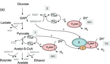
final electron acceptor and the reaction results in hydrogen formation (Li & Fang, 2007). During glucose fermentation, pyruvate is oxidized to acetyl-CoA and subsequently to acetate (Fig. 1a). Hydrogenases, which are linked to the thermodynamically favoured oxidation of reduced ferre-doxin e.g. pyruvate : ferreferre-doxin oxidoreductase, generate hydrogen using protons as terminal electron acceptors. A second pathway for hydrogen production is via NADH reoxidation during glycolysis, in which the cytosolic hydrogenase, coupled to NADH : ferredoxin oxido-reductase, uses NADH as the electron donor to reduce protons to hydrogen (Vardar-Schara et al., 2007). An alternative pathway was recently proposed for Thermotoga maritima, in which a trimeric bifurcating hydrogenase (similar multisubunit complexes are present in the genomes of sequenced members of the genus Clostridium) simultaneously oxidizes reduced ferredoxin and NADH under low partial hydrogen pressure (Schut & Adams, 2009).
Although as many as 12 mol hydrogen can theoretically be derived from glucose, there is no known natural metabolic pathway that could provide this yield, due to the presence of other products (Woodward et al., 2000). Ideally 1 mol glucose generates 4 mol hydrogen, with acetic acid, hydrogen and carbon dioxide as the only fermentation end products (Fig. 1b). With butyric acid only 2 mol hydrogen are produced. However, in a consortium composed of different Clostridium species, a mixture of products is obtained and the amount of hydrogen generated is determined by the acetate/butyrate ratio. In addition, the partial hydrogen pressure and the metabolic shift towards the production of more reduced products (e.g. alcohols) affect the final gas yield obtained. (Bartacek et al., 2007; Nath & Das, 2004; Levin et al., 2004).
Fig. 1. Fermentative hydrogen production and the role of [FeFe] hydrogenases. (a) Fermentative hydrogen production pathway. There are two possible pathways for hydrogen production in clostridia. One is linked to the oxidation of reduced ferredoxin catalysed by the enzyme complex pyruvate : Fd oxido-reductase (1). The second involves ferre-doxin-mediated NADH reoxidation catalysed by NADH : Fd oxidoreductase (2). An alter-native pathway involving trimeric bifurcating hydrogenase was proposed recently (see main text). MH, Monomeric hydrogenase; TH, tri-meric hydrogenase; Fd, ferredoxin; dashed lines, pathways competing for NADH+H+. (b) Stochiometric relations between glucose and the products formed during carbohydrate fermentation.
Hydrogenases
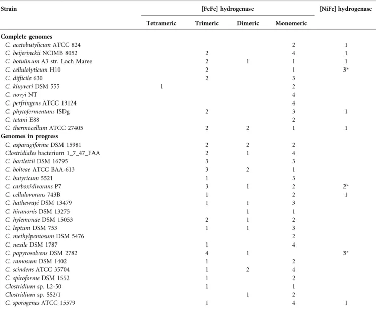
The process of biohydrogen production fundamentally depends on the presence of hydrogen-producing enzymes. Two groups of hydrogenases have been described in clostridia. [NiFe] hydrogenases are well-characterized and widely distributed among archaea and bacteria but only a few representatives of clostridia possess this type of enzyme (Table 1). In contrast, [FeFe] hydrogenases, restricted to bacteria and a few eukaryotic species, are much more abundant in clostridia.
From the first reported crystal structures of the [FeFe] hydrogenase from Clostridium pasteurianum (Peters et al., 1998) and Desulfovibrio desulfuricans (Nicolet et al., 1999) and of the [NiFe] hydrogenase from Desulfovibrio gigas (Volbeda et al., 1995), it was deduced that in both types of
enzymes, the bimetallic active sites are deeply buried within the protein matrix. They have similar frameworks with a unique coordination of the central Fe atom(s) by inorganic diatomic ligands. Another common feature is the existence of a [4Fe4S] cluster, proximal to the active centre and connected to the protein surface by means of additional FeS clusters. Finally, a hydrophobic gas channel, which runs from the molecular surface to the protein active site is characteristic for both classes of enzymes. Except for these similarities, the core polypep-tides do not share any similarity, neither in amino acid sequence nor in 3D folding (Nicolet et al., 2000). In addition, the protein maturation and assembly systems employed by the two classes of enzymes are phylogeneti-cally unrelated and biochemiphylogeneti-cally dissimilar (Bo¨ck et al., 2006).
Table 1. The hydrogenase content of the analysed genomes of members of the genus Clostridium
Strain [FeFe] hydrogenase [NiFe] hydrogenase Tetrameric Trimeric Dimeric Monomeric
Complete genomes
C. acetobutylicum ATCC 824 2 1
C. beijerinckii NCIMB 8052 2 4 1
C. botulinum A3 str. Loch Maree 2 1 1 1
C. cellulolyticum H10 2 1 3* C. difficile 630 2 3 C. kluyveri DSM 555 1 2 C. novyi NT 4 C. perfringens ATCC 13124 4 C. phytofermentans ISDg 2 3 1 C. tetani E88 2 C. thermocellum ATCC 27405 2 2 1 1 Genomes in progress C. asparagiforme DSM 15981 2 2 2 Clostridiales bacterium 1_7_47_FAA 2 1 4
C. bartlettii DSM 16795 3 3
C. bolteae ATCC BAA-613 3 2 1
C. butyricum 5521 1 3 C. carboxidivorans P7 3 1 2 2* C. cellulovorans 743B 1 2 1 C. hathewayi DSM 13479 1 1 3 C. hiranonis DSM 13275 1 1 C. hylemonae DSM 15053 2 1 2 C. leptum DSM 753 1 1 3 C. methylpentosum DSM 5476 2 C. nexile DSM 1787 1 4 C. papyrosolvens DSM 2782 4 1 3* C. ramosum DSM 1402 1 2 C. scindens ATCC 35704 1 2 4 C. spiroforme DSM 1552 1 2 Clostridium sp. L2-50 1 1 Clostridium sp. SS2/1 1 2 C. sporogenes ATCC 15579 1 4 1
*One of the[NiFe] hydrogenases of this organism is a putative [NiFe] hydrogenase complex; its catalytic subunit shows some similarity to the hydrogenase 4 (HyfE) from E. coli (Supplementary Table S1).
[NiFe] hydrogenases of clostridia
Based on the analysis of sequenced genomes of clostridia (Supplementary Table S1, available with the online version of this paper), only Clostridium acetobutylicum, Clostridium beijerinckii, Clostridium botulinum, Clostridium carboxidi-vorans, Clostridium cellulolyticum, Clostridium cellulovor-ans, Clostridium papyrosolvens and Clostridium sporogenes have genes encoding a [NiFe] hydrogenase. The core enzyme is composed of at least two subunits: a and b. The large subunit (a, ~60 kDa) hosts the Ni–Fe active centre. The small subunit (b, ~ 30 kDa) contains FeS clusters which function as an electron transfer pathway between the catalytic centre of the enzyme and the electron donor (acceptor). The average size of the large subunit ranges between 463 and 471 aa, and that of the small subunit between 290 and 306 aa. The structural genes encoding the periplasmic H2-uptake hydrogenases of
proteo-bacteria are clustered together with their accessory genes. In clostridia, the gene cluster includes the regulatory genes hypC, hypD and hypE, which control the expression of the structural genes. However, no orthologues of HyaE and HybE were found in the genomes of clostridia, despite the fact that in E. coli these proteins are chaperones that play a step by step controlling role in hydrogenase assembly by preventing premature Tat-dependent membrane targeting (Dubini & Sargent, 2003). Moreover, the small subunits of clostridial [NiFe] hydrogenases do not possess a large signal peptide containing a conserved RRxFxK motif which directs other hydrogenases towards Tat-dependent membrane location (Wu et al., 2000), suggesting a cytoplasmic localization for clostridial [NiFe] hydrogenases. On the other hand, the presence of a membrane-anchoring third subunit (cyto-chrome b5) and the predicted membrane localization (Supplementary Table S1, available with the online version of this paper) of the small subunit (PSORTb v.2.0. predicted results; Gardy et al., 2005) could indicate that this group of proteins is associated with a membrane. Additionally, in the genomes of Clostridium thermocellum, C. cellulolyticum, C. papyrosolvens and Clostridium phyto-fermentans, different six-subunit membrane-bound com-plexes were identified as belonging to the Ech hydrogenase group.
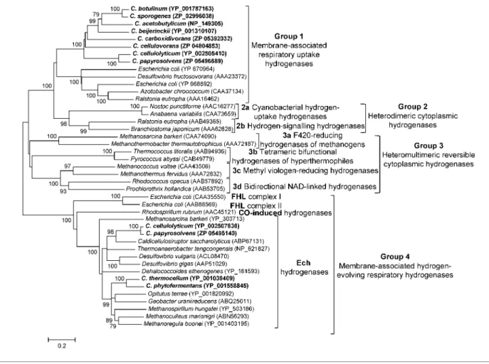
The analysis of sequence alignments of the small and large subunits led to a classification of[NiFe] hydrogenases into four main groups (Vignais et al., 2001). The bacterial and archaeal membrane-bound respiratory-uptake hydro-genases and periplasmic soluble hydrohydro-genases of sulfate reducers were classified together into the first group (Fig. 2). The second group includes the cyanobacterial uptake hydrogenases together with the cytoplasmic soluble hydrogenases involved in H2 sensing. The [NiFe]
hydrogenases of the third group contain subunits capable of binding soluble cofactors, e.g. F420, NAD or
NADP, and are reversible. The last and fourth group contains membrane-associated H2-evolving respiratory
[NiFe] hydrogenases, termed energy-converting
hydroge-nases (Ech). The hydrogehydroge-nases produced by members of the genus Clostridium clustered into groups 1 and 4 when aligned with representatives of other bacteria and archaea.
Clostridial [NiFe] hydrogenases clustering in group 1
Clostridial hydrogenases of group 1 form a homogeneous, well-separated cluster (Fig. 2). The amino acid sequence pattern present in all the small subunits of the [NiFe] hydrogenases contains most of the conserved cysteine residues ligating the proximal [4Fe4S], distal [4Fe4S] and medial[3Fe4S] clusters. However, a second cysteine residue of a conserved motif ligating a proximal[4Fe4S] cluster is replaced by asparagine in most of the clostridial proteins. Also, the two characteristic L1 (RICGICSTAH) and L2 (DPCxSCATH) signatures (regions of the sequence near the N- and C-termini of the large subunit) surrounding the two pairs of cysteine ligands at the NiFe site, are well conserved in clostridial[NiFe] hydrogenases. The terminal His residue of L2 is the predicted endopeptidase cleavage site at the C terminus of the large subunit.
Except for C. beijerinckii and C. cellulovorans, all the putative operons possess a gene encoding a di-haem cytochrome, which, in proteobacteria, connects the hetero-dimer to the quinone pool of the respiratory chain and maintains the enzyme in a reduced, active state (Meek & Arp, 2000). In clostridia, this protein has only two of four conserved haem b-binding histidine residues and the polypeptide is shorter.
The hydrogenase of C. acetobutylicum ATCC 824 is the only one encoded on a megaplasmid (pSOL1); all the others are encoded on bacterial chromosomes. In proteo-bacteria,[NiFe] hydrogenases allow the organism to use H2
as an energy source by linking its oxidation to the reduction of aerobic or anaerobic electron acceptors with energy recovery in the form of protonmotive force (Vignais, 2008). In nitrogen-fixing bacteria, e.g. Azotobacter chroococcum, [NiFe] hydrogenase is induced when hydrogen is produced as an intrinsic part of nitrogenase (Yates et al., 1997). By recycling the hydrogen produced in this process, [NiFe] hydrogenase recovers energy which would otherwise be lost by the cell. Therefore, this type of hydrogenase functions as a hydrogen uptake rather than as a hydrogen-evolving enzyme.[NiFe] hydrogenases from clostridia have not yet been studied and there is no information about their physiological function. Although many members of the genus Clostridium are recognized as non-symbiotic (free-living) nitrogen-fixing organisms, [NiFe] hydrogenase is found in only a few species of this genus. This would suggest that another type of enzyme, perhaps one of the numerous [FeFe] hydrogenases (see below), may be responsible for the uptake of the hydrogen generated during nitrogen fixation by clostridia.
Clostridial [NiFe] hydrogenases clustering in group 4
A small group of multisubunit membrane-associated [NiFe] hydrogenases, closely related to proton-pumping NADH : ubiquinone oxidoreductase (complex I), has been identified in several organisms including Escherichia coli
(hydrogenases 3 and 4; Maeda et al., 2007; Self et al., 2004), Rhodospirillum rubrum (Singer et al., 2006), Methanosarcina barkeri (Kurkin et al., 2002) and Desulfovibrio gigas (Rodrigues et al., 2003). These have been assigned to group 4 described by Vignais et al. (2001), which is characterized by membrane-associated H2-evolving respiratory [NiFe]
hydrogenases. The Ech hydrogenase from the methanogenic
Fig. 2. [NiFe] hydrogenase distance tree with special interest in clostridial enzymes. The evolutionary history was inferred using the neighbour-joining method. The percentages of replicate trees in which the associated taxa clustered together in the bootstrap test (1000 replicates) are shown next to the branches. The evolutionary distances were computed using the Poisson correction method. All positions containing alignment gaps and missing data were only eliminated in pairwise sequence comparisons (pairwise deletion option). Phylogenetic analyses were conducted inMEGA4 (Tamura et al., 2007). Proteins originating from
members of the genus Clostridium are indicated in bold type. FHL, Formate hydrogenylase; CO, carbon monoxide.
Fig. 3. Genomic organization of clostridial [NiFe] hydrogenases group Ech. (a) Genomic organization of Ech hydrogenase from M. barkeri; similar putative transcription units were found in sequenced genomes of clostridia. (b) Structural organization and localization of Ech hydrogenase from M. barkeri; an example of a possible arrangement of subunits encoding clostridial Ech hydrogenase (adapted from the paper by Hedderich & Forzi, 2005).
archaea M. barkeri is the best studied. It is an integral membrane protein composed of six subunits encoded by the echABCDEF operon (Fig. 3a and b). Surprisingly, analysis of the genomes available for members of the genus Clostridium revealed the presence of Ech-like hydrogenase genes in C. thermocellum, C. phytofermentans, C. papyrosolvens and C. cellulolyticum. The six genes encoding the putative subunits A–F (Fig. 3a) are organized into operons. The EchC polypeptide is composed of 144–149 residues and is at least 150 aa shorter than the small subunits of classical [NiFe] hydrogenases. It possesses only one (accommodating the [4Fe4S] proximal cluster) of the three conserved FeS cluster’s binding motifs typically present in classical[NiFe] hydro-genase small subunits. The large subunit, EchE, is composed of 359 aa. Two conserved patterns, containing cysteine residues involved in the binding of the Ni–Fe binuclear site, are present near the amino and carboxy termini of the polypeptide. A terminal histidine residue (L2 motif) is substituted by arginine, a feature typical of this group of enzymes. The hydropathy plots indicate that the clostridial EchA and EchB are integral membrane proteins with 18 and 8 internal helices, respectively, whereas the subunits EchC, EchD, EchE and EchF show a hydrophilic nature and are putatively cytoplasmically oriented membrane-associated components (Supplementary Table S1).
There are no data concerning a physiological function for the Ech hydrogenase in clostridia. In archaea, the Ech hydrogenase from M. barkeri was shown to play a central role in cell metabolism, providing reducing equivalents in the form of reduced ferredoxin for the first step of methanogenesis (the reduction of CO2to
formylmethano-furan by H2). A distinct role of Ech hydrogenase was
observed in the acetoclastic pathway (methanogenesis from acetate), where the enzyme catalyses an energy-conserving reaction: the production of hydrogen with reduced ferredoxin as the electron donor. Additionally it can provide the cell with reduced ferredoxin as the electron donor for the oxidoreductases that are required for the synthesis of acetyl-CoA and pyruvate (Meuer et al., 2002). In members of the family Desulfovibrionaceae, depending on the growth conditions, Ech hydrogenase can work bi-directionally. During growth in the presence of hydrogen Ech hydrogenase can reduce ferredoxin for carbon fixation, while during lactate metabolism, it produces H2 with a
ferredoxin as a redox partner (Pereira et al., 2008). In the genomes of C. cellulolyticum, C. carboxidivorans and C. papyrosolvens, another cluster was found (Supple-mentary Table S1) containing six ORFs and showing some similarity to E. coli hydrogenases 3 and 4. However, because of the lack of crucial cysteines involved in the coordination of the active centre in the putative large subunit, this enzyme complex was not analysed further in this review.
Highly modular [FeFe] hydrogenases of clostridia All available complete genomes and genomes in progress (February, 2009) for members of the genus Clostridium
were searched for the presence of hydrogenase- and maturation-encoding genes (Supplementary Table S2). As previously observed (Meyer, 2007), the catalytic subunits of clostridial[FeFe] hydrogenases display significant variations in size, mainly due to the presence of additional domains accommodating different numbers of FeS clusters (Fig. 4). The core of the[FeFe] hydrogenase consists of a ~40 kDa domain (H domain) which accommodates the H-cluster (a structure of six iron atoms with a unique arrangement of non-protein ligands). The latter is composed of an FeFe active site and one[4Fe4S] cluster bound to it by a bridging cysteine. The best conserved parts of the H-cluster domain are those surrounding the four cysteine ligands of the bimetallic active site: L1-TSCCPxW, L2-MPCxxKxxE and L3-ExMACxxGCxxG. Most of the clostridial hydrogenases, in addition to the H-cluster, have an N-terminal domain with two cysteine runs (Cx2Cx2Cx3C) homologous to
bacterial ferredoxins 2[4Fe4S] (also called the F-cluster) (Peters et al., 1998). The enzyme of C. pasteurianum is a typical ‘clostridial-type’ [FeFe] hydrogenase, in which the N-terminal domain coordinates two more FeS centres. One of these is a[2Fe2S] cluster similar to plant ferredoxins with a characteristic arrangement of cysteines, Cx2Cx9Cx31Cx3C.
The other one is a[4Fe4S] cluster with one iron bound to the N epsilon of the imidazole ring of the histidine ligand. A different group of clostridial hydrogenases seems to accommodate another type of FeS cluster, since their accessory domains contain six and eight cysteine residues distributed in two and three runs (CC, Cx2C,
Cx2Cx4Cx3C), respectively (Meyer, 2007).
Many clostridial [FeFe] hydrogenases are putatively dimeric, trimeric or even tetrameric enzymes (Table 1). The number and pattern of the conserved cysteine residues within the catalytic and accessory subunits suggests that an unusually high number of FeS clusters can be accommo-dated. This allows for efficient interaction with numerous redox partners.
Relatively little is known about the maturation and assembly of[FeFe] hydrogenases. Recent studies of mutants of Chlamydomonas reindhardtii revealed that two genes, hydEF (in most other organisms it splits into hydE and hydF) and hydG are involved in hydrogenase maturation (Posewitz et al., 2004). Clostridial [FeFe] hydrogenase maturation genes, unlike those of [NiFe]hydrogenases, do not cluster together with structural genes and are encoded in different locations on the genome (Meyer, 2007). This observation indicates that a pleiotrophic effect of the accessory enzymes on the assembly and maturation of the different FeS complexes could explain the highly modular forms of the[FeFe] hydrogenases found in clostridia. Clostridia contain numerous distinct[FeFe] hydrogenases, presumably to allow them to quickly respond to changing environmental conditions. The existence of multiple forms of this enzyme was confirmed as early as in 1963 when it was found that the pH optimum for the hydrogenase of D. desulfuricans varied depending on the chemical reagents
used for its activation (Sadana & Rittenberg, 1963). With the completion of genome sequences, a much higher variety of forms of [FeFe] hydrogenases was revealed in many organisms (Vignais et al., 2001; Meyer, 2007). For this review, sequences of more than 200 putative hydro-genases from different sources, with a focus on clostridial enzymes (Supplementary Table S2), were compared to each other (protein alignments were prepared by using the
MAFFT algorithm; Katoh & Toh, 2008). The resulting
distance tree (Fig. 5) shows the distribution of clostridial hydrogenases into four main groups, A, B, C and D, with
each group containing a different number of clusters. A similar distribution was previously observed by Meyer (2007). Group A includes organisms with the highest number of diverse proteins, but is the least robust section of the tree with lower bootstrap values and an internal topology that is sensitive to the number of sequences added or removed. Group B contains many sequences originating from members of the genus Clostridium and includes only a few representatives of other genera. Group C consists of mostly dimeric PAS/PAC sensory domains containing hydrogenases. Group D contains putative hydrogenases
Fig. 4. Schematic representation of modular structures of [FeFe] hydrogenases from members of the genus Clostridium. Modular structures were predicted with CDD (Marchler-Bauer et al., 2009). Structural frames were derived from the paper by Vignais et al. (2001), modified by Meyer (2007), with further modifications. M, Monomeric; D, dimeric; TR, trimeric; TE, tetrameric; bD, binding domain; Fd, ferredoxin; CODH, carbon monoxide dehydrogenase.
with much less well conserved L1, L2 and L3 motifs. Among the various structural frameworks (Fig. 4) and as previously observed (Vignais et al., 2001; Meyer, 2007), the modular structure M2 (H cluster plus F cluster) is the most widespread one as it occurs in all the main branches. The modular structure M3, typical for C. pasteurianum, is very frequent in group A. Tetrameric and trimeric hydrogenases cluster exclusively inside group A, whereas groups B and D contain monomeric enzymes, and most representatives of group C are dimeric.
Known hydrogen-producing eukaryotes are scattered in the different clusters inside group A. Proteins phylogenetically related to [FeFe] hydrogenases (e.g. human Nar1p) have also been identified in all mitochondrial eukaryotes studied so far (Balk et al., 2004). However, their further analysis is beyond the scope of this review.
The monomeric [FeFe] hydrogenases of group A In the distance tree (Fig. 5), monomeric hydrogenases of group A are distributed between three clusters: A2, A4 and A5. The best studied is a soluble, monomeric hydrogenase (AAA23248) from C. pasteurianum with an M3 modular structure that is characteristic for all the proteins in the (Clostridium-specific) A2 cluster. These enzymes catalyse hydrogen production by transferring electrons from ferre-doxins and flavoferre-doxins to protons (Demuez et al., 2007). The hydrogenase-encoding gene is located down-stream from another ORF which encodes a butyrate kinase. For C. perfringens, it has been shown that both genes are similarly regulated in response to glucose and other fermentative substrates (Kaji et al., 1999). In addition, the inactivation of the phosphotransacetylase gene (acetate formation pathway) in C. tyrobutyricum increased the expression of the butyrate kinase and hydrogenase. This resulted in a metabolic shift towards higher H2and butyrate
production, which was attributed to the reduced specific growth rate resulting from decreased acetate production (Liu et al., 2005). Although closely related to cluster A2, the C. beijerinckii hydrogenase (YP_001311066) is structurally a unique enzyme. Its C-terminal domain contains an insertion of 60 aa, not found in any other sequenced hydrogenase gene, with a conserved Cx2Cx2Cx3C motif binding an
additional 2[4Fe4S] ferredoxin-like domain. In cluster A4, most hydrogenases contain only one additional domain accommodating 2[4Fe4S] bacterial ferredoxin in the N terminus. However, a very long (270 aa) C terminus contains two small modular non-heme iron-binding domains containing a rubredoxin [Fe(SCys)4] centre, and one containing rubrerythrin (RRR). Rubredoxins are
detected in either the N- or C-termini of proteins such as flavin reductases, NAD(P)H-nitrite reductases or ferre-doxin–thioredoxin reductases (Pierik et al., 1993). An RRR domain is found in many air-sensitive bacteria and archaea. It is believed to reduce H2O2as a part of an oxidative stress
protection system, but its function is still poorly understood (Li et al., 2003). The catalytic subunit of the putative dimeric hydrogenase of Clostridium bolteae (ZP_02089645) does not contain any additional motifs in its C terminus and is shorter (451 aa). Interestingly, it seems to be a component of the CO-oxidizing system, since it is coupled to the carbon monoxide dehydrogenase gene (CODH). A similar CO-oxidation/H2-evolution enzyme complex, associated with
[NiFe] hydrogenase, was found in the membrane of Rhodospirillum rubrum and has been described as allowing the bacterium to metabolize carbon monoxide in the reaction CO+H2O A CO2+H2 (Singer et al., 2006).
Neither hydrogenase nor the CODH subunit are predicted membrane proteins in C. bolteae, therefore a function as a putative energy coupling site is hypothetical.
Cluster A5 forms a coherent set of clostridial hydrogenases with an N-terminal domain containing a recently described SLBB motif (soluble-ligand-binding b-grasp fold). This fold can be found in a diverse set of proteins performing different cellular functions. It also provides an effective scaffold for binding FeS clusters in the case of the [2Fe2S] ferrodoxins involved in electron transport (Burroughs et al., 2007).
Heteromultimeric [FeFe] hydrogenases of group A The first well-characterized hydrogenase known to contain more than one subunit was purified from the hyperther-mophilic, heterotrophic bacterium Thermotoga maritima (Verhagen et al., 1999). In clostridia, heteromultimeric hydrogenases are composed of catalytic subunits contain-ing an H cluster and FeS accessory domains (Fig. 4). The residual subunits accommodate additional FeS clusters. According to the predicted localization, almost all putative multimeric hydrogenases of clostridia are cytoplasmic enzymes (Supplementary Table S2). The only membrane-bound enzymes were assigned to the A3 cluster, associated with the formate dehydrogenase gene (Figs 4 and 5). Group A dimeric hydrogenases with a glutamate-synthase-binding domain
In group A, cluster A7 contains dimeric hydrogenases with a D(M3) modular structure. The catalytic subunits show homology to cluster A2, whereas the accessory subunits
Fig. 5. [FeFe] hydrogenase distance tree with special interest in clostridial enzymes. The tree was constructed using the same algorithm as for the[NiFe] hydrogenase distance tree (Fig. 2). Amino acids corresponding to the H-domain were used in the comparison. Proteins originating from members of the genus Clostridium are in bold type. The modular structure (Fig. 4) is indicated below the name of each cluster. *A subgroup of cluster A1 with a larger secondary subunit. 3No RRR motif, putative dimeric enzyme. The tree has been split into two parts along the dotted line for ease of viewing.
have two different domains, including one NuoF-like domain and another that interacts with the glutamate synthase NADPH small subunit. Glutamate synthases and glutamine synthases play a crucial role in microbial ammonium assimilation by catalysing the production of
L-glutamate fromL-glutamine and 2-oxoglutarate (Vanoni
& Curti, 2008). Reducing equivalents for this reaction are provided in the form of reduced pyridine nucleotides. Presumably, any H2 produced during the fermentative
processes could be reoxidized by this hydrogenase complex to generate NAD(P)H for glutamate synthase activity. The C-terminal domain of the catalytic subunit of a dimeric hydrogenase from C. thermocellum (YP_001039392) con-tains an uncharacterized motif (Cx18CxCx2C) binding a
putative FeS cluster. In addition, an NADPH-dependent glutamate synthase binding domain was identified in its N terminus.
Heterotrimeric bifurcating hydrogenases of group A
The trimeric clostridial hydrogenases of group A are distributed over four clusters. The proteins clustering into A1, A6 and A8 seem to be NAD-dependent, since their accessory subunits contain NADH-binding domains. The TR(M3) modular structure is characteristic for practically all of these protein complexes, except for those in cluster A3 which have the TR(M2) modular structure. The length of the catalytic a subunits is in the range of 560–600 aa. Two accessory subunits, a large b subunit (570–630 aa), similar to NuoF and a small c subunit (120–170 aa), which is homologous to[2Fe2S] thioredoxin, belong to the same operon as the catalytic subunits. There is evidence that the trimeric cytoplasmic hydrogenases in anaerobic species require both reduced ferredoxin and NADH in order to efficiently catalyse H2production (Schut & Adams, 2009).
By utilizing exergonic oxidation of ferredoxin, they drive the unfavourable oxidation of NADH (Fig. 1a). As a consequence of linked redox reactions, the oxidation of NADH is coupled to H2 production. This recently
described ‘electron bifurcation’ process was proposed as a third type of energy conservation in anaerobic bacteria (Herrmann et al., 2008). As regards gene organization, the genes encoding the trimeric hydrogenases in cluster A1 are usually preceded by a histidine protein kinase and are in close proximity to the other putative hydrogenases of cluster D. A similar situation was described for Ralstonia eutropha, where a histidine kinase and two [NiFe] hydrogenase complexes are involved in the H2-dependent
signal transduction cascade leading to regulated hydro-genase gene expression (Friedrich et al., 2005). The trimeric hydrogenases of cluster A6 have catalytic subunits of similar size to those of cluster A1, but their accessory subunits are slightly larger. At the genomic level, the trimeric enzymes of cluster A8 are usually associated with hydrogenases of group C containing PAS/PAC sensory domains.
Heterotrimeric hydrogenases of group A associated with the formate dehydrogenase complexes
Another trimeric hydrogenase complex [TR(M2), cluster A3] with two putative electron-transporting protein subunits seems to be present in the genome of Clostridium difficile. One of these subunits has the predicted post-translational cytoplasmic membrane local-ization, and probably anchors the active enzyme to the membrane, while the second one connects the enzyme to the formate dehydrogenase-like protein (FDH). Similar complexes have been detected in other bacterial genomes including C. beijerinckii, C. carboxidivorans, D. desulfur-icans, D. phychrophila and R. rubrum. A tungsten- and selenium-containing formate dehydrogenase associated with [FeFe] hydrogenase has also been reported for Eubacterium acidaminophilum (Graentzdoerffer et al., 2003). The putative hydrogenase operon contains four genes (hymA–D), three of which encode a trimeric hydrogenase. However, its catalytic subunit (hymA) is larger than those of cluster A3 (578 versus 461 aa) and has a modular M3 structure, clustering in A1 in the tree in Fig. 5. Formate is used by this bacterium as an electron donor during amino acid fermentation. It was reported that at least one of the FeS clusters of FDH could interact with similar clusters of the hydrogenase. This suggests that electrons derived from the formate oxidation could be transferred to the hydrogenase to produce H2. The catalytic
subunit of Clostridium kluyveri (YP_001394240) is a putative tetrameric enzyme and one accessory subunit has the predicted cytoplasmic membrane localization. This complex is not associated with any genes encoding formate dehydrogenase.
Monomeric hydrogenases of group B
Most of the sequences clustered within this group represent hydrogenases from members of the genus Clostridium. Proteins belonging to clusters B1 and B3 show a very similar structure to M3. A difference appears at the N-termini where the characteristic cysteine motifs binding the plant ferredoxin-like domain and the [4Fe4S] (HCCC) ferredoxin domain are replaced by a series of six and eight cysteines in clusters B1 and B3, respectively.
The average size of the polypeptide in cluster B2 is 450 aa, which corresponds to the predicted molecular mass of 50 kDa. The modular structure is similar to M2a. The characteristic feature of this group is the presence of an additional cysteine residue (TSCCCPxW) in the conserved L1 motif of the H cluster. In addition, the conserved pattern binding the [4Fe4S] cluster closest to the N-terminus is slightly different. This pattern is Cx1-4Cx 5-9Cx3C, whereas the canonical clostridial ferredoxin-binding
motif is Cx2Cx2Cx3C. A similar motif, Cx2Cx8Cx3C, is
observed in the nif-associated ferredoxin in Rhizobium meliloti. This ferrodoxin is a protein that is required for symbiotic nitrogen fixation. Six additional amino acids,
located between the second and third cysteine residues, form a loop which does not play any crucial role in protein activity since a deletion mutant exhibited almost 70 % of wild-type activity (Masepohl et al., 1992).
Group C dimeric hydrogenases, containing PAS/ PAC sensory domains, and group D monomeric hydrogenases
The characteristic feature of enzymes clustering into these two groups is the presence of an uncharacterized conserved motif Cx2Cx4Cx16C ligating the FeS centre. The N terminus
is much shorter than in the other enzymes and binds only one[4Fe4S] cluster. Group C contains enzymes with PAS/ PAC sensory domains in their C-termini. Most hydro-genases of this group are dimeric with a short (~80 aa) thioredoxin-like second subunit. In the genomes, they are localized in close proximity to the trimeric hydrogenases of cluster A8 and, in some cases, to the monomeric hydrogenases of cluster B3 (C. bolteae, C. phytofermentans and Clostridium leptum). The presence of PAS domains suggests that the enzyme may have a light sensing function or be associated with some aspects of redox changes (Kleihues et al., 2000). PAS domains may also be involved in protein–protein interactions. In bacteria, PAS domains are usually associated with the input domain of a histidine kinase or a sensor protein that regulates a histidine kinase (Zhulin et al., 1997). By detecting changes in the electron transport system, a PAS domain containing proteins can monitor the environment and thereby reduce cellular energy levels. This indicates that there are some regulatory interactions between both PAS/PAC sensory hydrogenases and other hydrogenases.
Within group D, the monomeric hydrogenases with M2e modular structure have L1, L2 and L3 motifs that are considerably less well conserved. However, all the cysteine residues necessary to link the [4Fe4S] cluster and the 2Fe centres in the H-cluster are present. This group of proteins is usually encoded upstream of the trimeric hydrogenases in cluster A1, separated by a histidine kinase gene in most cases.
Application of hydrogenases in biohydrogen production
Given continued concern about the future security and price of energy supplies and the climatic impact of increased carbon emissions, there is considerable interest in the processing of renewable resources to provide alternative fuels such as hydrogen. Nevertheless, despite several decades of extensive studies, bioenergy conversion technologies remain far too expensive to implement on an industrial scale.
Clostridia can be used for biohydrogen production and their possible application in this field has been reviewed (Lin et al., 2007). Clostridia can produce a theoretical 4 mol hydrogen from 1 mol glucose; however, this
potential yield is based on what we know about the cell metabolism. The discovery of a broad range of hydro-genases changes our perspective on how these anaerobes can produce hydrogen. Most of the hydrogenases described in this study are putative enzymes whose gene sequences were derived from sequenced genomes; information on their biochemical properties and function is limited to cluster A2 (Fig. 5) which contains C. pasteurianum-like [FeFe] hydrogenases. There are no experimental data concerning the remaining clostridial [FeFe] hydrogenases described in this study, making them somewhat of a ‘black box’ in fermentative hydrogen technology. Characterization of the organisms of interest in terms of their different hydrogenases would help to overcome several problems. One approach would be to design a mutant strain lacking an uptake hydrogenase, thereby removing its ability to recycle hydrogen. Another approach would be to screen for new enzymes with higher turnover rates and a broader range of redox partners, as well as lower sensitivity to oxygen inactivation, that could be used in in vitro systems. Clostridia possess a large number of interesting enzymes that could fulfil many of the engineering requirements. Many of the hydrogenases described in this study exhibit novel substitutions in the L1 and L2 sequence motifs, thus altering the redox properties of the[4Fe4S] cluster of the active centre. Other clostridial hydrogenases contain additional N- or C-terminal domains (Figs 4 and 5) coordinating different numbers of FeS clusters and thereby moderating the activity of the enzyme. Also, the recently described bifurcating hydrogenases, which are particularly abundant in clostridia, could offer new ways to overcome limiting bioengineering barriers.
The integration of hydrogen-producing enzymes into non-native hosts could increase hydrogen yields to a commer-cially competitive level. However, it should be considered that gene products functioning properly under cellular conditions may behave differently under conditions for which they did not evolved. Therefore, a precise character-ization of the biochemical properties of the key enzymes is required to improve the synthetic biology of an organism. It would be useful to be able to answer questions on the kind of regulation that is required and on whether the maturation system should also be introduced in order to synthesize the fully active protein e.g. heterologous expression.
The development of synthetic biology offers new perspec-tives for the construction of efficient hydrogen-producing strains and/or systems. It has already been shown that a yield far higher than the theoretical limit of 4 mol hydrogen can be obtained when using a cell-free artificial enzymic cascade composed of highly substrate-specific enzymes (Zhang et al., 2007). Further improvement could be made by characterizing and testing new hydrogenases. A future challenge would be to derive an oxygen-resistant iron hydrogenase from a highly reactive, but oxygen-sensitive one by substituting amino acid residues.
Another approach is to increase hydrogen production yields by engineering the metabolic pathways that convert glucose into hydrogen (Mathews & Wang, 2009). The discovery of multiple hydrogenases in clostridia suggests the existence of novel metabolic pathways involved in hydrogen production.
Several clostridia are also very interesting with respect to their ability to ferment cellulose, the most abundant and renewable polymer on Earth. Efficient degradation of lignocellulosic biomass is required for the production of biofuels (Liu et al., 2008). Interestingly, some of these cellulose-degrading species, such as C. cellulolyticum, C. phytofermentans or C. thermocellum, also possess a broad range of hydrogenases. In particular, the clostridial[NiFe] hydrogenases and the recently described Ech hydrogenases present in these organisms should be characterized in more detail.
Conclusions
This study demonstrates that the diversity of hydrogenases, especially the[FeFe] group, is severely under-reported in the literature, with the exception of two previous studies (Vignais et al., 2001; Meyer, 2007). Even members of the genus Clostridium, a genus widely studied for its ability to produce hydrogen, have mostly been described as possessing only one type of hydrogenase, commonly named HydA. Here, we have presented a comparative genomic perspect-ive of the clostridial hydrogenases that could set a new paradigm for their application in hydrogen-producing systems. Sequenced genomes give a completely new insight into the diversity of clostridial hydrogenases. In contrast with most of the past reports,[FeFe] hydrogenases are not a homogeneous group of enzymes but exist in multiple forms with different modular structures. However, very little information is available on the physiological and biochemical properties of[FeFe] hydrogenases in clostridia. There is a need for detailed studies on the catalytic mechanisms of the different[FeFe] hydrogenases in order to assess the potential for their application in artificial hydrogen-producing systems. A challenge ahead is to identify key enzymes in micro-organisms capable of hydrogen evolution, and to develop optimal microbial systems for biomass conversion by means of metabolic bioengineering. A precise knowledge of hydrogen metabolism and hydrogenases is essential to optimization of the biohydrogen production process and should therefore be a goal for future research.
Acknowledgements
This work was supported by an ARC grant, entitled ‘Micro-H2’, granted by the French-speaking Community of Belgium (Action de Recherches Concerte´es, ARC-07/12-04). A. W. is a Research Associate of the FNRS-FRS (Belgian Fund for Scientific Research). T. H. was supported by the Bundesministerium fuer Bildung and Forschung (BioH2 program). We would like to thank Bart Ghysels from the University of Lie`ge for valuable discussions and a critical reading of
the manuscript and to Ian Hamilton and Christopher Hamilton for English correction.
References
Balk, J., Pierik, A. J., Netz, D. J., Mu¨hlenhoff, U. & Lill, R. (2004).The hydrogenase-like Nar1p is essential for maturation of cytosolic and nuclear iron-sulphur proteins. EMBO J 23, 2105–2115.
Bartacek, J., Zabranska, J. & Lens, P. N. L. (2007).Developments and constraints in fermentative hydrogen production. Biofuels Bioprod Bioref 1, 201–214.
Bo¨ck, A., King, P. W., Blokesch, M. & Posewitz, M. C. (2006).
Maturation of hydrogenases. Adv Microb Physiol 51, 1–71.
Burroughs, A. M., Balaji, S., Iyer, L. M. & Aravind, L. (2007).Small but versatile: the extraordinary functional and structural diversity of the b-grasp fold. Biol Direct 2, 18.
Demuez, M., Cournac, L., Guerrini, O., Soucaille, P. & Girbal, L. (2007).Complete activity profile of Clostridium acetobutylicum [FeFe]-hydrogenase and kinetic parameters for endogenous redox partners. FEMS Microbiol Lett 275, 113–121.
Dubini, A. & Sargent, F. (2003).Assembly of Tat-dependent[NiFe] hydrogenases: identification of precursor-binding accessory proteins. FEBS Lett 549, 141–146.
Fang, H. H. & Liu, H. (2002).Effect of pH on hydrogen production from glucose by a mixed culture. Bioresour Technol 82, 87–93.
Friedrich, B., Buhrke, T., Burgdorf, T. & Lenz, O. (2005).A hydrogen-sensing multiprotein complex controls aerobic hydrogen metabolism in Ralstonia eutropha. Biochem Soc Trans 33, 97–101.
Gardy, J. L., Laird, M. R., Chen, F., Rey, S., Walsh, C. J., Ester, M. & Brinkman, F. S. L. (2005).PSORTb v.2.0: expanded prediction of bacterial protein subcellular localization and insights gained from comparative proteome analysis. Bioinformatics 21, 617–623.
Graentzdoerffer, A., Rauh, D., Pich, A. & Andreesen, J. R. (2003).
Molecular and biochemical characterization of two tungsten- and selenium-containing formate dehydrogenases from Eubacterium acidaminophilum that are associated with components of an iron-only hydrogenase. Arch Microbiol 179, 116–130.
Hallenbeck, P. C. & Benemann, J. R. (2002).Biological hydrogen production; fundamentals and limiting processes. Int J Hydrogen Energy 27, 1185–1193.
Hedderich, R. & Forzi, L. (2005).Energy-converting [NiFe] hydro-genases: more than just H2activation. J Mol Microbiol Biotechnol 10, 92–104.
Heinekey, D. M. (2009).Hydrogenase enzymes: recent studies and active site models. J Organomet Chem 694, 2671–2680.
Herrmann, G., Jayamani, E., Mai, G. & Buckel, W. (2008).Energy conservation via electron-transferring flavoprotein in anaerobic bacteria. J Bacteriol 190, 784–791.
Kaji, M., Taniguchi, Y., Matsushita, O., Katayama, S., Miyata, S., Morita, S. & Okabe, A. (1999). The hydA gene encoding the H2 -evolving hydrogenase of Clostridium perfringens: molecular character-ization and expression of the gene. FEMS Microbiol Lett 181, 329–336.
Katoh, K. & Toh, H. (2008). Recent developments in the MAFFT multiple sequence alignment program. Brief Bioinform 9, 286–298.
Khanal, S. K., Chen, W.-H., Li, L. & Sung, S. (2004). Biological hydrogen production: effects of pH and intermediate products. Int J Hydrogen Energy 29, 1123–1131.
Kleihues, L., Lenz, O., Bernhard, M., Buhrke, T. & Friedrich, B. (2000). The H2 sensor of Ralstonia eutropha is a member of the subclass of regulatory[NiFe] hydrogenases. J Bacteriol 182, 2716–2724.
Kurkin, S., Meuer, J., Koch, J., Hedderich, R. & Albracht, S. P. J. (2002). The membrane-bound [NiFe]-hydrogenase (Ech) from Methanosarcina barkeri: unusual properties of the iron-sulphur clusters. Eur J Biochem 269, 6101–6111.
Levin, D. B., Pitt, L. & Love, M. (2004). Biohydrogen production: prospects and limitations to practical application. Int J Hydrogen Energy 29, 173–185.
Li, C. & Fang, H. H. P. (2007). Fermentative hydrogen production from wastewater and solid wastes by mixed cultures. Crit Rev Environ Sci Technol 37, 1–39.
Li, M., Liu, M. Y., LeGall, J., Gui, L. L., Liao, J., Jiang, T., Zhang, J. P., Liang, D. C. & Chang, W. R. (2003). Crystal structure studies on rubrerythrin: enzymatic activity in relation to the zinc movement. J Biol Inorg Chem 8, 149–155.
Lin, P. Y., Whang, L. M., Wu, Y. R., Ren, W. J., Hsiao, C. J., Li, S. L. & Chang, J. S. (2007). Biological hydrogen production of the genus Clostridium: metabolic study and mathematical model simulation. Int J Hydrogen Energy 32, 1728–1735.
Liu, X., Zhu, Y. & Yang, S. T. (2005). Butyric acid and hydrogen production by Clostridium tyrobutyricum ATCC 25755 and mutants. Enzyme Microb Technol 38, 521–528.
Liu, Y., Yu, P., Song, X. & Qu, Y. (2008).Hydrogen production from cellulose by co-culture of Clostridium thermocellum JN4 and Thermoanaerobacterium thermosaccharolyticum GD17. Int J Hydrogen Energy 33, 2927–2933.
Maeda, T., Sanchez-Torres, V. & Wood, T. K. (2007).Escherichia coli hydrogenase 3 is a reversible enzyme possessing hydrogen uptake and synthesis activities. Appl Microbiol Biotechnol 76, 1035–1042.
Marchler-Bauer, A., Anderson, J. B., Chitsaz, F., Derbyshire, M. K., DeWeese-Scott, C., Fong, J. H., Geer, L. Y., Geer, R. C., Gonzales, N. R. & other authors (2009).CDD: specific functional annotation with the Conserved Domain Database. Nucleic Acids Res 37, D205–D210.
Masepohl, B., Kutsche, M., Riedel, K. U., Schmehl, M., Klipp, W. & Pu¨hler, A. (1992).Functional analysis of the cysteine motifs in the ferredoxin-like protein FdxN of Rhizobium meliloti involved in symbiotic nitrogen fixation. Mol Gen Genet 233, 33–41.
Masukawa, H., Mochimaru, M. & Sakura, H. (2002).Hydrogenases and photobiological hydrogen production utilizing nitrogenase system in cyanobacteria. Int J Hydrogen Energy 27, 1471–1474.
Mathews, J. & Wang, G. (2009).Metabolic pathway engineering for enhanced biohydrogen production. Int J Hydrogen Energy 34, 7404– 7416.
Meek, L. & Arp, D. J. (2000).The hydrogenase cytochrome b heme ligands of Azotobacter vinelandii are required for full H2oxidation capability. J Bacteriol 182, 3429–3436.
Meuer, J., Kuettner, H. C., Zhang, J. K., Hedderich, R. & Metcalf, W. (2002). Genetic analysis of the archaeon Methanosarcina barkeri Fusaro reveals a central role for Ech hydrogenase and ferredoxin in methanogenesis and carbon fixation. Proc Natl Acad Sci U S A 99, 5632–5637.
Meyer, J. (2007).[FeFe] hydrogenases and their evolution: a genomic
perspective. Cell Mol Life Sci 64, 1063–1084.
Morimoto, K., Kimura, T., Sakka, K. & Ohmiya, K. (2005). Overex-pression of a hydrogenase gene in Clostridium paraputrificum to enhance hydrogen gas production. FEMS Microbiol Lett 246, 229– 234.
Nath, K. & Das, D. (2004).Improvement for fermentative hydrogen production: various approaches. Appl Microbiol Biotechnol 65, 520– 529.
Nicolet, Y., Piras, C., Legrand, P., Hatchikian, C. E. & Fontecilla-Camps, J. C. (1999).Desulfovibrio desulfuricans iron hydrogenase: the
structure shows unusual coordination to an active site Fe binuclear center. Structure 7, 13–23.
Nicolet, Y., Lemon, B. J., Fontecilla-Camps, J. C. & Peters, J. W. (2000).A novel FeS cluster in Fe-only hydrogenases. Trends Biochem Sci 25, 138–143.
Pereira, P. M., He, Q., Valente, F. M. A., Xavier, A. V., Zhou, J., Pereira, I. A. C. & Louro, L. O. (2008).Energy metabolism in Desulfovibrio vulgaris Hildenborough: insights from transcriptome analysis. Antonie van Leeuwenhoek 93, 347–362.
Peters, J. W., Lanzilotta, W. N., Lemon, B. J. & Seefeldt, L. C. (1998). X-ray crystal structure of the Fe-only hydrogenase (CpI) from Clostridium pasteurianum to 1.8 Angstrom resolution. Science 282, 1853–1858.
Pierik, A. J., Wolbert, R. B., Portier, G. L., Verhagen, M. F. & Hagen, W. R. (1993).Nigerythrin and rubrerythrin from Desulfovibrio vulgaris each contain two mononuclear iron centers and two dinuclear iron clusters. Eur J Biochem 212, 237–245.
Pilak, O., Mamat, B., Vogt, S., Hagemeier, C. H., Thauer, R. K., Shima, S., Vonrhein, C., Warkentin, E. & Ermler, U. (2006).The crystal structure of the apoenzyme of the iron-sulfur cluster-free hydrogenase. J Mol Biol 358, 798–809.
Posewitz, M. C., King, P. W., Smolinski, S. L., Zhang, L., Seibert, M. & Ghirardi, M. L. (2004).Discovery of two novel radical S-adenosyl-methionine proteins required for the assembly of an active [Fe] hydrogenase. J Biol Chem 279, 25711–25720.
Rodrigues, R., Valente, F. M. A., Pereira, I. A. C., Oliveira, S. & Rodrigues-Pousada, C. (2003). A novel membrane-bound Ech [NiFe] hydrogenase in Desulfovibrio gigas. Biochem Biophys Res Commun 306, 366–375.
Sadana, J. C. & Rittenberg, D. (1963). Some observations on the enzyme hydrogenase of Desulfovibrio desulfuricans. Proc Natl Acad Sci U S A 50, 900–904.
Schut, G. J. & Adams, M. W. (2009). The iron-hydrogenase of Thermotoga maritima utilizes ferredoxin and NADH synergistically: a new perspective on anaerobic hydrogen production. J Bacteriol 191, 4451–4457.
Self, W. T., Hasona, A. & Shanmugam, K. T. (2004).Expression and regulation of a silent operon, hyf, coding for hydrogenase 4 isoenzyme in Escherichia coli. J Bacteriol 186, 580–587.
Singer, S. W., Hirst, M. B. & Ludden, P. W. (2006).CO-dependent H2 evolution by Rhodospirillum rubrum: role of CODH : CooF complex. Biochim Biophys Acta 1757, 1582–1591.
Tamura, K., Dudley, J., Nei, M. & Kumar, S. (2007).MEGA4: Molecular
Evolutionary Genetics Analysis (MEGA) software version 4.0. Mol Biol
Evol 24, 1596–1599.
Van Ginkel, S. W., Oh, S. E. & Logan, B. E. (2005).Biohydrogen gas production from food processing and domestic wastewaters. Int J Hydrogen Energy 30, 1535–1542.
Vanoni, M. A. & Curti, B. (2008). Structure–function studies of glutamate synthases: a class of self-regulated iron-sulfur flavoenzymes essential for nitrogen assimilation. IUBMB Life 60, 287–300.
Vardar-Schara, G., Maeda, T. & Wood, T. K. (2007). Metabolically engineered bacteria for producing hydrogen via fermentation. Microbiol Biotechnol 1, 107–125.
Verhagen, M. F., O’Rourke, T. & Adams, M. W. (1999).The hyper-thermophilic bacterium, Thermotoga maritima, contains an unusually complex iron-hydrogenase: amino acid sequence analyses versus biochemical characterization. Biochim Biophys Acta 1412, 212–229.
Vignais, P. M. (2008).Hydrogenases and H+-reduction in primary energy conservation. Results Probl Cell Differ 45, 223–252.
Vignais, P. M. & Colbeau, A. (2004).Molecular biology of microbial hydrogenases. Curr Issues Mol Biol 6, 159–188.
Vignais, P. M., Billoud, B. & Meyer, J. (2001).Classification and phylogeny of hydrogenases. FEMS Microbiol Rev 25, 455–501.
Volbeda, A., Charon, M. H., Piras, C., Hatchikian, E. C., Frey, M. & Fontecilla-Camps, J. C. (1995).Crystal structure of the nickel-iron hydrogenase from Desulfovibrio gigas. Nature 373, 580– 587.
Wang, M. Y., Tsai, Y. L., Olson, B. H. & Chang, J. S. (2008a).
Monitoring dark hydrogen fermentation performance of indigenous Clostridium butyricum by hydrogenase gene expression using RT-PCR and qPCR. Int J Hydrogen Energy 33, 4730–4738.
Wang, M. Y., Olson, B. H. & Chang, J. S. (2008b).Relationship among growth parameters for Clostridium butyricum, hydA gene expression, and biohydrogen production in a sucrose-supplemented batch reactor. Appl Microbiol Biotechnol 78, 525–532.
Woodward, J., Orr, M., Cordray, K. & Greenbaum, E. (2000).
Enzymatic production of biohydrogen. Nature 405, 1014–1015.
Wu, L. F., Ize, B., Chanal, A., Quentin, Y. & Fichant, G. (2000).
Bacterial twin-arginine signal peptide-dependent protein transloca-tion pathway: evolutransloca-tion and mechanism. J Mol Microbiol Biotechnol 2, 179–189.
Yang, K. & Metcalf, W. W. (2004).A new activity for an old enzyme: Escherichia coli bacterial alkaline phosphatase is a phosphite-dependent hydrogenase. Proc Natl Acad Sci U S A 101, 7919–7924.
Yates, M. G., De Souza, E. M. & Kahindi, J. H. (1997).Oxygen, hydrogen and nitrogen fixation in Azotobacter. Soil Biol Biochem 29, 863–869.
Zhang, Y. H. P., Evans, B. R., Mielenz, J. R., Hopkins, R. C. & Adams, M. W. W. (2007).High-yield hydrogen production from starch and water by a synthetic enzymatic pathway. PLoS One 2, e456.
Zhulin, I. B., Taylor, B. L. & Dixon, R. (1997).PAS domain S-boxes in archaea, bacteria and sensors for oxygen and redox. Trends Biochem Sci 22, 331–333.