Université de Sherbrooke
The hypoxic tumor microenvironment regulates the LPA signaling axis to promote cancer cell invasion and metastasis
Par Kelly Harper
Programme d’immunologie
Thèse présenté(e) à la Faculté de médecine et des sciences de la santé en vue de l’obtention du grade de philosophiae doctor (Ph.D.)
en immunologie
Sherbrooke, Québec, Canada May, 2018
Membres du jury d’évaluation
Pre Claire Dubois, programme d’immunologie Pre Jana Stankova, programme d’immunologie Pre Caroline Saucier, programme de biologie cellulaire
Pr Sylvain Bourgoin, Microbiologie-Infectologie et d’Immunologie, Faculté de Médecine, Université Laval
Le microenvironnement hypoxique tumoral régule la signalisation du LPA pour favoriser l'invasion des cellules cancéreuses et le développement des métastases
Par Kelly Harper
Programme d’Immunologie
Thèse présentée à la Faculté de médecine et des sciences de la santé en vue de l’obtention du diplôme de philosophiae doctor (Ph.D.) en immunologie, Faculté de médecine et des
sciences de la santé, Université de Sherbrooke, Sherbrooke, Québec, Canada, J1H 5N4 Le développement des métastases est la cause principale de mortalité des patients atteints de cancer, mais demeure un obstacle majeur aux traitements. L'hypoxie, une caractéristique commune des tumeurs solides, est fortement impliquée dans l'invasion cellulaire et le développement des métastases, mais les mécanismes sous-jacents demeurent méconnus. La signalisation du LPA joue un rôle important au cours de la tumorigenèse et du développement des métastases, les membres de cette voie étant souvent régulés à la hausse dans les cellules tumorales. La signalisation du LPA a également été impliquée dans la production de structures de dégradation, les invadopodes, qui sont nécessaires à la formation de métastases. Des études récentes indiquent que la formation d'invadopodes est également induite par l'hypoxie. Par conséquent, nous avons voulu élucider l'influence du microenvironnement hypoxique sur l'axe de signalisation du LPA et si celui-ci joue un rôle dans la production d'invadopodes et la formation de métastases.
Nous avons découvert que le LPA1 est un récepteur utilisé de façon commune et majoritaire pour la production d'invadopodes induite par l'hypoxie, et ce, dans diverses lignées cellulaires cancéreuses. Nous avons démontré que l'hypoxie favorise la formation d'invadopodes en utilisant une voie de signalisation distincte qui implique une communication croisée entre le récepteur LPA1 et l'EGFR, médiée par la kinase Src, Dans ce contexte, l'inhibition combinée du LPA1 et de l'EGFR agit en synergie afin d’empêcher la formation de métastases spontanées. Étant donné que la toxicité et la résistance aux inhibiteurs de l'EGFR représentent un défi important pour les patients atteints de cancer, ce travail permet l’identification d’une cible potentielle, le LPA1, qui pourrait être inhibée conjointement avec l'EGFR dans le but d’améliorer la survie de ces patients. D'autres études sur la régulation hypoxique de l'axe de signalisation du LPA ont démontré que l'hypoxie peut contrôler les niveaux d'expression des enzymes impliqués dans la production (ATX) et la dégradation (LPP1 / LPP3) du LPA, des évènements qui conduisent à une production accrue d'invadopodes. L'hypoxie permet également de modifier la localisation de ces protéines, ce qui pourrait constituer un mécanisme additionnel de régulation de l’axe de signalisation du LPA en hypoxie.
Notre travail suggère que l'hypoxie est un régulateur important de l'axe de signalisation du LPA menant à l’invasion et à la formation de métastases. Par conséquent, les thérapies ciblant cet axe pourraient être bénéfiques pour contrer les effets néfastes de l'hypoxie tumorale sur la survie des patients atteints de cancer. De plus, un traitement combiné, ciblant le LPA1 et l’EGFR, pourrait être utile afin de réduire les effets secondaires et la résistance aux inhibiteurs de l'EGFR. Des études supplémentaires seront nécessaires afin de valider le potentiel thérapeutique de ce type de traitement.
S
UMMARYThe hypoxic tumor microenvironment regulates the LPA signaling axis to promote cancer cell invasion and metastasis
By Kelly Harper Immunology Program
Thesis presented at the Faculty of Medicine and Health Sciences for the obtention of Doctor degree diploma philosophiae doctor (Ph.D.) in Immunology, Faculty of Medicine
and Health Sciences, Université de Sherbrooke, Sherbrooke, Québec, Canada, J1H 5N4 Metastasis is the leading cause of cancer patient mortality yet remains a major hurdle for treatment. Hypoxia, a common feature of solid tumors, has been critically involved in cell invasion and metastasis but the underlying mechanisms remain poorly understood. The LPA signaling axis plays an important role during tumorigenesis and metastasis, with members of this pathway often being upregulated in tumor cells. LPA signaling has also been implicated in production of the degradative structures invadopodia, which are known to be required for metastasis. Interestingly, formation of invadopodia can also be induced by hypoxia. Therefore, we endeavoured to elucidate the influence of the hypoxic tumor microenvironment on the LPA signaling axis and whether this could play a role in invadopodia production and metastasis.
We uncovered LPA1 as a common and major receptor used for hypoxia-induced
invadopodia production in various cancer cell lines. We demonstrated that hypoxia promotes invadopodia formation through a distinct signaling pathway that involves Src-mediated cross-communication between LPA1 and EGFR, and that combined inhibition of
LPA1 and EGFR acts synergistically to impede spontaneous metastasis. Since EGFR
inhibitor toxicity and resistance represents a current challenge for cancer patients, this work identifies a potential target, LPA1 that could be inhibited in conjunction with EGFR to
improve patient outcomes. Further studies into hypoxic regulation of the LPA signaling axis demonstrated that hypoxia can control the expression levels of LPA producing (ATX) and degrading (LPP1/LPP3) enzymes, events that lead to increased invadopodia production. Hypoxia was also found to alter the localization of these proteins, uncovering an additional mechanism of hypoxic regulation.
Our work suggests that hypoxia is a master regulator of the LPA signaling axis that leads to metastasis, therefore therapies targeting this axis could be beneficial to counteract the detrimental effects of tumor hypoxia on cancer patient survival. Furthermore, LPA1-EGFR combination therapy could be a useful strategy to reduce EGFR inhibitor side effects and resistance and therefore warrants further studies to evaluate the potential of combination therapies in cancer patients.
1 Résumé ... iii 2 Summary ... iv 3 Table of Contents ... v 4 List of figures ... vii 5 List of Abbreviations ... ix 1 Introduction ... 1 1.1 The Tumor microenvironment promotes invasion and metastasis ... 1 1.1.1 Hypoxia in the tumor microenvironment ... 3 1.1.2 Mechanisms of hypoxia-induced effects in cells ... 4 1.1.2.1 Gene expression ... 4 1.1.2.2 Metabolism and pH alterations ... 5 1.1.2.3 Endocytosis regulation ... 6 1.1.2.4 Activation of signaling pathways ... 7 1.1.3 Hypoxia as a driver of invasion and metastasis ... 8 1.2 Invadopodia mediate cancer cell invasion ... 9 1.2.1 The stages of invadopodia formation ... 11 1.2.2 Inducers of invadopodia ... 15 1.2.3 Implication of invadopodia in the metastatic process ... 17 1.3 The LPA signaling axis ... 18 1.3.1 LPA structure and sources ... 18 1.3.2 Production and degradation of LPA ... 19 1.3.2.1 Autotaxin as the main producer of LPA ... 21 1.3.2.1.1 Structure and activity of ATX ... 21 1.3.2.1.2 Regulation of ATX expression ... 23 1.3.2.1.3 ATX role in physiology and pathology ... 24 1.3.2.2 Lipid phosphate phosphatases ... 25 1.3.2.2.1 Structure and function of LPPs ... 25 1.3.2.2.2 Regulation of LPPs ... 27 1.3.2.2.3 Role of LPPs in physiology and pathology ... 27 1.3.3 LPA receptors and signaling ... 28 1.3.3.1 LPA receptor identification ... 29 1.3.3.2 Signaling pathways of LPARs ... 30
1.3.3.3 Crosstalk of LPARs with RTKs ... 32 1.3.3.4 Implication of LPA signaling in physiological and pathological processes ... 33 1.3.4 Implications of the ATX/LPA axis in cancer ... 34 1.4 Hypothesis ... 38 1.4.1 Objectives ... 38 2 Article 1 ... 39 3 Chapter 2: Hypoxia promotes invasion through regulation of LPA Synthesis and Degradation Enzymes ... 81 3.1 MATERIALS and METHODS ... 82 3.1.1 Reagents ... 82 3.1.2 Cell culture and transfection ... 82 3.1.3 Real time RT-PCR ... 83 3.1.4 Western blotting ... 83 3.1.5 Invadopodia assay ... 83 3.1.6 Immunofluorescence ... 84 3.1.7 3D invasion assay ... 84 3.2 RESULTS ... 85 3.2.1 Hypoxia induces ATX and represses LPP expression ... 85 3.2.2 Activity of the LPA producing enzyme ATX is required for hypoxia-induced invadopodia production ... 87 3.2.3 Lipid phosphate phosphatases modulate invadopodia production ... 88 3.2.4 Hypoxia affects the spatial localization of ATX and LPPs ... 90 3.2.5 ATX promotes cancer cell migration and invasion ... 92 4 Discussion ... 94 4.1 Discussion of Article ... 94 4.2 Discussion of Chapter 2 ... 102 4.3 Conclusions and Perspectives ... 113 5 References ... 115 6 Annexes ... 159 Annex 1: Discussion Figures ... 159
INTRODUCTION FIGURES
Figure 1 Invadopodia ... 10
Figure 2 Structure of LPC and LPA ... 19
Figure 3 Mechanisms of LPA production and degradation ... 20
Figure 4 Structural domains of ATX ... 22
Figure 5 Lipid phosphate phosphatase structure and catalytic domains ... 26
Figure 6 LPAR signaling ... 31
ARTICLE 1 FIGURES Figure 1 LPA1 is essential for hypoxia-induced cancer cell invasion. ... 52
Figure 2 LPA1 and GRK2 expression is not modulated under hypoxic conditions. ... 54
Figure 3 EGFR is transactivated by LPA1 under hypoxic conditions. ... 56
Figure 4 EGFR is necessary for hypoxia-induced invadopodia formation downstream of LPA1. ... 57
Figure 5 Mechanism of transactivation of EGFR by LPA1 under hypoxia. ... 59
Figure 6 LPA1 signals through PI3K/AKT to promote hypoxia induced invasion, an effect mediated by transactivation of EGFR. ... 62
Figure 7 LPA1 and EGFR are implicated in spontaneous metastasis in a chorioallantoic membrane. ... 65
CHAPTER 2 FIGURES Figure 1 ATX expression is modulated in hypoxia ... 86
Figure 2 Lipid phosphate phosphatase expression is modulated in hypoxia ... 87
Figure 3 ATX is essential for hypoxia-mediated invadopodia production ... 89
Figure 4 LPP1 and LPP3 contribute to invadopodia production ... 90
Figure 5 ATX localization is altered in hypoxic cells ... 91
Figure 6 LPP1 and LPP3 localization is altered in hypoxic cells ... 92
Figure 7 ATX is necessary for hypoxia-induced 3D invasion and migration ... 93
ANNEX 1 : DISCUSSION FIGURES Figure 1 Increased ROS production in hypoxia is dependent on LPA1 ... 159
AC adenylyl cyclase
ADAM a disintegrin and metalloproteinase ADF actin depolymerizing factor
AP-1 activator protein 1
Arg Abelson-related gene
Arp2/3 actin-related proteins ATP adenosine Triphosphate
ATX autotaxin
Bcl-2 B-cell lymphoma 2
bFGF basic fibroblast growth factor bHLH basic helix-loop-helix
BMDC bone marrow-derived cells Bmp-2 bone morphogenic protein 2
C1P ceramide 1-phosphate
CAF cancer-associated fibroblasts CAIX carbonic anhydrase 9
cAMP cyclic adenosine 3’,5’-monophosphate CDK cyclin-dependent kinase
cPA cyclic phosphatidic acid
CREB: cAMP response element-binding
CXCR chemokine receptor
DAG diacylglycerol
DDR1 Discoidin domain receptor family, member 1 DPP4 dipeptidyl dipeptidase IV
EBV Epstein-Barr virus ECM extracellular matrix
Edg endothelial differentiation gene EGF epidermal growth factor
EGFR epidermal growth factor receptor EMT epithelial-mesenchymal transition
E.R endoplasmic reticulum
ERK1/2 extracellular signal-regulated kinases F-actin filamentous actin
FAK focal adhesion kinase GAP GTPase activating protein GDP guanosine diphosphate GEF guanine exchange factor GLUT Glucose transporter
GM1 monosialotetrahexosylganglioside GPCR G-protein coupled receptor
GPR G-protein receptor
Grb2 Growth factor receptor-bound protein 2 GTP guanosine triphosphate
GTPase guanosine triphosphatase
HB-EGF heparin-binding EGF-like growth factor HDAC histone deacetylases
HGF hepatocyte growth factor
HIF Hypoxia-inducible factor
HUVECs human umbilical vascular endothelial cells
IFN interferon
IGFR1 insulin-like growth factor receptor 1
IL interleukin
IP3 inositol triphosphate IRS-1 insulin receptor substrate 1 ITGA integrin subunit alpha ITGB integrin subunit beta JNK c-Jun N-terminal kinases LDH lactate dehydrogenase
LOX lysyl oxidase
LPA lysophosphatidic acid LPAR lysophosphatidic receptor
LPAAT lysophosphatidic acid acyl transferases LPC lysophosphatidyl choline
LPP lipid phosphate phosphatase
LPS lipopolysaccharide
lysoPLD lysophospholipase D
MAG monoacylglycerol
MAPK mitogen activated protein kinase MβCD methyl-beta cyclodextrin
MCP-1 monocyte chemoattractant protein-1 MCT monocarboxylate transporter
MMP matrix metalloproteinase
MORFO modulator of oligodendrocyte remodeling and focal adhesion organization
mRNA messenger ribonucleic acid
MT1-MMP membrane-type 1 MMP
NAC N-acetyl-L-cysteine
NADPH nicotinamide adenine dinucleotide phosphate-oxidase NFAT nuclear factor of activated T-cells
NF-kB: nuclear factor kappa-light-chain-enhancer of activated B cells
NGF Nerve growth factor
NHE Na+/H+ exchanger
NHERF1/EBP50 Na+-H+exchanger regulatory factor/ ERM-binding protein 50 NPP2 nucleotide pyrophosphatase/phosphodiesterase 2
N-WASP neuronal Wiskott-Aldrich syndrome protein
PA phosphatidic acid
PAP phosphatidic acid phosphatase
PAP2 type 2 PAP
PDGF platelet-derived growth factor
PFK phosphofructokinase
PHD prolyl hydroxylase domain protein PI3K phosphoinositide 3-kinases
PKC protein kinase C
PLA1 or 2 phospholipase A1 or A2
PLC phospholipase C
PLD phospholipase D
PTP protein tyrosine phosphatase
PX phox homology
RGD arginine-glycine-aspartic acid
RNA ribonucleic acid
ROCK rho-associated coiled-coil-forming protein kinase ROR2 receptor tyrosine kinase-like orphan receptor 2 ROS reactive oxygen species
RPLP0 ribosomal protein lateral stalk subunit P0 RT-PCR reverse transcriptase polymerase chain reaction RTK receptor tyrosine kinase
S1P sphingosine-1-phosphate
SDF-1 stromal cell-derived factor 1
SH2 src-homology 2 domain
SH3 src-homology 3 domain
SHIP SH2 domain-containing inositol 5-phosphatase 2
SMB somatomedin B domain
SOS son of sevenless
shRNA short hairpin RNA
SRA steroid receptor RNA activator 1 SRE serum response element
STED Stimulated emission depletion TAM tumor-associated macrophage TGF-β transforming growth factor beta
TKS5/FISH tyrosine kinase substrate 5/five SH3 domains TLR toll-like receptor
TNF- α tumor necrosis factor-alpha TrkA tropomyosin receptor kinase A
tyr tyrosine
uPA urokinase-type plasminogen activator uPAR urokinase receptor
VATPase vacuolar-type H+-ATPase
VEGF vascular endothelial growth factor ZEB zinc finger E-box binding homeobox
Cancer is the deadliest disease for Canadians, accounting for 30% of deaths in Canada in 2012 (Canadian Cancer Society’s Advisory Committee on Cancer Statistics, 2017). Over 90% of cancer patients end up dying from metastatic cancer rather than the growth of the primary tumor. However treatments specifically targeting metastasis are not in clinical use due, in part, to the complexity of this process (Gandalovičová et al., 2017). For cancer cells to metastasize they must first acquire migratory and invasive capabilities in order to leave the site of the primary tumor by degrading their surrounding extracellular matrix (ECM) and/or underlying basement membrane. The cancer cells then eventually enter into lymphatic or blood vessels, allowing them to travel to distant sites in the body. At these distant sites, cancer cells will need to leave the blood vessels to form micrometastasis and eventually full-blown macrometastasis at the new site (Steeg and Theodorescu, 2008). The acquisition of cellular invasive capabilities is therefore an essential step for metastasis, which allows cancer cells to degrade ECM and leave the primary tumor, as well as intravasate into, and extravasate out of blood vessels. A better understanding of the mechanisms underlying the invasion-metastasis cascade should lead to the development of novel targeted therapeutics to inhibit this deadly aspect of tumor progression.
1.1 The Tumor microenvironment promotes invasion and metastasis
The tumor microenvironment has become acknowledged as a major player promoting tumorigenesis. It has been increasingly recognized that tumor cell intrinsic properties, such as immortality and sustained proliferation, alone are not enough to drive tumor progression and metastasis. Rather, as tumor cells exist in complex tissue environments, the surrounding tumor microenvironment has an important role to play, interacting with, or being modified by, tumor cells to promote tumorigenesis. The tumor microenvironment consists of many factors such as stromal, immune, and endothelial cells, as well as non-cellular components such as ECM, growth factors and cytokines (Sounni and Noel, 2013). Sites of chronic inflammation are often associated with the development of cancer, reinforcing the importance of the microenvironment during cancer development (Balkwill and Mantovani, 2001). For example, liver cirrhosis is associated with increased incidence
of hepatocellular carcinoma (Sangiovanni et al., 2004) and inflammatory bowel disease with increased risk of colorectal cancer (Beaugerie et al., 2013). The tumor microenvironment consequently contains many inflammatory cells, for example tumor-associated macrophages (TAMs), which support tumor progression and drive tumor cell invasion by supplying promigratory factors like epidermal growth factor (EGF) and ECM degrading proteases (Condeelis and Segall, 2003; Qian and Pollard, 2010; Quail and Joyce, 2013; Wyckoff et al., 2007). Other immune cell types in the microenvironment, such as regulatory T cells, act as immunosuppressants contributing to tumor immune evasion (Whiteside et al., 2012). Another important tumor microenvironment cell type are the cancer-associated fibroblasts (CAFs), which have been shown to affect tumorigenesis and metastasis by providing a major source of secreted growth factors such as vascular endothelial growth factor (VEGF), Transforming growth factor-β (TGF-β) and hepatocyte growth factor (HGF), pro-inflammatory factors such as monocyte chemoattractant protein-1 (MCP-protein-1) and Interleukin-protein-1 (IL-protein-1), and ECM degrading proteases such as matrix metalloproteinases (MMPs), particularly in breast cancer (Dumont et al., 2013; Kalluri and Zeisberg, 2006). Endothelial cells in the microenvironment can also aid tumor progression by forming new blood vessels to support the growth of the tumor (Du et al., 2008; Semenza, 2013; Weis and Cheresh, 2011). A major driver of angiogenesis is hypoxia, which is another important factor in the tumor microenvironment. Hypoxia promotes angiogenesis in part by inducing the expression of the major angiogenic factor, VEGF, to affect endothelial cells, pericytes and bone marrow-derived cells (BMDCs) to induce vessel growth (Chouaib et al., 2012; Du et al., 2008; Petrova et al., 2018; Semenza, 2013; Weis and Cheresh, 2011). Hypoxia in the tumor microenvironment can also induce the recruitment of immune cells, such as macrophages, through endothelin-2 and VEGF secretion from tumor cells. Hypoxia then promotes the switch of macrophages to a pro-tumorigenic phenotype through upregulation of genes affecting tumor growth, invasion, angiogenesis and immune evasion such as HGF, platelet-derived growth factor (PDGF), MMP7, VEGF, and tumor necrosis factor-α (TNF-α) (Chouaib et al., 2012; Lewis and Murdoch, 2005; Petrova et al., 2018). Hypoxic cancer cells also secrete paracrine signaling molecules like TGF-β and PDGF to promote the transformation of fibroblasts into CAFs, which subsequently secrete pro-tumorigenic factors like HGF and angiogenic factors such
as VEGF and angiopoietin (Murdoch et al., 2004; Petrova et al., 2018; Yan et al., 2015). In addition to these effects on the tumor cellular microenvironment, hypoxia has profound effects on the tumor cells themselves, which will be discussed in the following sections. It is therefore important to take into consideration the supportive role of the tumor microenvironment during cancer progression (Fang and Declerck, 2013).
1.1.1 Hypoxia in the tumor microenvironment
Hypoxia is a condition of low oxygen concentration, commonly found within solid tumors. While normal tissues have oxygen levels varying from 4% to 9.5%, depending on vascular networks and metabolic activity of the tissue in question, oxygen levels in various tumors have been shown to fall between 0.3% and 2% (Muz et al., 2015). However, the majority of these tumors have regions of hypoxia around 1% oxygen (Muz et al., 2015), which is the oxygen level commonly used in experimental settings to evaluate the effects of hypoxia. The level of oxygen within a tumor depends on many factors such as the initial oxygenation of the tissue as well as the size and stage of the tumor (Carreau et al., 2011; Höckel et al., 1991; Müller et al., 1998; Vaupel et al., 2007). Hypoxia first arises in solid tumors due to their rapid proliferation resulting in a high demand for oxygen and nutrients, to sustain their metabolic needs, that quickly exceeds the supply available from normal vasculature. The rapid tumor growth also results in increased distance between the cells and blood vessels further limiting access to oxygen and nutrients (Muz et al., 2015; Semenza, 2000; Thomlinson and Gray, 1955). This starts a vicious cycle as hypoxia then induces angiogenesis that is structurally and functionally abnormal, resulting in chaotic, immature and leaky blood vessels that are prone to collapse generating additional regions of hypoxia within the growing tumor mass (Vaupel and Harrison, 2004). The exposure to hypoxia within tumors may be acute or chronic. Acute hypoxia is a brief and abrupt exposure to low oxygen levels that could be due to a blood vessel occlusion and lasts several minutes. Acute hypoxia is therefore often reversible and may even be cycling, with several minutes of hypoxia followed by reoxygenation and then hypoxia again. In vitro, acute hypoxia is considered to be several minutes up to 72hr of hypoxic exposure, compared to chronic hypoxia, which is considered to be a few hours up to several weeks. Chronic hypoxia is observed more often in larger tumors where changes in blood flow and decreased oxygen availability cause a sustained lack of oxygen to the cells (Bayer and Vaupel, 2012; Vaupel
and Harrison, 2004). This tends to lead to more long-term effects on the cells, including increased DNA damage (Luoto et al., 2013).
1.1.2 Mechanisms of hypoxia-induced effects in cells
1.1.2.1 Gene expression
Hypoxia activates a diverse array of transcription factors to profoundly affect cellular gene expression promoting long-term cell survival and adaptation to hypoxic conditions. Some of the transcription factors activated in hypoxic cells are NF-κB, CREB, and AP-1, which regulate genes involved in cell proliferation, apoptosis, angiogenesis and inflammatory responses (Beitner-Johnson and Millhorn, 1998; Koong et al., 1994; Millhorn et al., 1997). For example, NF-κB reduces apoptosis through effects on Bcl-2 family members and induces angiogenesis by regulating the expression of important chemokines such as IL-8 (D’Ignazio and Rocha, 2016). However, many of the transcriptional responses to hypoxia are orchestrated by the hypoxia-inducible factors (HIFs), with HIF-1 being the most well known and studied. HIFs are heterodimers composed of a HIFα and HIFβ subunit that together bind to hypoxia-responsive elements in the promotor region of many genes. They are members of the basic helix-loop-helix (bHLH) family of transcription factors (Wang et al., 1995). These transcription factors are sensitive to hypoxia because the HIFα subunit is normally degraded under normoxic conditions due to the effects of oxygen-dependent prolyl hydroxylase domain proteins (PHDs) (Epstein et al., 2001). The HIF1α subunit is therefore stabilized under hypoxic conditions due to the inactivity of these PHD proteins (Schofield and Ratcliffe, 2004). HIF-1 regulates the transcription of 100s of genes affecting diverse physiological and pathological processes (Semenza, 2012). One of the most well-known effects of hypoxia is the induction of angiogenesis and HIF-1 can promote this effect through upregulation of pro-angiogenic factors such as VEGF and SDF-1 (Siemeister et al., SDF-1996; Zagzag et al., 2005). Hypoxia is also known to alter cell metabolism by inducing anaerobic glycolysis. This process is also affected by HIF-1-mediated gene expression of GLUT-1 and GLUT-3, glucose transporters that help supply the hypoxic cells with sufficient glucose for energy production through glycolysis (Iliopoulos et al., 1996; Iyer et al., 1998). Hypoxia modulates apoptosis and cell survival through HIF-1 regulation of p53, TGF-β and bFGF (An et al., 1998c; Semenza, 2000).
Hypoxia is a strong inducer of EMT in part through HIF-dependent regulation of E-cadherin, Zeb1 and Zeb2, regulating EMT, adhesion and motility (Krishnamachary et al., 2006). Finally, HIF-1 induces CXCR4, CAIX, LOX, MMP2 and MMP9 expression promoting migration and invasion (Erler et al., 2006; Grabmaier et al., 2004; Semenza, 2012; Staller et al., 2003). Transcription factors of the HIF family can therefore drive many aspects of tumorigenesis increasing cell survival and angiogenesis while reducing cell-cell attachment allowing cancer cells to migrate and invade. These slower transcriptional responses however are not responsible for all of the effects of hypoxia, especially not the acute responses.
1.1.2.2 Metabolism and pH alterations
One of the major and immediate adaptations to hypoxia is a change in metabolism to anaerobic glycolysis. This results in production of lactic acid, which, along with a decrease in CO2 dispersion, contributes to the acidification of the tumor microenvironment
(Cassavaugh and Lounsbury, 2011). In hypoxic cells the pH gradient is altered (reversed) with an acidic extracellular environment and a more alkaline intracellular environment. This is due to the action of a variety of proton pumps and transporters that are responsible for regulating cellular pH, such as Na+/H+ exchangers (NHEs), vacuolar-type H+-ATPase (VATPases), monocarboxylate transporters (MCTs) and CAIX. NHE-1, which is found at the cell membrane, is responsible for expelling protons, by exchanging one intracellular proton for one extracellular sodium ion. VATPases, similarly remove protons from the cytosol, however they move these protons into intracellular vesicles such as endosomes and lysosomes. MCTs move lactic acid and protons, produced by glycolysis, out of the cell. Finally, CAIX has an extracellular catalytic domain that catalyses the hydration of carbon dioxide to bicarbonate and protons. Therefore, all of these proteins contribute to the reversed pH gradient found in cancer cells, by acidifying the extracellular environment, while increasing the pH in the cell cytosol and can in turn be regulated by hypoxia (Chiche et al., 2010). The reversed pH gradient in hypoxia has profound effects on the cell. First, increased intracellular pH promotes proliferation and cell survival (Pouysségur et al., 1985). Proliferation is promoted in part by the increased activity of CDK2 (cyclin-dependent kinase 2), a major driver of cell cycle progression, under alkaline conditions.
This is due to reduced expression of the protein kinase Wee1 in alkaline conditions, resulting in a reduction of the inhibitory phosphorylation of CDK2 (Putney and Barber, 2003). The increased cell survival is mediated in part by limiting apoptosis, as apoptosis is associated with a lower more acidic pH, which results in conformational changes in the pro-apoptotic BAX protein (Lagadic-Gossmann et al., 2004). Increased intracellular pH also further promotes glycolysis, which will result in increased lactic acid production thereby sustaining the acidic microenvironment (Kuwata et al., 1991). The activity of several enzymes important for glycolysis is controlled by pH, such as phosphofructokinase (PFK) and lactate dehydrogenase (LDH), which are more active at slightly alkaline pH (Chiche et al., 2010; Halprin and Ohkawara, 1966). Finally, the alkaline intracellular pH also facilitates migration, as many actin-binding proteins including cofilin (Pope et al., 2004), profilin (McLachlan et al., 2007), villin (Grey et al., 2006), and talin (Srivastava et al., 2008), are pH sensors. These proteins adapt to altered pH due to conserved histidines found within their structure. Histidines are the only amino acid with a pKa close to physiological pH and can therefore be protonated or deprotonated by changes in pH. This can result in conformational changes in the histidine containing protein, which can affect their activity or binding partners (Webb et al., 2011). Cofilin, for example, dissociates from membrane lipids at high pH with an associated increase in its activity (Frantz et al., 2008). Finally, the acidic extracellular environment induced under hypoxic conditions also greatly affects cells. Most notably, lower extracellular pH promotes degradation of the ECM essential for cancer cell invasion by activating many proteases, such as MMP-3, urokinase-type plasminogen activator (uPAR) and cathepsins. Low pH also facilitates the conversion of pro-MMPs to active MMPs and the secretion of certain proteases such as MMP-9 and cathepsin B and L (Rozhin et al., 1994; Stock and Schwab, 2009).
1.1.2.3 Endocytosis regulation
Many endocytosis-associated proteins, such as clathrin, Rab25 and caveolin 1, are deregulated in cancer cells, with an associated alteration in receptor trafficking, that has been implicated in malignant transformation (Mosesson et al., 2008). Recent evidence demonstrates that hypoxia regulates endocytosis in several ways in order to mediate cellular effects. For example, while hypoxia is known to upregulate signaling through
EGFR and other receptor tyrosine kinases (RTKs), most notably by increasing their expression, hypoxia was also shown to increase EGFR signaling by prolonging the half-life of EGFR (Wang and Ohh, 2010). Endocytosis involves early and late endosomal fusion events that are controlled by Rabs, a group of Ras-like small guanosine triphosphatases (GTPases) (Wang et al., 2009). Under hypoxic conditions there is a decrease in Rab5-mediated early endosome fusion due to HIF-dependent downregulation of rabaptin-5. The delay of early endosome fusion results in a delay in EGFR movement through the endocytic pathway and degradation. This retention of EGFR in internalized vesicles results in prolonged signaling. The authors suggest that the deceleration of the endocytic cycle in hypoxia could therefore affect many signaling events due to delayed endocytosis-mediated deactivation of receptors (Wang et al., 2009). Hypoxia may also affect integrin recycling as it stimulated Rab11 recycling of integrin α6β4 to the plasma membrane (Yoon et al., 2005). This was associated with increased invasion and migration by maintaining integrins at the leading edge of cells (Caswell et al., 2007). Acute hypoxia induces endocytosis of N, K-ATPase thereby inhibiting its activity. Endocytosis and internalization of N, K-ATPase has been associated with metastasis in several cancers (Dada et al., 2003). Hypoxia was also found to promote the relocalization of the proprotein convertase furin to the plasma membrane where it can process proproteins involved in tumorigenesis. Importantly, this relocalization was associated with increased cellular invasion (Arsenault et al., 2012). More recently hypoxia was found to change global protein endocytosis via caveolin-1-dependent mechanisms. Acute hypoxia inhibited global endocytosis in a HIF-1 incaveolin-1-dependent manner (Bourseau-Guilmain et al., 2016). However certain proteins were found to have enhanced internalization under hypoxic conditions, including several RTKs such as EGFR, DDR1, IGFR1 and ROR2, and several integrins such as ITGA1, 2, or 3 and ITGB1 and 5 (Bourseau-Guilmain et al., 2016). Therefore, it seems that hypoxia can have differing effects on protein internalization and trafficking depending on the protein being studied so much remains to be discovered on this subject.
1.1.2.4 Activation of signaling pathways
Additionally, many important signaling pathways are activated in hypoxic cells, resulting in complex signaling networks that interact with each other to mediate the effects of hypoxia.
Two major pathways activated under hypoxic conditions are PI3K/AKT/mTOR and MAPK/ERK, which are the main pathways responsible for cell proliferation, survival, apoptosis, metabolism, migration and inflammation (Courtnay et al., 2015; Muz et al., 2015; Sanchez et al., 2012). For example, the PI3K/AKT pathway plays an important role in hypoxia-induced changes to metabolism by regulating glucose uptake (Courtnay et al., 2015). Hypoxia also induces activation of PI3K/AKT/mTOR cell survival pathways resulting in protection against apoptosis (Alvarez-Tejado et al., 2001). Interestingly, the mTOR pathway can also be activated independently of HIF-1 under hypoxia. In hypoxia, mTOR and its effectors were readily hypophosphorylated resulting in rapid inhibition of mRNA translation (Arsham et al., 2003). Activation of the MAPK/ERK pathway is probably most well-known for its major role in cell proliferation in part by increasing the expression of Myc and cyclin D (Zhang and Liu, 2002). The MAPK/ERK pathway was also found to be essential for hypoxia-induced effects on endothelial cells, potentially contributing to enhanced expression of PDGF, TGF-β and metalloproteinases by upregulating the gene expression of Egr-1 (Lo et al., 2001). The MAPK pathway is also a key regulator of hypoxia-induced effects on inflammation (Sanchez et al., 2012). These pathways can of course be activated independently of hypoxia by cytokines, chemokines and growth factors binding to RTKs, GPCRs and Toll-like receptors. Interestingly, activation of each of these pathways can in turn lead to activation of HIFs further complicating matters (Muz et al., 2015). For example, PI3K was shown to activate HIF under normoxic conditions (Agani and Jiang, 2013). On the other hand, ERK kinases activated in hypoxia were found to be involved in HIF activation in hypoxia by directly phosphorylating HIF (Minet et al., 2000).
1.1.3 Hypoxia as a driver of invasion and metastasis
As mentioned above, many of the genes regulated by hypoxia as well as other effects of hypoxia on the cells are implicated in various aspects of cancer progression. Hypoxic cells have repeatedly been demonstrated to be more aggressive, invasive and prone to recurrence (Hockel et al., 1996). For example, cervical carcinoma cells exposed to hypoxia had increased metastasis to lymph nodes in an orthotopic murine model (Cairns and Hill, 2004) and acute hypoxia was also found to increase lung metastasis of sarcoma cells in mice
(Cairns et al., 2001). Many studies have investigated the mechanisms responsible for hypoxia-induced cellular invasion and a multitude of pathways have been implicated so far, surely with many more to come, as hypoxia appears to be a master regulator of cell invasion through a diverse array of effects. Several HIF-dependent mechanisms of hypoxia-induced invasion have been delineated. These include increased melanoma invasion through PDGFR- and FAK-mediated activation of Src as well as increased ECM degradation via MT1-MMP and MMP-2 expression under hypoxia (Hanna et al., 2013). Another study found a role for the Rho-family activator β-Pix in matrix degradation dependent on HIF-1 (Md Hashim et al., 2013). Hypoxia, via HIF-1, was also found to promote metastasis by regulating the expression of Twist1, a major promotor of EMT (Yang et al., 2008). Other studies identified HIF-independent mechanisms of hypoxia-induced invasion, including many that depend on the altered pH observed in hypoxia. Hypoxia was found to induce cellular invasion through enhanced activity of HDAC6, via EGFR, promoting TGF-β signaling (Arsenault et al., 2013). Under hypoxia, HIF-independent activation of NHE-1 was found to induce cell invasion in fibrosarcoma cells (Lucien et al., 2011). Furthermore, NHE-1 was found to promote cell invasion by regulating cortactin-cofilin binding (Magalhaes et al., 2011) and through acidification of the extracellular space (Busco et al., 2010). Interestingly, in these reports, the increase in cellular invasion was associated with production of invadopodia, which are specialized cell structures required for cancer cell dissemination that will be discussed in the next section. All of this suggests that hypoxia is a tumor promoting force driving cancer cell invasion that we need to overcome in order to treat solid tumors and specifically block invasion and metastasis.
1.2 Invadopodia mediate cancer cell invasion
As previously discussed, cancer cells acquire invasive capabilities in order to metastasize and invadopodia have been shown to be the main structures mediating cancer cell invasion. Invadopodia have been implicated in many important steps of metastasis including crossing the basement membrane, invading through ECM and entering blood vessels (Chen, 1989; Kelly et al., 1998; Wolf and Friedl, 2009). Invadopodia can be defined as actin-rich protrusions formed at the ventral side of cells. These cell structures possess proteolytic
activity directly associated with sites of matrix degradation (Mueller and Chen, 1991). Electron microscopy studies have revealed that invadopodia structures consist of many protrusions, from 100 nm to several µm, extending from a deep invagination of approximately 8 µm in width and 2 µm in depth (Baldassarre et al., 2003; Chen, 1989). Invadopodia structures are stable with a long half-life of two hours or more and are found in proximity to the Golgi, which is orientated toward invadopodial protrusions, suggesting a possible relationship between membrane/protein transport and proteolytic activity (Baldassarre et al., 2003). The following sections will detail the stages of invadopodium production, including the important players involved, as well as the regulators of their formation and their in vivo relevance to metastasis.
Figure 1 Schematic representation of an invadopodium
The core structure of an invadopodium is composed of actin and cortactin along with the actin regulatory proteins Arp2/3, N-WASP and cofilin. Upstream signals from RhoGTPases activate N-WASP to promote actin polymerization. Recruitement of Tks5 to the core structure during the stabalization phase tethers these proteins to the plasma membrane. During the maturation phase β1 integrins and Src activate Arg, which
phosphorylates cortactin, further promoting actin polymerization. MT1-MMP is recruited to the plasma membrane and activates a cascade of MMPs to promote matrix degradation.
1.2.1 The stages of invadopodia formation
As research has focused on delineating the underlying mechanisms involved in invadopodia formation and function, distinct stages in their production have come to light (Artym et al., 2006; Beaty and Condeelis, 2014; Murphy and Courtneidge, 2011). There are three recognized steps necessary for fully functioning invadopodia: precursor formation, stabilization, and maturation. Invadopodia are initiated mainly by growth factors or ECM-rigidity signals, which will be further discussed in section 1.2.2. Similar to other cellular protrusions such as lamellipodia and filipodia, invadopodia require spatially and temporally regulated remodeling of actin, which constitutes the core of their structure (Alblazi and Siar, 2015). See Figure 1 for a schematic representation of an invadopodium and the important proteins involved in its formation and function.
Invadopodia precursor structures are formed within seconds and are inherently unstable (Artym et al., 2006; Sharma et al., 2013). As actin remodeling is essential for the formation of invadopodial protrusions, it is unsurprising that many essential actin regulatory proteins are recruited to the cell surface around an actin-cortactin complex during the formation of invadopodia precursor structures. These include Arp2/3 (Actin related proteins 2 and 3), N-WASP (neural Wiskott-Aldrich syndrome protein) and cofilin. These proteins have all been either localized to invadopodia or shown to be essential for invadopodia formation (Artym et al., 2006; Baldassarre et al., 2006; Yamaguchi et al., 2005a).
As mentioned, the core of the precursor structure is composed of actin and cortactin, an actin-binding scaffolding protein (Ammer and Weed, 2008). Cortactin is an essential element of invadopodia and is often used as a prominent marker of invadopodia structures. Cortactin clusters, located at the basement membrane near the center of the cell, and not in the cell periphery, can identify invadopodia structures (Gimona and Buccione, 2006). Cortactin binds to or interacts with filamentous actin (F-actin), Arp2/3, N-WASP, dynamin2 and Src to coordinate cell migration, cytoskeletal remodeling, and intracellular
protein transport (Ammer and Weed, 2008; Daly, 2004; Lua and Low, 2005; Weaver et al., 2001). It can act by stabilizing branched actin filaments and, therefore, regulates actin assembly mediated by Arp2/3 (Uruno et al., 2001). Cortactin is a substrate of Src and tyrosine phosphorylation of cortactin, driven by growth factor stimulation or integrin activation, has been implicated in motility and metastatic dissemination of breast cancer cells (Li et al., 2001) as well as being important for invadopodia function (Artym et al., 2006). Along with its cytoskeletal remodeling functions, cortactin might also regulate MMP secretion at focal sites of degradation during invadopodia maturation discussed below (Clark et al., 2007). Additionally, cortactin is frequently upregulated in many cancers including breast, head and neck, and bladder cancers suggesting that cortactin must play an important role in tumor progression, possibly through its role in invadopodia production (Schuuring, 1995).
Important actin regulatory proteins, Arp2/3 and N-WASP, are recruited to the actin-cortacin core of invadopodia. Proteins of the WASP family, including N-WASP, are responsible for functional activation of the Arp2/3 complex, which explains why these two proteins are recruited to invadopodia together (Mullins et al., 1998; Welch et al., 1998). The Arp2/3 complex serves as a nucleation site on an existing filament to initiate the growth of a new actin filament at a distinctive 70-degree angle, creating branched actin polymerization (Mullins et al., 1998; Welch et al., 1998). Therefore, Arp2/3 is responsible for actin rearrangement implicated in the formation of lamellipodia, filopodia, invadopodia and cell motility in general (Goley et al., 2004). Proteins of the WASP family integrate multiple upstream signals, from Rho GTPases such as Cdc42 and Rac1, to induce actin polymerization through the Arp2/3 complex (Eden et al., 2002; Millard et al., 2004). Besides affecting actin polymerization through Arp2/3, N-WASP is also implicated in endocytic and phagocytic processes and may promote internalization of degraded matrix components or recycling of invadopodia components (Innocenti et al., 2005; Lorenzi et al., 2000). Similar to cortactin, Arp2/3 and WASP family proteins are found to be up-regulated in some tumors and invasive cells (Otsubo et al., 2004; Semba et al., 2006; Yamaguchi and Condeelis, 2007).
Finally, another essential component for the assembly of nascent invadopodia is cofilin. Cofilin is a member of the actin depolymerizing factor (ADF)/cofilin family that binds monomeric and filamentous actin (Paavilainen et al., 2004). It is an essential regulator of actin dynamics at the plasma membrane through its ability to sever actin filaments. This results in disassembly of F-actin from the rear of migrating cells and recycling of actin monomers to the leading edge for further polymerization (Paavilainen et al., 2004). Depletion of cofilin results in small, short-lived and, therefore, poorly degrading invadopodia (Yamaguchi et al., 2005a). Cofilin is also implicated in tumor cell invasion and metastasis (Wang et al., 2007).
The next step in invadopodia formation is the stabilization of the precursor structure. This is achieved by recruitment of the scaffold protein Tks5/FISH (tyrosine kinase substrate 5/five sh3 domains), which tethers the proteins involved in the formation of nascent invadopodia structures to the lipid membrane via PI(3,4)P2 (Sharma et al., 2013). The adaptor protein Tks5 contains five SH3 domains and one Phox homology (PX) domain, bringing membrane and cellular components together (Saini and Courtneidge, 2018). Binding of Tks5 to N-WASP occurs via its SH3 domains (Oikawa et al., 2008), while its PX domain interacts with PI(3,4)P2 anchoring these proteins to the cell membrane. These events result in its specific localization to the invadopodium core structure. Furthermore, Tks5 has been shown to be essential for invadopodia formation in many different cell types (Abram et al., 2003; Mader et al., 2011; Oikawa et al., 2008; Seals et al., 2005; Stylli et al., 2009), and its colocalization with cortactin can identify invadopodia structures (Eddy et al., 2017). Additionally, Tks5 is involved in the generation of reactive oxygen species (ROS) that are implicated in signaling promoting invadopodia production (Saini and Courtneidge, 2018). The PI(3,4)P2, that Tks5 binds to, has been shown to accumulate at invadopodia sites a few minutes after the core initiation, this correlates with the arrival of SHIP2 at the same site. The increase in PI(3,4)P2 at invadopodia sites therefore seems to be regulated by SHIP2, a 5’-inositol phosphatase that is known to regulate PI(3,4)P2 levels (Sharma et al., 2013). Inhibition of SHIP2 or Tks5 has no effect on invadopodia precursor formation while significantly reducing the formation of mature invadopodia and blocking subsequent degradation, confirming their role in the maturation of the precursor structure (Sharma et
al., 2013).
The third and final stage of invadopodia production is maturation, which consists of further actin polymerization to elongate the protrusion as well as recruitment of MMPs and subsequent degradation of the ECM. Recently, β1 integrins have been shown to be required for the formation of mature degradation-competent invadopodia. Integrins of the β1 family are localized to invadopodia structures and promote metastasis of numerous cancer types including breast, ovarian, pancreatic and skin cancers (Grzesiak et al., 2011; Huck et al., 2010; Lahlou and Muller, 2011; Mitra et al., 2011; Trikha et al., 1994). Integrins of the β1 family promote the maturation of invadopodia through activation of Arg (Abelson-related gene). Cortactin is phosphorylated by Arg, which subsequently promotes the actin-severing activity of cofilin, resulting in actin polymerization at invadopodia sites. Cortactin phosphorylation mediated by Arg, triggered by β1 integrins, is therefore a key switch for invadopodia maturation (Beaty et al., 2013).
An equally important part of the maturation process is the recruitment and activation of proteases that degrade ECM proteins. Invadopodia have been shown to degrade multiple extracellular substrates such as collagen type I and IV, fibronectin and laminin (Kelly et al., 1994). The majority of proteolysis at invadopodial structures is due to the metalloproteinase family members, which includes matrix metalloproteinases (MMPs) and ADAMs (a disintegrin and metalloproteinase). A key metalloproteinase that is enriched at the invadopodia-associated plasma membrane is MT1-MMP/MMP14 (membrane type-1 matrix metalloproteinase), which plays an important role in activating many of the subsequently recruited and secreted MMPs. The metalloproteinase, MT1-MMP degrades collagen, fibronectin and laminins and is a master regulator of protease-mediated cell invasion through activation of a cascade of proteases including the gelatinase MMP-2 (Holmbeck et al., 2004; Nakahara et al., 1997). Therefore, recruitment of MT1-MMP to invadopodia might establish a focal zone of MMP activation around this structure. In fact, MT1-MMP over-expression or knockdown has been shown to increase or decrease invadopodia degrading ability (Artym et al., 2006; Nakahara et al., 1997). Two gelatinases MMP-2 and MMP-9 have also been localized to invadopodia and are known to degrade
type IV collagen, a major component of basement membrane (Redondo-Muñoz et al., 2006). Serine proteinases and the urokinase-type plasminogen activator (uPA) proteolytic system are also implicated in invadopodia-mediated matrix degradation. Seprase and DPP4 (dipeptidyl dipeptidase IV) are transmembrane serine proteinases implicated in ECM degradation, shown to localize at invadopodia through binding with β1 integrins (Artym et al., 2002; Ghersi et al., 2006; Kindzelskii et al., 2004; Monsky et al., 1994; Mueller et al., 1999). Urokinase-type plasminogen activator receptor (uPAR) is found in a complex with seprase at sites of invadopodia formation and activates plasminogen which can subsequently activate various MMPs (Lijnen, 2001).
Recruitment and docking of proteases to invadopodia structures can be mediated by β1 integrins, adding another aspect to their role in invadopodia maturation (Jacob and Prekeris, 2015). Integrins of the β1 family can also recruit NHE-1 to invadopodia sites, via talin and moesin, which contributes to invadopodia maturation in two ways. By acidifying the extracellular space, NHE-1 activates proteases around the invadopodia structure. The subsequent increase in intracellular pH, due to the extrusion of protons from the cytosol, results in cofilin activation that promotes actin polymerization (Beaty et al., 2014; Brisson et al., 2012). Cortactin has also be implicated in the localized delivery of proteases to invadopodia, as the plasma membrane delivery of MT1-MMP was found to correlate with cortactin expression levels (Clark et al., 2007).
1.2.2 Inducers of invadopodia
Many stimuli have now been identified that initiate invadopodia formation, most of them being microenvironmental signals. Several oncogenes have also been implicated in invadopodia initiation such as constitutively active mutants of Src or Ras. In pancreatic cancer invadopodia production was dependent on K-Ras (Neel et al., 2012), while Src has been repeatedly demonstrated to be essential for invadopodia production by phosphorylating cortactin and Tks5 (Bailey et al., 2016; Burger et al., 2014; Pignatelli et al., 2012; Williams and Coppolino, 2014). In certain contexts, such as hypoxia, Notch mediates cell contact-dependent signaling resulting in paracrine activation of EGFR to induce invadopodia (Díaz et al., 2013; Pignatelli et al., 2016). However, the most common stimuli are growth factors that activate receptor tyrosine kinases (RTK) or serine/threonine
receptors, such as TGF-βR. Numerous growth factors, including EGF (Mader et al., 2011), PDGF (Eckert et al., 2011), VEGF (Hoshino et al., 2013a), HGF (Rajadurai et al., 2012), HB-EGF (Díaz et al., 2013), TGF-β (Pignatelli et al., 2012), and SDF-1 (Hoshino et al., 2013a) can induce invadopodia production. For example, EGF activation of the EGFR was shown to induce invadopodia formation in breast cancer cells via Src- and Arg-induced phosphorylation of cortactin (Mader et al., 2011). Finally, ECM proteins can also initiate invadopodia maturation through the activity of adhesion receptors such as integrins, CD44 and discoidin domain receptors (DDRs) (Di Martino et al., 2016; Eddy et al., 2017). For example, Di Martino et al. found type I collagen to be a potent inducer of invadopodia via its activation of the discoidin domain receptor DDR1 (Di Martino et al., 2015). In another study, dense fibrillar collagen was found to induce invadopodia in human breast cancer cells through activation of α2β1 integrin, while alternative integrin α5β1 were necessary for invadopodia production in a fibronectin rich environment (Artym et al., 2015). Additionally, α6β1 activation promotes Src-dependent Tyr phosphorylation of p190RhoGAP, a key regulator of Rho GTPase signaling, affecting the actin cytoskeleton. This event activates membrane-protrusive and proteolytic activity leading to invadopodia formation and cell invasion (Nakahara et al., 1998). Matrix rigidity also promotes invadopodia production (Alexander et al., 2008; Parekh and Weaver, 2016; Parekh et al., 2011).
Activation of these integrins and RTKs induce many intracellular cascades involving PKC, Src, Rho GTPases and tyrosine kinases for invadopodia generation. In fact, increased total tyrosine phosphorylation is a marker of invadopodia (Mueller et al., 1992). Serine/threonine kinases, such as ERK1/2 and PAK, have also been implicated in invadopodia biogenesis, possibly through their involvement in cortactin phosphorylation (Ayala et al., 2008; Tague et al., 2004). Cell shape, morphology, polarization, motility and metastasis formation is influenced by RhoGTPases through their activation of protein kinases and actin nucleators (Hall, 2005). For example, Rac1 and Cdc42 can both activate Arp2/3 through their effectors Sra-1 and N-WASP, respectively (Caldieri et al., 2009). In particular, Cdc42 has been shown to act upstream of invadopodia formation, with constitutively active mutants of Cdc42 inducing dot-like degradation (Nakahara et al.,
2003). Also, RhoA has been suggested to play a role due to the fact that p190Rho-GAP activates membrane protrusive activity (Nakahara et al., 1998). Additionally, Rac1 can also promote invadopodia formation, possibly through activation of Arp2/3, inducing actin polymerization, or recruitment of cortactin, both essential to invadopodia formation (Fung et al., 2008; Revach et al., 2016). The Rho GTPase effector, ROCK, as well as Rac1 and Cdc42 also activate PAKs, which phosphorylate and activate LIMKs, which then phosphorylate cofilin to permit its role in actin dynamics at invadopodia (Ayala et al., 2008; Caldieri et al., 2009). In summary, microenvironment signals from growth factors, the ECM and also tumor hypoxia, discussed in the previous section (Arsenault et al., 2013; Díaz et al., 2013; Lucien et al., 2011; Md Hashim et al., 2013), promote the initiation of invadopodia in cancer cells under different cellular contexts.
1.2.3 Implication of invadopodia in the metastatic process
Recent progress suggests an essential role for invadopodia in tumor invasion and metastasis (Paz et al., 2014). Invadopodia biogenesis has previously been shown to correlate closely with well-established assays for invasive capability such as invasion through matrigel-coated transwell chambers and xenograft metastasis models (Bowden et al., 1999; Coopman et al., 1998; Thompson et al., 1992). Enrichment of invadopodia markers such as cortactin and Tks5 have also been found at the invading front of human tumors in tissue samples, consistent with invadopodia-mediated invasion (Seals et al., 2005; Zhang et al., 2006). Invadopodia-like structures have been imaged in migrating cancer cells undergoing intravasion, while intravital imaging has allowed real-time visualization of invadopodia-like protrusions in tumor cells invading through connective tissues (Condeelis and Segall, 2003; Yamaguchi et al., 2005b). The visualization and characterization of invadopodia in breast cancer models suggest that invadopodia are key mediators of intravasation (Eckert et al., 2011; Gligorijevic et al., 2012; Kedrin et al., 2008; Roh-Johnson et al., 2014). For example, 3D time-lapse imaging of breast cancer cells showed formation of protrusions that were positive for cortactin and proteolytic activity (Gligorijevic et al., 2012). Furthermore, invadopodia were recently found to be required for cancer cell extravasation (Leong et al., 2014). In this study by Leong et al, cancer cells were shown to extend invadopodia into the extravascular space prior to extravasation of the cells. This was dependent on essential invadopodia components such as Tks5 and cortactin, as their genetic or pharmacological
inhibition blocked cancer cell extravasation and metastatic colony formation in vivo (Leong et al., 2014). Therefore the ability to form invadopodia correlates with the invasive and metastatic potential of tumor cells making them ideal targets for anti-metastasis therapy (Yamaguchi, 2012). Also, these structures are not important for cell viability and their disruption may, therefore, have less side effects than some of the current treatments (Weaver, 2006).
1.3 The LPA signaling axis
Lysophosphatidic acid (LPA; 1 or 2-acyl-sn-glycerol-3-phosphate) is a bioactive lipid implicated in a plethora of biological activities and processes including cancer (van Meeteren and Moolenaar, 2007). It was the first lysophospholipid found to exert growth factor-like activities, at submicromolar concentrations, and is an important lipid mediator (Tokumura et al., 1978). In this section we will examine the major aspects of the LPA signaling axis including the enzymes controlling the production and degradation of LPA. LPA acts on cell surface GPCRs to mediate intracellular signaling (Shimizu, 2009) therefore this aspect will be addressed to provide an understanding of the biological effects of LPA. Finally, LPA has been found to promote many of the hallmarks of cancer through activation of its various LPA GPCRs (Houben and Moolenaar, 2011), therefore the implication of members of the LPA signaling axis during tumorigenesis will also be examined in this section.
1.3.1 LPA structure and sources
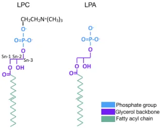
A glycerol backbone, a single carbon chain, and a polar headgroup compose the structure of LPA (Figure 2) (Meyer zu Heringdorf and Jakobs, 2007). Multiple different species of LPA with varying carbon chain lengths and degrees of unsaturation have been identified. Additionally, their carbon chains may be either acyl- or ether-linked with the acyl chain esterified at either the sn-1 or sn-2 position of the glycerol backbone (1-acyl-LPA or 2-acyl-LPA) while ether-linked LPAs carry an alkyl or alkenyl linkage at the sn-1 position (1-alkyl-LPA or 1-alkenyl-LPA). All of these factors contribute to the differing biological activities of individual LPA species (Meyer zu Heringdorf and Jakobs, 2007). The concentration of LPA is approximately 154 pmol in cells, 0.1-6.3 µM in blood and 80-100nM in plasma (Hosogaya et al., 2008; Kishimoto et al., 2003). This lipid has been
detected in many biological fluids including serum, plasma, saliva, follicular fluid, seminal fluid, and malignant effusions (Hama et al., 2002; Sugiura et al., 2002; Tokumura et al., 1999; Westermann et al., 1998). The major cellular sources of LPA include platelets and adipocytes (Eichholtz et al., 1993; Valet et al., 1998), while postmitotic neurons, lymphoid cells, endometrial cells, erythrocytes and cancer cells are also reported to produce LPA (Aoki et al., 2008; Smyth et al., 2008; Ye, 2008). Therefore, LPA may act as a circulating as well as a locally produced paracrine mediator (Takuwa et al., 2002).
Figure 2 Structure of LPC and LPA
Representation of the structures of LPC and LPA, each composed of a glycerol backbone, phosphate group, and fatty acyl chains. LPC has an additional choline headgroup. The lysophospholipase D activity of ATX is responsible for hydrolyzing the bond between choline and the phosphate group of LPC to produce LPA. Adapted from: (Mills and Moolenaar, 2003) with permission from Springer Nature, license # 4382541352781.
1.3.2 Production and degradation of LPA
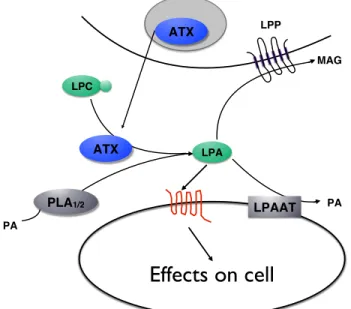
Various enzymes are involved in the production and degradation of LPA as detailed in Figure 3. Intracellular LPA can be produced by de novo LPA biosynthesis through intermediate lipid metabolism (Goetzl and An, 1998). Extracellular LPA can be produced from precursor glycerophospholipids by the action of many different enzymes including phospholipase A1 or A2 (PLA1 or 2), monoacylglycerol kinase or glycerol-3-phosphate acyltransferase (Pébay et al., 2007). For example, PLA1/2 produces LPA by deacylating phosphatidic acid (PA) that is first generated intracellularly from phospholipids or diacylglycerol (Aoki et al., 2008). However, the majority of LPA produced in vivo depends
on the lysophospholipase D activity of autotaxin, which will be discussed in the next subsection, 1.3.2.1 (Umezu-Goto et al., 2002). Additionally, two families of proteins, lipid phosphate phosphatases (LPP) and acyl transferases, are responsible for the rapid degradation of LPA resulting in a short half-life for this lipid mediator. The LPA Acyl transferase (LPAAT) family mediates the acylation of LPA to PA (Yamashita et al., 2001). The LPPs, integral membrane proteins, that dephosphorylate LPA to monoacylglycerol (MAG), are the major LPA degrading enzymes that terminate LPA signaling (see section 1.3.2.2) (Brindley et al., 2002). To counteract these degradative effects, extracellular LPA is normally bound to proteins such as albumin, fatty acid binding protein, or gelsolin which act to increase the stability and facilitate transport of LPA (Aoki, 2004; Gaits et al., 1997; Mills and Moolenaar, 2003; Pagès et al., 2001). Another mechanism regulating LPA levels in the blood is the rapid trans-cellular uptake of LPA into the liver, which therefore acts as an important buffering system controlling LPA bioavailability (Salous et al., 2013).
Figure 3 Mechanisms of LPA production and degradation
The major producer of LPA in vivo is secreted ATX which converts LPC to LPA. Other enzymes can also produce LPA such as PLA1/2, which can produce LPA from PA. LPA may be degraded to PA by the action of LPAAT, or to MAG by the action of LPPs. Adapted from:(Meyer zu Heringdorf and Jakobs, 2007) with permission from Elsevier, license # 4382550417670. ATX ATX
Effects on cell
LPC LPA LPAAT LPP MAG PA PLA1/2 PA1.3.2.1 Autotaxin as the main producer of LPA
Autotaxin (ATX), also known as ENPP2 (ectonucleotide pyrophosphatase/ phosphodiesterase 2), was originally identified as a novel 125-kDa autocrine motility stimulating factor after its isolation from the culture medium of human melanoma cells (A2058) in 1991 (Stracke et al., 1992). It was subsequently found to be present in the culture medium of several other cancer cell types including glioblastoma and breast cancer (Gaetano et al., 2009; Jansen et al., 2005; Kishi et al., 2006). The ATX protein is a secreted protein that is synthesized as a pre-protein and processed by a signal peptidase and pro-protein convertase (such as furin) to remove the pre, N-terminal 27-residue hydrophobic domain, and pro domain of ATX, respectively (Jansen et al., 2005; Koike et al., 2006). The ATX pro-protein follows the classical secretory pathway, where proteins are transported outside the cell from the E.R via the Golgi apparatus (Jansen et al., 2005). Four ATX isoforms have been identified, ATX α, β, γ, and δ with differing tissue distributions (Giganti et al., 2008; Hashimoto et al., 2012). High expression of ATXβ mRNA is found in peripheral tissues, ATXγ mRNA is expressed mostly in brain, ATXα mRNA has lower expression levels in all tissues, and ATXδ is highly expressed in the small intestine and spleen (Giganti et al., 2008; Hashimoto et al., 2012). Originally discovered in A2058 cells, ATXα lacks exon 21, while ATXβ, a splice variant reported in human teratocarcinoma (Lee et al., 1996) lacks exons 12 and 21 and is the major isoform of ATX. ATXγ (PD-1α) was isolated from the brain and lacks exon 12 and ATXδ is identical to ATXβ except for a deletion of four amino acids in the linker region and is the second most common isoform (Giganti et al., 2008; Hashimoto et al., 2012). All isoforms, except ATXα, were found to be fully active with no differences in catalytic efficiency or substrate specificity. The ATXα isoform is cleaved in exon 12, which is unique to this isoform, resulting in a less active cleaved 55-66kDa form (Giganti et al., 2008; Hashimoto et al., 2012).
1.3.2.1.1 Structure and activity of ATX
The ATX protein contains several structural domains illustrated in Figure 4. These include a Modulator of Oligodendrocyte Remodeling and Focal adhesion Organization (MORFO) domain, implicated in oligodendroglial process network formation and focal adhesion organization (Dennis et al., 2008), an EF-hand-like motif that contributes to the function of the MORFO domain, an inactive nuclease-like domain, and two cysteine-rich somatomedin
B domains (Yuelling and Fuss, 2008). The somatomedin B domain (SMB), which is derived from the amino terminus of vitronectin, forms a presumed binding site for type 1 plasminogen activator inhibitor (PAI-1), and uPAR (Seiffert and Loskutoff, 1991; Seiffert et al., 1994). The N-terminal SMB-2 domain has also been shown to bind to β1 and β3 integrins thereby localizing ATX to platelets and cells including lymphocytes (Fulkerson et al., 2011; Hausmann et al., 2011). Additionally, the ATXα isoform can bind to heparin sulfate proteoglycans, due to the arginine/lysine rich clusters in its 53 amino acid polybasic insertion. This isoform was therefore found to bind abundantly to cultured mammalian cells (Houben et al., 2013; Perrakis and Moolenaar, 2014). Finally, ATX has a catalytic domain which has lysophospholipase D (lysoPLD) activity, producing LPA from LPC (Umezu-Goto et al., 2002). The crystal structure of ATX has been solved and reveals a central catalytic domain interacting with the SMB domains on one side and the nuclease domain on the other side (Nishimasu et al., 2011).
Figure 4 Structural domains of ATX
ATX is processed by a signal peptidase and pro-protein convertase to remove pre and pro N-terminal domains. ATX contains a MORFO domain implicated in oligodendrocyte remodeling, an EF hand-like motif, an inactive nuclease-like domain and a putative ATP binding site. ATX also has two somatomedin B domains implicated in binding to integrins. Finally there is a catalytic domain which functions as a lysophospholipase D to produce LPA.
The main physiological substrate for ATX/lysoPLD is LPC, which subsequently produces LPA from LPC by hydrolysis removing the choline head group (Figure 2) (Umezu-Goto et al., 2002). Cyclic phosphatidic acid (cPA), an analog of LPA and intermediate in LPA formation, may also be produced by ATX from LPC (TSUDA et al., 2006). Furthermore, ATX/LysoPLD has a higher affinity for unsaturated acyl-LPCs as compared to saturated or