UNIVERSITE DE LIEGE
FACULTE DE MEDECINE VETERINAIRE
DEPARTEMENT DE PATHOLOGIE ET AUTOPSIES
SERVICE DE PATHOLOGIE SYSTEMIQUE
ETUDE DE LA PATHOGENIE DE L’INFECTION PAR LE VIRUS
USUTU IN OVO ET IN VIVO
STUDY OF THE PATHOGENISIS OF USUTU VIRUS INFECTION
IN OVO AND IN VIVO
Emna BENZARTI
THESE PRESENTEE EN VUE DE L’OBTENTION DU GRADE DE
DOCTEUR EN SCIENCES VETERINAIRES
Remerciements
Je remercie Allah pour tout ce que je suis et tout ce que j’ai.
Je tiens tout d’abord à remercier les Professeurs Mutien-Marie Garigliany et Daniel Desmecht pour m’avoir accueillie au sein du laboratoire de Pathologie et autopsies de la Faculté de Médecine vétérinaire de l'Université de Liège.
Pour son professionnalisme, sa disponibilité et sa confiance, je remercie encore le Professeur Mutien-Marie Garigliany.
Pour leur proche collaboration et leur aide précieuse dans le laboratoire, un merci tout particulier à Michaël Sarlet, Mathieu Franssen et Felipe Rivas,.
Un très grand merci à Nathalie Guillaume et Anne Wlasowzki pour la gestion des problèmes administratifs et leurs précieux conseils. Merci à Nathalie pour sa relecture attentive de ce manuscrit.
Merci aux autres membres du service pour les discussions constructives : Calixte, Adrienne, Dominique, Etienne, Michel, Audrey, Anne-Sophie, Caroline, Iskra, Thierry et Gautier.
Un grand merci aux membres du réseau de Surveillance de la Faune Sauvage de la Faculté de Médecine vétérinaire de l'Université de Liège pour leurs efforts de collecte de cadavres d’oiseaux référés à notre service.
A papa, maman, mon âme-sœur Ahmed, mon frère Haithem et mes sœurs Ihsène, Zeineb et Habiba, pour leur amour inconditionnel, pour le soutien de tous les jours et pour avoir toujours cru en moi.
Abbreviations
2AFAMD: 2 A peptide of Food And Mouth Disease virus aa: Amino acidADE: Antibody-dependent enhancement AMP: Adenosine monophosphate ATP: Adenosine triphosphate BAGV: Bagaza virus
BBB: Blood-brain-barrier C: Capsid
CAM : Chorio-allantoic membrane
CARD: Caspase activation and recruitment domains CCR1: Capsid-coding region 1
cDNA: Complementary DNA CFW: Carworth Farms White cGAS: Cyclic GMP-AMP synthase cHP: Capsid-coding region hairpin CLR: C-type lectin receptor CM: Convulted membrane CNS: Central nervous system CS: Cyclization sequence DAR: Downstream AUG region DB: Dumbbells structures Dc: Dendritic cell
DC-SIGN: Dendritic Cell-Specific Intercellular adhesion molecule-3-Grabbing Non-integrin DENV: Dengue virus
DI: Domain I DII: Domain II DIII: Domain III
DMEM: Dulbecco's minimal essential medium DNA: Desoxyribonucleic acid
E: Envelope
ECE: Embryonated chicken eggs eIF4F: Eukaryotic initiation factor
ELISA: Enzyme-Linked Immunosorbent Assay ER: Endoplasmic reticulum
GFP: Green fluorescent protein Gluc: Gaussia luciferase
GMP: Guanosine monophosphate
HALT: Head Associated Lymphoid Tissue
HDVr/SV40pA: Hepatitis delta virus ribozyme / Simian virus 40 polyadenylation signal HEK293: Human embryonic kidney 293 cells
HSP: Heat Shock protein IC: Intracranial
ICAM: Intercellular Adhesion Molecule
ICTV: International Committee on Taxonomy of Viruses ID: Intradermal
IFN: Interferon
IFNAR: Interferon-Alpha/Beta Receptor IHC: Immunohistochemistry
IKK: Inhibitor of nuclear factor kappa B kinase IN: Intranasal
IP: Intraperitoneal
IPS: IFN-β promoter stimulator
IRAK: Interleukin-1 receptor-associated kinase IRES: Internal ribosome entry site
IRF: Interferon regulatory factors ISA: Infectious subgenomic amplicons ISG: Interferon Stimulated Gene
ISRE: IFN-stimulated response elements ITV: Israel Turkey meningoencephalitis Virus JAK: Janus kinase
JEV: Japanese Encephalitis virus Kb: Kilobase
kDa: Kilodalton M: Membrane
MAVS: Mitochondrial antiviral-signaling protein MDA5: Melanoma differentiation-associated gene 5 MHC: Major histocompatibility complex
MIP: Macrophage Inflammatory Protein MMP: Matrix metalloproteinase
mosGCTL-1: Mosquito galactose-specific C-type lectin MR: Mannose receptor
mRNA: messager RNA
MyD88: Myeloid differentiation primary response 88
NF-kB: Nuclear factor kappa-light-chain-enhancer of activated B cells NK: Natural Killer
NMRI: Naval Medical Research Institute NPC: Neural progenitor cells
NS: Non-structural
OAS: Oligoadenylate synthetase ORF: Open reading frame PABP: Poly-A-binding protein PBS: Phosphate Buffered Saline PCR: Polymerase chain reaction PFU: Plaque forming unit PK: Porcine kidney prM: Membrane precursor
RCS: Repeated conserved sequence RIG: Retinoic acid-inducible gene RIP-1: Receptor-interacting protein 1 RNA: Ribonucleic acid
RSVP: Recombinant sub-viral particles
RT-PCR: Reverse transcription-polymerase chain reaction
RT-qPCR: Reverse transcription-quantitative polymerase chain reaction sHP: Small hairpin
SLA: Stem Loop A SLB: Stem Loop B
SPF: Specific pathogen-free
STAT: Signal Transducer and Activator of Transcription STING: Stimulator of interferon gene
SVP: Sub-viral particles TBK: TANK-binding kinase
TBEV: Tick-borne encephalitis virus TCID50: 50% Tissue infective dose TIA: T-lymphocyte Internal Antigen
TIAR: T-lymphocyte Internal Antigen Receptor TIM: T cell Immunoglobulin Mucin domain TL: Terminal Loop
TLR: Toll-like receptor TMUV: Tembusu virus TNF: Tumor Necrosis Factor TPR: Tetratricopeptide repeat
TRIF: TIR-domain-containing adapter-inducing interferon-β Tyk: Tyrosine Kinase
UAR: Upstream AUG regions UFS: UAR-flanking stem
UPR: Unfolded protein response USUV: Usutu virus
UTR: Untranslated Region VB: Virus bags
VP: Vesicle packets vRNA: viral RNA WNV: West Nile virus YFV: Yellow Fever virus ZIKV: Zika virus
Résumé – Abstract ...1
General preamble ...5
Introduction ...7
1. MOSQUITO-BORNE FLAVIVIRUSES PATHOGENIC FOR BIRDS ... 8
1.1 OVERVIEW ... 8
1.2 MANUSCRIPT N°1-MOSQUITO-BORNE EPORNITIC FLAVIVIRUSES: AN UPDATE AND REVIEW ... 10 2. EPIDEMIOLOGY ... 26 2.1 GEOGRAPHICAL DISTRIBUTION ... 26 2.2 PHYLOGENY ... 27 2.3 TRANSMISSION CYCLE ... 29 2.3.1 Vectors ... 31 2.3.2 Reservoirs ... 31 2.3.3 Accidental hosts ... 31
3. GENOMIC ORGANIZATION AND VIRAL PROTEINS ... 32
3.1 GENERAL GENOMIC ORGANIZATION ... 32
3.2 VIRAL PROTEINS ... 33
3.2.1 Structural proteins ... 34
3.2.2 Non-structural proteins ... 36
3.3 NON-CODING REGIONS ... 37
3.3.1 Non-coding region in 5’ (5’UTR) ... 38
3.3.2 Non-coding region in 3’ (3’UTR) ... 38
4. PATHOGENESIS OF THE FLAVIVIRAL INFECTION ... 38
4.1 4.1VIRAL CYCLE ... 39
4.1.1 Fixation of the virus to cellular receptors and internalization by endocytosis ... 39
4.1.2 Release of the viral RNA after decapsidation ... 42
4.1.3 Translation and replication of the vRNA ... 42
4.1.5 Maturation of virions in the Golgi apparatus ... 44
4.1.6 Release of virus particles in the extracellular compartment ... 45
4.1.7 Replication factories ... 45
4.2 THE VERTEBRATE HOST ANTIVIRAL RESPONSE ... 46
4.2.1 Innate immune response ... 46
4.2.2 Adaptive immune response ... 52
4.2.3 Cellular stress, autophagy and apoptosis ... 56
4.3 USUV INFECTION ... 57
4.3.1 Vectors ... 57
4.3.2 Humans and other mammals ... 59
4.3.3 Birds ... 61
4.4 EXPERIMENTAL MODELS FOR THE STUDY OF USUV ... 61
4.4.1 Cellular models ... 62
4.4.2 In ovo models ... 65
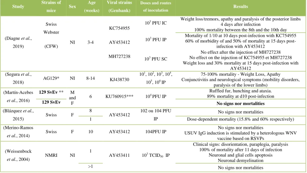
4.4.3 Vertebrate animal models ... 65
5. LABORATORY DIAGNOSIS ... 68
6. PREVENTION AND CONTROL OF USUV ... 69
6.1 ANTIVIRAL MOLECULES AGAINST USUV... 69
6.2 VACCINATION ... 69
Objectives ...71
Experimental section ...75
Study 1: Pathogenesis of USUV infection in spontaneous cases in wildlife and monitoring of the virus evolution ...76
Manuscript n°2: Usutu Virus Epizootic in Belgium in 2017 and 2018 : Evidence of Virus Endemization and Ongoing Introduction Events ...79
scoter (Melanitta nigra) ...88
Study 2: Development of experimental models of USUV infection ...93
a) Validation of the first in ovo model of USUV infection ...94
Manuscript n°4: Usutu virus infection of embryonated chicken eggs and a chicken embryo-derived primary cell line ………...………..96
b) Establishment of an in vivo avian model: Serinus canaria………..…..113
Manuscript n°5: Experimental Usutu Virus Infection in Domestic Canaries Serinus canaria ...116
c) Establishment of mammalian in vivo model (129/Sv Mouse)……….………131
Manuscript n°6: New insights into the susceptibility of immunocompetent mice to Usutu virus…134 General discussion – perspectives ...148
Supplemental material ...158
Appendix 1 ...159
Appendix 2 ...177
Annex ...180
1
Résumé
Le virus Usutu (USUV), un flavivirus zoonotique transmis par les moustiques et découvert en 1959 en Afrique de Sud, s’est propagé au cours des vingt dernières années sur une grande partie du continent européen, provoquant des mortalités aviaires importantes. Chez l’homme, l’infection est le plus souvent asymptomatique, ou d’une expression clinique bénigne. Toutefois, des complications neurologiques, telles que des encéphalites ou méningoencéphalites, ont été décrites, similaires àc equi est décrit pour le virus West Nile (WNV), un flavivirus apparenté au USUV. L’histoire récente de flambées épidémiques d’autres arboviroses invite la communauté scientifique à la plus grande vigilance quant à l’évolution génétique de ce virus, même si, à ce jour, les cas humains restent exceptionnels. Par ailleurs, le USUV présente une grande proximité sur le plan génétique avec les autres membres du sérogroupe de l’encéphalite japonaise (dont le WNV) et les autres flavivirus transmis par les moustiques, dont le virus de la Dengue et le virus Zika. A ce titre, le USUV constitue un modèle d’étude de premier plan pour la compréhension de la pathogénie et le développement de solutions prophylactiques et thérapeutiques pour ces flavivirus proches. En effet, il est le seul membre de ce groupe qui puisse être manipulé en conditions de biosécurité de niveau 2, les souches de terrain sont facilement accessibles et présentent un haut degré de variation génétique naturelle. En dépit de ces avantages, les connaissances concernant la physiopathologie de ce virus émergent sont, pour l’heure, très sommaires. Nos travaux ont, donc, visé à mieux comprendre la pathogenèse de son infection, en combinant une approche descriptive de cas spontanés chez des oiseaux sauvages et le développement de modèles expérimentaux.
Ayant débuté ce travail en pleine épizootie du USUV chez les oiseaux sauvages en Belgique en 2016, nous avons entrepris une étude descriptive et systématique de ces cas spontanés. Nous avons mis en évidence l’endémISAtion de ce virus en Belgique, avec la survenue fréquente d’épizooties aviaires en 2017 et 2018 et une co-circulation de souches génétiquement variables et en constante évolution. De plus, nous avons élargi le spectre d’hôtes au sein des hôtes aviaires, en détectant le virus chez une nouvelle série d’espèces, notamment la macreuse noire (Melanitta nigra), qui constitue, à l’heure actuelle, la seule espèce de la famille des Anatidae qui est hautement sensible au virus. Nous avons, également, isolé des souches virales de terrain qui nous ont permis d’établir des modèles d’infection. Ensuite, nous avons testé la sensibilité au virus de deux modèles aviaires d’infection, un modèle Gallus gallus in ovo et un modèle in vivo, le canari domestique (Serinus canaria) et d’un modèle « mammifère », les souris 129/Sv. Nous avons réussi à démontrer que, contrairement aux résultats de trois études indépendantes menées par des équipes européennes renommées, le USUV est capable, non seulement de se répliquer dans les œufs embryonnés de poulet, mais aussi d’éliciter une
des cellules de ce tissu et évalué la cinétique de réplication des souches virales en utilISAnt ce modèle
in vitro. Nous avons, ensuite, établi le premier modèle in vivo, le canari domestique, adéquat pour
l’étude du USUV et sa transmission. Enfin, notre infection expérimentale pilote des souris immunocompétentes 129/Sv a conclu à la pertinence de ce modèle murin pour l’étude de la neuroinvasivité du USUV et de la possibilité d’une transmission directe chez les mammifères.
Dans l’ensemble, à travers l’examen d’oiseaux infectés naturellement ou de différents modèles in ovo et in vivo infectés au laboratoire, nous avons réussi à mettre en exergue des différences majeures dans la pathogénie de l’infection par le USUV, selon qu’il s’agisse d’hôtes aviaires ou mammifères, ou même entre espèces aviaires différentes. Nous pensons que l'utilISAtion future de ces modèles favorisera une compréhension significative de la neuropathogenèse induite par le USUV et de sa réponse immunitaire et permettra le développement futur de médicaments et de vaccins contre le USUV ou d’autres virus apparentés d’importance zoonotique majeure, en bénéficiant de l’avantage de l’immunité croisée entre ces virus.
Summary
Usutu virus (USUV), a mosquito-borne zoonotic flavivirus discovered in South Africa in 1959, has spread to many European countries over the last twenty years, causing significant bird mortalities. Human infections most often remain asymptomatic, or with a benign clinical expression. However, neurological complications, such as encephalitis or meningoencephalitis, have been described, reminiscent of infections with West Nile Virus (WNV), a USUV-related flavivirus. The recent history of outbreaks linked to other arboviruses invites the scientific community to be extremely vigilant about the genetic evolution of USUV, even if, to date, human cases remain exceptional. In addition, USUV is genetically very close to other members of the Japanese encephalitis serogroup (including WNV) and other mosquito-borne flaviviruses, including Dengue virus and Zika virus. As such, USUV is a leading model for the study of the flaviviral pathogenesis and the development of prophylactic and therapeutic solutions against these more pathogenic flaviviruses. Indeed, it is the only member of this group that can be handled under level 2 biosafety conditions, field strains are easily accessible and have a high degree of natural genetic variation. Despite these advantages, knowledge about the pathophysiology of this emerging virus is, for the moment, very sketchy. Our work has, therefore, aimed to better understand the pathogenesis of its infection, by combining a descriptive approach of spontaneous cases in wild birds and the development of experimental models.
Indeed, since this work started during the USUV epizootic in wild birds in Belgium in 2016, we undertook a descriptive and systematic study of these spontaneous cases. We have highlighted the endemization of this virus in Belgium, with the frequent occurrence of avian epizootics in 2017 and 2018 and a co-circulation of genetically variable and constantly evolving strains. In addition, we have expanded the avian host spectrum by detecting the virus in a new series of species, including the common scoter (Melanitta nigra), which is currently the only known species of the Anatidae family that is highly susceptible to the virus. We also isolated several field viral strains which allowed us to properly establish models of infection. Then, we tested the susceptibility to the virus of two avian models, a Gallus gallus model in ovo and an in vivo model, the domestic canary (Serinus canaria), and a "mammalian" model, 129/Sv mice. We succeeded to demonstrate that, unlike the results of three independent studies conducted by renowned European teams, USUV is able not only to replicate in embryonated chicken eggs but also to elicit a marked virulence and an extended cellular tropism within the chick embryo. Subsequently, as we found that the chorioallantoic membrane was a site of predilection for viral replication, we isolated cells from this tissue and evaluated the replication kinetics of viral strains using this model in vitro. We, then, established the first avian in vivo model,
infection of 129/Sv immunocompetent mice concluded that this murine model is useful for the study of USUV neuroinvasivity and its possible direct transmission in mammals.
Overall, through the examination of naturally infected birds and different in ovo and in vivo models in the laboratory, we highlighted major differences in the pathogenesis of USUV infection, according to avian or mammal hosts, or even between different avian species. We believe that the future use of these models will promote a significant understanding of the USUV-induced neuropathogenesis and its immune response and allow the future development of drugs and vaccines against USUV or other related viruses of major zoonotic importance, based on the known cross-immunity between these viruses.
USUV is an arbovirus of the family Flaviviridae and genus Flavivirus. Among these viruses are some of the most important arboviruses for humans, such as the dengue virus (DENV), Zika virus (ZIKV), yellow fever virus (YFV), West Nile virus (WNV), or the Japanese encephalitis virus (JEV).
Responsible for recurrent epizootics since 1996 in the European avifauna, USUV is now recognized as the causative agent of potentially severe neurological disorders in humans. Its recent geographic spread to a large number of European countries, the frequent occurrence of USUV-associated bird epizootics and the co-circulation of several lineages of genetically different strains warrant specific research studies.
In this thesis, USUV surveillance in Belgium and research models were developed. The aim of this thesis was to better understand the pathogenesis of the infection with this virus linked to the genetic diversity of its strains. In this manuscript, we aim to (1) make a state of the art knowledge about USUV and other mosquito-borne flaviviruses pathogenic for birds and to (2) investigate the pathogenesis of USUV infection in naturally-infected birds or using laboratory models.
1. Mosquito-borne flaviviruses pathogenic for birds
1.1 Overview
According to the World Health Organization, 61% of all human pathogens are of an animal origin, and 75% of the emerging animal diseases can be transmitted to humans (WHO, 2006). Except for some emerging zoonoses such as the Severe Acute Respiratory Syndrome and the Highly Pathogenic Avian Influenza H5N1, the vast majority of zoonotic diseases are strikingly not a priority in health systems at both national and international levels and are, therefore, considered as neglected (WHO, 2006). Among these neglected pathogens, many are vector-borne, among which the arboviruses. The most important arboviroses in human health are those caused by the Alphavirus (Togaviridae family, including for example the Chikungunya virus), Orthobunyavirus and
Phlebovirus (Bunyaviridae family, including the California encephalitis virus and Rift Valley Fever
virus, respectively) and Flaviviruses (family Flaviviridae). Recent epidemics associated with the Zika virus (ZIKV) in South America, Chikungunya virus in the Indian Ocean or the West Nile Virus (WNV) in North America or Europe (Bakonyi et al., 2013) illustrate the severe consequences of the emergence of these neglected arboviruses for both public health and animal health.
The Flaviviridae family includes four genera: Flavivirus, Pestivirus, Pegivirus, and
Hepacivirus (Simmonds et al., 2017). The genus Flavivirus is the largest of the four, including more
than 70 species (MacKenzie and Williams, 2009), the majority of which are zoonotic arboviruses. This genus is divided into three distinct groups: mosquito-borne (about 50%), tick-borne (28%) and those whose vector is, to date, unknown (Moureau et al., 2015; Simmonds et al., 2017). The group of mosquito-borne viruses can be subdivided according to their reservoir/vector into two clades (ICTV, 10th report). Aedes clade viruses (anthropophilic mosquitoes), such as the Dengue (DENV), yellow fever (YFV) or ZIKV, have a primate reservoir and are responsible in most cases for hemorrhagic diseases in humans. Culex (mosquitoes that may feed on birds and many mammalian hosts) clade viruses have an avian reservoir, are neurotropic and frequently cause meningoencephalitis (Lindenbach et al., 2013; Mazeaud et al., 2018; Mazzon et al., 2009). Among these viruses are the WNV, Japanese encephalitis virus (JEV) and the Usutu virus (USUV).
From the antigenic side, flaviviruses are divided into 8 different serocomplexes (Simmonds et
al., 2017). A serocomplex is defined as a group of viruses sharing common antigenic sites on their
surface, which promotes serological cross-reactions (ICTV, 10th report). The JEV serocomplex includes 8 viral species, including the WNV and USUV (Table 1). The Ntaya serocomplex contains the Israel turkey meningoencephalitis virus (ITV), Bagaza virus (BAGV) and Tembusu virus (TMUV). These five viruses are the only mosquito-borne flaviviruses considered as “epornitic” (capable of causing epizootics in birds) (ICTV, 10th report).
Table 1: Main Characteristics of viruses from the JEV serocomplex
Virus Geographical
distribution Reservoir Main vector Disease
Cacipacoré virus Brazil Birds? Culex spp? A human case with
febrile syndromes
Japanese
encephalitis virus Asia Birds - pigs Culex spp
Neurological in humans, cattle and horses
Koutango virus Senegal - Central African Republic- Somalia Rodents? Ticks? Aedes aegypti? Neurological in experimental mice Murray Valley encephalitis virus Australia et New
Guinea Birds Culex spp
Neurological in humans and horses
Saint Louis encephalitis virus
United States of
America Birds Culex spp
Neurological in humans and horses
Usutu virus
Africa - Europe and the Middle
East
Birds Culex spp
Systemic in birds. Neurological (rare) in
humans
West Nile virus Worldwide Birds Culex spp
Systemic in birds. Neurological (rare) in
humans and horses
Yaoundé virus Cameroon - Ghana Birds Culex
1.2 Manuscript n°1 - Mosquito-borne epornitic flaviviruses: an update and review
Some vertebrates, such as birds, play the role of "reservoirs" in the flavivirus epidemiological cycle. They are hosts capable of replicating and transmitting the virus to other ones, thus maintaining the virus in the environment. These reservoir hosts can sometimes show clinical signs, even mortalities, following the infection. This has been described in birds infected with five mosquito-borne flaviviruses: WNV, USUV, ITV, BAGV, and TMUV. These arboviruses have had a significant impact on the health of birds and the poultry industry and are capable of infecting humans (Bondre et
al., 2009; Colpitts et al., 2012; Gaibani and Rossini, 2017; Tang et al., 2013b), except the ITV.
After a careful reading of the scientific literature on these flaviviruses, we found that there was no comprehensive review of virological, epidemiological, pathological, and prophylactic data for this particular group of viruses. Consequently, we developed a review of the literature incorporating these aspects. In particular, we analyzed different results from reports on the circulation of these pathogens in order to describe the specific host tropism of each of these viruses. In addition, by analyzing studies of vaccine candidates targeting these viruses in avian hosts, we developed an update on the advancement in the prophylactic strategies against these pathogens, for which there is currently no etiological treatment. This review is published in the Journal of General Virology*.
Mosquito-borne epornitic flaviviruses:
an update and review
Introduction
Review
J Gen Virol
100(14):119-32Benzarti Et al., Journal of General Virology 2019;100:119–132 DOI 10.1099/jgv.0.001203
Mosquito-borne epornitic flaviviruses: an update and review
Emna Benzarti,1 Annick Linden,2 Daniel Desmecht1 and Mutien Garigliany1,* Abstract
West Nile Virus, Usutu virus, Bagaza virus, Israel turkey encephalitis virus and Tembusu virus currently constitute the five flaviviruses transmitted by mosquito bites with marked pathogenicity for birds. They have been identified as the causative agents of severe neurological symptoms, drop in egg production and/or mortalities among avian hosts. They have also recently shown an expansion of their geographic distribution and/or a rise in cases of human infection. This paper is the first up-to-date review of the pathology of these flaviviruses in birds, with a special emphasis on the difference in susceptibility among avian species, in order to understand the specificity of the host spectrum of each of these viruses. Furthermore, given the lack of a clear prophylactic approach against these viruses in birds, a meta-analysis of vaccination trials conducted to date on these animals is given to constitute a solid platform from which designing future studies.
INTRODUCTION
West Nile virus (WNV), Usutu virus (USUV), Tembusu virus (TMUV), Bagaza virus (BAGV) and Israel turkey meningo-encephalitis virus (ITV) are positive-sense, single-stranded RNA viruses, included in the mosquito-borne clus-ter of the genus Flavivirus, family Flaviviridae [1]. Their natural life cycle mainly involves birds and mosquitoes, whereas humans and other vertebrates are considered inci-dental hosts [2–5]. A remarkable hallmark of these arbovi-ruses is their ability to invade new territories. The most recent examples of this feature are the introduction into Europe of USUV in 1996 [6], WNV lineage 2 in 2004 [7], BAGV in 2010 [8] and of TMUV into China in 2010 [9]. In avian hosts, these flaviviruses are considered as epornitic (capable of causing epizootics in birds). Consequently, we will refer to them in this review as mosquito-borne epornitic flaviviruses (MBEF). MBEF have been detected in an increasing number of bird species and can be deadly for a wide range of them. Moreover, when poultry flocks become infected by ITV and TMUV, high mortality, drop in egg production and heavy control measures constitute an eco-nomic burden for the infected countries.
Besides their impact on bird health and the poultry industry, MBEF are capable of infecting humans [10–13], except ITV,
of which the zoonotic potential is still to be determined. Most human infections remain asymptomatic, but symptoms rang-ing from transient flu-like syndrome (fever, headache) to severe neurological illness and death can be observed in some cases of WNV and USUV infections [13, 14].
In this article, we will review the genome structure, classifi-cation, eco-epidemiology, pathology and preventive meas-ures related to MBEF. We will list avian species currently known to be susceptible to the infection and we will provide an overview of vaccination trials conducted to date on birds to boost their immune system against these viruses.
Genome structure
The MBEF group are positive-sense, single-stranded RNA viruses [15]. Spherical and enveloped virions measure 40– 60 nm in diameter [1]. Their ~11 kb viral RNA genome contains a unique open reading frame (ORF) flanked by a capped 5¢-terminal non-coding region (NCR) and a 3¢-5¢-terminal NCR (Fig. 1). The two NCRs form specific secondary structures necessary for genome replication and translation and are implicated in the pathogenicity of flaviviruses [16]. The single polyprotein encoded by the ORF is processed by viral and host proteases into three structural and seven non-structural proteins [1]. The structural proteins comprise: (1) an envelope protein E, involved in receptor binding, viral entry and
Received 17 July 2018; Accepted 29 November 2018; Published 22 January 2019
Author affiliations: 1FARAH Research Center, Department of Pathology, Faculty of Veterinary Medicine, University of Liege, Sart Tilman B43, B-4000 Liege, Belgium; 2FARAH Research Center, Surveillance Network for Wildlife Diseases, Faculty of Veterinary Medicine, University of Liege, Sart Tilman B43, B-4000 Liege, Belgium.
*Correspondence: Mutien Garigliany, mmgarigliany@uliege.be Keywords: birds; flaviviruses; epornitic; mosquitoes; vaccine.
Abbreviations: AMCR, American crow; BAGV, Bagaza virus; CNS, central nervous system; C protein, capsid protein; E protein, envelope protein; HOSP, house sparrow; IFN, interferon; ITV, Israel Turkey meningo-encephalitis virus; MBEF, mosquito-borne epornitic flaviviruses; M protein, membrane protein; NCR, non-coding region; NK, natural killer; NS, non-structural; ORF, open reading frame; PAMP, pathogen-associated molecular patterns; prM, membrane precursor; SPF, specific-pathogen-free; TE, truncated envelope protein; TMUV, tembusu virus; USUV, usutu virus; WNV, West Nile virus.
, Journal of General Virology 2019;100:119–132
Fig. 1. Virion structure and genomic organization of epornitic mosquito-borne flaviviruses. The single-stranded, positive-sense RNA genome contains a single unique ORF, encoding for a polyprotein which is processed into three structural proteins (C, PrM, and E) and seven non-structural proteins (NS1, NS2A, NS2B, NS3, NS4A, NS4B, and NS5). UTR, untranslated transcribed region.
membrane fusion; (2) a membrane protein M, which results from the cleavage of a membrane precursor prM upon matu-ration of the virion; and (3) a capsid protein C, involved in the assembly of the nucleocapsid and its incorporation into new virions [17]. The E protein carries both flavivirus cross-reac-tive and virus-specific epitopes, and hence it constitutes the
can be used to increase secretion of the E protein ectodomain, carrying major immunogenic epitopes [18]. The prM protein protects the E protein from premature fusion during the exocytosis of viral particles and participates in the folding and assembly of viral particles [1]. The prM-E proteins of fla-viviruses can self-assemble into subviral particles, which share
The non-structural proteins (NS1, NS2A, NS2B, NS3, NS4A, NS4B, and NS5) regulate RNA transcription and replication [1], determine virus evasion mechanisms from the host immune system (e.g. limit interferon (IFN) gene expression by attenuating the signaling through the JAK/ STAT pathway) [19, 20] and play an important role in avian host competence and virulence [21, 22]. Among these proteins, NS3 is a serine protease that cleaves NS2A/B, NS2B/ NS3, NS3/NS4A and NS4B/NS5 [20]. This protein also has RNA helicase activity, allowing the genome to be unwound during viral replication, and RNA triphosphatase activity, involved in the dephosphorylation of the 5¢ end of the genome before the addition of a cap [1]. NS5 is a highly conserved protein among flaviviruses and is also a multi-functional protein: at the N-terminus, it has methyltransfer-ase activity required for the formation of mRNA (RNA capping); and at the C-terminus, it has an RNA-dependent– RNA-polymerase activity necessary for copying genomic RNA [1].
Lineages and strains
The MBEF members belong to the genus Flavivirus, family Flaviviridae [1]. This family is divided, according to the transmission routes of its members, into three clusters (Fig. 2): (1) arthropod-borne viruses, transmitted horizon-tally by mosquito or tick bites to vertebrate hosts and thus considered as dual-host viruses; (2) unknown vector flaviviruses, also called vertebrate-specific flaviviruses, presumed to infect only rodents or bats; and (3) insect-specific or mosquito-only flaviviruses that can replicate only in insects, especially mosquitoes [23].
The most important flaviviruses in regard to humans and animals belong to the first cluster, for which birds can act as the reservoir [23]. Among these, some are transmitted by ticks, mostly Ixodes sp. [24], and can severely impact the health of human (e.g. Tick-borne encephalitis virus) [25] or avian hosts, such as Louping-ill virus, which is deadly for the red grouse (Lagopus lagopus) [26].
Other arthropod-borne flaviviruses are transmitted by mos-quitoes, with some being non-pathogenic for birds but highly virulent in humans, such as Murray Valley encepha-litis virus [27] and Saint Louis encephaencepha-litis virus [28]. WNV, USUV, TMUV, BAGV, and TMEV are the only mosquito-borne viruses having known pathogenicity for birds (Table 1).
The MBEF members are serologically classified within two different groups: (1) the Japanese encephalitis serocomplex, including WNV and USUV, and (2) the Ntaya serocomplex, including AMEV and TMUV [29, 30] (Table 1).
Viruses from the Japanese encephalitis serocomplex: USUV and WNV
Isolates of USUV are currently classified into eight lineages (Africa 1, 2 and 3 and Europe 1, 2, 3, 4 and 5) [31]. Molecu-lar studies on nucleotide and amino acid sequences of these isolates from vectors, birds, and humans reveal significant
substitutions, some of which have been suggested as being related to viral neuro-invasiveness [32]. The effective role of such candidate mutations in the development of both viral infectivity and virulence remains to be determined. At present, up to nine lineages have been proposed to classify WNV strains [33]. Lineage 1 is subdivided into clades 1a and 1b (or Kunjin virus) and 1 c [34], and is the most widespread in the USA (NY99 strain), Africa (KN3829), Europe and the Middle East [33]. Virulence is highly variable among WNV lineages. For instance, lineage 3 (Rabensburg virus) has never been isolated from humans and did not experimentally infect mammalian or avian cell cultures, the house sparrow (Passer domesticus) (HOSPs) or specific-pathogen-free (SPF) chicken eggs [35]. On the contrary, WNV lineages 1 and 2 have been responsible for major outbreaks in animals and humans [35, 36]. Viral strains from the same lineage (and clade) can also express variation in pathogenicity. For instance, despite the high genetic relatedness between strains KN3829 and NY99 (a total of 11 amino acid differences between the strains) [22], the latter exhibits a strikingly different avian virulence phenotype, eliciting significantly higher viremia and mortality in the American crow (Corvus brachyrhyncos; AMCR) [22, 37]. A mutation in the NS3 gene resulting in a T249P amino acid substitution was involved in increased pathogenicity in AMCR [38], and this mutation was proposed as a key determinant of WNV pathogenicity. Furthermore, the NS3-249 residue was shown to be under strong positive selective pressure because birds can drive adaptive evolution in WNV [38]. However, the mere presence of Pro at NS3-249 was neither sufficient nor necessary to enhance the virulence of WNV strains in theHOSP [39, 40], red-legged partridge [41] and SPF chicken [42]. Variation in virulence for avian species in regard to this mutation remains unexplained. Nonetheless, one study showed that WNV virulence in AMCR is corre-lated with increased ATP hydrolysis due to direct interaction between the NS3-249 residue and unknown host factors [43]. Helicase activity, however, did not differ between NS3 proteins with proline or threonine at position 249, and thus could not explain the in vivo effects in AMCR [43]. Other studies showed that the NS3-249 residue modulates replication in avian leukocytes [22, 44] and hence could affect the host immune response in a temperature-dependent manner and under the control of NS proteins [22].
Viruses from the Ntaya serocomplex: BAGV, ITV, and TMUV
The BAGV strains comprise isolates from the Central African Republic, India, and Spain, with high nucleotide identity (>92 %) [8]. ITV includes strains from Israel and South Africa, with <0.9 % of divergence [30]. Both viruses have shown cross-neutralization activity and nucleotide sequence identity >84 % and were proposed to be considered as a single virus species, named avian meningoencephalitis virus [23, 30]. However, the Interna-tional Committee on Taxonomy of Viruses species
, Journal of General Virology 2019;100:119–132
Fig. 2. Phylogeny of conserved partial gene sequences coding for the non-structural protein 5 of certain representative strains from the family Flaviviridae. ClustalW (implemented in Geneious 10.2.3) was used to create multiple alignments for the sequences. The phylogenetic tree was constructed from the sequence alignment by the maximum likelihood method based on the Kimura 2-parameter model [149] with a gamma distribution (five categories) and invariant sites (G+I) computed with MEGA 7 [150]. The tree is drawn to scale, with branch lengths measured according to the number of substitutions per site. Data were bootstrap re-sampled 500 times; values 70 % are shown next to the branches. Mosquito-borne epornitic flaviviruses are framed.
demarcation criteria for viruses of the genus Flavivirus include geographic, vector, host and disease associations and ecological characteristics [45] and, thus, these viruses should still be considered as separate species [15] because they differ in some of these aspects (Table 1). TMUV is a genetically distinct member of the Ntaya virus group and includes highly homologous isolates that were previously considered separate virus species, including the Sitiwan
Genetic features underlying the infection and disease out-come associated with these viruses are still poorly under-stood. Recently, N-glycosylation on residue 154 of TMUV E protein has appeared as a determinant of pathogenicity in ducks, as shown for WNV in other avian species [50–53]. In fact, an S156P mutation in the E protein of one TMUV strain (FX2010) resulted in the loss of the E-glycosylation motif, leading to limited virus replication and the
, Journal of General Virology 2019;100:119–132
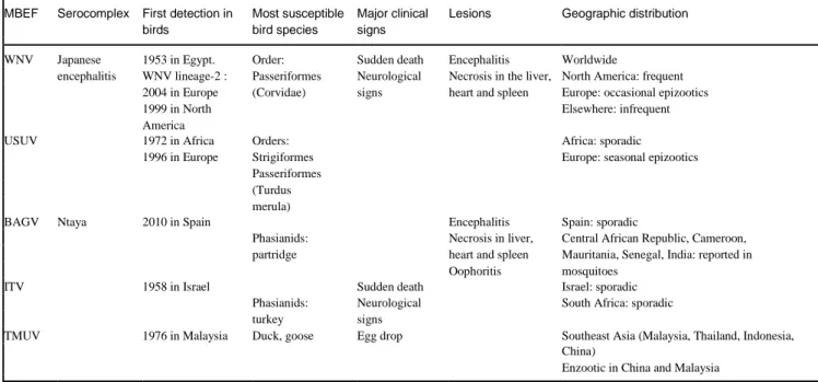
Table 1. Epornitic mosquito-borne flaviviruses: classification and main epidemiological and pathological features
MBEF Serocomplex First detection in Most susceptible Major clinical Lesions Geographic distribution birds bird species signs
WNV Japanese 1953 in Egypt. Order: Sudden death Encephalitis Worldwide
encephalitis WNV lineage-2 : Passeriformes Neurological Necrosis in the liver, North America: frequent 2004 in Europe (Corvidae) signs heart and spleen Europe: occasional epizootics
1999 in North Elsewhere: infrequent
America
USUV 1972 in Africa Orders: Africa: sporadic
1996 in Europe Strigiformes Europe: seasonal epizootics Passeriformes
(Turdus merula)
BAGV Ntaya 2010 in Spain Encephalitis Spain: sporadic
Phasianids: Necrosis in liver, Central African Republic, Cameroon, partridge heart and spleen Mauritania, Senegal, India: reported in
Oophoritis mosquitoes
ITV 1958 in Israel Sudden death Israel: sporadic
Phasianids: Neurological South Africa: sporadic turkey signs
TMUV 1976 in Malaysia Duck, goose Egg drop Southeast Asia (Malaysia, Thailand, Indonesia, China)
Enzootic in China and Malaysia
Geographic repartition USUV
USUV was detected for the first time in 1959, by B.R. McIn-toch, from Culex naevi (historically named Culex univitatus) captured near the Usutu river in Swaziland, South Africa [55]. The virus was later detected in mosquitoes in several African countries until its identification as the causative agent of mass mortality in the Eurasian blackbird (Turdus merula), barn swallow (Hirundo rustica) and great grey owl (Strix nebulosa) in and around Vienna (Austria) in 2001 [56]. Proof of the introduction of this virus in Europe prompted a retrospective analysis of tissue samples, collected from a dead blackbird in the Tuscany region of Italy in 1996 [6]. The results were positive for USUV, providing evidence of its circulation before its isolation in dead birds in Austria. In subsequent years, the virus range expanded to several European countries and it was detected in avian species (Appendix 1, available in the online version). Senegal has been suggested as the origin for virus introduction in Central Europe [57], and the identification of an African strain in August 2015 from the carcasses of two juvenile great grey owls in Berlin (Germany) has revealed the continuous introduction of the virus [58]. WNV
This virus has disseminated globally since it was first isolated in the West Nile province of Uganda in 1937 [59] and has had a major impact on human, equine and avian health
The virus was first isolated in avian species in Egypt in 1953 from the blood of two rock pigeons (Columba livia) and one hooded crow (Corvus cornix) [60]. It has since been associated with two major epornitics, the first in the migratory white stork (Ciconia ciconia) and domesticated goose
(Anser anser domesticus) in Israel, between 1997 and 2000 [61], and the second in AMCR in the USA, where strain NY99 was introduced in 1999 [62]. High mortality in birds has been a common feature of WNV activity in the USA, with infection detected in dead birds of to up to 342 species Besides, the virus has resulted in infection since its emergence in over 27 000 horses [64] and in neuro-invasive disease in 48 183 humans (2163 deaths), according to the Centers for Disease Control and Prevention [65]. In contrast, WNV only sporadically caused infections and neurological illnesses in humans and horses in Europe [36]. Wild bird mortality events have been even more infrequent, with small and isolated episodes and a limited number of avian species testing positive for WNV infection (24 species to date, as shown in Appendix 2). This variability in the clinical impact of WNV infections in humans, horses and birds has been linked to both intrinsic (e.g. vector competence, mosquito feeding preferences and longevity, and host immunity) and extrinsic factors (e.g. host and mosquito density, composition of host and vector populations and environmental conditions) [59, 66].
BAGV and ITV
Bagaza virus was first isolated in the Bagaza district of the Central African Republic (CAR) in 1966, from a pool of Culex spp. mosquitoes [67]. Subsequently, this virus has been isolated from various species of mosquito in Central and West African countries [68], and in India, where serological investigations implicated its involvement in human encephalitis [10]. In September 2010, BAGV was found to be associated with high mortality in game partridge and pheasant in southern Spain [8, 69]. This was the first time the virus had been detected in Europe and the first proof of BAGV adaptation to avian species. The closely related ITV
has been reported as affecting turkey (Meleagris gallipavo) since 1958, in Israel and in South Africa [70]. Apart from Israel, ITV has been reported only in South Africa, but also in the domesticated turkey [71].
TMUV
This virus was first detected in mosquitoes in Kuala Lumpur in 1955 [46], and it has frequently been isolated from Culex and Aedes mosquitoes in Malaysia [72] and Thailand [2]. Sitiawan virus was the first TMUV strain reported to cause encephalitis and retarded growth in broiler chickens in Malaysia [46]. In 2010, egg-drop syn-drome and encephalitis were observed in both meat and laying ducks in China, and TMUV was identified as the causative agent [73]. In addition, a similar TMUV dis-ease also emerged in duck flocks in Malaysia in 2012 [48] and in Thailand in 2013–2014 [74]. TMUV has not
been associated with human disease, but the detection of neutralizing antibodies to the virus has been reported in human sera from Malaysia and Indonesia [75]. Detection of antibodies against TMUV in healthy duck industry workers in Shandong, China provided evidence of TMUV duck-to-human transmission [12]. Although it has not been shown, to date, to result in either clinical manifestations or viremia in non-human primates [76], the potential emergence of strains virulent for humans should be considered [12].
Life cycle and host range
Viruses in the MBEF group are maintained in nature by a cycle (Fig. 3) involving adult ornithophilic mosquitoes, principally Culex spp., as vectors, and competent birds (those that express sufficiently high viremia levels to infect naive mosquitoes) as the reservoir [2, 4, 5, 13, 77]. BAGV,
WNV, USUV, and TMUV can incidentally infect many hosts, including humans [10–13], with varying degrees of pathogenicity, ranging from asymptomatic infection to severe neurological illness – attributed to WNV [14] and, less frequently, to USUV [13]. While little is known about other potential hosts of BAGAV, ITV, and TMUV, both WNV and USUV have been shown to naturally infect dog, bat [78], red deer [5, 79] and equids [80]. Only in equids have encephalitis and death following WNV infection been reported [64]. The vertebrate host range of WNV even encompasses other animals such as reptiles (e.g. alligator, snake) and amphibians (e.g. frog), yet only a small number of host species contribute to vector-borne transmission [5]. Some tick species can replicate WNV, but their role in the introduction and maintenance of WNV infections remains uncertain [81, 82].
Migratory birds are thought to be the principal agent for the global spread of WNV and the introduction of USUV to Europe. Avian migratory status did not appear to reduce WNV viremia titers or inhibit the migratory behavior of passerines, demonstrating that long-distance migratory birds can carry the virus to new territory [83, 84]. In addi-tion, infectious viremia was detected in birds during autumn migration in the Atlantic and Mississippi flyways in 2002 and 2003 [83]. Isolation of WNV and detection of virus activity by RT-PCR in the brain of white stork in Israel, during migration from Europe within two days of arrival at a stop-over site, provides further evidence of virus dispersal via these hosts [61]. A dispersal pattern of WNV across the USA via avian flyways was phylogenetically predicted [85]. Similarly, long-distance migratory birds were suggested as playing a key role in the introduction of USUV in Europe, because the genetic structure of the virus follows the geographical location and pattern of migratory flyways [57].
The MBEF group has a heterogeneous spectrum of pathoge-nicity according to avian species. Since its emergence in Europe, evidence of USUV circulation has been detected in at least 93 species from 35 families (Appendix 1). Some of these species showed evidence of silent infection, which was revealed by anti-USUV antibodies. However, the presence of viral RNA in dead birds of 36 species, mainly from the orders Passeriformes and Strigiformes, may indicate a specific virulence of the virus towards these avian species (Appendix 1). Eurasian blackbird (Turdus merula) is the species most affected in Europe (Appendix 1). In Germany, USUV has been demonstrated as causing a 15.7 % decline in the population of T. merula during the five years following its first detection in the southwest of that country in 2011
As a general rule, Passeriformes (especially Corvidae) and Charadriiformes (Laridae) are considered highly susceptible to WNV infection, with differences in viremia lev-els depending on the species and viral strain [70]. The emergence of BAGV in Spain in 2010 resulted in high mortality rates in two game bird species, red-legged partridge (Alectoris rufa) and common pheasant (Phasianus colchicus)
(Appendix 3). Following experimental infection with BAGV, red-legged partridges showed a mortality rate of 30 % [87], while grey partridges (Perdix perdix) showed 40% of mortality with severe neurological symptoms, but the level of viremia was not sufficiently high in the latter spe-cies for it to be considered a competent host, in contrast to the former [88].
Fatal disease has been reported in turkeys infected with ITV (Appendix 4), while TMUV has frequently been reported in ducks and occasionally in chickens and geese [46, 90] (Appendix 5).
The age of birds also seems to be an important factor in determining the course of mosquito-borne viral infections. Increased duration or intensity of viremia in nestlings and juveniles, compared to adult birds, was noted after infection with different lineages of WNV [70]. Young ducks and tur-keys are more susceptible to infection by TMUV and ITV, respectively, as they show more severe symptoms and lesions along with a lower neutralizing antibody response and a higher mortality rate [71, 91, 92]. There are no studies to date addressing the effect of age in regard to susceptibility to USUV and BAGV infections. Besides age, there is an influence of gender on the morbidity and severity of ITV- and BAGV-associated diseases, with the female being more susceptible than the male in turkey [71], partridge [87] and pheasant [93]. Non-vector-borne transmission
The capability of MBEF to be transmitted in a vector-borne free manner is variable.
USUV
Contact transmission of USUV did was not possible in laboratory experiments in chicken (Gallus domesticus) [94] and domestic goose [95], species in which lethal infection has not been described to date. The use of susceptible bird species, including Passerines or Strigiformes, might be more useful in investigating the occurrence of direct USUV transmission.
WNV
In humans, cases of transmission of WNV through blood transfusion, organ transplantation, intrauterine exposure, and breastfeeding have been reported [11]. In avian hosts, contact transmission of WNV has been demonstrated in six bird species: common goose [96], chicken [97], ring-billed gull (Larus delawarensis), blue jay (Cyanociatta cristata), black-billed magpie (Pica hudsonia) and AMCR [98]. WNV-contaminated water infected the common grackle (Quiscalus quiscula), HOSP and AMCR [98]. Besides, oral transmission was experimentally demonstrated after ingestion of WNV-infected mice by five bird species: great horned owl (Bubo virginianus) [98, 99], eastern screech owl (Megascops asio) [100], black-billed magpie (Pica hudsonia), AMCR [98] and American kestrel (Falco sparverius) [99]. An AMCR showed viremia after ingestion of an infected HOSP carcass, and the same was observed in a house finch
after the consumption of an infected mosquito [98]. This observation supports the hypothesis that WNV-infected birds in nature, especially corvids, constitute a source of contamination for birds of prey via the oral route [101]. BAGV and ITV
Direct transmission of BAGV in experimentally infected partridge remains controversial. While some researchers have demonstrated direct transmission in red-legged par-tridge [87], a recent study confirmed the absence of this transmission path in grey partridge [88]. Interestingly, the presence and persistence of viral load in feather pulp was found in Gyr–Saker hybrid falcon (Falco rusticolus Falco cherrug) infected with WNV [102], in red-legged partridge and in grey partridge [88] infected with BAGV, suggesting possible transmission via feather-picking. Further-more, ITV was detected and amplified from feather pulp and this technique was proposed to evaluate the proper administration of live vaccines [103]. However, contagion did not occur in turkey experimentally infected with ITV Similarly, the vertical passage of this virus was not found using the turkey as experimental models [71].
TMUV
TMUV is considered a contagious virus since horizontal transmission through direct contact, ingestion or inhalation of contaminated materials in duck (Anas platyrhynchos) and the goose was demonstrated under both field and laboratory conditions [9, 91, 105–107]. Besides, the vertical transmission was demonstrated in TMUV-infected duck [108]. The transmissibility of TMUV in duck is largely attributable to the E protein. Recently, the I domain of E protein has been found to directly impact virus replication in duck lung, thereby modulating virus shedding which is crucial for vector-free transmissibility of TMUVs in duck [54]. Besides, the amino acid Ser at position 156 in the E protein was shown to be responsible for virus tropism and transmission in duck, because a mutation of this residue led to the loss of N-linked glycosylation and the abrogation of non-vector-borne transmission of TMUV in duck [54].
Pathogenesis and immune response
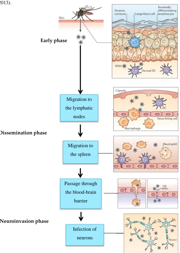
The pathogenesis of MBEF proceeds in three major phases: (1) local infection and primary viremia, (2) virus spread and peripheral replication and (3) neuro-invasion (infection of the central nervous system (CNS) and neurovirulence (damage to neuronal cells) [109] (Fig. 3).
After experimental inoculation, primary viremia usually develops in less than 24 h [91, 104, 110, 111]. A viraemia level of 105p.f.u. ml 1 is necessary to infect mosquitoes with WNV after a blood meal [112]. The dose and number of feeding mosquitoes directly affect the speed at which WNV spreads systemically [113]. Development of the disease results
inflammation and necrosis [1]. Typical neurological signs appear at this stage, such as ataxia and paralysis [48, 87, 93, 114, 115] and non-specific signs, such as lethargy, ruffled feathers and weight loss [8, 69, 90, 91, 116]. Lesions are like-wise developed and include necrotizing hepatitis, splenitis, myocardial degeneration and/or myocarditis, necrosis of striated muscles, non-suppurative encephalitis and neuronal necrosis [29, 48, 69, 74, 108, 110]. Haematogenous and/or neuronal dissemination of WNV and BAGV to the eye has been described in birds showing blindness [117, 118]. Severe egg drop (up to 90 %) and mortality (up to 30 %) in laying turkeys infected with ITV, and in layer chicken, ducks and geese infected with TMUV, have been reported [49, 71, 73]. The corresponding lesions are oophoritis, ovarian atrophy, hemorrhage and necrosis [9, 49, 71]. Although egg produc-tion can recover within 3–4 weeks after epizootic TMUV infection, both fertility and hatchability rates of eggs from breeding ducks were permanently lowered [119]. Reduced sperm production, spermatocyte swelling, and vacuolar degeneration occurred in the testes of infected male ducks, with focal lymphocytic infiltration in the later stages [111]. In ducklings, TMUV infection caused hyperglycemia (due to acute pancreatitis), neurological disease [47] and multi-organ failure leading to death [91].
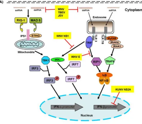
In a manner similar to humans and horses, birds utilize the 2¢ 5¢-oligoadenylate synthase pathway in the innate immune response against these flaviviruses [120]. This pathway ultimately induces apoptosis with other components of the innate immune response, including IFNs, inflammatory cytokines, complement factors, natural killer cells (NK) and autophagy to inhibit viral replication [1, 76]. Neutralizing antibodies, which primarily target the viral E glycoprotein, and antibodies against NS proteins constitute the major humoral immune response to flavivirus infection
Seroconversion, as well as the persistence of antibodies, is variable among birds. Importantly, maternal antibodies in young chicks can serve for rapid protection from WNV and TMUV infections [120, 122]. In addition to effective host humoral immunity, cellular immunity is triggered to control viral infection and dissemination [1]. Flaviviruses have developed numerous strategies to avoid the host immune system, including the limitation of initial steps of PAMP detection, type I IFN signaling by blocking the host gene expression and inhibition of the complement system and NK cells [19].
Once infected with WNV, most susceptible birds remain asymptomatic because the immune response eliminates the virus from the organism within two or three weeks [98]. In some cases, infection with WNV can become persistent and viral RNA may be detected for several months after infection, as has been demonstrated for house finch (Haemorhous mexicanus), HOSP, western scrub-jay (Aphelocoma californica), kea (Nestor notabilis) and rock
persistently infected birds could trigger a mosquito–bird transmission cycle remains unresolved [123].
Prevention and control
To monitor MBEF circulation, several approaches have been used in many European countries, including sero-sur-veillance in birds and viral identification in dead birds and in pooled mosquito samples [126, 127].
Given the lack of specific treatment for MBEF infection in birds and mammals, preventive measures should be applied to decrease the risk of infection. Mosquito control and indoor housing of captive animals is suggested to prevent mosquito bites [128]. The use of pyrethroid-based insecticides and the elimination of mosquito habitats where these insects can lay eggs should be implemented in affected areas
Widespread ultra-low-volume application of insecticides has been successfully applied to reduce human WNV infection [129, 130], but this alternative is challenging in wild territories in regard to free-ranging birds. Lowering viremia in competent avian hosts is another solution to pre-vent infection following mosquito bite [131] and, thus, to prevent human infections with the two major MBEF mem-bers, WNV and USUV. Biosecurity measures and the development of vaccines are crucial in preventing major economic losses in the poultry industry due to ITV and TMUV infections. While no vaccine against USUV or BAGAV has been tested on birds to date, many others have been developed against WNV, ITV, and TMUV and tested in these animals.
Vaccines against WNV Inactivated vaccines
The first licensed WNV vaccine for veterinary use was dedicated to the horse. A formalin-inactivated WNV lineage 1 vaccine was developed in 2003 by Fort Dodge Animal Health and commercialized in the USA under the trade name West Nile-Innovator (in Europe: Equip WNV Zoetis, previously Duvaxyn WNV). This vaccine elicited variable antibody responses across bird species and the majority of vaccine trials were not conclusive, as they lacked a virus challenge test (Appendix 6). A three-injection scheme with this vaccine was, however, suggested for falcons as it was able to provide protection from lethal testing, although minor clinical signs and lesions, as well as viremia and virus shedding, occurred following the vaccination/challenge test [132].
Subunit/DNA vaccines
Subunit vaccines based on WNV TE proteins were trialed in domestic goose, red-legged partridge and Hawaiian goose ēnē (Branta sandvicensis), but protection was assessed only in partridge, which remained fully protected after a challenge test (Appendix 7A).
Two DNA vaccines encoding the TE protein of WNV line-ages 1 and 2 without prM caused local inflammation at the site of injection and did not prevent death in all vaccinated falcons after lethal testing [133]. DNA vaccines expressing
WNV prM and E proteins were trialed in birds, including the pCBWN vaccine and the Fort Dodge WN-Innovator DNA equine vaccine (Overland Park, KS) (Appendix 7B). The former was shown to fully protect fish crow (Corvus ossifragus) via the intramuscular route [134]. In contrast, the latter failed to induce an antibody response in island scrub-jays (Aphelocoma insularis) [135] and did not prevent mor-tality, lesions, and high viremia levels after a challenge test in western scrub-jay (Aphelocoma californica) [133]. For large-scale immunization, oral administration of pCBWN was trialed in AMCR [136] and fish crow [134] but failed to provide protection in either species.
Chimeric vaccines
Using live attenuated strains of other viruses as a genetic backbone, multiple versions of chimeric vaccines against WNV have been designed and explored for immunogenicity in birds (Appendix 7C). A recombinant live canarypox ALVAC viral vector expressing WNV prM and E proteins, RecombiTEK, Merial-Sanofi Aventis, was licensed in 2004 for veterinary use [137]. Vaccine safety was not satisfactory as the vaccine induced local inflammatory and necrotic lesions at the injection site. Besides, it failed to induce an immune response in western scrub-jay [138]. However, three injections succeeded in reducing mortality after the virus challenge in falcon [132]. A recombinant adenovirus vaccine, expressing WNV E or NS3 proteins, induced a specific antibody response in Japanese quail (Coturnix japonica) but the protection level was not assessed [139].
Three chimeric vaccine candidates, currently under trial for humans, the use were tested in birds. The first was ChimeriVax-WN, where WNV prM and E protein-coding genes were incorporated into the genome of the 17D non-structural genes of the yellow fever virus. In the second, chimeric WN/ DEN4, prM and E protein-coding genes of dengue virus type 4 were replaced with the corresponding genes from WNV while in the third, WN/DEN4-3’D30, a 30-nucleotide deletion in the non-coding region of the DEN4 component of chimeric WN/DEN4 was introduced. These vaccines failed to prevent clinical symptoms, viremia or death after the challenge test as they could not be replicated in these avian hosts, probably due to the fact that the backbone viruses were not adapted to these hosts [140, 141].
Heterologous vaccines
To assess the advantage of flavivirus cross-reactivity for heterologous protection, an attenuated vaccine against ITV was tried in goose, and resulted in 39–72 % protection against WNV challenge in field-vaccinated birds [142].
Vaccines against ITV and TMUV
Since the emergence of ITV in Israel, commercial attenuated virus vaccines (Biovac Biological Laboratories, Akiva, Israel and Phibro, Beth Shemesh, Israel) based on virus strain JQ4E4 [143, 144] have been used in that country as a routine control strategy for the disease. Minor clinical signs have often been observed after vaccination [143].
To date, attenuated and killed vaccines have been commer-cialized to protect ducklings and layer ducks against TMUV, including Duck Tembusu Virus Vaccine Live (FX2010-180P strain) (ZHENGYE, Jilin, China), attenuated by serial passage in chicken embryo fibroblasts [107], and an inactivated TMUV vaccine (HB strain, Rinpu, Tianjin, China) (Appendix 8).
Attenuated Salmonella typhimurium SL7207 (pVAX-C) has been used as a vehicle in oral delivery of TMUV prM and E antigens to ducks [18]. Alternatively, another study used this attenuated bacteria to immunize ducks with TMUV C protein to induce a systemic immune response [145]. These two vaccines showed 100 % survival among duck, with minor clinical signs after lethal testing [18, 145].
To develop multivalent vaccines, recombinant avian viruses, such as Duck enteritis virus and Newcastle disease viruses, were used as vectors for prM/E [146–148] and succeeded in fully protecting duck following a challenge test.
Conclusions
Birds play a key role in the life cycle of many flaviviruses as amplifying hosts, with an important contribution to their transmission and spread either locally or to new territories. MBEF are highly pathogenic for certain avian species. Fur-thermore, WNV, and USUV occasionally cause severe neu-rological disease in humans and, thus, constitute a concern for both veterinary and public health.
Eradication of these pathogens is virtually impossible because the viruses are maintained in a complex life cycle involving several animal reservoirs, some of which remain unknown. Preventive measures remain the only solution to help reduce and control their circulation, but such measures are hampered by the unresolved transmission routes of these viruses, the limited cost-effectiveness of vaccination and the underestimation of seasonal infection and mortality rates. In fact, MBEF infections often occur unnoticed, because many birds develop an asymptomatic form of the disease or die without collection by competent authorities. Formulating cheap and completely protective single-dose or oral vaccines would be the golden goal for simple and large-scale immunization of domestic and wild birds. More studies need to be carried out to evaluate the actual prevalence and incidence of these MBEF, to study their pathogenesis and to fully elucidate their life cycles and transmission routes, as preliminary steps towards the preservation of wild bird species, the reduction of the impact on domestic birds and the prevention of human infections.
Author bio
The author is currently a Ph.D. student in the Morphology and Pathology Department of the Veterinary Faculty of Liege. Her primary research interest is the pathogenesis of USUV.
Funding information
References
Lindenbach BD, Murray CL, Thiel H-J, Rice CM. Flaviviridae. In: Knipe DM, Howley PM (editors). Fields Virology. Philadelphia: Lip-pincottWilliams &Wilkins; 2013. pp. 712–746.
O’Guinn ML, Turell MJ, Kengluecha A, Jaichapor B, Kankaew P Et al. Field detection of Tembusu virus in Western Thailand byrt-PCR and vector competence determination of select culex mosquitoes for transmission of the virus. Am J Trop Med Hyg 2013;89:1023–1028.
Nikolay B, Diallo M, Boye CS, Sall AA. Usutu virus in Africa. Vector Borne Zoonotic Dis 2011;11:1417–1423.
Sudeep AB, Bondre VP, Mavale MS, Ghodke YS, George RP Et al.
Preliminary findings on Bagaza virus (Flavivirus: Flaviviri-dae) growth kinetics, transmission potential & transovarial transmission in three species of mosquitoes. Indian J Med Res 2013;138:257–261.
Meulen KM Van Der, Pensaert MB. West Nile virus in the verte-brate world brief review. Arch Virol 2005;150:637–657.
Weissenböck H, Bakonyi T, Rossi G, Mani P, Nowotny N ET al. Emerg Infect Dis 1996;2013:1996–1999.
Erdely K, Ursu K, Ferenczi E, Szeredi L, Fer Ratz Et al. Clinical and
pathologic features of lineage 2 West Nile virus infections in birds of Prey in Hungary. Vector borne zoonotic dis 2007;7: 181–188.
Agüero M, Fernandez-Pinero J, Buitrago D, Sanchez A, Elizalde M
Et al. Bagaza virus in partridges and pheasants, Spain, 2010. Emerg Infect
Dis 2011;17:1498–1501.
Cao Z, Zhang C, Liu Y, Liu Y, Ye W Et al. Tembusu virus in ducks,
China. Emerg Infect Dis 2011;17:1873–1875.
Bondre VP, Sapkal GN, Yergolkar PN, Fulmali P V, Sankararaman V Et al. Genetic characterization of Bagaza virus (BAGV) isolated in India
and evidence of anti-BAGV antibodies in sera collected from encephalitis patients. J Gen Virol 2009;90: 2644–2649.
Colpitts TM, Conway MJ, Montgomery RR, Fikrig E. West Nile virus: biology, transmission, and human infection. Clin Microbiol Rev 2012;25:635–648.
Tang Y, Gao X, Diao Y, Feng Q, Chen H Et al. Tembusu virus in
Human, China. Trans Emerg Dis 2013;60:193–196.
Gaibani P, Rossini G. An overview of Usutu virus. Microbes Infect 2017;19:382–387.
Petersen LR, Brault AC, Nasci RS. West Nile virus: review of the literature. Jama 2013;310:308–315.
Simmonds P, Becher P, Bukh J, Gould EA, Meyers G Et al. ICTV virus taxonomy profile: Flaviviridae. J Gen Virol 2017;98:2–3.
Pijlman GP, Funk A, Kondratieva N, Leung J, Torres S Et al. A highly
structured, nuclease-resistant, noncoding RNA produced by Flaviviruses is required for pathogenicity. Cell Host Microbe 2008;4:579–591. Mukhopadhyay S, Kuhn RJ, Rossmann MG. A structural per-spective of the Flavivirus life cycle. Nat Rev 2005;3:13–22.
Huang J, Jia R, Shen H, Wang M, Zhu D. Oral delivery of a DNA vaccine expressing the PrM and E genes: a promising vaccine strategy against Flavivirus in Ducks. Sci Rep 2018;8:22360.
Ye J, Zhu B, Fu ZF, Chen H, Cao S. Immune evasion strategies of flaviviruses. Vaccine 2013;31:461–471.
Chen S, Wu Z, Wang M, Cheng A. Innate immune evasion medi-ated by flaviviridae non-structural proteins. Viruses 2017;9:1– 19.
Maharaj PD, Bosco-Lauth AM, Langevin SA, Anishchenko M, Bowen RA Et al. West Nile and St. Louis encephalitis viral genetic
determinants of avian host competence. PLoS Negl Trop Dis 2018;12:e0006302.