Validation of a nonaqueous capillary electrophoretic method for the
enantiomeric purity determination of R-flurbiprofen using a single-isomer
amino cyclodextrin derivative
A. Rousseaua, P. Chiapb, R. Ivanyic, J. Crommena, M. Filleta, A.-C. Servaisaa Department of Analytical Pharmaceutical Chemistry, Institute of Pharmacy, University of Liège, CHU, B36, B-4000 Liège, Belgium b Advanced Technology Corporation (ATC), CHU, B36, B-4000 Liège, Belgium
c Cyclolab, Cyclodextrin R&D Laboratory Ltd., P.O. Box 435, H-1525 Budapest, Hungary
ABSTRACT
Nonaqueous capillary electrophoresis (NACE) was successfully applied to the enantiomeric purity determination of R-flurbiprofen using 6-monodeoxy-6-mono(2-hydroxy)propylamino-β-cyclodextrin (IPA-β-CD) as chiral selector. The nonaqueous BGE was made up of 20 mM IPA-β-CD, 20 mM ammonium camphorsulfonate and 40 mM ammonium acetate in methanol. Flufenamic acid was selected as internal standard. The CE method was carefully optimized in order to prevent the adsorption of the cationic CD onto the capillary wall, and therefore, to avoid loss of peak efficiency and enantioresolution. To achieve this goal, the addition of ammonium
camphorsulfonate was found to be necessary. In the selected conditions, the determination of 0.1 % of S-flurbiprofen in R-S-flurbiprofen could be performed using the method of standard additions. The NACE method was then fully validated by applying a novel strategy using accuracy profiles.
Keywords: nonaqueous capillary electrophoresis ; enantiomeric purity determination ; R-Flurbiprofen ; cationic
cyclodextrin ; validation
1. Introduction
For many years, in the field of chiral compounds, the pharmacopoeia mainly deals with racemic drugs. However, it is well-known that large differences can exist between both enantiomers in terms of pharmacokinetics, activity and toxicity [1]. Pharmaceutical companies have to make the appropriate choice concerning the development of chiral drugs in their single-isomer or racemate forms. In the first case, rapid, sensitive and selective analytical methods are essential to control the chiral purity of the products. Currently, the separation of enantiomers can be performed in high-performance liquid chromatography, thin-layer chromatography, gas chromatography and more recently capillary electrophoresis (CE) [2-5]. As indicated by the number of papers published in this field, CE has become a powerful tool for enantioseparations due to its high separation efficiency, low operation cost and speed of analysis [6-9].
Most of CE enantioseparations are still performed in aqueous media or aqueous background electrolytes (BGEs) modified with organic solvents such as methanol or acetonitrile. However, the introduction of nonaqueous BGEs in CE has proved to be a very useful tool for the enantiomeric resolution of chiral drugs [10]. The possibility of using not only aqueous BGEs but also nonaqueous CE (NACE) has offered new opportunities for changes in separation selectivity, due to the extension of the range of solvent parameters such as the dielectric constant, viscosity, polarity and auto protolysis [11-18].
Recently, a family of single-isomer β-CD derivatives containing an amino or (hydroxy)alkylamino group in one of the primary positions has been synthesized [19]. Each amino-β-CD derivative showed a higher
enantioselectivity towards the studied acidic compounds, namely mandelic acid, permethrinic acid, and cis-deltamethrinic acid, than the native β-CD. Moreover, the presence of one hydroxyalkyl group attached to the primary amino N-atom significantly increased both the enantioselectivity and the resolution compared to the primary amino-β-CD. Some of these derivatives, such as 6-monodeoxy-6-mono(3-hydroxy)propylamino-β-CD (PA-β-CD), hydroxy)propylamino-β-CD (IPA-β-CD) and 6-monodeoxy-6-mono(2-hydroxy)ethylamino-β-CD, are soluble in methanol and have been successfully applied for the enantioseparation of some non-steroidal anti-inflammatory drugs (NSAIDs) in NACE [20].
Flurbiprofen is a NSAID belonging to the class of the profens or 2-arylpropionic acid derivatives, currently administrated as racemic mixture. It is used for the treatment of several inflammatory diseases. The inhibition of prostaglandin production and inflammatory events resides in the S-enantiomer. The R-enantiomer of flurbiprofen being inactive on COX, it does not produce the gastrointestinal and renal side effects of the racemate [21]. Moreover, it presents a great interest in Alzheimer's disease, due to its ability to selectively lower the production of Aβ42 [22-29]. Indeed, phase III clinical trials are carried out for the potential treatment of Alzheimer's disease
[30].
Several separation techniques using high-performance liquid chromatographic [31-34], capillary liquid chromatographic [35,36], gas chromatographic [37,38], micellar electrokinetic chromatographic [39], capillary electrophoretic [40-44] and thin-layer chromatographic methods [45] have been reported for the determination of flurbiprofen enantiomers.
A few of these analytical methods were applied to the enantiomeric purity testing of S-flurbiprofen [38,44] but none has been validated. Moreover, to the best of our knowledge, the enantiomeric purity determination of R-flurbiprofen has never been investigated.
In the present study, the possibility of using NACE for the enantiomeric purity determination of R-flurbiprofen using IPA-β-CD has been demonstrated. Moreover, the method has been fully validated according to the strategy proposed by a Commission of the Société Française des Sciences et Techniques Pharmaceutiques (SFSTP) for the validation of quantitative analytical procedures [46]. From a statistical point of view, this strategy is based on the use of accuracy profiles which take into account the total error, i.e., estimation of systematic and random errors of measurement.
2. Materials and methods 2.1. Instrumentation
All experiments were carried out on a HP3DCE system (Agilent Technologies, Waldbronn, Germany) equipped
with an autosampler, an on-column diode-array detector and a temperature control system (15-60 ± 0.1 °C). A CE Chemstation (Hewlett-Packard, Palo Alto, CA, USA) was used for instrument control, data acquisition and data handling. Fused-silica capillaries were provided by ThermoSeparation Products (San Jose, CA, USA). The statistical calculations for validation were performed by means of e.noval version 2.0 software (Arlenda, Liège, Belgium).
2.2. Chemicals and reagents
(R,S)-Flurbiprofen, R-flurbiprofen and flufenamic acid were supplied by Sigma-Aldrich (Saint-Louis, MO, USA). Their chemical structures are presented in Fig. 1. 6-Monodeoxy-6-mono(3-hydroxy)propylamino-β-CD (PA-β-6-Monodeoxy-6-mono(3-hydroxy)propylamino-β-CD) hydrochloride and 6-mono (2-hydroxy)propylamino-β-6-Monodeoxy-6-mono(3-hydroxy)propylamino-β-CD (IPA-β-6-Monodeoxy-6-mono(3-hydroxy)propylamino-β-CD) hydrochloride were kindly provided by Cyclolab (Budapest, Hungary). The optimized BGE consisted in a methanolic solution of 40 mM ammonium acetate, 20 mM ammonium camphorsulfonate and 20 mM IPA-β-CD. Ammonium acetate was obtained from Merck (Darmstadt, Germany). Trifluoroacetic acid was from Acros-Organics (Geel, Belgium). (1R)-(-)-10-Camphorsulfonic acid was purchased from Sigma-Aldrich. All reagents were of analytical grade. In order to convert (1R)-(-)-10-camphorsulfonic acid to ammonium salt, a quantity of ammonium acetate,
corresponding to the concentration of acid, was added. Methanol (Merck) was of HPLC grade. The BGE and samples solutions were filtered through a Polypure polypropylene membrane filter (0.2 µm) from Alltech (Laarne, Belgium) before use.
Fig. 1. Chemical structures of (A) flurbiprofen and (B) flufenamic acid.
2.3. Electrophoretic technique
diameter and 48.5 cm length (40 cm to the detector). At the beginning of each working day, the capillary was washed with methanol and the BGE without CD for 15 min. Before each injection, the capillary was washed successively with 1 M trifluoroacetic acid in methanol for 4 min, with the BGE without CD for 2 min, and then equilibrated with the BGE-CD for 2 min. At the end of each working day, the capillary was rinsed for 30 min with 1 M trifluoroacetic acid in methanol, 30 min with the BGE without CD and 15 min with methanol.
Capillary wash cycles were performed at a pressure of approximately 1 bar. The applied voltage was -30 kV and UV detection was set at 250 nm. Injections were made by applying a pressure of 50 mbar for a period of 3 s (corresponding to 8.8 nL, i.e. 0.9% of the total volume of the capillary) and the capillary was thermostated at 15°C. The resolution (Rs) was calculated according to the standard expression based on peak width at half height
[47].
2.4. Standard solutions
2.4.1. Sample solutions used for method development
A stock solution of S-flurbiprofen was prepared by dissolving an accurately weighed amount of approximately 20 mg of racemic flurbiprofen in 20.0 mL methanol. This stock solution was then diluted 100-fold and R-flurbiprofen was added in order to obtain a reference solution representing 0.25% (5 µg/mL) of impurity level and containing 2 mg/mL of R-flurbiprofen.
2.4.2. Sample solutions used for method validation
A stock solution of S-flurbiprofen was prepared by dissolving an accurately weighed amount of approximately 20 mg of racemic flurbiprofen in 10.0 mL methanol. Then, subsequent dilutions were carried out in order to obtain three calibration curves (k = 3) ranging from 2.0 to 24 µg/mL (m = 3) (cf. Table 1 ) and containing 2 mg/mL of R-flurbiprofen and 50 µg/mL of flufenamic acid, the internal standard. Two replicates (n = 2) were prepared per concentration level (m = 3). Each solution was injected two times (n = 2). The means were considered. The number of concentration levels was sufficient to generate different regression models. Three independent series of validation standards were also prepared in methanol, in the same way, with final concentrations of 2, 5, 12 and 24 µg/mL (cf. Table 1 ) in S-flurbiprofen. These validation standards also contained 2 mg/mL of R-flurbiprofen and 50 µg/mL of flufenamic acid. Three replicates (n = 3) were prepared per concentration level (m = 4).
Table 1 Preparation of standard solutions related to S-flurbiprofen for validation
Concentration level (% relative to
2.0mg/mL of R-flurbiprofen) Concentration of S-flurbiprofen (µg/mL) Calibration standards Validation standards
0.10 2.0 2.0
0.25 - 5.0
0.60 12.0 12.0
1.20 24.0 24.0
Total 6 samples/day 12samples/day
3. Results and discussion
3.1. Optimization of the CE method
In a previous work, an experimental design was applied to the enantiomeric resolution of nonsteroidal anti-inflammatory drugs in NACE systems using PA- and IPA-β-CD [20]. Generic NACE conditions were then deduced in order to obtain the highest Rs values in a minimum analysis time. The appropriate BGE for the
separation of flurbiprofen enantiomers consisted in a methanolic solution of 10 mM PA-β-CD and 40 mM ammonium acetate (Rs value: 4.8). Very recently, this NACE system was successfully applied for the
determination of flurbiprofen enantiomers in plasma after solid-phase extraction [48]. It is noteworthy that, in this study, the first migrating peak was identified as the S-enantiomer of flurbiprofen and the second one as the
R-enantiomer.
This BGE was also tested in the present study devoted to the enantiomeric purity determination of
R-flurbiprofen. The applied voltage was -25 kV and an injection time of 8 s was initially selected. Nevertheless, by contrast to what is observed with the racemic mixture, no enantioseparation was obtained when a solution of R-flurbiprofen (2 mg/mL) spiked with S-R-flurbiprofen (0.25%) was analyzed. Therefore, the PA-β-CD concentration was increased until 35 mM but the enantiomers could still not be separated. Higher CD concentrations could not be tested since the maximum solubility of the chiral selector in this BGE was reached.
IPA-β-CD was then investigated and was found to be more appropriate since a slight (but not baseline)
separation of flurbiprofen enantiomers was observed at a CD concentration of 30 mM. The enantiomer migration order was found to be the same as that observed with PA-β-CD. The CD concentration was then increased until 40 mM and at this concentration, a baseline enantioseparation was observed. Table 2 presents the influence of the applied voltage, the injection time and the type of BGE on the migration times and efficiency of S-flurbiprofen when a series of 30 consecutive runs was performed. The sample consisted in a R-S-flurbiprofen solution (2 mg/mL) spiked with S-flurbiprofen (0.25%). As can be seen in this table, despite the use of a drastic post-conditioning wash (1 M trifluoroacetic acid in methanol), a dramatic loss of peak efficiency and important variations in the migration times (RSD value: 9.8%) were observed, impairing the enantiomeric resolution. In order to increase peak efficiency, the injection time was reduced from 8 s to 3 s and the applied voltage was actually increased from -25 kV to -30 kV. A gain in analysis time as well as in peak efficiency was actually observed but the drift of the migration times and efficiency was even more important (cf. Table 2). This effect was attributed to a strong adsorption of IPA-β-CD onto the surface of the capillary wall. Nevertheless, it had to be strongly decreased in order to achieve stable conditions.
With this aim in view, ammonium camphorsulfonate (camphorSO3-) was then added to the BGE. In that
electrolyte, the IPA-β-CD concentration had to be decreased in order to achieve solubility. Therefore, the tested BGE consisted in a mixture of 20 mM IPA-β-CD, 40 mM ammonium acetate and 20 mM ammonium
camphorSO3- in methanol. Even though the CD concentration is lower, the enantioseparation was complete (cf.
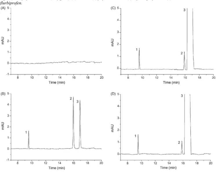
Fig. 2C), which demonstrates the importance of ammonium camphorSO3- in the enantiomeric separation. The
role of ammonium camphorSO3- seems to be related to the adsorption of the ammonium cation onto the capillary
wall, preventing that of the cationic CD, and, therefore, the decrease in peak efficiency of flurbiprofen. Indeed, despite the lower CD concentration, the value of the EOF is still lower than previously observed with the first BGE employed (cf. Table 2). As can be seen in this table, even if the RSD value of the migration times was 4.4%, rather constant peak efficiency and, therefore, constant enantioresolution, were observed.
Table 2 Influence of the applied voltage, the injection time and the type of BGE on the migration times (MT) and efficiency (N) of S-flurbiprofen with their corresponding RSD values (%, n = 30) and on the electroosmotic flow (EOF)
CE conditions
40 mM CD in BGE: -25 kV;
Inj. time: 8 s 40 mM CD in BGE: -30 kV; Inj. time: 3 s 20 mM CD + 20 mM camphorSO3
- in
BGE: -30 kV; Inj. time: 3 s MT(min) Run 1 25.3 16.0 15.7 Run 30 33.9 22.3 17.8 Mean (RSD in %) 28.5 (9.8) 19.4(10.3) 16.2 (4.4) N Run 1 75.000 97.000 102.000 Run 30 29.000 24.000 91.000 Mean (RSD in %) 43.000(29.3) 59.000 (43.3) 95.000 (3.8) EOF (x10-5 cm2 V-1 s-1) Run 1 6.5 5.8 4.4 Run 30 4.6 3.5 3.7
BGE: 40 mM ammonium acetate in MeOH; sample: methanolic solution of R-flurbiprofen (2 mg/mL) containing S-flurbiprofen (0.25%). Post-conditioning wash: 1 M trifluoroacetic acid in methanol (4 min). CE conditions as described in Section 2.
3.2. Method validation
An original strategy, based on total measurement error and accuracy profiles as a decision tool, was applied to demonstrate that the developed method is suited for its intended purpose [46,49,50]. This approach has been elaborated by a SFSTP (Société Française des Sciences et Techniques Pharmaceutiques) commission. The concept of accuracy profile was also used to select the most appropriate regression model for calibration, to determine the lower limit of quantitation and the range over which the method can be considered as valid. 3.2.1. Selectivity
The method selectivity was evaluated with respect to two aspects. Firstly, typical electropherograms obtained by analyzing methanol and a diluted solution of flufenamic acid, S- and R-flurbiprofen were compared in order to check the absence of peaks likely to interfere in the electrophoretic region of flurbiprofen enantiomers and flufenamic acid. As shown in Fig. 2A and B, no interference was observed at the migration times of the peaks corresponding to the three analytes, which demonstrates the selectivity of this method. Moreover, the
enantiomeric resolution of flurbiprofen enantiomers could be calculated from Fig. 2B and was found to be equal to 4.2.
Selectivity was also checked by evaluating the ability to easily integrate and therefore quantify a low amount of
S-flurbiprofen in the presence of the large peak corresponding to R-flurbiprofen (2.0 mg/mL). Fig. 2C illustrates
that the peak corresponding to S-flurbiprofen could be well integrated and quantified at a low concentration level.
Moreover, a solution of 2.0 mg/mL of R-flurbiprofen was also analyzed (cf. Fig. 2D). As can be seen from this figure, a small peak, identified as S-flurbiprofen by comparison of the migration times, was observed. For that reason, 2 mg/mL of R-flurbiprofen was added in the calibration standards.
Fig. 2. Typical electropherograms of methanol (A), a methanolic solution of flufenamic acid (50 µg/mL) and racemic flurbiprofen (48 µg/mL) (B), a methanolic solution of R-flurbiprofen (2 mg/mL) containing S-flurbiprofen (5 µg/mL) and flufenamic acid (50 µg/mL) (C) and a methanolic solution of R-S-flurbiprofen (2 mg/mL) and flufenamic acid (50 µg/mL) (D). Peaks: (1) flufenamic acid, (2) S-flurbiprofen and (3) R-flurbiprofen.
3.2.2. Selection of the calibration model
Several regression models were fitted to the calibration standards. From each regression line obtained, the concentrations of the validation standards were back-calculated in order to determine, at each concentration level, the mean relative bias, the relative standard deviation for intermediate precision as well as the upper and lower β-expectation tolerance limits at 90% [46].
From these data, different accuracy profiles were plotted to select the most appropriate regression model for the intended use of the analytical method, as illustrated in Fig. 3 [46,49,50]. Six response functions, namely the weighted (1/X) and (1/X2) linear regression models, the simple linear regression model, the weighted 1/X2 and
1/X quadratic regression models and the quadratic regression model, were tested. The acceptance limits were set at ± 10% according to the regulatory requirements [51 -54]. Only the weighted (1/X) linear regression allowed to demonstrate the capability of the method over the concentration range considered, since the tolerance intervals were totally included inside the acceptance limits as shown in Fig. 3A. The regression equations obtained by applying this model are presented in Table 3. With the other models, either a greater variance or a negative bias close to 10% was obtained.
As previously shown in Fig. 2D, the used batch of R-flurbiprofen contained a small amount of S-flurbiprofen. The obtained regression equations were used to determine the quantity of the enantiomeric impurity (method of standard additions). Taking into account the mean over the 3 days, it was established that the used batch of R-flurbiprofen contained 0.2% of S-R-flurbiprofen.
Table 3 Validation results referred to S-flurbiprofen using the weighted (1/X) linear regression model
Validation criteria S-Flurbiprofen
Response function Slope Intercept r2
(k = 3; m = 3; n = 2)(0.1-1.2%)
Day 1 0.170 0.643 0.9974
Day 2 0.143 0.700 0.9942
Day 3 0.140 0.691 0.9962
Trueness (k = 3; n = 3) Relative bias (%)
0.1% <0.1
0.25% 1.6
0.6% -3.1
1.2% 2.9
Precision RSD (%)
Repeatability Intermediate precision (k = 3;n = 3)
0.1% 1.3 3.0
0.25% 2.5 2.9
0.6% 3.5 3.5
1.2% 3.3 3.3
Accuracy (k = 3; n = 3) β-Expectation tolerance limits (%)
0.1% -8.4/8.4 0.25% -4.4/7.6 0.6% -9.9/3.8 1.2% -4.5/8.6 Linearity (k = 3; m = 4; n = 3)(0.1-1.2%) Slope 1.02 Intercept -0.160 r2 0.9966 LOD (%) 0.03 LOQ(%) 0.1
Fig. 3. Accuracy profiles obtained by using different calibration models. (A) Weighted linear regression model
with a weight equal to 1/X. (B) Weighted linear regression model with a weight equal to 1/X2. (C) Linear
regression model. (D) Quadratic regression model with a weight equal to 1/X2. (E) Quadratic regression models
with a weight equal to 1/X. (F) Quadratic regression model. The dotted lines represent the acceptance limits (10%), the dashed lines correspond to the accuracy profile, i.e. to the β-expectation tolerance limits expressed in relative error, and the relative bias values are located on the plain line.
3.2.3. Trueness
Trueness refers to the closeness of agreement between a conventionally accepted value and a mean experimental one. Trueness expressed in term of relative bias (%) was assessed from the validation standards at four
concentration levels, ranging from 0.1 to 1.2% (k = 3, n = 3) as can be seen in Table 3. According to the
regulatory requirements, the results show that the relative biases of the developed method were found acceptable since they are below the maximum value of 10%, irrespective of the concentration level.
3.2.4. Precision
The precision of the analytical method was then estimated by calculating repeatability and intermediate precision at each concentration level. The variance of repeatability and time-dependent intermediate precision, as well as their corresponding relative standard deviation (RSD) values, were calculated from the estimated concentrations. The RSD values, presented in Table 3, were rather low (between 1.3% and 3.5%).
3.2.5. Accuracy
Accuracy refers to the closeness of agreement between the test result and the accepted reference value, namely the conventionally true value. The accuracy of the analytical method takes into account the total error, i.e. the sum of systematic and random errors, related to the test results. The upper and lower β-expectation tolerance limits expressed in relative bias (%) as a function of the introduced concentrations are presented in Table 3. It is worth noting that the maximum risk (1 - β) to have future measurements outside the acceptance limits was set at 10%.
The different tolerance limits of the mean relative bias did not exceed the acceptance limits settled at 10% for each concentration level, including the lowest one (0.1%). Therefore, the proposed method can be considered as accurate over the concentration range investigated.
3.2.6. Linearity
The linearity of an analytical method is its ability, within a definite range, to obtain results directly proportional to the concentrations of the analyte in the sample. For all series, a regression line was fitted on the estimated or back-calculated concentrations of the validation standards as a function of the introduced concentrations by applying the linear regression model based on the least squares method.
The results attesting the method linearity, namely the regression equation corresponding to that relationship with the coefficient of determination, are presented in Table 3.
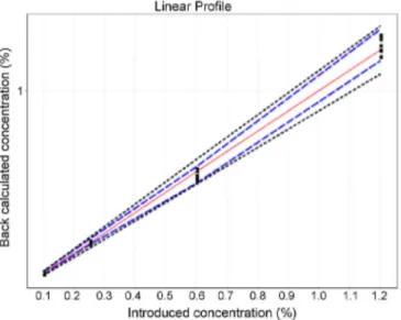
Moreover, in order to demonstrate the method linearity, the approach based on the absolute β-expectation tolerance limits can be applied. As can be seen in Fig. 4, the linearity of the present method was demonstrated since the absolute β-expectation tolerance limits were within the absolute acceptance limits.
Fig. 4. Linear profile of the developed method. The dashed limits on this graph correspond to the accuracy profile, i.e. the β-expectation tolerance limits expressed in absolute values. The dotted curves represent the acceptance limits at 10% expressed in the concentration unit.
3.2.7. Detection and quantitation limits
The limit of detection (LOD) is the smallest quantity of the targeted substance that can be detected, but not accurately quantified in the sample. In the present study, the LOD was estimated using the mean intercept of the
calibration model and the residual variance of the regression. By applying this computation method, the LOD of the developed method was found to be equal to 0.03%. The lower limit of quantitation (LOQ) of an analytical procedure is the smallest quantity of the targeted substance in the sample which can be quantitatively determined under the experimental conditions prescribed with a well-defined accuracy, i.e., taking into account the
systematic and random errors. The LOQ was obtained by calculating the lowest concentration beyond which the β-expectation tolerance limits fall outside the acceptance limits. As the accuracy profile was included inside the acceptance limits over the whole concentration range investigated (Fig. 3A), the first concentration level (0.1%) was considered as the LOQ taking into account the selected regression model. Indeed, precision and trueness were demonstrated at this concentration level.
4. Concluding remarks
A NACE method using IPA-β-CD for the enantiomeric purity determination of R-flurbiprofen was developed and validated. The method development was focused not only on the chiral separation but also on the
reproducibility of the results. With this aim in view, ammonium camphorsulfonate was found to be essential. Indeed, it reduces the adsorption of the cationic CD onto the capillary wall, leading to constant peak efficiency and enantioresolution.
Using the method of standard additions, the determination of 0.1% of S-flurbiprofen in R-flurbiprofen could be performed with the optimized NACE method which was fully validated according to the strategy based on the accuracy profiles. The method showed suitable performance in terms of selectivity, trueness, precision, accuracy and linearity.
It can be concluded that this simple and fast NACE method is appropriate for the enantiomeric purity testing of
R-flurbiprofen in routine. Acknowledgements
Research grants from the Belgium National Fund for Scientific Research (FNRS) to two of us (A.-C. S. and M. F.) are gratefully acknowledged. Many thanks are also due to FNRS and to the Léon Fredericq foundation for their financial support. The authors would like to thank Catherine Dumont for her technical support.
References
[ 1 ] E. Francotte, W. Lindner (Eds.), Chirality in Drug Research, Methods and Principles in Medicinal Chemistry, vol. 33, Wiley-VCH, Weinheim, 2006.
[2] R. Kuhn, S. Hoffstetter-Kuhn, Chromatographia 34 (1992) 505.
[3] I.E. Valko, H.A.H. Billiet, H.A.L. Corstjens, J. Frank, LC-GC Int. 6 (1993) 420. [4] S. Fanali, J. Chromatogr. 545 (1991) 437.
[5] K.D. Altria, A.R. Walsh, N.W. Smith, J. Chromatogr. 645 (1993) 193. [6] G. Blaschke, B. Chankvetadze, J. Chromatogr. A 875 (2000) 3. [7] S. Zaugg, W. Thormann, J. Chromatogr. A 875 (2000) 7.
[8] M.R. Hadley, P. Camilleri, A.J. Hutt, Electrophoresis 21 (2000) 193. [9] T. Ward, Anal. Chem. 74 (2002) 2863.
[10] B. Chankvetadze, G. Blaschke, Electrophoresis 21 (2000) 4159.
[11] I. Bjornsdottir.J. Tjornelund, S.H. Hansen, J. Cap. Electrophoresis 2 (1996) 83. [12] M.-L. Riekkola, S.K. Wiedmer, I.E. Valkó, H. Sirén, J. Chromatogr. A 792 (1997) 13. [13] K.D. Altria, S.M. Bryant, Chromatographia 46 (1997) 122.
[14] A. Karbaum, T. Jira, Electrophoresis 20 (1999) 3396.
[15] M.-L. Riekkola, M. Jussila, S.P. Porras, I.E.Valkó, J. Chromatogr. A892 (2000) 155. [16] F. Wang, M.G. Khaledi, J. Chromatogr. A 875 (2000) 277.
[17] F. Steiner, M. Hassel, Electrophoresis 21 (2000) 3994.
[18] M. Fillet, A.-C. Servais, J. Crommen, Electrophoresis 24 (2003) 1499.
[19] R. Iványi, L. Jicsinszky, Z. Juvancz, N. Roos, K. Otta, J. Szejtli, Electrophoresis 25 (2004) 2675.
[20] I. Fradi, A.-C. Servais, M. Pedrini, P. Chiap, R. Iványi, J. Crommen, M. Fillet, Electrophoresis 27 (2006) 3434. [21] K. Brune, W.S. Beck, G. Geisslinger, S. Menzel-Soglowek, B.M. Peskar, B.A. Peskar, Experientia 47 (1991) 257.
[22] M. Zemskova, W. Wechter, S. Bashkirova, C.-S. Chen, R. Reiter, M.B. Lilly, Biochem. Pharmacol. 72 (2006) 1257.
[23] W.J. Wechter, D.D. Leipold, E.D. Murray, D.D. Quiggle, J.D. McCracken, R.S. Barrios, N.M. Greenberg, Cancer Res. 60 (2000) 2203. [24] S. Grösch, K. Schilling, A. Janssen, J.M. Thorsten, E. Niederberger, G. Geisslinger, Biochem. Pharmacol. 69 (2005) 831.
[25] N. Stock, B. Munoz, J.D.J. Wrigley, M.S. Shearman, D. Beher, J. Peachey, T.L Williamson, G. Bain, W. Chen, X. Jiang, R. St-Jacques, P. Prasit, Bioorg. Med. Chem. Lett. 16 (2006) 2219.
[26] L. Gasparini, E. Ongini, W. Donna, D. Morgan, Brain Res. Rev. 48 (2005) 400.
[27] G.K. Wilcock, S.E. Black, J. Haworth, S.B. Hendrix, M. Laughlin, K.H. Zavitz, D. Christensen, S. Bass, E. Swabb, Alzheimer's Dementia 2 (2006) S81.
[28] J. Mintzer, G.K. Wilcock, S.E. Black, K.H. Zavitz, S.B. Hendrix, Alzheimer's Dementia 2 (2006) S368.
[29] J∙L. Eriksen, S.A. Sagi, T.E. Smith, S. Weggen, P. Das, D.C. McLendon, V.V. Ozols, K.W. Jessing, K.H. Zavitz, E.H. Koo, T.E. Golde, J. Clin. Invest. 112 (2003) 440.
[30] H. Geerts, IDrugs 10 (2007) 121.
[31] X.W. Teng, S.W.J. Wang, N.M. Davies, J. Pharm. Biomed. Anal. 33 (2003) 95.
[32] G. Geisslinger, S. Menzel-Soglowek, O. Schuster, K. Brune, J. Chromatogr. 573 (1992) 163. [33] J.-M. Maître, G. Boss, B. Testa, J. Chromatogr. 299 (1984) 397.
[34] M.P. Knadler, S.D. Hall, J. Chromatogr. 494 (1989) 173.
[35] B. Kafkova, Z. Bosakova, E. Tesarova, P. Coufal, J. Chromatogr. A 1088 (2005) 82. [36] B. Kafkova, Z. Bosakova, E. Tesarova, Chirality 18 (2006) 531.
[37] N.N. Singh, FM. Pasutto, R.T. Coutts, F. Jamali, J. Chromatogr. 378 (1986) 125. [38] M.J. Paik, D.T. Nguyen, K.R. Kim, Arch. Pharm. Res. 27 (2004) 1295. [39] J∙ Dey, A. Mohanty, S. Roy, D. Khatua, J. Chromatogr. A 1048 (2004) 127. [40] S. Fanali, Z. Aturki, J. Chromatogr. A 694 (1995) 297.
[41] F.K. Gówka, M. Karaźniewicz, J. Pharm. Biomed. Anal. 35 (2004) 807. [42] F. Lelièvre, P. Gareil, J. Chromatogr. A 735 (1996) 311.
[43] R∙ Hamoudová, M. Pospíšilová, J. Pharm. Biomed. Anal. 41 (2006) 1463. [44] S. La, J. Kim, J.-H. Kim, J. Goto, K.-R. Kim, Electrophoresis 24 (2003) 2642.
[45] M. Sajewicz, M. Gontarska, D. Kronenbach, L Wojtal, G. Grygierczyk, T.M. Kowalska, Acta Chromatogr. 18 (2007) 226.
[46] Ph. Hubert, J.-J. Nguyen-Huu, B. Boulanger, E. Chapuzet, P. Chiap, N. Cohen, P-A. Compagnon, W. Dewe, M. Feinberg, M. Lallier, M. Laurentie, N. Mercier, G. Muzard, C. Nivet, L. Valat, STP Pharma Pratiques 13 (2003) 101.
[47] The European Pharmacopoeia, 5th ed., Council of Europe, Strasbourg, France, 2005.
[48] A. Rousseau, M. Pedrini, P. Chiap, R. Ivanyi, J. Crommen, M. Fillet, A.-C. Servais, Electrophoresis, accepted.
[49] Ph. Hubert, J.-J. Nguyen-Huu, B. Boulanger, E. Chapuzet, P. Chiap, N. Cohen, P-A. Compagnon, W. Dewe, M. Feinberg, M. Lallier, M. Laurentie, N. Mercier, G. Muzard, C. Nivet, L. Valat, J. Pharm. Biomed. Anal. 36 (2004) 579.
[50] Ph. Hubert, J.-J. Nguyen-Huu, B. Boulanger, E. Chapuzet, P. Chiap, N. Cohen, P-A. Compagnon, W. Dewe, M. Feinberg, M. Lallier, M. Laurentie, N. Mercier, G. Muzard, C. Nivet, L. Valat, STP Pharma Pratiques 16 (2006) 28.
[51] Guidance for industry: Bioanalytical Method Validation, US Department of Health and Human Services, Food and Drug Administration, Center for Drug Evaluation and Research (CDER), Center for Biologics Evaluation and Research (CBER), May 2001. [52] C.T. Viswanathan, S. Bansal, B. Booth, A.J. DeStefano, M.J. Rose, J. Sailstad, V.P. Shah, J.P. Skelly, P.G. Swann, R. Weiner, AAPS J 9 (2007) E30.
[53] V.P. Shah, K.K. Midha, S. Dighe, I.J. McGilveray, J.P. Skelly, A. Yacobi, T. Layloff, C.T. Viswanathan, C.E. Cook, R.D. McDowall, K.A. Pittman, S. Spector, J. Pharm. Sci. 81 (1992) 309.
[54] V.P. Shah, K.K. Midha, J.W.A. Findlay, H.M. Hill, J.D. Hulse, I.J. McGilveray, G. McKay, K.J. Miller, R.N. Patnaik, M.L. Powell, A. Tonelli, C.T. Viswanathan, A. Yacobi, Pharm. Res. 17 (2000) 1551.


