ii Université de Sherbrooke
The NOD-Like Receptors as Endogenous Inhibitors of Multiple Sclerosis
Par
Marjan Gharagozloo Programme d’Immunologie
Thèse présentée à la Faculté de médecine et des sciences de la santé en vue de l’obtention du grade de philosophiae doctor (Ph.D.)
en Immunologie
Sherbrooke, Québec, Canada January, 2019
Membres du jury d’évaluation Dr. Denis Gris, programme d’Immunologie Dr. Abdelaziz Amrani, programme d’Immunologie
Dr. Xavier Roucou, external evaluator program Dr. Samuel David, external evaluator at the University
iii
This thesis is dedicated to
My dear husband, Behrooz, who inspired me for MS research. His endless sacrifice and love were a valuable source of strength in my life, My lovely twin sons, who gave me joy and encouraged me to overcome all the challenges, And my kind parents, who were always by my side to give me love, support, and confidenceLes récepteurs de type NOD en tant qu'inhibiteurs endogènes de la sclérose en plaques
Par
Marjan Gharagozloo Programmes d’Immunologie
Thèse présentée à la Faculté de médecine et des sciences de la santé en vue de l’obtention du grade de philosophiae doctor (Ph.D.) en Immunologie, Faculté de médecine et des sciences de la santé, Université de Sherbrooke, Sherbrooke, Québec, Canada, J1H 5N4 La sclérose en plaques (SEP) est une maladie chronique du système nerveux central (SNC) qui se manifeste par des rechutes dans la phase précoce de la maladie suivies d'une invalidité progressive dans la phase ultérieure. Les réponses immunitaires innées et adaptatives jouent un rôle crucial dans l'apparition et la progression de la maladie. Cependant, les traitements disponibles actuellement pour la SEP ciblent principalement la réponse immunitaire adaptative avec un effet limité sur la réponse immunitaire innée et l'inflammation du SNC. Par conséquent, les molécules anti-inflammatoires endogènes sont des cibles thérapeutiques potentielles pour prévenir la pathologie de la SEP. Les récepteurs de type NOD (NLR) sont une famille de récepteurs immunitaires innés qui sont des régulateurs de l'inflammation dans les cellules. Certains membres de cette famille, tels que NLRP1 et NLRP3, induisent des réponses inflammatoires, tandis que d'autres membres, comme NLRP12 et NLRX1, inhibent les voies inflammatoires. L'objectif de cette étude est d’investiguer si les NLRs anti-inflammatoires préviennent l'apparition et la progression de la SEP. Pour atteindre notre objectif, nous avons examiné les rôles anti-inflammatoires de NLRP12 et NLRX1 dans la pathogenèse de l’encéphalomyélite auto-immune expérimentale (EAE), le modèle murin de la SEP. NLRP12 est un inhibiteur de la voie NF-κB et est exprimé dans les cellules immunitaires, y compris les cellules myéloïdes et les cellules T. À l'aide de souris Nlrp12-/-, cette étude démontre la double fonction immunorégulatrice de NLRP12 dans la pathogenèse de l'EAE. NLRP12 inhibe la progression de l'EAE induite par l'immunisation avec un adjuvant à la myéline, mais n'empêche pas l'apparition de l’EAE spontanée (spEAE) chez les souris transgéniques du récepteur de cellules T spécifique à la myéline (TCR) (2D2). Des expériences in vitro démontrent la fonction inhibitrice de NLRP12 dans l'activation des microglies et des cellules T. NLRP12 inhibe les événements précoces en aval de la voie de signalisation du TCR, indiquant son rôle immunorégulateur de manière intrinsèque aux cellules T.
Contrairement à NLRP12, NLRX1 est un NLR situé au niveau des mitochondries qui est exprimé de manière omniprésente. NLRX1 est un inhibiteur de la voie NF-κB et peut empêcher l’apparition de la spEAE chez les souris 2D2. Considérant que les cellules immunitaires innées sont les principaux activateurs de la réponse des cellules T, cette partie de l'étude se concentre sur l'effet anti-inflammatoire de NLRX1 dans les cellules immunitaires innées. Des expériences de transfert adoptif utilisant des souris Nlrx1-/- Rag-/- déficientes en lymphocytes démontrent que NLRX1 inhibe l'inflammation dans les compartiments immunitaires innés. Plus spécifiquement, au tout début de l'inflammation dans le SNC, NLRX1 inhibe l'activation des microglies et l'induction d'astrocytes
v neurotoxiques qui provoquent la mort des oligodendrocytes et des neurones. Les résultats de cette étude démontrent que les processus neurodégénératifs au sein du SNC sont des déclencheurs possibles de la réponse des cellules T autoréactives. De plus, plusieurs polymorphismes de NLRX1 ont été découverts dans des familles affectées par la SEP, suggérant que NLRX1 serait un facteur de risque potentiel pour la SEP. Pris dans leur ensemble, ces résultats démontrent que les NLR sont des inhibiteurs endogènes de l'inflammation et peuvent être considérés comme des cibles thérapeutiques potentielles pour la prévention de l'inflammation du SNC dans la SEP.
vi
S
UMMARYThe NOD-Like Receptors as Endogenous Inhibitors of Multiple Sclerosis By
Marjan Gharagozloo Immunology Program
Thesis presented at the Faculty of medicine and health sciences for the obtention of Doctor of Philosophy (Ph.D.) in Immunology, Faculty of medicine and health sciences, Université
de Sherbrooke, Sherbrooke, Québec, Canada, J1H 5N4
Multiple sclerosis (MS) is a chronic disease of the CNS, manifested by relapses in the early phase followed by progressive disability in the later phase. Innate and adaptive immune responses play a critical role in the onset and progression of the disease; however, the current MS treatments mainly target adaptive immune response with limited effect on innate immune response and inflammation within the CNS. Therefore, the endogenous anti-inflammatory molecules are potential therapeutic targets to prevent MS pathology. NOD-like receptors (NLRs) are a family of innate immune receptors that are smart regulators of inflammation within the cells. Some members of the NLR family induce inflammatory responses such as NLRP1 and NLRP3, while some other members inhibit inflammatory pathways such as NLRP12 and NLRX1. The focus of this study is to evaluate the hypothesis that anti-inflammatory NLRs prevent the onset and progression of MS. The hypothesis was addressed by investigating the anti-inflammatory roles of NLRP12 and NLRX1 in the pathogenesis of Experimental Autoimmune Encephalomyelitis (EAE), the mouse model of MS. NLRP12 is a negative regulator of NF-κB pathway and is expressed in immune cells including myeloid cells and T cells. Using Nlrp12-/- mice, this study demonstrates the dual immunoregulatory
function of NLRP12 in the pathogenesis of EAE. NLRP12 inhibits the progression of EAE induced by myelin-adjuvant immunization but does not prevent the onset of spontaneous EAE (spEAE) in myelin-specific T cell receptor (TCR) transgenic mice (2D2). In vitro experiments demonstrate the negative regulatory function of NLRP12 in the activation of microglia and T cells. NLRP12 inhibits the very early events downstream of TCR signaling pathway, indicating its immunoregulatory role in a T cell-intrinsic manner.
Unlike NLRP12, NLRX1 is a mitochondria-located NLR that ubiquitously expressed. NLRX1 is an inhibitor of NF-κB pathway and can prevent the onset of spEAE in 2D2 mice. Considering the innate immune cells as the crucial activator of T cell response, this part of the study focuses on the anti-inflammatory effect of NLRX1 in innate immune cells. Adoptive transfer experiments using lymphocyte-deficient Nlrx1-/-Rag-/- mice demonstrate
that NLRX1 inhibits the inflammation in innate immune compartments. More specifically, in the very early stages of CNS inflammation, NLRX1 inhibits the activation of microglia and the induction of neurotoxic astrocytes that cause oligodendrocytes and neuronal death. The results of this study support the inside-out model of MS, in which neurodegenerative processes within the CNS are the possible triggers of autoreactive T cell response. Additionally, several polymorphisms of NLRX1 have been discovered in MS-affected families that suggest NLRX1 as a potential risk factor for MS. Taken together, these findings demonstrate that NLRs are endogenous inhibitors of inflammation and can be considered as potential therapeutic targets for preventing CNS inflammation in MS. Keywords: NLRX1, NLRP12, NLR, multiple sclerosis, EAE.
vii
T
ABLE OF CONTENTSRésumé ... iv
Summary ... vi
Table of contents ... vii
List of figures ... viii
List of tables ... ix
List of abbreviations ... 1
1. Introduction ... 5
1.1 Immune response in the CNS ... 5
1.1.1 Innate immune response ... 5
1.1.2 Adaptive immune response ... 7
1.2 Multiple sclerosis and its pathogenesis ... 8
1.2.1 Multiple Sclerosis ... 8
1.2.2 Risk factors of MS ... 9
1.2.3 MS: an autoimmune or neurodegenerative disease? ... 11
1.2.4 Immunopathogenesis of MS ... 12
1.2.5 MS animal models ... 15
1.2.6 MS treatments... 16
1.3 The Regulators of Inflammation:NLRs ... 18
1.3.1 NLRs as positive regulators of inflammation ... 18
1.3.2 NLRs as negative regulators of inflammation ... 22
1.3.2.1 NLRP12 ... 23
1.3.2.2 NLRX1 ... 25
2. Hypothesis and Objectives ... 29
3. Articles 3.1 Article 1 ... 30
3.2. Article 2 ... 57
3.3. Article 3 ... 87
4. Discussion ... 118
4.1 NLRP12 and the inhibition of CNS inflammation ... 118
4.2.1 NLRP12 and EAE ... 118
4.2.2 NLRP12 and the CNS immune response ... 119
4.2.3 NLRP12 and the peripheral immune response ... 120
4.2. NLRX1 and the inhibition of CNS inflammation ... 122
4.2.1 NLRX1 in EAE and MS ... 122
4.2.2 NLRX1 and the CNS immune response ... 125
4.2.3 NLRX1 and the peripheral immune response ... 127
4.3 Concluding remarks and future directions ... 128
Acknowledgments ... 131
THESIS
Figure 1 Outside-in and inside-out models of MS ... 12
Figure 2 Innate and adaptive immunity in the pathogenesis of MS ... 14
Figure 3 NLRs structure ... 20
Figure 4 Functional Characterisation of NLRs. ... 21
Figure 5 Anti-inflammatory NLRs inhibit NF-B activation ... 25
Figure 6 NLRs balance the inflammatory response in the CNS. ... 130
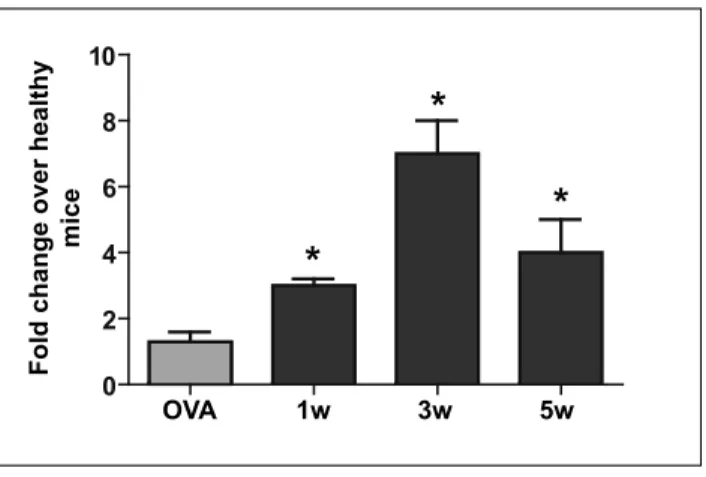
ARTICLE 1 Figure 1 Nlrp12 mRNA expression reaches a peak at third week post injection ... 40
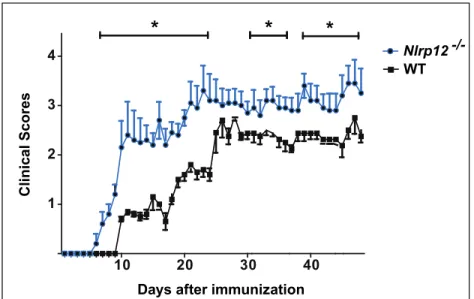
Figure 2 Nlrp12-/- mice exhibit exacerbated form of disease compared to WT mice ... 41
Figure 3 Photomicrograph pictures of Spinal Cords stained with GFAP ... 42
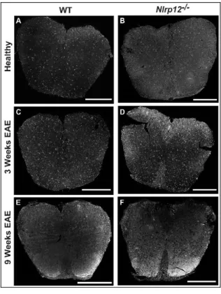
Figure 4 Photomicrograph pictures of Spinal Cords stained with Iba1 ... 43
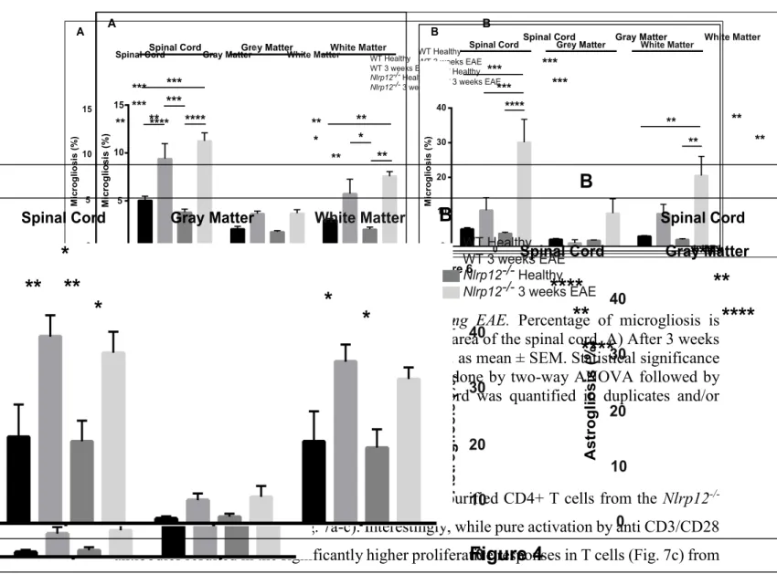
Figure 5 Percent level of Astrogliosis following EAE ... 43
Figure 6 Precent level of Microgliosis following EAE ... 44
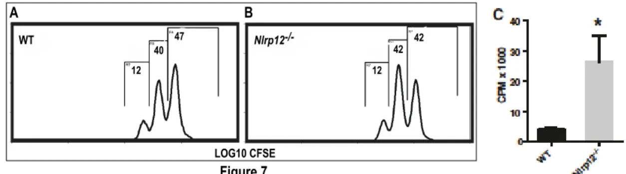
Figure 7 The proliferation of activated T cells from WT and Nlrp12-/- mice in vitro ... 45
Figure 8 IL-4 production by activated T cells from WT and Nlrp12-/- mice in vitro and in vivo ... 46
Figure 9 Nlrp12 deficiency augments expression of pro-inflammatory molecules in the CNS after EAE ... 47
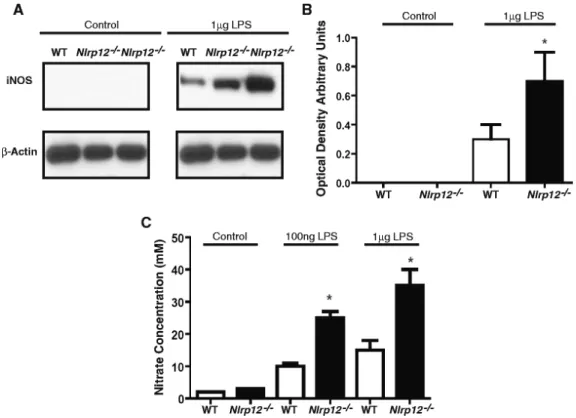
Figure 10 Expression of iNOS in primary microglia cells ... 48
Figure 11 TNF- and IL-6 concentrations following treatment with LPS in primary microglia cell ... 49
ARTICLE 2 Figure 1 Nlrp12 inhibits Th1 response in EAE. ... 67
Figure 2 Nlrp12 inhibits T cell proliferation and cytokine production by CD4+ T cells ... 68
Figure 3 Nlrp12 does not affect the differentiation of naïve CD4+ T cells to Th1 or Th17.69 Figure 4 T cell activation increases Nlrp12 mRNA expression ... 70
Figure 5 Nlrp12 inhibits phosphorylation of Akt and p65 in CD4+ T cells ... 71
Figure 6 Nlrp12 inhibits IL-2 synthesis by activated T cells ... 72
Figure 7 Nlrp12 does not modify mTOR activity ... 72
Figure 8 SpEAE in WT 2D2 mice is associated with spinal cord inflammation and demyelination. ... 74
Figure 9 Myelin-specific T cells infiltrate in the spinal cord of spEAE mice ... 75
Figure 10 Possible mechanism of action of Nlrp12 in regulating early TCR signaling pathways, T cell activation and proliferation. ... 78
Figure 11 Dual immunoregulatory function of Nlrp12 in EAE. ... 81
Figure S1 Nlrp12 genotyping of a Nlrp12-/- 2D2 mouse and a WT 2D2 mouse. ... 82
ix ARTICLE 3
Figure 1 Nlrx1-/-2D2 mice develop spEAE ... 89
Figure 2 Increased levels of IgG and frequency of B cells in the spinal cord of SpEAE mice ... 90 Figure 3 CNS inflammation associated with spEAE is more severe in Nlrx1-/-2D2 compared
to 2D2 mice ... 91 Figure 4 Activation of innate immune cells induce CNS inflammation and severe paralysis in Nlrx1-/-2D2 mice ... 93
Figure 5 Inflammation at the preclinical stages of spEAE is enhanced in the CNS of Nlrx1
-/-2D2 mice. ... 94 Figure 6 NLRX1 inhibits subclinical tissue damage and the generation of neurotoxic glia in the CNS. ... 95 Figure 7 NLRX1: implications for human MS. ... 97 Figure 8 NLRX1 inhibits early stages of CNS inflammation, prevents toxic glia activation,
and the onset of EAE ... 101 Figure S1 Nlrx1-/-APC activate T cells as efficiently as WT APC. ... 112
Figure S2 Nlrx1 inhibits T cell activation, proliferation, and differentiation to inflammatory subsets. ... 112
THESIS
Table 1 The immunopathogenesis of inflammatory and anti-inflammatory NLRs in MS patients and EAE mice.. ... 28
ARTICLE 2
Table 1 Nlrp12 does not prevent the development of spEAE. ... 73
ARTICLE 3
Table S1 Nlrx1-/- 2D2 model compared to previous models with CD4 T cells involvement.
... 114 Table S2 Demographic data of MS patients and healthy individuals. RRMS: replacing remitting MS. ... 115 Table S3 NLRX1 mutations identified in MS patients ... 115 Table S4 The primer sequences used for qPCR experiments ... 116
A
ADCC ALS ASC ATPB
BBB BIRC
CARD CATERPILLAR CCL5 CCL20 CCL21 CCR2 CCR5 CD CFA CIITA CLRs CNS CSFD
DAMPs DCs DD DMT DNA DRP1E
EAE ERKG
GDNF GFAPA
Antigen dependent cell cytotoxicity Amyotrophic Lateral Sclerosis
Apoptosis-associated Speck-like protein containing CARD Adenosine Triphosphate
A
Blood Brain Barrier
Baculoviral Inhibitory Repeat
B
Caspase-Recruitment Domain
CARD Transcription Enhancer, R (Purine)-binding, Pyrin, Lots of Leucine Repeats
Chemokine (C-C motif) Ligand 5 Chemokine (C-C motif) Ligand 20 Chemokine (C-C motif) Ligand 21 Chemokine (C-C motif) Receptor 2 Chemokine (C-C motif) Receptor 5 Cluster of Differentiation
Complete Freund’s Adjuvant Class II Transactivator C-type Lectin Receptors Central Nervous System Cerebrospinal Fluid
B
Danger Associated Molecular Patterns Dendritic Cells
Death Domain
Disease Modifying treatment Deoxyribonucleic Acid Dynamin related protein 1
B
Experimental Autoimmune Encephalomyelitis Extracellular Signal-Regulated Kinase
B
Glial-Derived Neurotrophic Factor Glial Fibrillary Acidic Protein
2 GWAS
H
HL60 HLA HMGB1I
IAP IBD ICAM-1 IFIH1 IFN IFN-1 IFN-1 IFN-1a IFN-1b IFN- IGF-1 IB IKK IKK IL IL-1 iNOS IP-10 IRAKL
LPS LRRsM
MAPK MBP MCP1 MHC MIP1 MMP-9 MOG MS MRI MyD88Genome-Wide Association Scan
B
Human Leukemia cell line 60 Human Leukocyte Antigen
High mobility group box 1 protein
B
Inhibitor of apoptosis protein Inflammatory Bowel Disease Intracellular Molecule-1
Interferon Induced with Helicase C domain 1 Interferon Interferon-1 Interferon-1 Interferon--1a Interferon--1b Interferon-
Insulin-like 1 Growth Factors Inhibitor of B Inhibitor of B kinase Inhibitor of B kinase Interleukin Interleukin-1 Interleukin-18
Inducible Nitric Oxide Synthase Interferon gamma-induced protein 10 IL-1R Associated Kinase
B
Lipopolysaccharide Leucine-rich repeats
B
Mitogen-Activated Protein Kinase
Myelin Binding Protein/Myelin Basic Protein Monocyte Chemoattractant Protein 1
Major Histocompatibility Complex Macrophage Inflammatory Protein 1 Matrix Metalloproteinase 9
Myelin Oligodendrocyte Glycoprotein Multiple Sclerosis
Magnetic Resonance Imaging
3
N
NACHT NAIP NAIP1 NAIP4 NBD NBS NF-B NIK NK NLR NLRC4 NLRP12 NLRX1 NO NODO
OPCsP
PAMPs PLP PML PMN PNS PPMS PRRs PYD PYPAF7R
RAG RANTES RIG1 RLR RNA ROS RRMSNAIP, CIITA, HET-E and TP1 domain NLP family Apoptosis Inhibitor Protein NLP family Apoptosis Inhibitor Protein 1 NLP family Apoptosis Inhibitor Protein 4 Nucleotide-Binding Domain Nucleotide-Binding Site Nuclear Factor-Kappa B NF-κB inducing kinase Natural Killer NOD-Like Receptor
NLR family CARD domain containing 4 NLR family Pyrin domain containing 12 NLR family X1
Nitric Oxide
Nucleotide-binding Oligomerization Domain
Oligodendrocyte Precursor Cells
Pathogen Associated Molecular Patterns Proteolipid Protein
Progressive Multifocal Leukoencephalopathy Polymorphonuclear
Peripheral Nervous System
Primary-Progressive Multiple Sclerosis Pathogen Recognition Receptors Pyrin domain
Pyrin-containing Apaf1-like protein 7
Recombination activating gene
Regulated upon Activation, Normal T cell Expressed and Secreted
Retinoic Acid Inducible Gene I RIG-Like Receptor
Ribonucleic Acid
Reactive Oxygen Species
4
S
SDF-1 SPEAET
TCR TGF- Th1 Th2 Th17 TIR TLR TNF TRAF Treg TUFMV
VCAMW
WTStromal cell-derived factor-1α Spontaneous EAE
T cell receptor
Transforming Growth Factor T helper 1
T helper 2 T helper 17
Toll/Interleukin-1 Receptor Toll-Like Receptor
Tumor Necrosis Factor-
TNF Receptor Associated Factor 3 Regulatory T cells
Tu Translation Elongation Factor, Mitochondrial
Vascular Cell Adhesion Molecule
B
5
1. I
NTRODUCTION1.1 Immune response in the CNS
Over the past decades, it was believed that the CNS is an immune-privileged site and peripheral immune cells were not allowed to enter to the CNS under physiological condition (Carson et al., 2006). Recently, this viewpoint was dramatically changed by the discovery of the lymphatic system in the brain's meninges, where immune cells and soluble mediators drain into the deep cervical lymph nodes and communicate with peripheral immune cells under both pathological and homeostatic conditions (Louveau et al., 2015). Instead of being surrounded by physical barriers that shield the CNS from any outside contacts, the CNS is protected by dynamic biological barriers that tightly regulate the crosstalk between the CNS and the immune system (Engelhardt et al., 2017). Here I summarize the mechanisms of the CNS-immune system crosstalk in the context of innate and adaptive immunity.
1.1.1. Innate immune response
The CNS innate immune compartment consists of biological barriers and the specialized resident cells that respond into the CNS infections and injuries. The dynamic biological barrier, known as the blood–brain barrier (BBB), controls the trafficking of the cells and molecules to the CNS. The endothelial cells are held together by tight junctions that control the diffusion of large (molecular weight bigger than 450 Da) and non-lipophilic molecules (Petty and Lo, 2002).
The CNS-resident cells are categorized in two groups: the nerve cells and the glial cells. Neurons receive synaptic inputs and transmit electrical signals down to the axons, the long projection of neurons that are covered by a protective myelin sheath. Glia cells are 10 times more numerous than neurons and include oligodendrocytes, astrocytes, and microglia (Nolte, 2002). Oligodendrocytes derived from oligodendrocyte precursor cells/progenitors (OPCs) are the cells that tightly wrapped around axons and make myelin sheath (Jacobson & Marcus, 2008). The axonal insulation by myelin increase the speed of signal transmission and serves as a protective layer against axonal degeneration (Fitzner et al., 2006).
Astrocytes are the most abundant glia in the CNS. Some of their processes, known as endfeet, make the last layer of the BBB in the brain parenchyma. Additionally, astrocytes support homeostasis and metabolism of neurons in several ways including formation and
6 maintenance of the BBB integrity; the regulation of extracellular ions and neurotransmitters; and synthesis of the neuronal metabolic substrates such as glycogen (Sofroniew, 2015). Astrocyte-mediated inflammation is associated with inflammatory responses that are characterized by robust proliferation and hypertrophy of astrocytes that is termed astrogliosis. Activated astrocytes release inflammatory cytokines such as IL-6, IL-1β, and TNF-α that affect the tight junctions of endothelial cells and control the passage of immune cells through the BBB. Moreover, astrocytes secrete different chemokines such as MCP-1, RANTES, IP-10, SDF-1, CCL20 and IL-8, which actively recruit a variety of leukocytes such as monocytes, neutrophils, DCs, and lymphocytes from the periphery into the CNS parenchyma. Therefore, astrocytes have all the features to regulate both an inflammatory response within the CNS and the influx and activity of leucocytes. Proinflammatory cytokines and chemokines also activate the major player of the CNS innate immunity, microglia (Correale and Farez, 2015a).
Microglia constantly monitor the CNS for various insults and orchestrates innate immune response within the CNS parenchyma (Herz et al., 2017). They are the only tissue-resident macrophages that origin from embryonic yolk sac at the very early stage of development, seed the brain and stay there into adulthood (Ginhoux et al., 2013). Interestingly, microglia have the ability for self-renewal and are not replenished from circulating monocytic progenitors (Lampron et al., 2013). The morphology of microglia differs from conventional macrophages by the presence of highly motile projections that constantly monitor their environment. Activated microglia increase the ability of phagocytosis and antigen presentation within the CNS (Hernandez-Pedro et al., 2013, Ransohoff and Cardona, 2010, Xanthos and Sandkuhler, 2014). In many CNS pathologies, the number of microglia often increases in a phenomenon that is called reactive microgliosis (Huber and Irani, 2015, Dendrou et al., 2015a). Furthermore, they release inflammatory mediators such as inducible nitric oxide synthase (iNOS), TNFα, IL-1β, IL-6, IL-12, which control the infections and recruit adaptive immune cells into the CNS (Gonzalez et al., 2014). The CNS also contains myeloid cells including macrophages and DCs that strategically positioned at the CNS barriers such as choroid plexus, perivascular space, and meninges (Hernandez-Pedro et al., 2013). These cells serve role of the antigen presenting cells (APC) that process the antigen and present peptide-MHC II complex to the T cells. Unlike other
7 organs such as liver, skin or intestine where dendritic cells (DCs) uptake the antigen and travel to the local lymph node, under normal conditions, there are no DCs in the CNS parenchyma and even microglia never leave the CNS (Hussain et al., 2014). Instead, there are other myeloid cells including macrophages and DCs that monitor the brain from outside of the parenchyma. These are the cells that communicate with adaptive immune cells such as T cells, which patrol the CNS from the outside of the tissue at the particular gateways; the meninges, perivascular spaces, and choroid plexus (Herz et al., 2017)
1.1.2. Adaptive immune response
Naïve T cells do not enter the normal or inflamed CNS (Goverman, 2009), indicating that activated T cells at the CNS barriers are licensed to enter the CNS parenchyma (Ransohoff and Engelhardt, 2012). T cell activation is initiated within the regional lymph nodes (cervical lymph nodes), where DCs process and present the antigen in both classes I and class II MHCs to naïve T cells, leading to the specific activation of CD4+ and CD8+ T cells. Depending on the type of cytokines present in T cell environment, naïve CD4+ T cells may differentiate into different T helper (Th) subsets including Th1, Th2, Th17, and regulatory T cells (Treg) that produce various cytokines including IFN, IL-4, and IL-17 respectively. Th subsets may affect CNS inflammation in different manners, in which Th1 and Th17 responses promote CNS inflammation, while Th2 response and regulatory T cells dampen the inflammatory response of T cells and protect CNS (Luckheeram et al., 2012). Th2 cells help B cells to form plasma cells that produce high-affinity pathogen-specific antibodies (Pennock et al., 2013). As a routine immunosurveillance activity, a small number of memory T cells can access the CNS via the blood–CSF barrier at subarachnoid space (Ransohoff et al., 2003). Macrophages at perivascular space present the CNS-derived antigens to memory T cells. In pathological condition, endothelial cells of the BBB increase the expression of several adhesion molecules including Vascular Cell Adhesion Molecule (VCAM-1) that support the recruitment of T cells into the CNS (Goverman, 2009, Selewski et al., 2010). As a result, lymphocytes including T cells and B cells are found in the CNS parenchyma only after CNS injuries, infections, neurodegenerative and autoimmune diseases such as multiple sclerosis (Kamm and Zettl, 2012).
8 1.2 Multiple sclerosis and its immunopathogenesis
1.2.1. Multiple sclerosis
Multiple sclerosis (MS) is a chronic autoimmune disease of the CNS, which is characterized by inflammation, demyelination, and axonal damage. It is the leading cause of chronic neurological disability in young adults (Dendrou et al., 2015a). Besides MS, there are rare forms of autoimmune demyelinating diseases including neuromyelitis optica (NMO), in which there is an adaptive immune response directed against the water channel aquaporin 4 (AQP4), and acute disseminated encephalomyelitis (ADEM), which is characterized by a monophasic and rapidly progressive immune mediated attack against the CNS (Hu and Lucchinetti, 2009).
The name of MS was inspired by the pathological signature of the disease, the presence of multiple sclerotic plaques in the white and gray matter of the brain and spinal cord (Murray, 2009). The first description of MS dates back to 1868 when the French neurologist, Jean-Martin Charcot, described MS symptoms and plaques (McAlpine and Compston, 2005, Ntranos and Lublin, 2016). As we know today, the pathological hallmark of MS is the formation of sclerotic plaques in the white and gray matter of the CNS, which is caused by several processes including inflammation, demyelination, astrocytic glial scars, axonal damage, and neuronal loss (Yadav et al., 2015, Kutzelnigg and Lassmann, 2014). Demyelinated axons can no longer support rapid conduction of action potential, which results in a disturbed transmission of nerve impulses (Ciccarelli et al., 2014). Therefore, MS manifests in a variety of clinical symptoms such as blurred or double vision, lack of neuronal muscle coordination, loss of balance, numbness, tingling, pain, fatigue, and difficulties in moving (Compston and Coles, 2002).
MS affects more than 2.5 million people worldwide (Marrie et al., 2015) and Canada has one of the highest rates of MS in the world (197/100,000 population) (Gilmour et al., 2018). It is most frequently diagnosed between the ages of 20 and 49. MS affects women 3 times more than men(Harbo et al., 2013). The clinical course of MS is different among patients. Relapsing-remitting MS (RRMS) is the common form of MS, which is found in about 70–80% of patients. In RRMS, there are clear episodes of inflammatory activity or relapses followed by remission. However, people with RRMS may develop a secondary progressive form of MS after a disease duration of about 10 years
.
A small percentage of9 patients may develop primary progressive MS (About 10%), which is characterized by continuous neurodegeneration from the onset without remission (Ciccarelli et al., 2014, Wegner, 2013).
1.2.2. Risk factors of MS ➢ Genetic factors
The prominent role of genetic factors in MS is shown in familial MS. For example, the monozygotic twins have a higher risk of developing MS (25-30%)(Hansen et al., 2005). Similarly, siblings of an MS individual are at least 7 times more likely to develop MS than the general population (Baranzini and Oksenberg, 2017). More than 200 loci associated with MS susceptibility have been discovered by genome-wide association scan (GWAS) performed by the International Multiple Sclerosis Genetic Consortium (Baranzini and Oksenberg, 2017). The main susceptibility locus is located within the Major Histocompatibility Complex (MHC) class II region. MHC or Human Leukocyte Antigen (HLA) genes code for cell surface molecules involved in antigen presentation to T cells and triggering the immune response (Files et al., 2015). Several studies have demonstrated that the Human Leukocyte Antigen (HLA)-DRB1*1501 has been constantly associated with MS in many populations (Alcina et al., 2012, Barcellos et al., 2006, Baranzini and Oksenberg, 2017). Moreover, many other immunologically relevant genes, particularly those involved in T cell response have been discovered, providing evidence that dysregulated T cell response is associated with MS (Sawcer et al., 2011).
➢ Gender factors
It is shown that women are at higher risk of developing MS, with a sex ratio of 3:1 female to male (Reich et al., 2018). Moreover, the predisposition to many autoimmune conditions is increased amongst women (Tiniakou, Costenbader, & Kriegel, 2013), suggesting that sex hormones may play a role in higher susceptibility of women to autoimmune diseases. A growing number of studies using EAE show that male hormone testosterone and the pregnancy hormone estriol induce anti-inflammatory and neuroprotective effects, supporting the potential therapeutic effect of these hormones in MS (Gold and Voskuhl, 2009). Findings from clinical studies of testosterone therapy in male MS patients and oral estriol in female MS patients are encouraging (Voskuhl et al., 2016, Sicotte et al., 2007). However, there are
10 concerns regarding potential side effects of sex hormone therapy in MS patients, such as carcinogenesis, thrombogensis, and changes in reproductive behavior in long-term use of these agents (Houtchens and Desai, 2018). More investigations need to be done before sex hormones can be used as MS therapeutics.
➢ Environmental factors
Many studies have proposed the influence of microbial infection such as viruses or bacteria on the susceptibility to MS. Among viral infections associated with MS, herpesviruses or retroviruses have been widely studied in MS (Gilden, 2005). Human herpesvirus 6 (HHV-6) and Epstein-Barr virus (EBV) are detected in MS plaques (Soldan et al., 1997). Additionally, a higher seroprevalence and higher titer of anti-EBV and anti-HHV-6 are reported in MS patients compared to age-matched controls (Virtanen and Jacobson, 2012). A recent study demonstrates fungal DNA from different species including Trichosporon mucoides in the CNS of MS patients (Alonso et al., 2018). Among bacterial agents, Mycoplasma pneumoniae, Chlamydia pneumoniae, and Staphylococcus aureus enterotoxins are associated with the development or exacerbation of MS (Libbey et al., 2014).
The gut microbiome is another environmental factor that has been demonstrated to significantly influence CNS health and disease (Zhu et al., 2017). Microbiota consists of all microbes including bacteria, archaea, fungi, and viruses that exist in human body and microbiome includes genomic, protein, or metabolite content of all the microbes (Shahi et al., 2017). It is shown that gut microbiome maintains the proper functions of CNS, including brain circuitry, neurophysiology, and behavior (Sharon et al., 2016). Gut microbiome also regulates the permeability of the blood-brain barrier (BBB)(Braniste et al., 2014). In addition, gut microbiota affects the function of immune cells, directing the immune response towards homeostasis or autoimmunity. For instance, gut microbiota influences the differentiation of T cells to inflammatory subsets (Th1, Th17) or regulatory T cells (Wu and Wu, 2012). Additionally, several reports demonstrate the critical role of gut microbiota in regulating the development of APCs, in which stimulates a subset of DCs that induce the differentiation of Th17 cells (Atarashi et al., 2008). These studies support the notion that gut microbiome contributes to the development or exacerbation of CNS inflammation.
In the context of MS, several studies have demonstrated gut microbial dysbiosis in MS patients with both enhancement and reduction of certain bacteria compared to healthy
11 controls (Chu et al., 2018). For example, Jangi et al. showed that Methanobrevibacter and Akkermansia are increased in MS patients, while Butyricimonas is decreased. The microbiota variations correlate with the expression of genes involved in interferon and NF-kB signaling, as well as DC maturation (Jangi et al., 2016). Interestingly, in transferring fecal content isolated from MS patients to a transgenic mouse model increased the incidence or severity of MS-like autoimmune disease. This finding suggests that gut microbiota from MS patients contain factors that exacerbate CNS inflammation and autoimmunity (Yadav et al., 2017). Global prevalence of MS shows a latitudinal gradient, where the farthest countries from the equator including the populations located at northern latitudes of Europe and North America including Canada and southern areas of Australia and New Zealand have a higher incidence of MS (Files et al., 2015; Rosati, 2001). Latitude gradient was explained by the lower levels of vitamin D in the countries located at northern latitude (O'Gorman et al., 2012). This is explained by the fact that the primary source of vitamin D in human is UVB radiation from sunlight, which varies according to latitude and season. Low vitamin D intake and its low serum level are associated with increased risk of MS (Munger et al., 2004, Munger et al., 2006). Interestingly, lower levels of vitamin D during MS relapses suggest the immunosuppressive role of vitamin D progression of MS (Alharbi, 2015). Mechanistically, vitamin D suppresses the activity of pro-inflammatory T cells including Th1 and Th17 cells and promotes the activity of regulatory T cells(da Costa et al., 2016, Zeitelhofer et al., 2017). In addition to its immunosuppressive activity, vitamin D also plays a role in the repair of damaged oligodendrocytes and induce remyelination(Shirazi et al., 2015). Therefore, vitamin D supplementation may have a therapeutic role in the treatment of MS (Alharbi, 2015).
Taken together, the complex interaction between genetic factors and environmental factors contributes in the etiology of MS.
1.2.3. MS: an autoimmune or a neurodegenerative disease?
Despite the plenty of research on the immunopathology of MS, the origin of the disease is still a matter of debates. The presence of autoreactive T and B cells in the CNS strongly supports the hypothesis that MS is primarily caused by an aberrant immune response against the CNS antigens, particularly myelin, in which chronic immune response cause
12 oligodendrocyte death and progressive demyelination (outside-in model of MS) (Figure 2) (Stys et al., 2012, McAlpine and Compston, 2005, Ntranos and Lublin, 2016). It is still unknown how myelin-specific T cells are activated in the periphery. There are studies that support the activation of myelin-specific T cells by infectious agents (molecular mimicry) or non-specific T cells by superantigens (bystander activation) (Cusick et al., 2012, Libbey et al., 2014). Molecular mimicry is the term coined for the activation of autoreactive cells by cross-reactivity between self-antigens and foreign agents (Oldstone, 2005), while bystander activation assumes that autoreactive cells are activated due to nonspecific inflammatory events that occur during infections. The gut microbiome is also important to balance and regulate the immune response (Bhargava and Mowry, 2014).
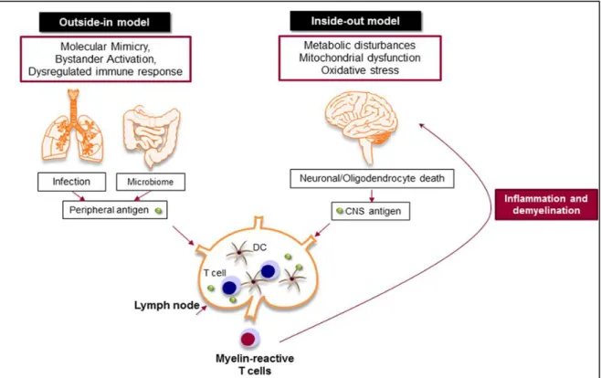
Inside-out model of MS supports the idea that MS is primarily a neurodegenerative disorder (Figure 1). In this model, the oligodendrocyte injury or death would be the trigger of the CNS inflammation that presumably begins in the absence of a direct immune attack (Stys et al., 2012, Patel and Balabanov, 2012). Oligodendrocytes are extremely vulnerable to oxidative stress due to their high metabolic rate, large intracellular iron stores, and low levels of anti-oxidative enzymes. Exposure to any stress reactions or metabolic disturbances leads to caspase activation and oligodendrocyte death (Patel and Balabanov, 2012). Oxidative stress also results in mitochondrial dysfunction, which causes axonal damage and oligodendrocyte apoptosis (Lassmann et al., 2012, Su et al., 2013). As a result, myelin antigens are released to the periphery (van Zwam et al., 2009, Engelhardt et al., 2016) and activate autoreactive T and B cells that cause further CNS damage (Figure 1) (Yadav et al., 2015, Kutzelnigg and Lassmann, 2014, Ciccarelli et al., 2014).
13
Figure 1. Outside-in and inside-out models of MS. Outside-in model supports the idea that T cells are activated in the periphery by pathogen-derived molecules (molecular mimicry) or non-specific bystander activation. The digestive-tract associated microbes are also important to balance and regulate the immune response. The activated T cells attack CNS and cause inflammation and neurodegeneration. Inside-out model argues that the CNS inflammation primarily begins in the absence of a direct immune attack, in which tissue damage or cell death within the CNS releases myelin antigen that triggers the immune response in the periphery (Gharagozloo et al., 2018a).
1.2.4. Immunopathogenesis of MS
The presence of autoreactive CD4+ T cells within CNS lesions is detectable in the early stages
of MS (Dendrou et al., 2015b). Th1 and Th17 cells are the main CD4+ T cell subsets implicated in disease (Legroux and Arbour, 2015). Th17 cells induce neuroinflammation by producing pro-inflammatory mediators such as IL-17, granulocyte/macrophage colony-stimulating factor (GM-CSF)(McGeachy, 2011), matrix metalloproteinases and CXCL8, a potent chemokine for recruiting neutrophils (Waisman et al., 2015). Interestingly, a recent study has identified T cells that express both IL-17 and IFN-γ cytokines in MS brain tissue, suggesting the pathological role of IL-17+ IFNγ+ T cells in human MS (Kebir et al., 2009).
14 CD8+ T cells represent the predominant T cell population in the lesions of human MS
(Sinha et al., 2015). CD8 cytotoxicity is mediated through cell surface Fas ligand or release of soluble molecules such as IFNγ, TNF-α, lymphotoxin, granzyme B and perforin. During inflammation, oligodendrocytes upregulate MHC I expression, Fas, IFN-γ and TNF-α receptors, result in direct cytotoxicity of CD8+ cells (Patel and Balabanov, 2012).
B cells and antibodies are also implicated in the pathogenesis of MS. Plasma cells produce specific antibodies to myelin antigens that initiate the complement cascade, leading to destruction, opsonization, and subsequent phagocytosis of the myelin sheath (von Büdingen et al., 2011). B cells also act as APCs to capture and present the antigens to autoreactive T cells.
Inflammatory mediators from activated T cells such as TNFα and IFNγ stimulate macrophages and microglia to produce more proinflammatory mediators such as iNOS, TNFα, and IL-1β. Activated macrophages and microglia produce reactive oxygen species (ROS) and NO radicals that are associated with demyelination and neuronal injury in MS lesions (Luo et al., 2017). There is also evidence that astrocytes promote demyelination by regulating peripheral immune cell trafficking, modulating BBB integrity, and being a source of chemokines and cytokines such as IL-12, IL-23, and IL-15, that mediate differentiation CD4+T cells into Th1 or Th17 and activates the cytotoxicity of CD8+ T cells. Astrocyte
activation induces the down-regulation of important proteins that compose the tight junctions of endothelial cells, such as claudin-5 and occludin, leading to the compromise of the BBB (Argaw et al., 2012). Astrocytes are the main source of the chemokine CCL20 or monocyte chemoattractant protein 1 (MCP1) that is a crucial chemoattractant for monocytes and T cells in EAE and MS (Correale and Farez, 2015b). The role of innate and adaptive immune cells in the immunopathogenesis of MS is summarized in Figure 2.
The above immunopathological mechanisms are involved in developing MS plaques, which are the hallmark of MS pathology and histologically classified as acute and chronic lesions (Popescu and Lucchinetti, 2012). Acute lesions are more common in the early stage of MS, characterized by demyelination, reactive astrocytes, and microglia (glial scar tissue), and perivascular and parenchymal inflammation. The majority of infiltrated cells in active lesions are macrophages, followed by lymphocytes including CD4+ and CD8+ T cells, B cells
15 associated with severe demyelination and axonal loss, oligodendrocytes death, while there are very few infiltrating leukocytes (Popescu et al., 2013).
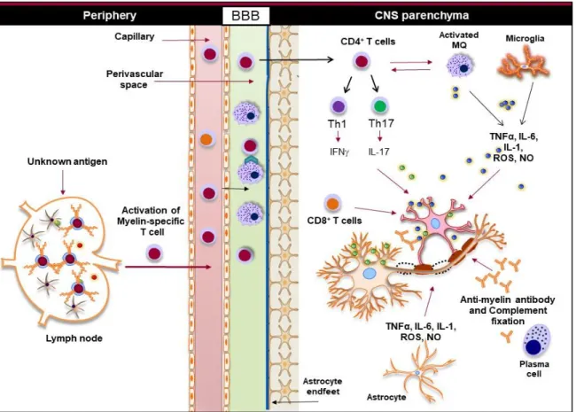
Figure 2. Innate and adaptive immunity in the pathogenesis of MS. Myelin-reactive T cells are activated in the periphery and accumulate in the perivascular spaces, where they are reactivated by the CNS myeloid cells, such as macrophages, and enter the CNS parenchyma. CD4+ T cells are differentiated to different inflammatory subsets, such as Th1, Th17, and
Th9, and once in the CNS, they promote the activation of CNS-resident innate immune cells, such as macrophages and microglia. Inflammatory mediators, such as TNFα, IL-6, IL-1, ROS, and NO, released from activated macrophages, microglia, and astrocytes damage oligodendrocytes and neurons, leading to demyelination. Activated cytotoxic CD8+ T cells
directly induce apoptosis in oligodendrocytes, while plasma cells produce anti-myelin antibodies that activate the complement system and damage oligodendrocytes (Gharagozloo et al., 2018a).
1.2.5. MS animal models
The understanding of MS immunopathogenesis and current drug therapies have been mainly derived from the studies in experimental autoimmune encephalomyelitis (EAE). EAE is a CD4+ T cell-mediated autoimmune disease consisting of Th1- and Th17-mediated tissue
16 damage, mononuclear cell infiltration and demyelination within the CNS (Miller, Karpus, & Davidson, 2010). Major histopathological features of EAE is the presence of multiple focal lesions within the CNS, particularly, in the spinal cord, associated with astrogliosis and microgliosis. The EAE clinical signs are scored from 0-5, depending on the severity and progression of the clinical symptoms. Clinical scores are given using the following scale: 0, no sign of disease; 1, limp tail or weakness in limbs; 2, limp tail and weakness in limb; 3, partial limb paralysis; 4, complete limb paralysis (Miller and Karpus, 2007). Depending on the research design, EAE can be induced by different protocols.
Active EAE consists of two phases; the induction and the effector phase. The induction phase involves the priming of CD4+ T cells following subcutaneous immunization of mice
with myelin antigens such as Myelin Oligodendrocyte Glycoprotein (MOG), emulsified in Complete Freund's Adjuvant (CFA) (Miller et al., 2010), followed by a pertussis toxin injection intraperitoneally (Miller and Karpus, 2007). Typical EAE onset is 9 to 14 days after
immunization. The clinical scores reach the highest usually 20-27 days after immunization. Passive EAE is induced by adoptive transfer of myelin-specific T cells to the recipient mice. Myelin specific T cells are purified from lymph nodes and spleen of animals with active EAE and transferred intravenously or intraperitoneally into the recipient mice (Stromnes and Goverman, 2006). To study the biology of T cell response, T cells are transferred to the Rag -/- recipients, which are lymphocyte-deficient mice (Miller and Karpus, 2007).
Spontaneous EAE develops in a variety of T cell receptor (TCR) transgenic mice including the MBP TCR transgenic (Lafaille et al., 1994), the 5B6 SJL/PLP TCR transgenic (Waldner et al., 2000), and the 2D2 C57BL/6/MOG35-55 TCR transgenic (Bettelli et al.,
2003). TCR transgenic animals develop a variety of symptoms ranging from conventional clinical and histological signs of EAE to optic neuritis (2D2 mice) (Bettelli et al., 2003). Since TCR transgenic mice have already high numbers of myelin-specific T cells, the mice do not need to be immunized to develop clinical signs of EAE. Therefore, this model is very useful for the study of predisposing factors of MS such as genetic and environmental factors (Ben-Nun et al., 2014).
17 To date, MS has no cure but there are approved medications for MS. Approved medications in this category mainly reduce the replace rates and delay the progression of the disease (Thorpe et al., 2015). They have mainly anti-inflammatory effects and are more effective in the early phases of disease development. Therefore, the majority of disease-modifying treatments (DMT) that are currently used are effective in the treatment of RRMS (Dörr and Paul, 2015). Based on their risk-benefit profile, DMT can be divided into first-line or second-line of treatment. First-second-line includes interferon β, glatiramer acetate, dimethyl fumarate, teriflunomide. Second-line of treatment includes fingolimod, natalizumab, alemtuzumab, and ocrelizumab. The only approved DMT for PPMS is ocrelizumab (Zimmermann et al., 2018).
Interferon β is the first approved DMD for RRMS that includes interferon β-1a (Avonex®, Rebif®), interferon β-1b (Betaseron®, Extavia®), and peginterferon β-1a
(Plegridy®) (Dörr and Paul, 2015). Interferon β-1a is a recombinant cytokine produced by
mammalian cells, while interferon β-1b is produced in bacteria. Peginterferon β-1a is an interferon β-1a conjugated to a polyethylene glycol (PEG) molecule to increased its bioavailability(Hoy, 2015). All forms of interferon β are injected subcutaneously except Avonex® that is injected intramuscularly. The exact mechanism of interferon β therapeutic
function is still unclear. However, it is shown that interferon β negatively controls a wide range of immunological processes, such as Th1 and Th17 response, production of inflammatory cytokines, and BBB permeability (Boiko et al., 2002).
Glatiramer acetate (Copaxone®) is a synthetic peptide composed of 4 amino acids and
structurally similar to the myelin basic protein. Its exact mechanisms of action are still unknown, however, there is evidence showing that it shifts the T cell response towards Th2, thereby suppress inflammatory Th1 response (Ziemssen and Schrempf, 2007).
Dimethyl fumarate (DMF, Tecfidera®) is an oral therapy for RRMS (Montes Diaz et al., 2018). DMF exert multiple functions including immunomodulating, anti-inflammatory and anti-oxidative activities. DMF is neuroprotective and improves cell survival via activating the transcription factor nuclear factor erythroid-derived 2 (Nrf2) that induces the transcription of antioxidant genes. Moreover, DMF inhibits NF-B signaling pathway (Linker et al., 2011).
18 Teriflunomide (Aubagio®) is an oral therapy that inhibits pyrimidine synthesis in proliferating lymphocytes, limiting inflammatory effects on the CNS. Importantly, teriflunomide is not a DNA intercalating agent and has no effect on cell viability (Bar-Or et al., 2014). However, it is a specific blocker of the mitochondrial enzyme dihydro-orotate dehydrogenase (DHODH) that are highly expressed in proliferating lymphocytes(Bar-Or et al., 2014).
Fingolimod (FTY720, Gilenya®) is the first approved oral treatment for MS. Structurally, it is the analog of sphingosine-1-phosphate (S1P) and blocks lymphocyte trafficking. When Fingolimod binds to S1P receptor, it causes receptor internalization and prevents the egress of activated lymphocytes from the lymph node (Wingerchuk & Lucchinetti, 2007).
Natalizumab (Tysabri®) is the first humanized monoclonal antibody approved for MS treatment, delivered by intravenous infusion. It binds to alpha-4-integrin and blocks leukocyte adhesion to vascular cell adhesion molecule (VCAM), resulting in the prevention of T cell infiltration into the CNS (Alper & Wang, 2009; Clerico et al., 2017). The serious side effect of this drugs is its potential to reactivate latent JC virus, which can result in progressive multifocal leukoencephalopathy (PML) and death (Banwell & Anderson, 2005; Clerico et al., 2017).
Alemtuzumab (Lemtrada®) is a humanized monoclonal antibody targeting CD52-antigen on the surface on leukocytes, mainly mature lymphocytes, monocytes, dendritic cells, and granulocytes. The effector mechanisms of alemtuzumab involve immunomodulation through the resetting the immune system through the depletion and repopulation of lymphocytes. Depletion of CD52 positive cells is performed via antibody-dependent cellular cytotoxicity, complement-mediated cell lysis, and apoptosis. (Willis and Robertson, 2016) Ocrelizumab (Ocrevus®) is an anti-CD20 humanized monoclonal antibody that is recently approved for the treatment of RRMS. Ocrelizumab is the only approved treatment for PPMS. It targets mature B cells by binding to CD20 and results in depletion of circulating B cells (Mulero et al., 2018).
The DMT that I listed above mainly target the immune response in the periphery through depleting lymphocytes, inhibiting their proliferation or blocking their activity (Torkildsen et al., 2016). None of the DMT directly target inflammation in the CNS cells. For that reason,
19 they are inefficient in the treatment of progressive MS, which is driven mainly by the inflammation within the CNS (Mahad et al., 2015). A better understanding of the pathological mechanisms of inflammation and neurodegeneration within the CNS is needed to develop therapies that effectively treat patients with progressive MS. Next, I review the regulators of inflammation and their contributions to MS immunopathogenesis.
1.3 The Regulators of Inflammation: NLRs
Neuroinflammation in MS is thought to be triggered by infection and/or tissue damage (Hernandez-Pedro et al., 2016). Pattern recognition receptors (PRRs) are a complex network of receptors that are mainly expressed in innate immune cells and can detect both triggers of infection and stress. PRRs are able to recognize unique molecular structures in the cell wall or nuclei components of microorganisms, known as pathogen-associated molecular patterns (PAMPs) as well as molecules that are released from damaged or stressed cells, known as damage-associated molecular patterns (DAMPs). There are four major families of PRRs in the cells, including Toll-like receptors (TLRs), C-type lectin receptors (CLRs), RIG-I-like receptors (RLRs), and the nucleotide-binding domain (NOD) receptors (NLRs) that are expressed either on the cell surface or inside the cells (Mogensen, 2009). The PRRs initiate an inflammatory response upon recognition of PAMPs and/or DAMPs, which leads to the secretion of cytokines and chemokines, recruitment of phagocytes, and induction of autophagy or pyroptotic cell death (Bortoluci and Medzhitov, 2010).
TLRs are expressed in most cell types, either at the cell surface (TLR1, 2, 4, 5, 6, 10) or in endosomes (TLR3, 7, 8, 9). TLRs can detect a variety of molecules, including proteins, lipopeptides, and nucleic acids (single-stranded RNA, double-stranded RNA, or CpG DNA). Ligand detection by TLRs initiates intracellular signaling cascades that activate interferon regulatory factor (IRF) family members or NF-B. In macrophages, the activation of TLRs initiates phagocytosis and increases the production of ROS, which facilitates rapid clearance of microbes. Moreover, activation of NF-B signaling pathway leads to production of chemokines that recruit additional neutrophils and monocytes to the site of immunological challenge. In DCs, TLR stimulation induces the expression of co-stimulatory receptors and causes their migration to the draining lymph nodes, where they meet and activate naïve antigen-specific T cells. Depending on the type of cell that receives the stimuli, TLR
20 activation can have several outcomes that result in inflammation and activated immune response (Dowling and Mansell, 2016).
CLRs are another family of PRRs. CLRs bind to carbohydrate structures, including mannose, fucose, and glucan on pathogens. They are mainly expressed by APC such as monocytes, macrophages, and DCs (Dunkelberger and Song, 2010). Binding of pathogens to CLRs leads to its internalization and degradation and subsequent antigen presentation. Mannose-binding lectin is a CLR that activates the complement system. This multipotent system of innate immunity generates three broad effector pathways: direct lysis of pathogens, generation of potent pro-inflammatory anaphylatoxins, and opsonization and clearance of target cells (Dunkelberger and Song, 2010).
RLRs are cytosolic nucleic acid PRRs expressed in both immune and non-immune cells that sense cytoplasmic RNA.When cells are infected by a virus, viral dsRNA is generated in the cytoplasm during the course of replication. Infected cells sense dsRNA by RLRs and activate anti-viral signaling pathways. There are three members in RLRs family; RIG-I, melanoma differentiation associated gene 5 (MDA5), and laboratory of genetics and physiology 2 (LGP2) (Sparrer and Gack, 2015).
NLRs are the most recently discovered group of PRRs (Martinon and Tschopp, 2005, Inohara and Nunez, 2003) that sense a wide variety of PAMPs and DAMPs inside the cells. NLRs are evolutionarily conserved in many species, from sea urchins to plants and mammals, which suggests that NLRs are efficient and successful molecules in innate immunity against a wide variety of invaders. NLRs include 23 members in humans and at least 34 members in mice (Franchi et al., 2009).
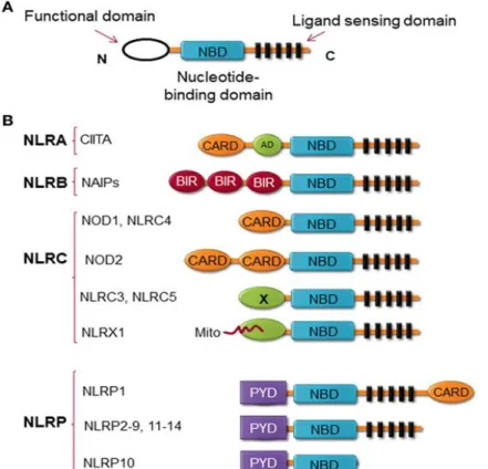
Structurally, NLRs consist of three highly conserved domains characterized by the C-terminal leucine-rich repeat (LRR), which is essential for ligand sensing; the central nucleotide binding ATPase domain NACHT/NBD (also known as NOD), which promotes oligomerization and activation, and the N-terminal domain, which contains either a caspase recruitment domain (CARD) or pyrin domain (PYD) and drives downstream signaling (Figure 3) (Allen, 2014). Members of the NLR family are categorized into at least 4 sub-families based on the structure of the N-terminal domain (Figure 3), including (i) NLRA or Class II transactivator (CIITA) contains acidic transactivation domain, (ii) NLRBs contains baculovirus inhibitor of apoptosis protein repeat (BIR), (iii) NLRC possesses CARD or an
21 undefined domain, (iv) and NLRP contains pyrin domain (Kawai and Akira, 2009, Di Virgilio, 2013, Barbe et al., 2014).
NLRs are also categorized based on their function, either as positive or negative regulators of inflammation (Figure 4). Additionally, there are NLRs such as CIITA and NLRC5 that indirectly regulate the immune response by tuning the expression of MHC II and I on APCs (Downs et al., 2016). Since inflammation is the hallmark of MS pathology, next we discuss how NLRs regulate the inflammation in MS.
Figure 3. NLRs structure. (a) The general structure of NLRs, consists of 3 domains, including functional domain, nucleotide binding and oligomerization domain, and ligand sensing domain. (b) Classification of NLRs based on the nature of their functional domain: NLRA, an acidic transactivation (AD) domain; NLRB, a baculovirus inhibitor of apoptosis protein (IAP) repeat (BIR); NLRC, a caspase-recruitment and activation domain (CARD); and NLRP, a Pyrin domain (PYD). In NLRC subfamily, the X displays an unknown domain that has no homology with the other NLR members. Mito is the mitochondria-localization sequence that directs NLRX1 to the mitochondria (Gharagozloo et al., 2018a).
22 Figure 4. Functional Characterisation of NLRs. NLRs can be classified depending on their mechanism of action to inflammasome and non-inflammasome forming NLRs. The inflammasome forming NLRs assemble inflammasome that activates Caspase-1 and promotes the production of inflammatory cytokines, IL-1β and IL-18. In the group of non-inflammasome forming NLRs, some NLRs regulate MHC II expression, while other NLRs regulate NF-B signaling. The regulators of NF-B consist of NLRs that enhance (NOD-1, NOD-2) or inhibit (NLRP12, NLRX1) NF-B signaling pathway. The negative NLRs, NLRP12 and NLRX1, can inhibit both inflammasome-dependent and -independent cytokine production. NLRC5 and NLRP12 have been described to influence both inflammasome and non-inflammasome signaling pathways in a cell- and stimuli-dependent fashion (Gharagozloo et al., 2018a).
1.3.1 NLRs as positive regulators of inflammation
NLRs such as NLRP1, NLRP3, NLRP6, NLRP7, NLRP12, NLRC4, and NAIP are positive regulators of inflammation through the formation of inflammasomes (Latz et al., 2013). NLRP3 inflammasome is the best-characterized form of the inflammasome, which is activated in two steps; the first step is priming the cells by PAMPs or DAMPs via TLRs, leading to the activation of nuclear factor-kB (NF-B) signaling that triggers the expression of inflammasome-related components, including NLRP3, pro-IL-1β, and pro-IL-18. The second step is oligomerization of NLRP3 and its association with an adaptor protein ASC, and pro-caspase-1. This complex triggers the activation of caspase-1 that cleaves pro-IL-1β and pro-IL-18 into their mature and secretable forms, IL-1β and IL-18 (Shao et al., 2015). Activation of the NLRP3 inflammasome also results in pyroptosis, a caspase 1-dependent cell death, which is a highly inflammatory form of cell death. Pyroptosis results in cell lysis
23 and the release of cytosolic components into the extracellular environment (Miao et al., 2011).
Previous studies showed that NLRs and their adaptors could positively influence the development and the severity of EAE (Gris et al., 2010). Deletion of Nlrp3, ASC, or the caspase-1 gene resulted in protection against EAE (Shaw et al., 2010, Gris et al., 2010). NLRP3 causes severe inflammatory symptoms in EAE by producing more IL-1β and IL-18, which stimulate the development and activation of Th1/Th17 cells and enhance their infiltration into the spinal cords (Gris et al., 2010). In an alternative pathway, NLRP3 inflammasome engages caspase-8 instead of caspase-1 (Antonopoulos et al., 2015). The importance of NLRP3-caspase-8 inflammasome was recently shown in the production of IL-1β by T cells that support the survival of Th17 cells in EAE (Martin et al., 2016). Moreover, NLRP3 inflammasome in APCs played a critical role in upregulating chemotactic proteins, such as osteopontin, CCR2, and CXCR6 in Th1 and Th17 cells, thereby inducing T cell migration to the CNS in EAE (Inoue et al., 2012).
Many reports demonstrate the role of the NLRP3 inflammasome in MS patients. The expression of caspase-1 and IL-18 are elevated in peripheral mononuclear cells from MS patients compared to cells from healthy controls (Huang et al., 2004). Moreover, the levels of IL-1β are upregulated in CSF of MS patients and correlated with the progression of MS (de Jong et al., 2002). MS treatments such as glatiramer acetate or IFNβ elevate the levels of endogenous IL-1 receptor antagonist in MS patients (Burger et al., 2009, Nicoletti et al., 1996). IFNβ treatment is also shown to attenuate the course and severity of MS by reducing the activity of NLRP3 inflammasomes via suppressing caspase-1 dependent IL-1β secretion (Malhotra et al., 2015). These findings collectively demonstrate that NLR proteins can exacerbate MS, either via formation of inflammasome or stimulation of inflammatory pathways such as NF-B and MAPK. Here, I review the biological activities of NLRs that negatively regulate inflammation. .
1.3.2 NLRs as negative regulators of inflammation
Some members of NLRs family including NLRP12, NLRX1, NLRP6, and NLRC3 inhibit the inflammatory signals that are triggered by PRRs following the recognition of PAMPs and DAMPs in the cells. Here I discuss the biological features of 2 inhibitory NLRs, NLRP12 and NLRX1.
24 1.3.2.1 NLRP12
NLRP12 is a pyrin-containing NLR protein that initially was identified in HL60 human leukemic cell line(Shami et al., 2001). Later, two research groups simultaneously cloned the full-length sequence of human NLRP12 naming it PYPAF7(Wang et al., 2002) or Monarch-1 (Williams et al., 2003), which eventually the HUGO Gene Nomenclature Committee (HGNC) approved the name of NLRP12 for this gene. The Nlrp12 expression is highly restricted to bone marrow and peripheral blood leukocytes, particularly polymorphonuclear cells that express much higher levels of Nlrp12 than mononuclear cells (Shami et al., 2001, Zamoshnikova et al., 2016, Lich et al., 2007).
Since the discovery of NLRP12, there have been contrasting reports that demonstrate both pro-inflammatory and anti-inflammatory roles of NLRP12 in cell-type and stimuli-specific manners (Lukens et al., 2015, Allen et al., 2012, Vladimer et al., 2012, Silveira et al., 2016). Early studies showed that NLRP12 is an inflammatory NLR that interacts with ASC to form inflammasome, leading to caspase-1 activation and release of mature IL-1β. Evidence for the involvement of NLRP12 in inflammasome formation and activation are largely derived from in vitro studies where the expression of NLRP12 was significantly elevated (Wang et al., 2002). Recent studies show the role of NLRP12 in activation of inflammasome by intracellular pathogens such as Yersinia Pestis and Plasmodium infection (Vladimer et al., 2012, Ataide et al., 2014). The formation of the inflammasome by NLRP12 seems to be pathogen specific, as NLRP12 is not involved in activating the inflammasome by other pathogens such as Salmonella, Klebsiella, Escherichia, Mycobacterium, and Listeria species (Tsuchiya et al., 2010, Zaki et al., 2014, Allen et al., 2013). Taken together, these studies establish a biologically relevant role for the NLRP12 inflammasome in innate immune responses against pathogens; however, the exact molecule that triggers NLRP12 inflammasome remains unknown.
On the other hand, there are studies that demonstrate NLRP12 as a negative regulator of inflammation through the inhibition of NF-B signaling in innate immune cells. It is shown that the activation of human peripheral blood granulocytes and monocytes by TLR4 or TLR2 agonists (E. coli LPS or synthetic lipopeptide Pam3Cys respectively) reduces the expression of NLRP12 (Williams et al., 2003). Moreover, the expression of NLRP12 declines significantly in myeloid cells (THP-1 human monocytic cell line) after in vitro stimulation
25 with live bacteria such as Mycobacterium or Plasmodium or cytokines such as TNFα or IFNγ (Williams et al., 2005). When NLRP12 is knocked down in THP1 cells using shRNA, the expression levels of pro-inflammatory cytokines significantly increase following LPS or M. tuberculosis treatment (Williams et al., 2005). A transcriptional repressor called B lymphocyte-induced maturation protein-1 (Blimp-1) is induced by TLR stimulation and downregulates NLRP12 expression by binding to NLRP12 promoter and recruiting histone deacetylases (Lord et al., 2009, Shi et al., 2016). These findings suggest that NLRP12 plays an anti-inflammatory role during inflammation and its expression is down regulated to induce the most effective immune response against pathogens.
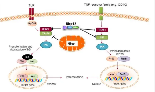
Mechanistically, NLRP12 was shown to suppress both canonical and non-canonical NF-κB signaling pathways. Canonical pathway is activated by a number of signals through TNFR, IL-1R, or TLR, which results in translocation of the RelA/p50 subunits to the nucleus. NLRP12 inhibits hyperphosphorylation of receptor-associated kinase (IRAK-1) that triggers IκBα degradation and p50 nuclear translocation (Zaki et al., 2011, Williams et al., 2005). The non-canonical NF-B pathway is triggered by signaling through receptors such as CD40, LT𝛽R, or BAFF-R. The signal activates NF-κB inducing kinase (NIK) and IKKα, which leads to p100 cleavage and nuclear translocation of p52 dimers. In non-canonical NF-κB signaling pathway, NLRP12 interacts with TRAF3 and NIK, which leads to the degradation of NIK and subsequent reduction of p100 cleavage to p52 (Figure 5)(Lich et al., 2007, Allen et al., 2012). ATP binding to NLRP12 is crucial for its inhibitory function, as the cells with an NBD mutant form of NLRP12 are not able to inhibit NF-κB activation. As a result, they produce high levels of pro-inflammatory cytokines and chemokines (Ye et al., 2008). Considering the regulatory function of NLRP12 in innate cells, Nlrp12-/- mice were
shown to be highly susceptible to the inflammatory disease of intestine such as experimental colitis and colon cancer (Allen et al., 2012, Zaki et al., 2011). This is due to the increased activation of NF-κB in macrophages of Nlrp12-/- mice, which results in the production of
26
Figure 5. Anti-inflammatory NLRs inhibit NF-B activation. Both NLRX1 and NLRP12 inhibit the activation of NF-κB canonical pathway following TLR stimulation. Nlrx1 interacts and inhibits TRAF6, while NLRP12 inhibits the phosphorylation of IRAK-1. NLRP12 can also inhibit non-canonical NF-κB signaling through regulation of TRAF3 and NIK (Gharagozloo et al., 2018a).
1.3.2.2 NLRX1
NLRX1 is the recently characterized member of NLRs that is ubiquitously expressed and uniquely localized in the mitochondria (Moore et al., 2008). Initial studies showed that NLRX1 was located in the outer membrane of mitochondria (Moore et al., 2008), however, later studies by two independent groups demonstrated that NLRX1 is located in the matrix of mitochondria (Rhee et al., 2013, Arnoult et al., 2009). The localization of NLRX1 in mitochondria is due to the presence of a functional N-terminal mitochondrial-addressing sequence (UQCRC2), which allows the targeting of NLRX1 to the mitochondrial matrix (Arnoult et al., 2009). A study by Tattoli et al., showed that NLRX1 induces the production of ROS in the cells treated by TNFα and double-stranded RNA, which result in increased activation of NF-κB inflammatory pathway (Tattoli et al., 2008). On the other hand, Xia et al. reported that NLRX1 acts as a negative regulator of NF-κB signaling.They showed that