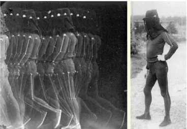
Figure 1. Decomposition of a motion thanks to a succession of pictures. (E. Muybridge).
Figure 2. Decomposition of a motion thanks to a series of photographic pictures on a single plate. (E.-J. Marrey).
Using 3D to understand human motion
C. Schwartz, B. Forthomme, O. Brüls, V. Denoël, S. Cescotto, J.L. Croisier
LAMH, Laboratoire d’Analyse du Mouvement Humain University of Liège
Liège, Belgium Abstract—The understanding and tracking of human motion has
been a subject of interest in the scientific community for more than one century. The long history of human motion analysis comes from the large scope of applications of such measurement that can be found in medicine, biomechanics, sport, ergonomics, and even civil engineering. More recently, those technologies have also been widely exploited for the development of animation movies and games. Needless to say, the techniques used one century ago significantly differ from those used today. This paper describes in a first part the evolution of the technological capabilities for motion analysis and the actual limitations. From this analysis and in a second part, we describe the experience related to the creation of a motion analysis laboratory at the University of Liège and show how such a platform could be the center of a multidisciplinary research and provide valuable information to various communities.
Keywords- biomechanics; motion capture; sport gesture I. INTRODUCTION: WHY MEASURING HUMAN MOTION?
The first and obvious application of measuring the human body is to provide information about how the bony segments move relatively one to another for life science. Thanks to the development of new technologies, researches on the human body were progressively extended from the understanding of the whole body behavior to the physiological study of muscle actuation, bone development, and related pathologies. However, the understanding of how muscles are recruited during a motion, of what happens in a joint and even the definition of “normal” vs. “pathological” gesture is still incomplete. In this view, a better understanding, through appropriate measurement of the human motion could have important implications in topics such as rehabilitation, orthopedics, and the development of specific training programs for athletes.
The measurement of the human motion is also needed to understand the way we interact with our environment. The everyday objects we use (tools, chairs, handles, etc.) as well as our surrounding environment (buildings, roads…) are mostly developed on computers and designed for our own use. That is one of the reasons for which ergonomics has become more and more important during the conception process. As an input to these models and/or simulations, the knowledge of what a “normal” or an extreme motion would be in specific situations
is needed. Recording the real motion of subjects is therefore very valuable in many engineering concerns.
Other applications of human motion recording also exist such as animation. For games or movies it is useful to record separately the motion of actors and then combine real-life shots with the captured data. Actually, a number of recent movies (such as Harry Potter, Pirate of the Caribbean, Star Wars III) using special effects have been realized using motion capture cameras during their development.
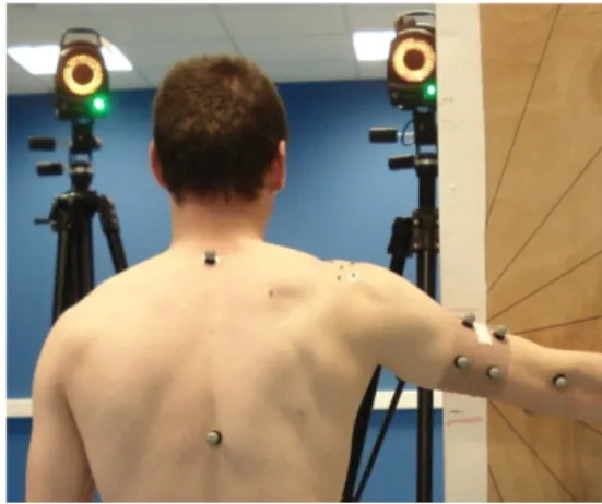
Figure 3. Passive markers.
Figure 4. Infrared camera.
II. HOW TO FOLLOW HUMAN MOTION?A HISTORICAL POINT OF VIEW
The history of motion analysis is closely linked to the technological evolutions. A motion, quick or slow, is a succession of momentary positions. To be analyzed, the motion needs to be stopped or say in another way expressed as a succession of instantaneous positions. Photography was the first way to follow motions. At the end of the XIXth century, E. Muybridge has become famous for his photographic decomposition of motion (Fig. 1).
His technique inspired E.-J. Marrey, who was probably one of the first to define a scientific approach for motion analysis. One of his numerous contributions was the use of marked suits (Fig. 2), which is strikingly reminding skin markers, as commonly used nowadays.
The discovery of X-rays in 1895 and its application for medical imaging offered a completely new approach for motion analysis. Indeed, for the first time, it was possible to actually see the bones. However, the main disadvantage of this technique is the loss of the dynamic nature of the motion, as the subject has to be still during acquisition. Fluoroscopy now allows “dynamic” X-rays acquisitions up to 30 Hz. However the aforementioned methods only allow 2D analysis of the motion and projection artifacts make it difficult to obtain an accurate description of the bone motion in space [1].
The apparition of 3D imaging techniques during the second half of the XXth century like CT scans and MRI gave access to 3D representation of the bones. Medical imaging has consequently been a real breakthrough but these systems still do not allow 3D dynamic acquisitions. Palpation of landmarks on the bones presents the same kind of limitations.
Later, progress in surgery and anesthesia has allowed one of the only available 3D method for direct dynamic capture of the bone motion. This method consists in drilling pins in the bones [2]. A localization system attached to the pins gives an accurate description of the bone motion. However, special care must be taken in the placement of the pins in order to avoid disturbing the natural subject motion. However, in spite of its interest, this methodology cannot be widely used because of its invasivity.
In order to get a 3D and dynamic measurement of bones motion in a non-invasive way, other systems have been developed. One inexpensive and simple solution is the use of electromagnetic systems (like Isotrak – Polhemus Inc., Flock of Birds – Ascension Technology), which transmit their position and orientation. The motion analysis requires the fixation of one sensor on each segment of the human body. This technology is easy to use but unfortunately quite sensitive to the motion of the skin, especially during longitudinal rotations.
Most of the recent studies concerning motion tracking use optical systems. Optical systems are based on the localization of 3 markers per bony segments. The markers are placed on the surface of the skin. Contrary to the electromagnetic systems, which do not need direct view between the emitter and the receptor, markers have to be seen by the cameras. As a consequence, the tracking of complex motions may involve multiplying the number of cameras in order to be sure to cover
the whole field under investigation. There are two main types of optical systems. The first type, and probably most common one, includes passive systems (like Vicon - Oxford Metrics, Elite – BTS Engineering, Oqus – Qualisys Medical AB), see Figs. 3 and 4. They use reflective markers, which reflect the infrared light emitted by the cameras. When using this system, the markers have to be identified manually and if two markers are too close, the system may be unable to distinguish them. The other possibility is to use active markers [3] (like Coda Motion – Charnwood Dynamics, Optotrak – Northern Digital). The cameras identify the markers one by one when they emit their own infrared signal. The main disadvantages of this method are the limited number of markers that can be used at the same time as well as the necessity to connect energy cables to the markers. For all passive and active systems, the placement of the markers on the skin should avoid as much as possible areas sensitive to large skin motions.
III. MULTIDISCIPLINARY RESEARCH AT LAMH
(LABORATOIRE D’ANALYSE DU MOUVEMENT HUMAIN)
A. Description of the laboratory
The creation of a human motion analysis laboratory (Laboratoire d’Analyse du Mouvement Humain – LAMH) at the University of Liège (ULg) aims to develop a multidisciplinary research, covering various topics of the 3-D analysis of human motion. This transversal project is carried out by the Faculty of Medicine (Prof. J.L. Croisier, Prof. B. Forthomme) and the Faculty of Applied Science (Profs. O. Brüls, S. Cescotto, V. Denoël).
Figure 5. Coda Motion unit CX1.
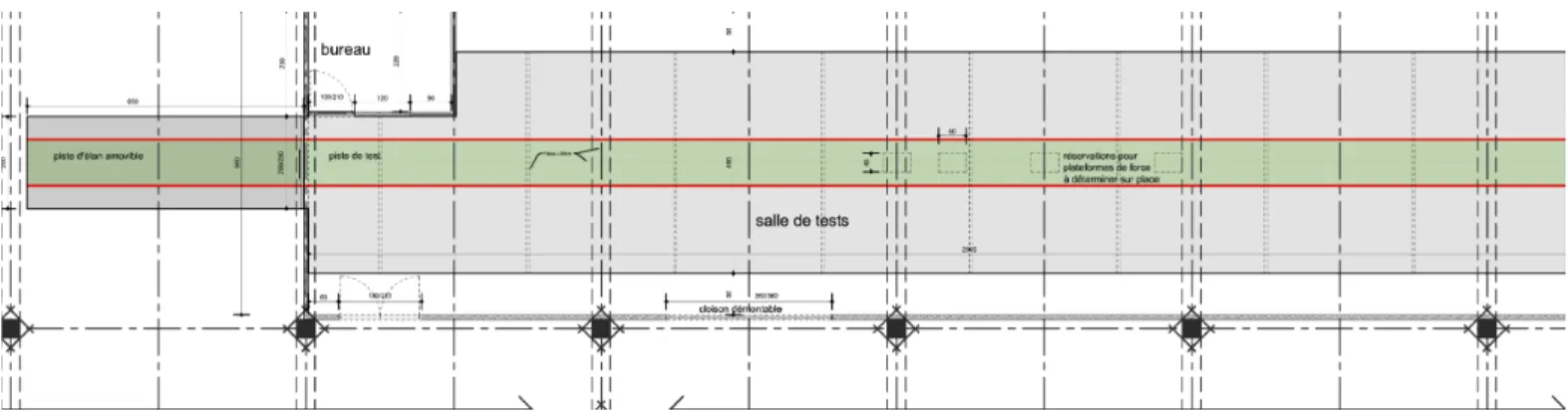
Figure 6. Running track design.
B. Technological choices
The 3D technology, which was chosen for the laboratory, is an active optical system (Coda Motion – Charnwood Dynamics), see Fig. 5. This system offers several benefits:
• the use of active markers makes their identification automatic and marker labels cannot be misidentified; • the easiness to move, install and calibrate the cameras
makes it possible to use them on the field and not only in the laboratory. Indoor measurements are however preferred for optimal lighting.
The standard deviation in position of a static marker at 3m range is equal at 0.05mm for width and height axes and 0.3mm for range axis. It is therefore possible to follow accurately the position of the markers in space. The sampling rate of the system depends on the number of markers which are used. The largest acquisition frequency is 100 Hz for 56 markers, and 800 Hz for 6 markers. As a minimum of 3 markers is needed to describe the 3D position of a segment, the position of only 2 segments can be measured at 800 Hz.
The LAMH is equipped with 4 units (cameras). Therefore, wide ranges of configurations are possible to cover a large field of view and follow complex motions. In addition to the cameras, the lab is currently equipped with one force platform, which will be particularly usefully for studies focusing on gait, run or the interaction of a subject with the ground. It is expected that other force platforms could complement this equipment in the future.
The size of the laboratory is large enough to allow various activities including sprint and throw. The length of the running track is equal to 27m and can be extended to 33m (Fig. 6).
In conclusion, concerning the technological choices, emphasis was given to the modularity of the system and the possibility of adaptation to various studies and objectives. This, once again, aims to answer to the transversal context of the laboratory for future operations.
It is important to underline that the French Community of Belgium grants funds for the equipment of the lab. The lab will also operate as an expertise center for professional athletes.
C. Objectives of LAMH
The LAMH aims to be an open platform for researchers of ULg coming from different fields, allowing therefore collaborations with an enlarged scientific community. The central kernel of the research team is precisely composed of the authors of this paper and is responsible for the coordination of the research projects to be developed by the LAMH.
The first aim of the laboratory is to study elite athletes for injuries prevention and performance enhancement for both upper and lower limbs. In particular, a short-term objective is to analyze the complex motion of the shoulder and particularly the scapula motion. The latest is typically difficult to follow because of the important relative motion between the bone and the skin. However, the knowledge of its behavior would be particularly relevant for specialized athletes, who have usually an asymmetrical positioning of the scapula between the dominant upper limb and the contra-lateral shoulder [4]. This asymmetry results from an adaptation to a specific and repetitive motion. This scapula dyskinesis frequently accompanies the pathological shoulder context. The comprehension of these processes would help in understanding the factors of risk and provide some prevention schemes. In the case of pathology, the evaluation of shoulder motion alteration could help the therapist to define specific rehabilitation goals. Medium-term objectives of the laboratory are related to the analysis of other human motions including the study of the lower limb. In particular, focus will be made on sprint for elite athletes.
Measurement artifacts caused by the relative motion between the skin and the bones are an inherent difficulty of 3D motion analysis systems. In order to reduce the influence of those soft-tissue artifacts, rigorous experimental protocols will
be established and relevant post-treatment algorithms will be investigated [5]. For example, a promising idea is to fit the experimental data on a kinematic model of the musculoskeletal system, including a description of soft-tissue motions. For that purpose, advanced biomechanical models of the articulated human body will be an important field of activity of the laboratory.
Another challenge for the understanding of human motion is related with the evaluation of joint forces and torques, as well as muscle efforts. This kind of information appears especially useful to improve the performance of a sport gesture, to detect muscle failure and to prevent injuries. Inverse dynamics methods have been recently proposed in order to evaluate muscle efforts by combining 3D measurement data with a full dynamic model of the patient. We intend to explore those techniques in the lab and to address many open questions regarding the patient-specific modeling of the human body, the influence of muscle activation dynamics and muscle forces, or the development of reliable numerical methods to solve inverse problems in biomechanics.
Yet another axis of development of the laboratory is the study of the interaction of a person or a group of persons with their environment, more precisely in the context of civil structures as footbridges where this interaction is systematically disregarded for the lack of available information.
A general scope of researches taking place in the context of the LAMH is to answer with efficiency and pragmatism questions arising in various biomechanical and other engineering applications. Furthermore, numerical simulations and inverse dynamic modeling of the human motion are expected to be regularly developed as a supplementary analysis tool throughout these researches.
People involved in the development of the laboratory have brought together a series of complementary skills that cover a
wide range of knowledge to complete successfully these research objectives. The objective is not only to answer to questions specific to the different fields involved in the project but also, thanks to the interaction between different scientific cultures, to provide new approaches to address these problems. In the medium term, other research developments and fields of investigation are expected to arise depending on the emerging interest and demand of other researchers, medical staff, athletes, etc.
ACKNOWLEDGMENT
The authors would like to warmly acknowledge the support of the Belgium French Community that allowed the start of the LAHM.
REFERENCES
[1] J.H. De Groot, “The scapulo-humeral rythm: effects of 2-D roentgen projection, ” Clin Biomech, vol.14, pp. 63-68, 1999.
[2] P.W. McClure, L.A. Michener, B.J. Sennett, “Direct 3-dimentional measurement of scapular kinematics during dynamic movements in vivo,” J Shoulder Elbow Surg, vol. 10, pp. 269-77, 2001.
[3] C. Anglin and U.P. Wyss, “Arm motion and load analysis of sit-to-stand, stand-to-sit, cane walking and lifting,” Clin Biomech, vol. 15, pp. 441-448, 2000.
[4] B. Forthomme, J.-M. Crielaard, J.-L. Croisier, “Scapular positioning in athlete’s shoulder: particularities, clinical measurements and implications,” Sports Med, vol. 38(5), pp. 369-386, 2008.
[5] C. Schwartz, M. Lempereur, V. Burdin, J.-J. Jacq, O. Rémy-Néris “Shoulder motion analysis using a marker cluster registration,” Proceedings of the 2007 IEEE Engineering in Medicine and Biology 29th, Lyon, France, 2007.