Université de Montréal
Élucidation du rôle de la voie Hippo dans l’ovaire chez la souris
par Mayra Tsoi
Département de biomédecine vétérinaire Faculté de médecine vétérinaire
Thèse présentée à la Faculté de médecine vétérinaire en vue de l’obtention du grade de philosophiae doctor (Ph.D.)
en sciences vétérinaires, option reproduction
Mai 2018 © Mayra Tsoi, 2018
i Résumé
La voie de signalisation Hippo est une voie conservée entre espèces avec des rôles bien établis dans le développement embryonnaire, l’homéostasie tissulaire et le cancer. La voie ne possède ni ligand ni récepteur spécifique, mais semble être régulée par une variété de signaux extracellulaires et intracellulaires qui diffèrent selon le type cellulaire. L’activation de la voie Hippo débute avec la phosphorylation de MST1/2 qui phosphoryle et lie la protéine adaptatrice SAV1. Ensemble, ils phosphorylent et activent LATS1/2 et la protéine adaptatrice MOB1. Ce complexe phosphoryle et inactive les effecteurs principaux de la voie, c’est-à-dire les co-activateurs transcriptionnels YAP et TAZ. Inactivation de la voie permet à YAP et TAZ de se déplacer vers le noyau et de se lier à des facteurs de transcription, notamment ceux de la famille TEAD, afin de moduler la transcription de gènes cibles impliqués dans la prolifération cellulaire et l’inhibition de l’apoptose. De plus en plus de publications suggèrent l’implication de la voie Hippo dans l’ovaire postnatal, cependant, les facteurs qui régulent la voie et les rôles spécifiques de ses effecteurs demeurent inconnus. L’objectif global des deux études présentées dans cette thèse était d’élucider la régulation et les rôles de LATS1, LATS2, YAP et TAZ spécifiquement dans les cellules de la granulosa. Les résultats de la première étude ont démontré que l’hormone lutéinisante (LH) induit la phosphorylation de LATS1 et YAP et que cette dernière s’effectue par l’action de la protéine kinase A (PKA). De plus, nous avons identifié qu’en absence de Yap/Taz, la LH est incapable d’induire la transcription de ses gènes cibles et que ceci serait dû, au moins en partie, à la perte de l’expression du gène codant pour le récepteur à la LH (Lhcgr). Dans notre deuxième étude, la génération et les analyses de souris Lats1flox/flox; Lats2flox/flox;CYP19-cre ont révélé que LATS1/2 sont essentiels pour le maintien du destin des cellules de la granulosa. En effet, en absence de Lats1/2, celles-ci perdent leur identité et leur fonction, subissent une transition épithéliale-mésenchymale (EMT) et se transdifférencient en cellules de Sertoli, en ostéoblastes ainsi qu’en cellules dérivées de la crête neurale. Nous soupçonnons que ces processus cellulaires surviennent à cause d’une activité transcriptionnelle aberrante induite par une accumulation de YAP/TAZ. Ces deux études dévoilent de nouveaux rôles pour les effecteurs de la voie Hippo dans la cascade de signalisation de la LH et dans le maintien de la gonade femelle, en plus d’établir une solide base de connaissances sur laquelle les études subséquentes visant l’élucidation des mécanismes en cause pourront s’appuyer.
ii
Mots clés : la voie Hippo, Lats1, Lats2, Yap, Taz, l’ovaire, les cellules de la granulosa, les souris transgéniques
iii Abstract
The Hippo signaling pathway is an evolutionarily conserved pathway with well-defined roles in embryonic development, tissue homeostasis, and cancer. The Hippo pathway has no specific ligands or receptors but is regulated by a variety of extracellular and intracellular cues that vary depending on the cell type. Activation of the Hippo pathway begins with phosphorylation of MST1/2 that phosphorylate and bind to an adaptor protein SAV1. Together, they phosphorylate and activate LATS1/2 and its adaptor protein MOB1. This complex then phosphorylates and inactivates the key downstream effectors of the pathway, the transcriptional co-activators YAP and TAZ. Disruption of Hippo signaling allows YAP and TAZ to translocate to the nucleus to bind notably to members of the TEAD family of transcription factors to mediate the transcription of target genes that promote cell proliferation and inhibit apoptosis. An increasing amount of evidence in the literature suggests a role for Hippo signaling in the postnatal ovary, however, regulators of (and specific roles for) Hippo effectors remain unknown. The global objective of this thesis was therefore to characterize Hippo signaling in the murine ovary by investigating the regulation of and roles of LATS1, LATS2, YAP, and TAZ specifically in granulosa cells. Results from our first study identified that luteinizing hormone (LH) activates Hippo signaling by inducing the phosphorylation of LATS1 and YAP and that this occurs via protein kinase A (PKA). In addition, we found that LH is unable to induce the transcription of its target genes in the absence of Yap/Taz, and that this might be due in part to the loss of the expression of the gene encoding the LH receptor (Lhcgr). In our second study, generation and analyses of Lats1flox/flox;Lats2flox/flox;CYP19-cre mice revealed that LATS1/2 are critical mediators of granulosa cell fate maintenance and in their absence, granulosa cells lose their identity and function, undergo epithelial-to-mesenchymal transition (EMT), and transdifferentiate into Sertoli-like cells, osteoblasts, and neural crest cell derivatives. We suspect that these cell processes occur as a result of aberrant transcriptional activity induced by an overaccumulation of YAP/TAZ. This thesis presents novel and exciting findings that confirm important roles for Hippo pathway effectors in the LH signaling cascade and in the maintenance of the female gonad, as well as pave the way for future studies that will elucidate the mechanisms underlying these processes.
iv Table of contents Résumé ... i Abstract ... iii Table of contents ... iv List of Tables ... ix List of figures ... x
List of abbreviations ... xii
Acknowledgements ... xvi
Chapter 1. Literature review ... 1
1 Introduction ... 2
1.1 Anatomy of the mouse female reproductive tract ... 3
1.2 Histology of the mouse ovary ... 4
1.3 Embryology of the mouse ovary ... 6
1.3.1 Female sex determination ... 6
1.3.2 Female gonadal development ... 8
1.4 Physiology of ovarian follicle development ... 9
1.4.1 The regulation of ovarian follicle development by the hypothalamic-pituitary-gonadal (HPG) axis ... 9
1.4.2 Gonadotropin signaling in the ovary ... 11
1.4.2.1 FSH signaling ... 11
1.4.2.2 LH signaling ... 13
1.4.3 Follicle development ... 15
1.4.3.1 Crosstalk between germ cells and gonadal somatic cells during follicle development ... 15
v
1.4.3.3 Primary follicle development ... 17
1.4.3.4 Secondary follicle development ... 18
1.4.3.5 Antral follicle development ... 18
1.4.3.6 Events initiated by the LH surge ... 20
1.4.3.6.1 Oocyte maturation ... 20 1.4.3.6.2 Cumulus expansion ... 21 1.4.3.6.3 Ovulation ... 22 1.4.3.6.4 Luteinization ... 23 1.4.4 Steroidogenesis ... 25 1.4.4.1 Estradiol ... 25 1.4.4.2 Progesterone ... 27
2 Hippo signaling pathway ... 29
2.1 Introduction ... 29
2.2 Discovery of the Hippo signaling pathway ... 29
2.3 Canonical Hippo signaling pathway ... 30
2.4 Biological functions of Hippo in the mouse ... 32
2.4.1 Germline knockouts ... 32
2.4.2 Early embryonic development ... 33
2.4.3 Stem cells ... 33
2.4.4 Organ development, homeostasis, and regeneration ... 33
2.4.4.1 Liver ... 34
2.4.4.2 Heart ... 34
2.4.4.3 Intestines ... 34
2.4.5 Cancer ... 35
vi
2.4.5.2 Evasion of apoptosis ... 36
2.4.5.3 Indefinite replicative potential ... 37
2.4.5.4 Angiogenesis ... 37
2.4.5.5 Metastasis ... 37
2.5 Regulation of the Hippo pathway ... 38
2.5.1 Cell-cell contact ... 38 2.5.2 Cell-cell adhesion ... 38 2.5.3 Cell polarity ... 39 2.5.4 Mechanical inputs ... 39 2.5.5 G protein-coupled receptors ... 40 2.5.6 Actin cytoskeleton ... 40
2.6 Crosstalk with other pathways ... 41
2.6.1 Crosstalk between the Hippo and Wnt/β-catenin pathways ... 41
2.6.2 Crosstalk between the Hippo and TGF-β/SMAD pathways ... 44
2.7 Large tumor suppressors ... 46
2.7.1 Regulation of LATS ... 46
2.7.2 Functions of LATS ... 47
2.8 YAP and TAZ ... 48
2.8.1 Molecular structure of YAP and TAZ ... 48
2.8.2 Regulation of YAP/TAZ ... 49
2.8.3 YAP and TAZ as transcriptional co-activators ... 49
2.8.4 YAP/TAZ target genes ... 50
2.9 Hippo signaling in the ovary ... 51
Chapter 2. Hypotheses and objectives ... 53
vii
3.1 ABSTRACT ... 57
3.2 INTRODUCTION ... 57
3.3 MATERIALS AND METHODS ... 60
3.4 RESULTS ... 63 3.5 DISCUSSION ... 65 3.6 ACKNOWLEDGEMENTS ... 67 3.7 REFERENCES ... 67 3.8 FIGURES ... 71 3.9 SUPPLEMENTAL FIGURES ... 78 3.10 TABLES ... 80 Chapter 4. Article 2 ... 82 4.1 ABSTRACT ... 84 4.2 INTRODUCTION ... 84
4.3 MATERIALS AND METHODS ... 87
4.4 RESULTS ... 91 4.5 DISCUSSION ... 95 4.6 ACKNOWLEDGEMENTS ... 99 4.7 REFERENCES ... 99 4.8 FIGURES ... 105 4.9 SUPPLEMENTAL FIGURES ... 113 4.10 TABLES ... 116 4.11 SUPPLEMENTAL TABLE ... 117
Chapter 5. General discussion ... 119
5.1 Why study Hippo signaling in the ovary? ... 120
viii
5.2.1 Challenges ... 121
5.2.2 In retrospect ... 122
5.2.3 Perspectives ... 123
5.2.4 Comparison to the literature ... 123
5.3 Discussion topics Article 2 ... 124
5.3.1 Further discussion topics ... 124
5.3.2 Challenges ... 125
5.3.3 In retrospect ... 126
5.3.4 Perspectives ... 127
5.3.5 Comparison to the literature ... 128
5.4 A global model of Hippo signaling in the ovary ... 128
5.4.1 Comparison of results from both studies ... 128
5.4.2 Importance of findings for the scientific community ... 129
ix List of tables
TABLE 2.1 Evolutionarily conserved Hippo pathway components ... 30 TABLE 2.2 Regulators of Hippo signaling ... 41
Article 1
Table 1. Primer list ... 81 Table 2. Antibody list ... 81
Article 2
Table 1. Mating trials ... 116 Table 2. Microarray analysis of gene expression in Lats1flox/flox;Lats2flox/flox;CYP19-cre
granulosa cells infected with Ad-eGFP vs Ad-cre for 30h ... 116 Table S1. Primer list ... 118
x List of figures
FIGURE 1.1 Anatomy of the female reproductive tract ... 4
FIGURE 1.2 Histology of immature and mature ovaries ... 6
FIGURE 1.3 Female sex determination ... 8
FIGURE 1.4 Timeline of female gonadal development ... 9
FIGURE 1.6 FSH signaling in granulosa cells ... 13
FIGURE 1.7 LH signaling in the preovulatory follicle ... 15
FIGURE 1.8 Regulation of folliculogenesis ... 20
FIGURE 1.9 Maintenance of oocyte meiotic arrest (on left) vs resumption of meiosis (on right) ... 21
FIGURE 1.10 LH signaling that drives oocyte maturation, cumulus expansion, ovulation, and luteinization ... 25
FIGURE 1.11 Estradiol biosynthesis ... 27
FIGURE 1.12 Progesterone biosynthesis ... 28
FIGURE 2.1 Canonical Hippo signaling pathway ... 32
FIGURE 2.2 Crosstalk between the Hippo and Wnt/β-catenin pathways ... 44
FIGURE 2.3 Crosstalk between the Hippo and TGF-β/SMAD pathways ... 46
FIGURE 2.4 Molecular structure of YAP and TAZ ... 49
Article 1 Figure 1. LH activates the Hippo pathway in GCs in vivo ... 71
Figure 2. LH activates the Hippo pathway in GCs in vitro ... 73
Figure 3. LH acts via PKA to phosphorylate YAP ... 74
xi
Figure 5. Yap and Taz are required for expression of Lhcgr ... 77 Figure S1. No additional effects on LH-induced YAP phosphorylation are observed when inhibitors are used in combination ... 78 Figure S2. Two PKA inhibitors similarly reduce LH-induced YAP phosphorylation ... 79
Article 2
Figure 1. Lats1flox/flox;Lats2flox/flox;CYP19-cre ovaries ... 105 Figure 2. Loss of Lats1 and Lats2 causes loss of granulosa cell identity, transdifferentiation into Sertoli cells and epithelial-to-mesenchymal transition ... 107 Figure 3. Hippo signaling is disrupted in Lats1flox/flox;Lats2flox/flox;CYP19-cre ovaries ... 108 Figure 4. Lats1flox/flox;Lats2flox/flox;CYP19-cre granulosa cells transdifferentiate into multiple cell lineages ... 110 Figure 5. Knockdown of Lats1 and Lats2 in vitro causes a loss of granulosa cell identity and responsiveness to gonadotropins ... 111 Figure 6. Knockdown of Lats1 and Lats2 in granulosa cells in vitro induces
transdifferentiation into multiple cell lineages ... 112 Figure S1. FSH and LH serum levels from Lats1flox/flox;Lats2flox/flox (control) vs
Lats1flox/flox;Lats2flox/flox;CYP19-cre (mutant) female mice ... 113 Figure S2. Knockdown of Lats1 or Lats2 alone has less effect on the transcription of LH target genes or on markers of Sertoli cells (relative to knockdown of Lats1/2) ... 114
Final conclusions
xii List of abbreviations
Adamts1 A disintegrin and metalloproteinase with thrombospondin-like motifs-1 Amh Anti-Müllerian hormone
Apc Adenomatous polyposis coli Areg Amphiregulin
AS Activation segment motif Axin Axis inhibition protein 1/2 Bcl-2 B cell lymphoma 2
Birc Baculoviral inhibitor of apoptosis repeat containing Bmp Bone morphogenetic protein
Btc Betacellulin
β-TrCP β-transducin repeat-containing protein E3 ubiquitin ligase cAMP Cyclic adenosine monophosphate
CCN Family of regulatory proteins including Cyr61, Ctgf, Nov Ccnd1 Cyclin D1
Ccnd2 Cyclin D2
Cdk Cyclin dependent kinase
Cdkn Cyclin dependent kinase inhibitor Cebpb CAAT enhancer binding protein beta cGMP Cyclic guanosine monophosphate CK1 Casein kinase 1
CL Corpus luteum
COC Cumulus cell oocyte complex
Creb cAMP responsive element binding protein Ctgf Connective tissue growth factor, or Ccn2
Cyp11a1 Cytochrome P450 family 11 subfamily A member 1 Cyp17a1 Cytochrome P450 family 17 subfamily A member 1
Cyp19a1 Cytochrome P450 family 19 subfamily A member 1 or aromatase Cyr61 Cysteine-rich 61, or Ccn1 DHEA Dehydroepiandrosterone Dpp Days post-partum Dvl Dishevelled E Embryonic day E2 Estradiol or 17-βestradiol ECM Extracellular matrix
Egfr Epidermal growth factor receptor EMT Epithelial-to-mesenchymal transition Ep2 Prostaglandin E receptor 2
Ereg Epiregulin
Erk Extracellular signal-regulated kinase Esr1/2 Estrogen receptor 1 and 2
Fgf9 Fibroblast growth factor 9 Figla Factor in the germline α
xiii Foxl2 Forkhead box L2
Foxo1/3 Forkhead box protein 01 FSH Follicle-stimulating hormone Fshr FSH receptor
Fshb FSH beta subunit
Fzd Frizzled
Gdf Growth differentiation factor
Gja1 Gap junction protein 1, or connexin 43 Gja4 Gap junction protein 4, or connexin 37 GnRH Gonadotropin-releasing hormone GPCR G protein-coupled receptor GSK3β Glycogen synthase kinase 3 β Has2 Hyaluronan synthase 2
Hif1a Hypoxia inducible factor 1 alpha subunit HM Hydrophobic motif
Hpg Hypothalamic-pituitary-gonadal
Hpo Hippo
HSD Hydroxysteroid dehydrogenase
hTERT Human telomerase reverse transcriptase IaI Inter-alpha trypsin inhibitor
ICM Inner cell mass
Igf1 Insulin-like growth factor 1 IHC Immunohistochemistry Il6 Interleukin 6
Inha Inhibin alpha subunit Kit Kit receptor
Kitl Kit ligand
Lats1/2 Large tumor suppressors 1 and 2 LH Luteinizing hormone
Lhcgr LH/choriogonadotropin receptor Lhb LH beta subunit
LPA Lysophosphatidic acid
LRP5/6 Low-density lipoprotein receptor-related protein 5/6 MAPK Mitogen-activated protein kinase
Mats Mob-as-tumor-suppressor mESCs Mouse embryonic stem cells Mob1 MOB kinase activator 1A and 1B
Mst1/2 Mammalian STE20-like protein kinase 1 and 2 mTORC1 Mammalian target of rapamycin complex 1 Nf2 Merlin, or neurofibromin 2
Nobox Newborn ovary homeobox
Nov Nephroblastoma overexpressed, or Ccn3 Nr5a2 Nuclear receptor member 5a2, or Lhr1
xiv Nrip1 Nuclear receptor interaction protein 1 OSE Ovarian surface epithelium
P4 Progesterone
PCR Polymerase chain reaction PDE3A Phosphodiesterase
PGC Primordial germ cell PGE1/2 Prostaglandins E1 and E2 Pgr Progesterone receptor PI3K Phosphoinositide 3-kinase PKA Protein kinase A
PP1A/2A Protein phosphatase 1A or 2A
PPARγ Peroxisome proliferator-activated receptor gamma Prlr Prolactin receptor
Pten Phosphatase and tensin homolog Ptgs2 Prostaglandin-endoperoxide synthase 2 Ptp Protein tyrosine phosphatase
Ptx3 Pentraxin 3 Rspo1 R-spondin-1
RT-qPCR Quantitative reverse transcription PCR Runx2 Runt related transcription factor 2 S1P Sphingosine-1 phosphate
Sav Salvador
Sav1 Salvador homologue 1 Scrib Scribble
Sd Scalloped
Ser Serine
Sohlh1/2 Spermatogenesis and oogenesis basic helix-loop-helix
Sox9 SRY-Box 9
Src Sarcoma
Sry Sex-determining region on Y chromosome StAR Steroidogenic acute regulatory protein Stra8 Retinoic acid gene 8
Taz Transcriptional co-activator with PDZ-binding motif, or Wwtr1
Tbx5 T-box 5
Tcf/Lef T-cell factor/lymphoid enhancer factor
TE Trophectoderm
Tead1-4 TEA domain family members 1-4 TGF-β Transforming growth factor beta
Thr Threonine
Tnfaip6 TNF-α-induced protein 6
Tsc1/2 Tuberous sclerosis complex 1 or 2 Vcan Versican
xv WB Western blotting
Wnt4 Wingless-type MMTV integration site family, member 4
Wts Warts
Yap Yes-associated protein
Yki Yorkie
xvi Acknowledgements
To members of the CRRF, the RQR, the SSR, and professors from the DMV program, thank you for constantly providing words of encouragement.
To members of FANI and CDEVQ, thank you for your invaluable technical support.
To members of the Boerboom laboratory, thank you for making it a pleasure to come to work every single day.
To Marilène, thank you for sharing with me your contagious enthusiasm for pathology.
To Derek, thank you for passing on to me your passion for research (and for everything else). To my mom, thank you for always telling the truth.
2 1 Introduction
The study of ovarian follicle development and its regulation by gonadotropins, follicle-stimulating hormone (FSH) and luteinizing hormone (LH) has been an active area of research over the course of several decades. It is well-known that FSH stimulates granulosa cell proliferation and estradiol production, while LH regulates ovulation and formation of the corpus luteum (CL)(Richards, 1980). FSH and LH bind to their respective receptors, FSHR and LH/choriogonadotropin receptor (LHCGR). This activates a variety of signaling cascades that lead to the transcription of FSH and LH target genes, respectively, which are ultimately responsible for mediating their effects (Richards & Pangas, 2010b). There still, however, remain important gaps in our knowledge regarding, for instance, the factors that link FSHR and LHCGR signaling pathways to the transcription of their respective target genes. This has driven the investigation of novel signaling pathways (known for their roles in other contexts) and their potential involvement in folliculogenesis.
One such pathway is the Hippo pathway, that is widely recognized as a regulator of organ size and tissue growth in embryonic development; its roles in the adult are still being uncovered (Zhao et al, 2010a). The Hippo pathway does not possess any specific ligands or receptors but instead, is regulated by a wide variety of cues (Piccolo et al, 2014). Recent studies have hinted at roles for Hippo effectors in the ovary. The generation of knockout mouse models for certain Hippo pathway effectors have revealed that in these animals, female fertility is severely affected (Hossain et al, 2007; St John et al, 1999). Other studies suggest that Hippo effectors might be involved in ovarian follicle development and ovarian cancer (Fu et al, 2014; Kawamura et al, 2013; St John et al, 1999). Very few studies, however, have investigated the specific roles of Hippo signaling effectors over the course of ovarian follicle development. No studies to date have investigated how Hippo signaling is regulated in granulosa cells.
This thesis describes two studies that addressed these questions: 1) How is Hippo signaling regulated in ovarian granulosa cells? and 2) What roles do Hippo effectors play in ovarian follicle development? using conditional knockout mouse models and primary granulosa cell cultures.
3
1.1 Anatomy of the mouse female reproductive tract
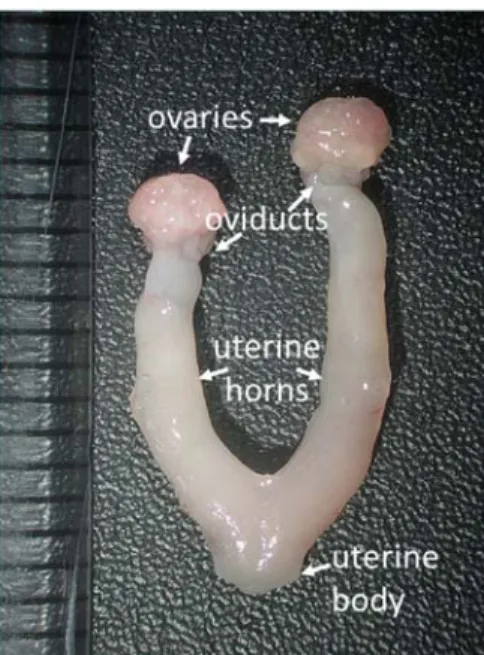
The female reproductive tract consists of two ovaries, two oviducts, two uterine horns, one uterine body, the cervix, the vagina, a ventral and two dorsal labia, and the vulva. The ovaries are located at the caudal pole of the kidneys (Bertolin & Murphy, 2014). Each ovary is enclosed within an ovarian bursa, that is suspended from the dorsal body wall by the mesovarium. Blood vessels and nerves pass through a small opening in the ovarian bursa that enter the ovary at the ovarian hilus (Rendi et al, 2012).
Each oviduct provides a passageway for the cumulus oocyte complexes (COCs) to travel from the ovary at the time of ovulation to the ipsilateral uterine horn (Evans & DeLahunta, 2004). The oviducts are narrow, coiled tubes, approximately 1.8cm long composed of three anatomic portions: the infundibulum, the ampulla, and the isthmus (Rendi et al, 2012). The infundibulum is the funnel-shaped, fimbriated end of the oviduct that catches the COCs at ovulation. The ampulla is the middle segment where fertilization occurs. The isthmus is the posterior end of the oviduct that opens into the uterine horn (Evans & DeLahunta, 2004). The oviducts are suspended from the dorsal body wall by the mesotubarium (that is continuous with the mesovarium, ovarian bursa, and mesometrium)(Rendi et al, 2012).
The uterus is a tubular organ composed of two horns that join distally to form one body (FIGURE 1.1)(Pasquini et al, 2007). The uterus is suspended by the mesometrium from the dorsal body wall. The uterine body opens up into the cervix, which is continuous with the vagina. The vestibule is common to both urinary and genital systems that connects the vagina to the vulva, which is the external genital orifice (Pasquini et al, 2007).
The ovaries are vascularized by ovarian arteries and veins. The ovarian arteries arise from the aorta distally from the renal arteries (Evans & DeLahunta, 2004). The ovarian veins leave the ovary and enter the caudal vena cava. The uterus is vascularized by the uterine branch of the ovarian artery and the uterine artery (Bertolin & Murphy, 2014; Evans & DeLahunta, 2004).
4
FIGURE 1.1 Anatomy of the mouse female reproductive tract
1.2 Histology of the mouse ovary
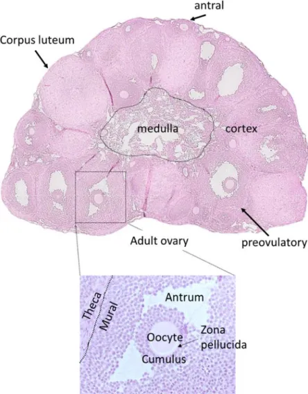
The ovarian surface epithelium (OSE) is a simple cuboidal to columnar epithelium that covers the surface of the ovary (Rendi et al, 2012). A dense connective tissue located right below the OSE is the tunica albuginea. Once primordial follicles begin to form directly beneath the OSE, the ovary becomes organized into two morphologically distinct compartments, the outer cortex and the inner medulla (Rendi et al, 2012; Wilhelm et al, 2007). The cortex contains the ovarian follicles at all stages of development, corpora lutea (CLs), and connective tissue. As follicles develop, they migrate towards the medulla (Sforza et al, 2003). The medulla contains blood vessels, nerves, lymphatics, smooth muscle fibers, and connective tissue fibers (Pasquini et al, 2007).
An ovarian follicle is made up of a central germ cell (oocyte) surrounded by varying layers of somatic cells (granulosa and theca) depending on the stage of follicle development. A primordial follicle contains a small oocyte surrounded by a layer of squamous pre-granulosa cells (Rendi et al, 2012). Once the primordial follicle is activated, the pre-granulosa cells differentiate into cuboidal granulosa cells to form primary follicles. The oocyte secretes a matrix of glycosylated zona proteins that directly surrounds itself, called the zona pellucida (Rankin et al, 2000; Richards et al, 2015a). A secondary follicle contains two or more layers of granulosa
5
cells that are surrounded by a layer of theca cells, the two separated by a basal lamina. The follicle (and oocyte) grow considerably in size as granulosa and theca cells proliferate. Dispersed areas of interstitial fluid develop between granulosa cells and form the antrum, marking the antral stage of follicle development. The theca cell layer differentiates into the steroidogenic theca interna and the theca externa (Pangas & Rajkovic, 2015; Richards et al, 2015a). Adjacent to the theca externa lies a second basal lamina (Richards et al, 2015a).
A large proportion of small antral follicles are not selected to continue maturation and undergo atresia (Richards, 1980). The preovulatory follicle is characterized by a large antrum and granulosa cells that have differentiated into two distinct populations: the cumulus cells surround the oocyte forming the COC while the mural granulosa cells line the basement membrane of the follicle. At ovulation, the follicle wall breaks down and releases the COC. After ovulation, the remaining follicular cells hypertrophy, differentiate into luteal cells, and become vascularized, forming the corpus luteum (CL). An ovary from a pre-pubertal mouse (whose first ovulations occur around 30 days post-partum (30 dpp)) contains primordial, primary, and secondary follicles (Bertolin & Murphy, 2014). An ovary from an adult mouse contains follicles of all stages, with CLs in different stages of regression (FIGURE 1.2).
6
FIGURE 1.2 Histology of ovaries from pre-pubertal and adult mice
1.3 Embryology of the mouse ovary
1.3.1 Female sex determination
As in other mammals, sex determination in the mouse begins in bipotential precursor cells within the fetal gonad (Wilhelm et al, 2007). Expression of the Y-linked gene sex-determining region on Y chromosome (Sry) around embryonic day 10.5 (e10.5) leads to activation of its target gene Sox9, which initiates the differentiation of bipotential precursor cells into Sertoli cells and thus, the differentiation of the bipotential gonad into a testis (Gubbay et al,
7
1990; Koopman et al, 1991; Vidal et al, 2001). Overexpression of Sox9 or Sry in XX mice is sufficient to drive male sexual development (Koopman et al, 1991; Vidal et al, 2001).
In the absence of Sry or Sox9, the bipotential precursor cells differentiate into granulosa cells (Wilhelm et al, 2007). While no single ovarian-determining factor has been identified in the female gonad, evidence suggests that ovarian development is orchestrated by multiple genes (Wilhelm et al, 2007). Primary female sex-determining factors were identified using knockout mouse models in which loss-of-function of these genes led to partial or complete sex reversal. These genes are primarily components of the Wnt/β-catenin signaling pathway, including Wnt4, Rspo1, β-catenin, in addition to Foxl2 (Richards & Pangas, 2010b). At e11.0, Wnt4 is expressed in the indifferent gonad, is downregulated around e11.5 in the male gonad and upregulated in the female gonad (Vainio et al, 1999; Wilhelm et al, 2007). Wnt4 knockout XX mice develop testicular-like structures (Vainio et al, 1999). At e11.5, Foxl2 is activated exclusively in the female gonad with the highest expression occurring in pre-granulosa cells (Ottolenghi et al, 2005; Ottolenghi et al, 2007). Foxl2 knockout XX mice lack ovarian follicles while still containing oocytes, and develop testicular cord-like structures (Ottolenghi et al, 2005). Ablation of Rspo1 (which stabilizes β-catenin) from XX mice leads to partial gonadal sex reversal and oocyte loss (Chassot et al, 2008; Tomizuka et al, 2008). Conditional expression of stable β-catenin in XY mice induces partial sex reversal, with testes containing ovarian-like structures devoid of germ cells (Maatouk et al, 2008). Interestingly, double Wnt4/Foxl2 knockout XX mice undergo complete sex reversal from an ovary to a functional testis containing tubules and spermatogonia, suggesting that separate mechanisms regulate somatic cell vs germ cell specification in the ovary (Ottolenghi et al, 2007). While the expression of female-specific markers has been identified (starting around e11.5), the precise moment at which follicle differentiation occurs is unclear.
During this critical period, a battle between the expression of male vs female-specific genes determines the fate of the bipotential gonad. SOX9 antagonizes β-catenin to prevent ovarian development in males (Chassot et al, 2008). β-catenin and FOXL2 antagonize SOX9 to prevent testis development in females (Matzuk & Burns, 2012). WNT4 and FGF9 (a male-specific gene that enhances Sox9 expression) antagonize one another (FIGURE 1.3)(Kim et al, 2006). Whether there is a hierarchy in terms of dominance of one factor over another (for
8
example if one male-specific gene and one female-specific gene are simultaneously expressed, will the gonad develop as a male or female?) has yet to be determined.
FIGURE 1.3 Female sex determination. Inspired by Schlessinger et al 2010.
1.3.2 Female gonadal development
During embryonic development, the bipotential gonads are derived from the intermediate mesoderm and develop next to the urogenital ridge (Richards & Pangas, 2010a; Richards & Pangas, 2010b; Wilhelm et al, 2007). The urogenital ridge is made up of three sections that include the pronephros (that will give rise to the adrenals), the mesonephros (the future gonads and genital ducts), and the metanephros (the future kidney)(Wilhelm et al, 2007). More specifically, the gonads develop on the surface of the mesonephros around e9.5-10.5 as paired thickenings of the epithelium in conjunction with cells from the mesonephros and the coelomic epithelium, which give rise to the somatic precursor cells (Bertolin & Murphy, 2014; Tanaka & Nishinakamura, 2014; Wilhelm et al, 2007).
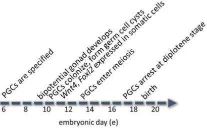
Primordial germ cells (PGCs) are specified from epiblast cells (that arise from the inner cell mass) around e6.0-8.0, they proliferate and migrate from the region of the hindgut to colonize the genital ridge around e9.5-11.5 (Richards & Pangas, 2010b; Wilhelm et al, 2007). Colonization of the bipotential gonad by PGCs is followed by the differentiation of somatic precursor cells into granulosa cells (Richards & Pangas, 2010b; Tanaka & Nishinakamura, 2014; Wilhelm et al, 2007). Between e10.5-13.5, PGCs undergo mitosis without complete cytokinesis, which forms syncytia of germ cells, called germ cell cysts, that are connected by cytoplasmic bridges (Pepling & Spradling, 2001). In females, meiosis is induced in PGCs by
9
retinoic acid induction of retinoic acid gene 8 (Stra8)(Koubova et al, 2006). PGCs enter meiosis around e13.5, arrest at the diplotene stage of meiotic prophase I at approximately e17.5, and remain arrested until the LH surge induces the resumption of meiosis in pubertal mice (FIGURE 1.4)(Bertolin & Murphy, 2014; Pepling & Spradling, 2001; Richards & Pangas, 2010b).
Members of the transforming growth factor beta (TGF-β) superfamily appear to be the major regulators of PGC proliferation and survival. Bone morphogenetic protein (BMP) -4 and -7 promote PGC proliferation, in contrast to activin and TGF-β that inhibit proliferation, while follistatin (an activin inhibitor) is required for PGC survival (Pesce et al, 2002; Richards et al, 1999; Richards & Pangas, 2010b; Ross et al, 2007; Yao et al, 2004).
FIGURE 1.4 Timeline of female gonadal development
1.4 Physiology of ovarian follicle development
1.4.1 The regulation of ovarian follicle development by the hypothalamic-pituitary-gonadal (HPG) axis
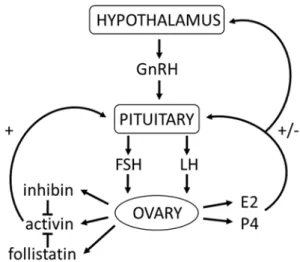
Gonadotropin-releasing hormone (GnRH) is secreted by the hypothalamus and acts on the anterior pituitary to stimulate the secretion of FSH and LH from gonadotrope cells. FSH and LH are dimeric glycoproteins composed of a common α subunit and a unique β subunit (Pangas & Rajkovic, 2015). Once released in the bloodstream, FSH and LH reach the ovary to stimulate the production of steroid hormones depending on the phase of the estrous cycle. The secretion
10
of FSH and LH is controlled (in part) by ovarian-derived hormones (steroidal and non-steroidal) that exert positive and negative feedback mechanisms on the hypothalamus and pituitary (Messinis, 2006).
In mice, the estrous cycle lasts 4-5 days and is divided into phases: proestrus (lasting approximately 32.4h) and estrus (20.7h), metestrus (21.8h), and diestrus (21.8h)(Bertolin & Murphy, 2014; Van Ebbenhorst Tengbergen, 1955). During proestrus, FSH and LH stimulate antral follicles to produce increasing levels of estradiol (E2), which exerts a positive feedback on the hypothalamus and pituitary to induce the preovulatory LH surge (Richards, 1980; Richards, 1994). During estrus, ovulation occurs 12-16h after the LH surge (Richards, 2005). Lower estradiol levels during the other phases of the cycle, exert a negative feedback at the level of the hypothalamus and the pituitary to suppress gonadotropin secretion (Herbison, 2015). During metestrus, the CL produces increasing amounts of progesterone (P4), which inhibits LH secretion. During diestrus (in the absence of mating), the CL regresses and the levels of P4 decrease, which releases LH inhibition, allowing GnRH secretion and proestrus to continue (Bertolin & Murphy, 2014).
Granulosa cells produce activins, inhibins (A and B isoforms), and follistatins that regulate pituitary function. As their names suggest, activin stimulates FSH secretion while inhibin inhibits FSH by antagonizing activin (McArdle & Roberson, 2015). Follistatin binds and inhibits activin activity; it is expressed starting in antral stage follicles (Pangas & Rajkovic, 2015). Inhibin B appears to be produced by small follicles and its levels are inversely correlated with FSH levels during the estrous cycle, while inhibin A levels correlate with estradiol production (FIGURE 1.5)(Pangas & Rajkovic, 2015).
11
FIGURE 1.5 Hypothalamic-pituitary-gonadal axis
1.4.2 Gonadotropin signaling in the ovary
FSH and LH bind their respective receptors, the FSHR and LHCGR, which are Gαs protein-coupled receptors located on plasma membranes of their target cells (Pangas & Rajkovic, 2015). FSHR is expressed exclusively on granulosa cells (starting in preantral stage follicles), while LHCGR is located on theca cells (starting in secondary stage follicles), mural granulosa cells of large preovulatory follicles, and luteal cells (Hunzicker-Dunn & Mayo, 2015; Richards, 1980; Richards & Midgley, 1976). Gonadotropin binding to their receptors initiates an intracellular signaling cascade leading to the transcription of FSH and LH-specific target genes. Why FSHR expression is limited to granulosa cells in the female remains unknown.
1.4.2.1 FSH signaling
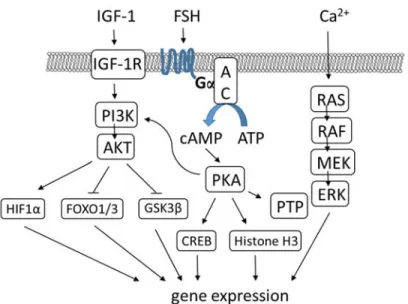
FSH regulates the expression of around 500 target genes that mediate granulosa cell proliferation, survival, estradiol synthesis, and differentiation (Hunzicker-Dunn & Mayo, 2015; Richards & Pangas, 2010b). FSH exerts its functions primarily through the cAMP/PKA pathway. FSH binds to FSHR to activate adenylyl cyclase, which converts ATP to cAMP (Simoni et al, 1997). Cyclic AMP accumulates, binds to, and activates PKA (Taylor, 1989). PKA phosphorylates most notably CREB(Ser133), which has binding sites on the promoters of some FSH target genes, including inhibin alpha (Inha) and Cyp19a1 (Carlone & Richards, 1997; Pei et al, 1991). PKA also rapidly phosphorylates histone H3, which is suspected to promote the
12
transcription of FSH target genes (by facilitating the access of transcription factors to loosened chromatin)(Hunzicker-Dunn & Mayo, 2015)
FSH signals via cAMP/PKA to activate ERK. More specifically in preantral granulosa cells, PKA stimulates the phosphorylation/inactivation of protein tyrosine phosphatase (PTP) to dissociate it from ERK, which allows ERK to be activated by RAS/RAF/MEK (Cottom et al, 2003). ERK phosphorylates or promotes the phosphorylation of transcription factors and co-activators involved in FSH target gene expression, including Ccnd2 (Hunzicker-Dunn & Mayo, 2015; Kayampilly & Menon, 2004). The mechanism responsible for regulating RAS/RAF/MEK (upstream of PKA) in granulosa cells is unclear, but appears to involve calcium entry into the cell (Hunzicker-Dunn & Mayo, 2015)
FSH signals via cAMP/PKA to activate PI3K in immature granulosa cells, which activates AKT (FIGURE 1.6)(Gonzalez-Robayna et al, 2000). Important AKT targets include HIF1α, FOXO1 and FOXO3, and GSK3β. HIF1α is a transcription factor required by FSH to induce the transcription of Lhcgr, Vegf, and Inha. FOXO1 and FOXO3 are transcription factors that are negatively regulated by AKT; phosphorylation by AKT targets them for degradation. In their active state (in the absence of PI3K/AKT signaling), they promote cell cycle arrest, and repress genes involved in steroidogenesis and Lhcgr expression. Finally, the protein kinase GSK3β is also inactivated by AKT; in its active state, GSK3β regulates glucose production, cell survival, and cell motility (Alam et al, 2004; Cunningham et al, 2003; Diehl et al, 1998; Hunzicker-Dunn & Mayo, 2015). The regulation of PI3K/AKT signaling (upstream of PKA) in granulosa cells is still under investigation, but seems to involve insulin-like growth factor 1 (IGF1) activation of its receptor (Hunzicker-Dunn & Mayo, 2015).
FSH is also able to activate signaling cascades independently from cAMP/PKA in granulosa cells, including PI3K, RAS, and GSK3β (Gonzalez-Robayna et al, 2000; Richards & Pangas, 2010b; Wayne et al, 2007). Their importance relative to cAMP/PKA signaling for the induction of FSH target genes is unclear.
Many questions still remain unanswered regarding FSH signaling in granulosa cells. For instance, is PKA is responsible for mediating the majority of FSH target genes and if so, by interacting with which signaling pathways, and how? While many transcription factors and regulators responsible for regulating the transcription of several FSH target genes have been identified, the majority still remain unknown (Hunzicker-Dunn & Mayo, 2015).
13
FIGURE 1.6 FSH signaling in granulosa cells. Inspired by Hunzicker-Dunn 2015.
1.4.2.2 LH signaling
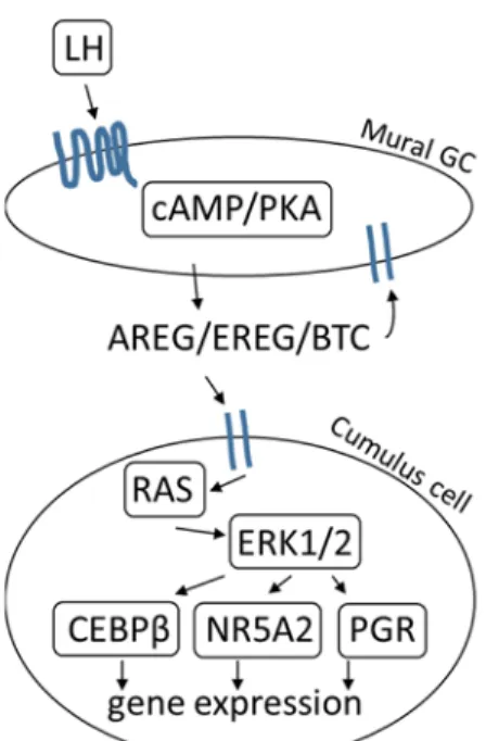
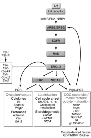
LH regulates the expression of more than 500 target genes that mediate oocyte maturation, cumulus expansion, ovulation, and luteinization in preovulatory follicles (Richards & Pangas, 2010b). LH signals via cAMP/PKA to induce the expression of epidermal growth factor receptor (EGFR) ligands amphiregulin (Areg), betacellulin (Btc), and epiregulin (Ereg) in mural granulosa cells (Conti et al, 2006). These ligands are first synthesized as transmembrane precursors, are cleaved by a metalloproteinase, and then shed as mature proteins (Hsieh & Conti, 2005). They bind EGFRs on mural granulosa and cumulus cells, which activates RAS and ERK1/2 signaling (Fan et al, 2008b). ERK1/2 turns off the FSH genetic program (including Inha, Cyp19a1, Lhcgr)(Fan et al, 2009a). ERK1/2 also activates transcription factors CAAT enhancer binding protein beta (Cebpb), nuclear receptor member 5a2 (Nr5a2), and the progesterone receptor (Pgr), which are required for the transcription of specific target genes involved in oocyte maturation, cumulus expansion, ovulation, and luteinization (FIGURE 1.7)(Duggavathi et al, 2008; Fan et al, 2009b). One critical CEBPβ target is Ptgs2, which is responsible for prostaglandin E2 (PGE2) synthesis. In a positive feedback loop, Areg induces the expression of Ptgs2, which promotes PGE2 synthesis, allowing it to bind to its receptor on cumulus cells to promote increased Areg expression (Shimada et al, 2006).
14
Briefly, cumulus expansion and oocyte maturation involves the induction of Has2, Ptx3, and Tnfaip6 in cumulus granulosa cells (Ochsner et al, 2003a). Ovulation requires induction of Ptgs2 and Pgr in cumulus and mural granulosa cells (Lim et al, 1997; Lydon et al, 1995). Luteinization (that converts mural granulosa cells and theca cells into luteal cells) results in the expression of Cyp11a1, Steroidogenic acute regulatory protein (Star), and the downregulation of FSH target genes that promote proliferation (Espey & Richards, 2002; Goldring et al, 1987; Hunzicker-Dunn & Mayo, 2015). Disruption of EGFR or ERK1/2 signaling is sufficient to block oocyte maturation, cumulus expansion, ovulation, and luteinization (Fan et al, 2009b; Hsieh et al, 2011), illustrating the fact that these two signaling pathways are upstream regulators essential for mediating all of the events triggered by the LH surge.
Although LH primarily signals via cAMP, LH also exerts its effects in a PKA-independent manner to activate RAS, p38 MAPK (MAPK14), and PI3K/AKT signaling. LH signals via a SRC tyrosine kinase to activate RAS, which also leads to the activation of MEK1 and ERK1/2 (Wayne et al, 2007). LH signaling induces a rapid phosphorylation of p38 MAPK (MAPK14) in preovulatory granulosa cells that is involved in COC expansion (Sela-Abramovich et al, 2005). LH has been found to synergize with IGF1 to activate PI3K/AKT signaling, however, the mechanism by which this occurs remains unknown (Vanhaesebroeck et al, 2010). As with FSH signaling, the relative importance of PKA-independent signaling in comparison to PKA-dependent signaling in the transcription of LH target genes remains to be determined.
15
FIGURE 1.7 LH signaling in the preovulatory follicle. Inspired by Conti et al 2006.
1.4.3 Follicle development
1.4.3.1 Crosstalk between germ cells and gonadal somatic cells during follicle development Communication between the oocyte and granulosa cells (cumulus cells and mural granulosa cells) throughout all stages of follicle development ensures that oogenesis and folliculogenesis are coordinated, leading to the release of a fertilizable oocyte. This is especially critical during the early stages of follicle development when intra-ovarian factors drive follicle growth (Pangas & Rajkovic, 2015). Communication is primarily achieved via gap junctions and paracrine signaling. Gap junctions are composed of connexin proteins that form membrane channels between cells which allow passage of ions and small molecules. Gap junction protein 1 (GJA1, or connexin 43) forms gap junctions between granulosa cells, while GJA4 (connexin 37) forms gap junctions between the oocyte and granulosa cells (Ackert et al, 2001). Paracrine signaling is when one cell type produces a signal (ex. growth factor) that binds receptors on a neighboring cell type to elicit a response (Russell et al, 2016). One classic example of this involves the expression of KIT ligand (KITL) by granulosa cells that act on the tyrosine kinase
16
receptor KIT localized on the plasma membrane of the oocyte. Binding of KITL to KIT activates PI3K signaling within the oocyte to stimulate oocyte growth (Thomas & Vanderhyden, 2006).
1.4.3.2 Primordial follicle formation
Primordial follicle formation begins with the breakdown of the cytoplasmic bridges that make up the germ cell cysts between e17.5 and 4dpp, and is accompanied by significant germ cell loss (with approximately only one third of oocytes surviving past this stage)(Pangas & Rajkovic, 2015; Pepling & Spradling, 2001; Richards & Pangas, 2010a). Squamous pre-granulosa cells migrate and surround the oocytes to form primordial follicles, that become enclosed within a basement membrane (Pepling & Spradling, 2001; Rajah et al, 1992; Tingen et al, 2009; Wilhelm et al, 2007). This pool of dormant primordial follicles provides the source of germ cells available for recruitment during the reproductive lifespan of the female, known as the ovarian reserve (Pangas & Rajkovic, 2015; Richards, 1980; Richards & Pangas, 2010a). The mechanisms responsible for germ cell cyst breakdown and primordial follicle formation are incompletely understood, but appear to be regulated, at least in part, by members of the TGF-β family, oocyte-specific transcription factors, and steroid hormone signaling.
The proper formation of a primordial follicle requires complete germ cell cyst breakdown so that pre-granulosa cells can enclose a single oocyte. Numerous factors regulate this process, as evidenced by the mouse models that exhibit multi-oocyte follicles (which is the result of incomplete germ cell cyst breakdown), including granulosa cell-specific conditional knockouts of activin, Bmp15 knockouts in oocytes, and overexpression of Inha in granulosa cells (Jorgez et al, 2004; McMullen et al, 2001; Pangas et al, 2007; Yan et al, 2001).
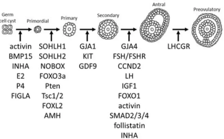
Primordial follicle formation requires a healthy oocyte, which depends on the expression of oocyte-specific transcription factors. Ablation of factor in the germline alpha (Figla) results in significant oocyte loss and a decrease in the formation of primordial follicles (Soyal et al, 2000). In addition, ablation of Sohlh1, Sohlh2, or Nobox also causes increased oocyte loss and disrupts the formation of primordial and primary follicles (FIGURE 1.8)(Choi et al, 2008; Pangas et al, 2006a; Rajkovic et al, 2004).
Appropriate levels of hormone signaling is also crucial. For instance, treatment of newborn mouse ovaries with estrogens, estrogen-mimetics, or progesterone inhibits the
17
formation of primordial follicles (Chen et al, 2007; Tingen et al, 2009). One hypothesis that has been advanced is that elevated levels of estrogens and progesterone during pregnancy maintain germ cell cysts, but at birth, the drop in circulating hormones triggers germ cell cyst breakdown (Chen et al, 2007). Any pathologies therefore that cause aberrant elevated levels of steroid hormones can prevent primordial follicle formation.
1.4.3.3 Primary follicle development
Primordial follicles have three possible fates: to remain dormant, to be activated to join the growing follicle pool, or to directly undergo atresia (McGee & Hsueh, 2000; Reddy et al, 2010). Cohorts of primordial follicles (approximately 3-6) become activated daily to develop to the primary follicle stage (Fortune, 2003; Richards & Pangas, 2010a). Primary follicles are detectable by 3 dpp and consist of a growing oocyte surrounded by proliferating cuboidal granulosa cells (Bertolin & Murphy, 2014; Pangas & Rajkovic, 2015; Richards, 1980).
Activation of primordial follicles appears to be regulated by mTORC1 and PI3K/AKT signaling in granulosa cells (AKT acts upstream of mTORC1 by repressing its inhibitor, TSC1/2)(Reddy et al, 2010). Inhibition of mTORC1 signaling in squamous pre-granulosa cells prevented their differentiation into cuboidal granulosa cells and maintained oocytes in a quiescent state, whereas overactivation of mTORC1 signaling accelerated granulosa cell differentiation and prematurely activated primordial follicles (Zhang et al, 2014a).
Continuous inhibitory signals are required in order to maintain primordial follicles in a dormant state and therefore preserve the ovarian reserve. Oocyte-specific ablation of Foxo3a (negatively regulated by AKT), Pten (a PI3K inhibitor), or Tsc1/2 (an mTORC1 inhibitor) caused widespread premature activation of primordial follicles and consequently depletion of the primordial follicle reserve (Adhikari et al, 2009; Castrillon et al, 2003; Reddy et al, 2008). How PTEN/PI3K/AKT signaling is regulated in this context remains unknown.
Inhibitory signals also originate from granulosa cells. Total knockout of Foxl2 impaired squamous pre-granulosa cell differentiation into cuboidal granulosa cells, with oocytes undergoing premature growth, leading to follicle depletion and oocyte death (Schmidt et al, 2004; Uda et al, 2004). Ablation of anti-Müllerian hormone (Amh) resulted in overactivation of primordial follicles and premature primordial follicle depletion (FIGURE 1.8)(Durlinger et al,
18
1999). Despite what we do know, the regulation of the dormant primordial follicle pool remains one of the least understood mechanisms in ovarian physiology.
1.4.3.4 Secondary follicle development
By 7 dpp, secondary follicles can be detected within the ovary, which consist of a growing oocyte surrounded by more than a single layer of proliferating granulosa cells and a second somatic cell layer consisting of theca cells. The regulation of theca cell differentiation is poorly understood, but appears to involve the differentiation of interstitial cells mediated by granulosa cell-derived signals (Gardiner & Swain, 2015). Follicle development is still dependent at this stage on intra-ovarian factors; the oocyte continues to supply cumulus cells with growth factors, while the latter contribute to maintaining meiotic arrest in the oocyte (Monniaux, 2016).
Loss-of-function of either gap junction proteins or growth factor signaling impede follicle growth beyond the primary stage. Ablation of Gja1 from granulosa cells caused follicle arrest at the primary follicle stage (Ackert et al, 2001), in contrast to ablation of Gja4 from oocytes caused arrest at the preantral follicle stage (Simon et al, 1997). Loss-of-function of KIT caused follicle arrest at the primary follicle stage (John et al, 2009). Ablation of growth differentiation factor 9 (Gdf9) resulted in follicle arrest at the primary follicle stage, in which large oocytes were surrounded by a single layer of cuboidal granulosa cells (FIGURE 1.8)(Dong et al, 1996). These conditional knockout models illustrate, without any doubt, how communication between the oocyte and granulosa cells is essential in order for normal follicle development to proceed.
1.4.3.5 Antral follicle development
Antral follicles become apparent around 13 dpp, wherein the oocyte has completed its growth and becomes suspended in the antrum surrounded by cumulus granulosa cells (Bertolin & Murphy, 2014; Conti et al, 2006; Pangas & Rajkovic, 2015; Richards, 1980). At the antral stage, follicles are now dependent on gonadotropins, FSH and LH, for their continued development (Pangas & Rajkovic, 2015; Richards & Pangas, 2010a). A small pool of growing follicles is capable of responding to FSH and LH to reach the preovulatory stage around 21 dpp
19
(Pepling & Spradling, 2001). LH stimulates theca cells to produce androgens and FSH stimulates granulosa cells to aromatize androgens into estradiol, which then exerts a positive feedback on the HPG axis to trigger the LH surge (Richards, 1980; Richards, 1994). Estradiol acts in synergy with FSH to stimulate granulosa cell proliferation (by induction of Ccnd2 and stimulation of IGF1 synthesis) and granulosa cell differentiation (by induction of Lhcgr, increased CYP19A1 activity, and increased inhibin production)(Pangas & Rajkovic, 2015). In the absence of FSH stimulation, widespread apoptosis of granulosa cells in early antral follicles occurs, resulting in follicular atresia (Chun et al, 1996; Richards, 1980). Fshb, Fshr, Ccnd2, Lhb, and Igf1 knockout mice are all unable to form antral follicles and arrest at the preantral stage (Dierich et al, 1998; Kumar et al, 1997; Ma et al, 2004; Sicinski et al, 1996; Zhou et al, 1997). Lhcgr knockout mice do not develop past the antral stage (Lei et al, 2001). Constitutive activation of Foxo1 (a downstream effector of FSH and IGF1) in granulosa cells results in suppression of genes involved in granulosa cell proliferation (Ccnd2), steroidogenesis (Cyp19a1, cholesterol biosynthesis), and gonadotropin signaling (Fshr, Lhcgr) (Liu et al, 2009b; Park et al, 2005; Richards & Pangas, 2010b).
TGF-β/SMAD signaling stimulated by growth factors produced by granulosa cells, is particularly critical during the preantral and antral stages of follicle development. Activin enhances FSH activity and signals through SMAD2/3/4 to promote granulosa cell proliferation (by regulating Ccnd2) and steroidogenesis (by regulating Cyp19a1)(Hunzicker-Dunn & Maizels, 2006; Park et al, 2005). Ablation of activin subunits lead to premature luteinization of preantral follicles and accumulation of CLs (Pangas et al, 2007). Conditional knockouts of Smad2/3 or Smad4 exhibit increased preantral follicle atresia, premature luteinization, defective cumulus cells, and decreased ovulation (Li et al, 2008; Pangas et al, 2006b). On the other hand, conditional knockout of follistatin (an activin inhibitor) leads to follicle depletion by eight months of age (Jorgez et al, 2004). Ablation of Inha (an activin antagonist) results in granulosa cell proliferation in the absence of oocyte growth, and consequently the absence of late-stage follicles (FIGURE 1.8)(Myers et al, 2009). Clearly, gonadotropin-dependent follicle development (past the antral stage) involves a complex network in which hormones enhance or repress the effects of FSH to induce key target genes that drive granulosa cell proliferation, differentiation, survival, and steroidogenesis. While several of the key mediators required for growth beyond the antral stage have been identified, many still have yet to be discovered.
20
FIGURE 1.8 Regulation of folliculogenesis. Inspired by Richards and Pangas 2010, and Pangas 2015.
1.4.3.6 Events initiated by the LH surge
The LH surge acts on large preovulatory follicles to trigger key events that lead to the release of a fertilisable oocyte, which include cumulus expansion, oocyte maturation, ovulation, and luteinization (Richards et al, 2015b). Only preovulatory follicles are able to respond to the LH surge and ovulate because only their mural granulosa cells selectively express high levels of LHCGR that bind LH/hCG (Jeppesen et al, 2012). While LH signaling via its receptor to activate EGFR signaling is well-established (Espey & Richards, 2002; Park et al, 2004; Shimada et al, 2006), many of the downstream targets have yet to be identified.
1.4.3.6.1 Oocyte maturation
Throughout follicle development, oocyte meiotic arrest is maintained by elevated cAMP and cGMP levels within the oocyte (Conti et al, 2012). cAMP/PKA maintains oocyte arrest by inhibiting the CDK1-Cyclin B complex (Chesnel & Eppig, 1995). cAMP levels are regulated by phosphodiesterase 3A (PDE3A) that breaks down cAMP and adenylyl cyclase 3 that converts ATP to cAMP (Vaccari et al, 2008). cGMP originates from cumulus cells that reach the oocyte via gap junctions (made up of GJA4) to inhibit PDE3A (Simon et al, 1997).
The LH surge causes a disruption of GJA4 leading to a drop in cGMP in the oocyte, which derepresses PDE3A. PDE3A, now active, can hydrolyze cAMP. The drop in cAMP
21
derepresses CDK1-Cyclin B, allowing meiosis to resume (FIGURE 1.9)(Conti et al, 2012; Oh et al, 2010). This results in the breakdown of the oocyte nuclear membrane (commonly referred to as germinal vesicle breakdown)(Hunzicker-Dunn & Mayo, 2015). The oocyte completes meiosis I, releases the first polar body, and progresses to metaphase II where it arrests again until fertilization (Pangas & Rajkovic, 2015; Richards, 1980).
FIGURE 1.9 Maintenance of oocyte meiotic arrest (on left) vs resumption of meiosis (on right). Inspired by Hunzicker-Dunn and Mayo 2015.
1.4.3.6.2 Cumulus expansion
Cumulus cells produce a complex hyaluronan-rich extracellular matrix that is stabilized by numerous hyaluronan-binding proteins, in which the cumulus cells dissociate from one another and migrate outwards away from the oocyte in a process referred to as cumulus expansion (Richards, 2005; Richards et al, 2015a). The main drivers of cumulus expansion include granulosa cell expression of hyaluronan synthase 2 (Has2), EGFR ligands, prostaglandin synthase 2 (Ptgs2), and oocyte-derived Gdf9 and Bmp15 (Richards et al, 2015b).
In mural granulosa cells, LH induces the expression of EGFR ligands Areg, Ereg, and Btc, which promote expression of Ptgs2, leading to the production of prostaglandins PGE1/2 that act on cumulus cells (Park et al, 2004; Shimada et al, 2006). A Ptgs2 autoregulatory loop promotes increased expression of EGFR ligands to further enhance its own expression (Shimada et al, 2006). EGFR ligands will then act in an autocrine and paracrine manner. In the latter, they regulate the expression of Has2, which synthesizes hyaluronan polymers that are stabilized by versican (VCAN), A disintegrin and metalloproteinase with thrombospondin-like motifs-1
22
(ADAMTS1), TNF-α-induced protein 6 (TNFAIP6), inter-alpha trypsin inhibitor (IAI), and pentraxin 3 (PTX3)(Richards, 2005). Areg, Ereg, Btc signaling through EGFR are sufficient to drive cumulus expansion (Park et al, 2004). Disruption of Ptgs2, Has2, Adamts1, Tnfaip6, or Ptx3 impairs cumulus expansion and oocytes remain trapped within luteinized follicles (Dinchuk et al, 1995; Mittaz et al, 2004; Ochsner et al, 2003a; Sugiura et al, 2009; Varani et al, 2002). The phenotype of these mice illustrates how COC expansion/ovulation vs luteinization are regulated by separate mechanisms and that a proper sequence of events is essential in order for ovulation to occur.
Oocyte-derived Gdf9 and Bmp15 promote cumulus cell expression of genes that will provide the oocyte with the metabolic substrates it requires, such as cholesterol and glucose (Su et al, 2008; Sugiura et al, 2007). Ablation of Bmp15 results in defective cumulus cell expansion and reduced ovulation (Su et al, 2004).
In addition, several innate immune response components also mediate cumulus expansion, including Toll-like receptors 2 and 4, and interleukin-6 (IL6)(Richards et al, 2008); IL6 alone is sufficient to stimulate COC expansion (FIGURE 1.10)(Liu et al, 2009a).
1.4.3.6.3 Ovulation
Ovulation entails the rupture of the preovulatory follicle wall and the release of the COC from the ovary into the oviduct (Richards et al, 2008). The breakdown of the follicle wall is carried out by proteases and collagenases that loosen and dissolve the numerous layers separating the COC from the peritoneal cavity (Richards et al, 2015a; Robker et al, 2000). Smooth muscle contractions in the theca layer and intra-follicular positive pressure assist the release of the COC out of the ruptured follicle (Richards et al, 2015a; Rose et al, 1999). Genes that mediate ovulation must be expressed prior to genes that mediate luteinization, otherwise the oocyte remains trapped within a luteinized follicle (Robker et al, 2000).
Two transcription factors are key for the processes of ovulation and luteinization, NR5A2 and CEBPB. Conditional knockout of either Nr5a2 or Cebpb in granulosa cells exhibit impaired ovulation and luteinization (Duggavathi et al, 2008; Sterneck et al, 1997). LH also induces expression of the transcriptional co-regulator Nrip1 that regulates expression of Areg (Nautiyal et al, 2010). Nrip1 knockout mice also fail to ovulate (Tullet et al, 2005).
23
The LH surge rapidly induces the expression of two key genes, Pgr and Ptgs2 that mediate ovulation-related events (Park & Mayo, 1991; Wong & Richards, 1991). In cumulus cells PTGS2 binds to one of its receptors, EP2, and induces the expression of Tnfaip6 (Ochsner et al, 2003b). PGR induces the expression of proteases such as Adamts1 and cathepsin L (FIGURE 1.10)(Robker et al, 2000). Beyond these two proteases, the other key proteases that mediate the “breakdown” of the follicle wall remain to be identified. Mice deficient in Pgr, Ptgs2, or Ep2 are anovulatory (Dinchuk et al, 1995; Hizaki et al, 1999; Lydon et al, 1995).
1.4.3.6.4 Luteinization
Following the release of the COC, the remaining mural granulosa and theca cells in the follicle stop proliferating (exit the cell cycle), become hypertrophic, and terminally differentiate into luteal cells that synthesize P4 (Richards et al, 1998). This process requires the expression of transcription factors Cebpb, Nr5a2, and Runx2, steroidogenic enzymes Cyp11a1 and Star, Prlr, cell cycle inhibitor genes Cdkn1a/b, and downregulation of FSH genes involved in follicle maturation (Ccnd2, Fshr, Esr2, Cyp19a1, Inha, Foxo1)(Hunzicker-Dunn & Mayo, 2015).
Major remodelling occurs with the breakdown of the basal lamina between the theca and granulosa cells and the infolding of the follicle wall. Several proteases are involved in extracellular matrix (ECM) remodeling: serine proteases, matrix metalloproteinases, their tissue inhibitors, and ADAMTS-1 (Curry & Osteen, 2003). This process can be described as an inflammatory response given that many innate immunity-related molecules (cytokines, chemokines) are recruited, the previously avascular granulosa layer becomes infiltrated with new blood vessels, and large amounts of prostaglandins (mostly PGE2) are produced (FIGURE 1.10)(Richards et al, 2008; Richards et al, 2015a; Robker et al, 2000) .
The differentiated luteal cells will then form the CL. The CL has a limited lifespan which depends on whether pregnancy occurs or not. If pregnancy occurs, the CL is functional and secretes P4 throughout the duration of gestation to maintain pregnancy (Bertolin & Murphy, 2014; Stouffer & Hennebold, 2015). In rodents, “pseudopregnancy” can occur in which imitation of mating by cervical stimulation at estrus induces the development of a functional CL that produces progesterone for 12-14 days. In the absence of either pregnancy or pseudopregnancy, no functional CL is formed in rodents, and the insufficient amount of
24
progesterone produced is unable to induce uterine decidualization (Stouffer & Hennebold, 2015).
Luteotropic factors are essential to develop and maintain the functional CL, which include prolactin (from the anterior pituitary), prolactin-like hormones (from the uterine decidua and placenta), and estradiol (produced locally) (Binart et al, 2000). Luteolysis is the process by which prostaglandin F2α (the “uterine luteolytic factor”) promotes the loss of CL function and (eventually) structure either at the end of an ovarian cycle (in the absence of pregnancy) or at the end of gestation, allowing for the start of the subsequent ovarian cycle (Horton & Poyser, 1976). Luteal regression is marked by a drop in progesterone production (Niswender et al, 1994). In the mouse, three or more generations of non-functional CLs may be present during the same cycle (Stouffer & Hennebold, 2015).
25
FIGURE 1.10 LH signaling that drives oocyte maturation, cumulus expansion, ovulation, and luteinization. Revised from Richards and Pangas 2010.
1.4.4 Steroidogenesis
1.4.4.1 Estradiol
Ovarian estrogen synthesis requires theca and granulosa cells that express cell-specific enzymes to convert cholesterol into estrogens (Richards, 1980). The main ovarian sex steroid hormones are 18-carbon estrogens, 19-carbon androgens, and 21-carbon progestins (McKenna, 2015). Steroidogenic enzymes/proteins can be classified into three classes: cytochromes P450
26
(CYP11A1, CYP17A1, CYP19A1), oxidoreductases (17βHSD, 3βHSD), and transport proteins (STAR)(Auchus, 2015).
Cholesterol is the precursor of all steroids (Auchus, 2015). Within granulosa and theca cells, STAR transports cholesterol from the outer mitochondrial membrane to the inner mitochondrial membrane, where CYP11A1 is located (Auchus, 2015; Clark et al, 1994). The first (rate-limiting) step is the side-chain cleavage of 27-carbon cholesterol to 21-carbon pregnenolone by CYP11A1 (Miller & Auchus, 2011). Pregnenolone is converted to progesterone by 3βHSD2 (Miller & Auchus, 2011). In theca cells only, CYP17A1 converts pregnenolone to 17-hydroxypregnenolone and then to DHEA (Gupta et al, 2003). Progesterone is converted to androstenedione by CYP17A1 (Gupta et al, 2003). Oxidoreductase reactions carried out by 17βHSD1 and 3βHSD2 convert DHEA to androstenedione and testosterone (Miller & Auchus, 2011). These two 19-carbon androgens are then aromatized to 18-carbon estrone or 17β-estradiol (commonly referred to as estradiol) by CYP19A1 in granulosa cells only (Simpson et al, 1994). An additional step converts estrone to estradiol by 17βHSD1 (FIGURE 1.11)(Miller & Auchus, 2011).
Steroidogenesis is stimulated by gonadotropins. In theca cells, LH binds to its receptor to induce conversion of cholesterol to androgens (Hillier et al, 1991). In granulosa cells, FSH binds to its receptor to induce progesterone production and CYP19A1 expression (Escamilla-Hernandez et al, 2008).
A distinction between granulosa vs theca cell expression of steroidogenic enzymes dictates which hormones are produced within each cell type. Granulosa cells do not express CYP17A1 and therefore are unable to convert progesterone to androgens. Thecal cells do not express CYP19A1, which explains why they are unable to aromatize androgens to estrogens (Auchus, 2015).
Estradiol binds to estrogen receptors 1 and 2 (ESR1/2) that are nuclear transcription factors expressed in the hypothalamus, anterior pituitary, and ovary (Couse et al, 1999). Within the ovary, ESR1 is predominantly expressed in theca cells, while ESR2 is expressed in granulosa cells (Couse & Korach, 2001).
27
FIGURE 1.11 Estradiol biosynthesis. Adapted from Auchus 2015.
1.4.4.2 Progesterone
After ovulation, granulosa and theca cells within the ovulated follicle luteinize to form a functional CL that secretes larger amounts of P4 (Richards, 1980). The same steps required for progesterone synthesis in granulosa and theca cells apply to progesterone synthesis in luteal cells: cholesterol is transported by STAR to CYP11A1, which cleaves it to pregnenolone, which is then converted to progesterone by 3βHSD2 (FIGURE 1.12)(Auchus, 2015). This occurs under the control of LH (Goldring et al, 1987).
In the ovary, PGRs are expressed in theca cells of large preovulatory follicles, OSE, stroma, and transiently in mural granulosa cells of preovulatory follicles (induced by the LH surge) (Gava et al, 2004). Pgr expression is low during folliculogenesis except during a short 4-6h window after the LH surge (Park & Mayo, 1991). Its primary role (as described in section
28
1.4.3.6.3 Ovulation) is to mediate follicle rupture by inducing expression of key proteases that break down the follicle wall and the ECM (Lydon et al, 1995).
FIGURE 1.12 Progesterone biosynthesis. Adapted from Auchus 2015.
As outlined above, many critical steps during ovarian follicle development still remain incompletely understood. These gaps in our knowledge are what prompt us to investigate novel signaling pathways that are well-known in other contexts but whose study in the ovary is only just beginning. The Hippo signaling pathway is one such pathway, as outlined below.
29 2 Hippo signaling pathway
2.1 Introduction
How are cells instructed to stop growing when an organ has reached its proper size? How do cells know whether to proliferate or to differentiate? How do cells detect that a part of an organ is missing? How do damaged tissues regenerate? These are all long-standing questions that have puzzled researchers for many years until recently; with the discovery of the Hippo pathway, these enigmas are beginning to be resolved.
2.2 Discovery of the Hippo signaling pathway
Components of the Hippo pathway were first discovered in 1995 in genetic screens for tumor suppressor genes in the fruit fly Drosophila melanogaster. The screens identified two kinases Hippo (Hpo) and Warts (Wts), and the scaffold proteins Salvador (Sav) and Mob-as-tumor-suppressor (Mats)(Justice et al, 1995; Lai et al, 2005; Tapon et al, 2002; Wu et al, 2003). Loss-of-function mutations in any of these genes led to massive tissue overgrowth as a result of excessive cell proliferation and resistance to apoptosis. For example, the eye discs of hpo-null flies developed into oversized eyes and heads that resembled a hippopotamus hide, which eventually inspired the name of the pathway (Udan et al, 2003). Hpo-Sav formed a kinase complex that phosphorylated and activated Wts-Mats kinase complex, which was named the “Salvador-Warts-Hippo” (SWH) pathway (Wei et al, 2007). Yorkie (Yki) was later identified as a Wts-binding protein whose activity is negatively regulated by Hpo and Wts (Huang et al, 2005). Yki is unable to bind DNA directly, which led to the discovery of Scalloped (Sd), a member of the TEA domain (TEAD) family of transcription factors, as the mediator required for Yki to exert its functions (Wu et al, 2008).
In parallel with the discovery of the SWH pathway in Drosophila, several of the mammalian homologs were also being identified, however, it would take years before they became linked to what is now known as the Hippo pathway. In 1994, Yes-associated protein (YAP) was identified as a Yes tyrosine kinase-binding protein (Sudol, 1994). In 1998, mammalian STE20-like protein kinase 1 (MST1) was found to promote apoptosis (Graves et al, 1998). In 1999, large tumor suppressor 1 (LATS1) was found to regulate the cell cycle (Tao et