HAL Id: hal-00376093
https://hal.archives-ouvertes.fr/hal-00376093
Submitted on 14 Oct 2019
HAL is a multi-disciplinary open access
archive for the deposit and dissemination of sci-entific research documents, whether they are pub-lished or not. The documents may come from teaching and research institutions in France or abroad, or from public or private research centers.
L’archive ouverte pluridisciplinaire HAL, est destinée au dépôt et à la diffusion de documents scientifiques de niveau recherche, publiés ou non, émanant des établissements d’enseignement et de recherche français ou étrangers, des laboratoires publics ou privés.
The replacement of a phenol group by an aniline or
acetanilide group enhances the cytotoxicity of
2-ferrocenyl-1,1-diphenyl-but-1-ene compounds against
breast cancer cells
Pascal Pigeon, Siden Top, Ouardia Zekri, Elisabeth A. Hillard, Anne
Vessieres, M.A. Plamont, Olivier Buriez, Eric Labbé, Michel Huché, Sultana
Boutamine, et al.
To cite this version:
Pascal Pigeon, Siden Top, Ouardia Zekri, Elisabeth A. Hillard, Anne Vessieres, et al.. The replacement of a phenol group by an aniline or acetanilide group enhances the cytotoxicity of 2-ferrocenyl-1,1-diphenyl-but-1-ene compounds against breast cancer cells. Journal of Organometallic Chemistry, Elsevier, 2009, 694, pp.895-901. �10.1016/j.jorganchem.2008.11.035�. �hal-00376093�
The replacement of a phenol group by an aniline or acetanilide group enhances
the cytotoxicity of 2-ferrocenyl-1,1-diphenyl-but-l-ene compounds against
breast cancer cells
Pascal Pigeon a, Siden Top a, Ouardia Zekri a,c, Elizabeth A. Hillard a, Anne Vessières a,*, Marie-Aude Plamont a, Olivier Buriez b, Eric Labbé b, Michel Huché a, Sultana Boutamine c, Christian Amatore b, Gérard Jaouen a
a Laboratoire de Chimie et Biochimie des Complexes Moléculaires, UMR CNRS 7576, Ecole Nationale
Supérieure de Chimie de Paris, 11 rue Pierre et Marie Curie, 75231 Paris Cedex 05, France
b Ecole Normale Supérieure, Département de Chimie, UMR CNRS-ENS-UPMC 8640, 24 rue Lhomond, 75231
Paris Cedex 05, France
c
Université des Sciences et de la Technologie Houari Boumediene, Faculté de Chimie, BP32, El Alia Bab Ezzouar, Alger, Algeria
Keywords: Bioorganometallic chemistry ; Ferrocene ; Anti-cancer drugs ; Breast cancer ; Iron * Corresponding author. Tel.: +33 01 44 27 67 29; fax: +33 01 43 26 00 61.
E-mail address: a-vessieres@enscp.fr (A. Vessières).
Abstract
We have previously shown that conjugated ferrocenyl p-phenols show strong cytotoxic effects against both the hormone-dependent MCF-7 and hormone-independent MDA-MB-231 breast cancer cell lines, possibly via oxidative quinone methide formation. We now present a series of analogous amine and acetamide compounds: 2-ferrocenyl-1-(4-aminophenyl)-1-phenyl-but-1-ene (Z+E-2), 2-ferrocenyl-1-(4-N-acetylaminophenyl)-1-phenyl-2-ferrocenyl-1-(4-aminophenyl)-1-phenyl-but-1-ene (Z-3), and their corresponding organic molecules 1-(4-aminophenyl)-1,2-bis-phenyl-but-1-ene (Z+E-4) and 1-(4-N-acetamidophenyl)-1,2-bis-phenyl-but-1-ene (Z+E-5). All of the compounds have adequate relative binding affinity values for the estrogen receptor; between 2.8% and 5.7% for ERα, and between 0.18% and 15.5% for ERβ, as well as exothermic ligand binding in in silico ER docking experiments. Compounds 2 and 3 show dual estrogenic/cytotoxic activity on the MCF-7 cell line; they are proliferative at low concentrations (0.1 µM) and antiproliferative at high concentrations (10 µM). On the MDA-MB-231 cell line, the ferrocenyl complexes 2 and 3 are antiproliferative with IC50 values of 0.8 µM for 2 and 0.65
µM for 3, while the purely organic molecules 4 and 5 show no effect. Electrochemical experiments suggest that both 2 and 3 can be transformed to oxidized quinoid-type species, analogous to what had previously been observed for the ferrocene phenols.
1. Introduction
Compounds that can be activated by the redox environment of a cellular target to produce reactive oxygen species (ROS) [1] or reactive intermediates [2] have recently gained much attention, particularly in cancer treatment [3]. For example, compounds containing phenol moieties, such as resveratrol, can modify the oxidative stress level of the cell to cause necrosis and apoptosis [4,5]. Tamoxifen (Chart 1), a leading treatment for all phases of hormone-dependent breast cancer [6], also shows a mild non-genomic cytotoxic activity, which has been attributed to its aromatic hydroxylation and biooxidation to radical or electrophilic species [7]. We have been studying a series of conjugated ferrocenyl phenols, which show antiproliferative effects in vitro on both hormone-independent (ER–) and hormone-dependent (ER+) breast cancer cells [8,9] and in vivo against glioma [10]. Structure activity relationship studies have demonstrated the importance of the presence of a ferrocene group [11,12], a
p-phenol [13], and a -system linking the two moieties [14], for a strong cytotoxic activity in vitro. Electrochemical experiments have suggested that the active species in cell cultures could be a quinone methide, which may be formed via an intramolecular oxidation of the phenol moiety by the in situ-generated ferricenium cation [15]. Other investigators have also shown a correlation between the toxicity of a tamoxifen–ferrocene conjugate and the production of ROS [16]. Ferricenium-promoted intramolecular electron transfer in phenolic systems has been recognized [17–21], although our work is the first application of this mechanism to oxidation-activated drug development.
According to the proposed mechanism of activation [15], we expect that the substitution of the hydroxyl group by other protic and oxidizable functionalities should retain the molecules’ cytotoxic properties and specific electrochemical signature. However, in a previous study, we found that the dithioacetyl prodrug analog 1a (Chart 1) displayed only proliferative effects on dependent breast cancer cells (MCF-7), and no (cytotoxic) effect on hormone-independent breast cancer cells (MDA-MB-231) [22]. This was in marked contrast to the diacetoxy compound, 1b, which showed cytotoxic effects similar to that of the diphenol 1c, suggesting that intracellular hydrolysis of the compound to the active hydroxylated species takes place. As thioesterases have been isolated from breast cancer cells [23], we attributed this lack of activity not to the protection of the thiol, but to the poor overlap between the 3p S and 2p C atomic orbitals, which disfavors thioquinone methide formation.
Chart 1. The breast cancer drug tamoxifen, the amino derivative of tamoxifen, previously studied ferrocene complexes 1a–1d and 6 and new compounds 2–5.
We now investigate the synthesis, cell proliferation effects, and electrochemical behavior of the ferrocenyl aniline 2 and acetanilide 3 shown in Chart 1, particularly as compared to the cytotoxic monophenolic analog 6 [8]. We have also studied their purely organic counterparts,
4 and 5 to evaluate the importance of the ferrocenyl moiety. These later compounds are
relevant, in that the literature on the biological effects of aryl amines or amides based on the tamoxifen or triphenylethylene skeleton is scarce; to the best of our knowledge, only one
study on the uterotrophic effects of the amino derivative of tamoxifen (Chart 1) has been carried out [24].
2. Results and discussion
2.1. Preparation of compounds
The synthesis of the organometallic compounds was based on a McMurry cross-coupling reaction, the method we commonly use to form alkenes from the reaction of two ketones in the presence of TiCl4/Zn [25–27]. Compound 2 was synthesized as a mixture of Z and E
isomers via the reaction between propionyl ferrocene and 4-aminobenzophenone, as previously reported [28]. The moderate yield (26%) is consistent with the previously observed low activity of aminobenzophenone in McMurry reactions [29]. The Z and E isomers of 2 could not be separated using preparative HPLC, and thus the mixture of (Z+E)-2 was used in all further experiments. Similarly, its organic analog 4 was synthesized by a reaction between propiophenone and aminobenzophenone as a mixture of Z and E isomers in 38% yield (see Scheme 1).
An alternative pathway via a coupling reaction between propionyl ferrocene or propiophenone and 4-nitrobenzophenone also gave access to 2 and 4. Originally we used this reaction in an attempt to synthesize the corresponding nitro species, but found that an in situ reduction of the nitro group to an amino group furnished 2 and 4. However, depending on the batch of 4-nitrobenzophenone used, the yield of the reaction varied significantly from high (>50%) to poor.
Treating 2 with acetyl chloride and pyridine gave 3 as a mixture of Z and E isomers (60/40) in 91% yield, as shown in Scheme 2. The Z isomer was isolated by fractional crystallization and all further tests were performed with the pure isomer. Compound 5 was similarly synthesized from 4 in 81% yield as a mixture of Z and E isomers (85/15).
Scheme 1. Synthesis of the ferrocenyl and phenyl aniline derivatives 2 and 4.
Scheme 2. Synthesis of the ferrocenyl and phenyl acetanilide derivatives 3 and 5.
2.2. Biological studies
2.2.1. Determination of the relative binding affinity (RBA) values of the compounds for the two forms of estrogen receptors (ERα and ERβ)
The RBA values obtained for the new compounds are given in Table 1. The values found for the ferrocenyl aniline and acetanilide derivatives 2 and 3 on ERα were only slightly lower than that found for the ferrocenyl monophenol 6 (RBA = 4.6%). This is quite surprising for molecules having no phenol group, which is considered to be essential for ER recognition [30]. Although all compounds have RBA values on the same order of magnitude, the purely organic compounds 4 and 5, lacking the bulky ferrocene group, have slightly higher affinities for ERα. Interestingly, both 2 and 4 showed a greater affinity for ERα than that found for the amino derivative of tamoxifen (0.2%), evidently due to the absence of the amino side-chain
[24]. The binding affinities for ERβ are much more variable than for ERα, with the organic compounds showing significantly higher values than the ferrocenyl compounds. Nonetheless, all compounds have non-zero RBA values, and thus would be expected to interact with the ERs.
Table 1
Relative binding affinity (RBA) of the compounds on the two isoforms of the estrogen receptor (ERα and ERβ) and IC50 values for the compounds on hormone-independent breast cancer cells, MDA-MB-231.
Compound RBA (%) IC50 [µM]
ERα ERβ
(Z+E)-2 2.8 ± 0.1 1.08 ± 0.07 0.8 ± 0.1
(Z)-3 3.5 ± 0.3 0.18 ± 0.02 0.65 ± 0.03
(Z+E)-4 7 ± 0.3 15.5 ± 2.5 Not toxic at 1 µM
(Z+E)-5 5.7 ± 0.1 7.7 ± 0.8 Not toxic at 1 µM
(Z+E)-6 4.6 ± 0.1a 11 ± 1a 1.13 ± 0.07b
a
Value from Ref. [8].
b Value from Ref. [13].
2.2.2. Molecular modeling of receptor interactions
Molecular docking experiments using the crystal structure of ERα crystallized with DES (pdb erd.ent) [31], and of ERβ crystallized with 5,11-cis-diethyl-5,6,11,12-tetrahydrochrysene-2,8-diol (THC) (pdb 112j.ent) [32] were performed. Only the amino acids that constitute the wall of the cavity have been retained. The DES or THC molecules were removed from the cavity and replaced successively with the different bioligands. Energy minimization was then carried out using Merck molecular force field (MMFF). All the heavy atoms of the amino acids of the cavity wall were then immobilized and the side chain of His524 for ERα and His475 for ERβ were liberated. This allowed the ideal positions of the bioligands to be determined. Quantum mechanical semi-empirical PM3 methods were then used to determine the affinity of the bioligands for the cavity. This requires calculation of the energies of bioligand-cavity group, of the cavity itself, and of the ligand, the latter two in the conformations they had in the molecular assemblies to give the rH° enthalpy variations of the reactions: bioligand + cavity molecular assembly (Table 2). For all compounds, binding with both isoforms of the ER is thermodynamically favored, as evidenced by the negative enthalpy of formation for the ligand–receptor complex.
Table 2
Enthalpy variation values for the bioligand docked into ERα and ERβ.
Compound rH° (kcal mol-1)
ERα ERβ (Z)-2 –17.5 –17.3 (E)-2 –16.4 –31.1 (Z)-3 –16.5 –16.7 (Z)-4 –13.3 –8.4 (Z)-5 –13.5 –13.2 (E)-5 –8.8 –10.3
Fig. 1 shows (E)-2 in the cavity of ERα. The NH2 group forms a hydrogen bond with the
group interacts with His524. This triple anchorage demonstrates the good association of this isomer with the cavity.
Fig. 1. (E)-2 in the cavity of ERα.
Fig. 2. Effect of estradiol (E2, 10 nM), ferrocenyl aniline 2 and ferrocenyl acetanilide 3 at
cancer cells) after 5 days of culture. Non-treated cells are used as the control (C). Representative data of one experiment performed twice with similar results (six measurements ± confidence limit; P = 0.1, t = 1.895).
2.2.3. Effect on hormone-dependent breast cancer cells MCF-7
The effect of 2 and 3 on MCF-7 cells is shown on Fig. 2. At the low concentration of 0.1 µM, 2 showed a marked proliferative effect while 3 was only slightly proliferative. At the high concentration of 10 µM 2 had almost no effect while 3 gave rise to a moderate antiproliferative effect. It should be mentioned that the experimental conditions used (cell medium without phenol red, an agent normally used for MCF-7 cell culture, but containing an impurity known to be estrogenic) was chosen to favor the expression of the estrogenic component of the tested molecules. However, the estrogenic effects are not consistent with the observed RBA values, which suggest that 3, with a stronger binding affinity, should be more estrogenic than 2. This is because the observed effect is actually the net result of the estrogenic (proliferative) effect which is expressed at low concentrations, combined with the cytotoxic (antiproliferative) effect which begins to appear at higher concentrations. The observed results are consistent with the higher cytotoxicity of 3 compared with 2 (vide infra).
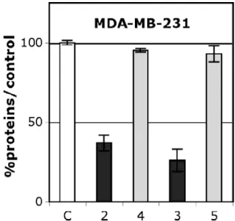
2.2.4. Effect on hormone-independent breast cancer cells MDA-MB-231
On MDA-MB-231 2 and 3 showed a strong antiproliferative effect (Fig. 3) with IC50
values of 0.8 and 0.65 µM, respectively (Table 1). Remarkably, these compounds are more cytotoxic than the ferrocenyl monophenol 6 and approach the antiproliferative potency of the previously reported ferrocenyl diphenol 1c (IC50 = 0.6 µM) [33]. Consequently, they join the
group of the ferrocenyl derivatives having the lowest IC50 values (<1 µM) found so far [9]. It
should be noted that the unsubstituted ferrocenyl compound 1d showed no effect even at concentrations as high as 10 µM, demonstrating the participation of the functional groups in the cytotoxic activity [13]. Interestingly, neither the organic amine 4 nor the acetylamide 5 was cytotoxic at 1 µM, although the oxidative metabolism of aromatic amines is known to produce toxic species [34,35], including in breast cancer cells [36]. Nonetheless, in our experiments, the presence of the ferrocenyl group was essential for a strong cytotoxic effect.
Fig. 3. Effect of 1 µM of the ferrocenyl aniline 2 and acetanilide 3 and of their corresponding purely organic molecules 4 and 5 on the growth of MDA-MB-231 (hormone-independent breast cancer cells) after 5 days of culture. Non-treated cells are
used as the control (C). Representative data of one experiment performed twice with similar results (six measurements ± confidence limit; P = 0.1, t = 1.895).
Xenobiotics are metabolized in vivo, generally to more polar, water-soluble products [37]. Hydrolyzing enzymes as esterases and amidases, are ubiquitous, [38] including in breast cancer cells [39–44]. These enzymes compete with arylamine N-acetyltransferase enzymes (NATs), which catalyze the acetylation of arylamines as a detoxification mechanism, and are also expressed in breast cancer cells [45,46]. Thus it is difficult to predict whether the active form of the ferrocenyl compound is the amine, the amide, or both.
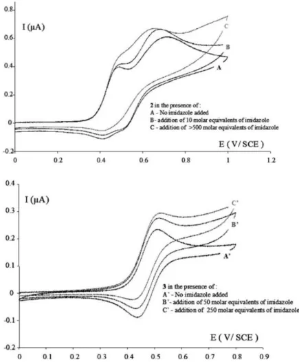
Fig. 4. Cyclic voltammograms for (a) 2 and (b) 3, in DMF. Pt (0.5 mm) working electrode, platinum mesh counter electrode, SCE reference electrode, scan rate = 100 mV/s. For 2, the first wave corresponds to the ferrocene oxidation, while the second corresponds to the amine oxidation.
2.3. Electrochemistry
Electrochemical results suggest that both 2 and 3 can be transformed into potentially toxic oxidized products via the previously proposed ferricenium-mediated proton-coupled electron transfer mechanism; the cyclic voltammograms in DMF and DMF/imidazole are shown in Fig. 4. A characteristic feature of this mechanism is a base-dependent enhancement of the wave appearing at the ferrocene oxidation potential, coupled with loss of reversibility, demonstrating electron transfer from the organic part of the molecule to the electrochemically-generated ferricenium moiety. The addition of imidazole enhanced the
intensity and irreversibility of this wave for both compounds. The base-promoted electron transfer in compound 2 is further evidenced by the decrease in intensity and potential of the oxidation wave of the amino group at 0.7 V upon addition of a base. This reveals the transition of an amino group to an aminyl radical triggered by the oxidation of the ferrocene at the first wave. In Fig. 4b, this is not apparent, because the amide oxidation wave lies outside the displayed window.
Electronic communication between a ferrocene group and an electrode via a conjugated system has been previously demonstrated by electron exchange when such compounds are anchored by a sulfur atom to a Au electrode [47]. This communication is very fast (t1/2 ca. 0.5
µs), compared to the scan rates we use in this report, and thus an equilibrium between the ferrocene-centered radical cation and the phenol-centered radical cation is reached essentially immediately. When the substituted phenyl radical cation can react (for example with a base present in the system), this equilibrium acts as an activation barrier for the overall reaction, such that, according to the Hammond postulate, the rate can be described as k = k0 exp(–
G°/RT), where k0 is the intrinsic rate constant of the deprotonation step and G° >> 0, the
free Gibbs energy difference between the two forms of the cation radical of the molecule, as shown in Scheme 3.
Scheme 3. Proposed mechanism of activation of 2 and 3.
The oxidation potential of the functional group is related to the position of the equilibrium and the height of the activation barrier, G°. Due to the electron withdrawing character of the
acetyl group, the oxidation of anilines occurs at less positive potentials than those of acetanilides [48–50], thus G° would be expected to be larger in the case of 3 compared to 2.
In our experiments, we found that the irreversible oxidation wave of the amino group in the organic molecule 4 occurred at +0.84 V (vs. SCE) and that of the amido group in 5 occurred irreversibly at +1.40 V. On the other hand, the value of k0 will be influenced by the acidity of
the amine or acetamide radical cation. The radical cation of phenylamine is very reactive [51], and that of acetanilide likely even more so, considering the difference in their pKa values (aniline = 30.6, aniline radical cation = 6.4 [52] and acetanilide = 21.45, acetanilide radical cation = –5 [53]). A recent electrochemical study demonstrated that 2 could be covalently bonded to an electrode surface through the nitrogen atom by electrolysis at the ferrocene oxidation potential (+0.4 V) in the presence of 2,4,6-pyridine [28]. This clearly demonstrates an intramolecular electron transfer for 2 even in the presence of a weak base. Due to the similarity of the CVs, it appears that the high k0 value for [3]+ may be enough to compensate
for the much smaller value of exp(–G°/RT), compared to 2, although further experiments
will have to be carried out to address this point.
In terms of the putative active agent, imino methides are known [54], and indeed have been implicated in cytotoxic processes [55–60]. However, while acetylated quinone imines are also important toxic metabolites, for example of the Parkinson’s disease drug tolcapone [48] and
the analgesic acetaminophen [61], acetylated imine methides are rare. To our knowledge, the only evidence of an acetylated p-imine methide was a short lived species (1 ms) generated from laser flash photolysis of 4-acetylaminostilbene [62]. These compounds are probably less stable due to the electron withdrawing acetyl group, because donation of the nitrogen atom -electrons into the ring favors the participation of the quinoid form over the aromatic form [63]. We are currently attempting to isolate the oxidation products of 2 and 3 and examine their reactivity with nucleophiles such as glutathione.
3. Conclusion
We have demonstrated that the oxidative activation of ferrocenyl compounds to toxic species is not limited to the series of ferrocenyl phenols. The ferrocenyl aniline 2 and acetanilide 3 showed estrogenic activity on the ER + MCF-7 breast cancer cell line, due to their affinity for the estrogen receptor, and a pronounced cytotoxicity against the ER-MDA-MB-231 breast cancer cell line. Notably, the purely organic aniline 4 and acetanilide 5 did not give rise to cytotoxic effects. The mechanism of activation seems to follow the intramolecular electron transfer process previously observed for the series of conjugated ferrocenyl p-phenols. This study thus broadens the perspectives for useful functional groups in the design of redox activated cancer drug candidates.
4. Experimental
4.1. General remarks
The synthesis of all compounds was performed under an argon atmosphere, using standard Schlenk techniques. Anhydrous THF was obtained by distillation from sodium/benzophenone. Thin layer chromatography was performed on silica gel 60 GF254. Infrared spectra were obtained on an IR-FT BOMEM Michelson-100 spectrometer equipped with a DTGS detector as a KBr plate. 1H and 13C NMR spectra were recorded on a 300 MHz Bruker spectrometer. Mass spectrometry was performed with a Nermag R 10-10C spectrometer. High resolution mass spectrometry (HRMS) was performed on a JEOL MS 700 instrument. Melting points were measured with a Kofler device. Elemental analyses were performed by the microanalysis service of CNRS at Gif sur Yvette. The semi-preparative HPLC separations were performed on a Shimadzu apparatus with a Kromasil C18 column (length of 25 cm, diameter of 2 cm, and particle size of 10 µm). Cyclic voltammograms were obtained utilizing a Princeton Applied Research potentiostat. Analyte solutions were 1 mM in DMF with 0.1 M Bu4NBF4
supporting electrolyte. Cyclic voltammetry experiments were performed at room temperature under an argon atmosphere in a three-electrode cell. The reference electrode was an SCE (Tacussel), which was separated from the solution by a bridge compartment filled with the same solvent/supporting electrolyte solution as used in the cell. The counter electrode was a platinum mesh (Goodfellow). The platinum working electrode was home-made (0.5 mm diameter; Goodfellow).
4.2. Synthesis and characterization of compounds
4.2.1. 2-Ferrocenyl-1-(4-aminophenyl)-1-phenyl-but-1-ene, (Z+E)-2
The synthesis of 2 via the reaction between propionyl ferrocene and 4-aminobenzophenone was reported in Ref. [28].
4.2.1.1. Alternative pathway. Zn powder (3.92 g, 60 mmol) was suspended in 30 mL of THF
at 5–10 °C in a Schlenk tube under argon. While stirring, TiCl4 (5.69 g, 30 mmol) was added
slowly via a syringe. The reaction mixture was removed from the cold bath and refluxed for 1h30. To the reaction mixture was added 15 mL of a THF solution containing propionyl ferrocene (2.42 g, 10 mmol) and 4-nitrobenzophenone (2.27 g, 10 mmol), and reflux conditions were maintained for 4 h. The reaction mixture was poured into 100 mL of water, and extracted with 3 x 100 mL of dichloromethane. The organic phase was washed with 100 mL of water, dried over magnesium sulfate, filtered, and the solvent was evaporated. The brown oil was purified on a silica gel column using dichloromethane as an eluent. Compound
2 (1.31 g, 54% yield) was recrystallized from ethanol to give a mixture of Z and E isomers
(45/55, major isomer was not identified). The yield varies significantly from one batch of 4-nitrobenzophenone to another.
4.2.2. 2-Ferrocenyl-1-(4-acetylaminophenyl)-1-phenyl-but-1-ene, 3
In a Schlenk flask under argon, 2 (407 mg, 1 mmol) was dissolved in 15 mL of anhydrous THF. Acetyl chloride (78 mg, 1 mmol) and pyridine (79 mg, 1 mmol) were added and the reaction mixture was stirred for 3 h. Water (50 mL) was added and the product was extracted with dichloromethane (3 x 50 mL). The organic phase was washed with 50 mL of water, dried over magnesium sulfate, filtered, and the solvent was evaporated. The product was purified on a silica gel column with ether/pentane (1/1) as the eluent. Compound 3 was obtained as a brown oil (372 mg, 91% yield) consisting of a mixture of Z and E isomers (60/40, respectively). The
identity of the major (Z) isomer was determined by 2D NMR.
(Z)-3: 1H NMR (300 MHz, DMSO-d6): 1.00 (t, J = 7.5 Hz, 3H, CH3), 2.02 (s, 3H,
CH3CO), 2.47 (q, J = 7.5 Hz, 2H, CH2), 3.82 (t, J = 1.0 Hz, 2H, C5H4), 4.11 (t, J = 1.0 Hz,
2H, C5H4), 4.13 (s, 5H, C5H5), 6.99 (d, J = 8.5 Hz, 2H, Harom), 7.22 (m, 3H, Harom), 6.99 (t, J =
8.5 Hz, 2H, Harom), 7.46 (d, J = 8.5 Hz, 2H, Harom), 9.89 (s, 1H, NH); 13C NMR (75 MHz,
DMSO-d6): 15.3 (CH3), 23.9 (CH3CO), 27.0 (CH2), 68.0 (C5H4), 68.7 (C5H4), 69.0 (C5H5),
85.4 (C C5H4), 118.9 (2 CHarom), 126.1 (CHarom), 128.3 (2 CHarom), 128.7 (2 CHarom), 129.4 (2
CHarom), 136.6 (C), 136.9 (C), 137.5 (C), 139.0 (C), 144.3 (C), 168.1 (CO). (E)-3: 1H NMR (300 MHz, DMSO-d6): 1.00 (t, J = 7.5 Hz, 3H, CH3), 2.06 (s, 3H, CH3CO), 2.47 (q, J = 7.5 Hz, 2H, CH2), 3.80 (t, J = 1.0 Hz, 2H, C5H4), 4.10 (t, J = 1.0 Hz, 2H, C5H4), 4.13 (s, 5H, C5H5), 7.08 (d, J = 8.5 Hz, 2H, Harom), 7.15 (d, J = 8.5 Hz, 2H, Harom), 7.26 (m, 3H, Harom), 7.54 (d, J = 8.5 Hz, 2H, Harom), 9.93 (s, 1H, NH); 13C NMR (75 MHz, DMSO-d6): 15.3 (CH3), 23.9 (CH3CO), 27.0 (CH2), 68.0 (C5H4), 68.7 (C5H4), 69.0 (C5H5),
85.4 (C C5H4), 118.9 (2 CHarom), 126.1 (CHarom), 128.3 (2 CHarom), 128.9 (2 CHarom), 129.1 (2
CHarom), 136.6 (C), 136.9 (C), 137.5 (C), 139.0 (C), 144.3 (C), 168.1 (CO).
The product was recrystallized from a mixture of ether/pentane to give the pure Z isomer: M.p.: 206 °C; Rf: 0.35 (ether/pentane = 1/1); IR (KBr, cm-1): 1656 (CON); MS (EI, 70 eV) m/z: 449 [M]+, 407, 384 [M-C5H5]+, 342, 326, 121 [FeC5H5]+; Anal. Calc. for C28H27FeNO:
C, 74.84; H, 6.06; N, 3.12. Found: C, 74.82; H, 5.85; N, 3.02%.
4.2.3. 1-(4-Aminophenyl)-1,2-bis-phenyl-but-1-ene, (Z+E)-4
Zinc powder (3.92 g, 60 mmol) was suspended in 30 ml of THF at 5–10 °C in a Schlenk tube under argon. While stirring, titanium tetrachloride (5.69 g, 30 mmol) was added slowly via a syringe. The reaction mixture was removed from the cold bath and refluxed for 1h30. To the reaction mixture was added 15 mL of a THF solution containing propiophenone (1.34 g, 10 mmol) and 4-aminobenzophenone (1.97 g, 10 mmol), and reflux conditions were maintained for three days. The reaction mixture was poured into 100 mL of water, and extracted with 3 x 100 mL of dichloromethane. The organic phase was washed with 100 mL
of water, dried over magnesium sulfate, filtered, and the solvent was evaporated. The oil was purified on a silica gel column using dichloromethane as an eluent. Compound 4 was obtained as an 85/15 mixture of Z and E isomers (major isomer was not identified) and was recrystallized from ethanol; Yield 38%.
4.2.3.1. Alternative pathway. The synthesis was identical to the alternative pathway of 2,
except that propiophenone (0.134 g, 1 mmol) was used in place of propionyl ferrocene, to furnish a mixture of Z and E isomers (85/15); Yield 55%. As for 2 the yield varies greatly with the batch of 4-nitrobenzophenone used.
1
H NMR (300 MHz, CDCl3): 0.84 and 0.87 (t, J = 7.4 Hz, 3H, CH3), 2.37 and 2.45 (q, J
= 7.4 Hz, 2H, CH2), 4.29 (s broad, 2H, NH2), 6.34 and 6.65 (d, J = 8.5 Hz, 2H, Harom), 6.60
and 6.95 (d, J = 8.5 Hz, 2H, Harom), 6.70–7.35 (m, 10H, Harom); 13C NMR (75MHz, CDCl3):
13.6 (CH3), 29.0 (CH2), 114.4 and 114.9 (2 CHarom), 125.5 and 125.9 (CHarom), 125.9 and
126.4 (CHarom), 127.2 and 127.8 (2 CHarom), 127.7 and 128.0 (2 CHarom), 129.5 and 129.7 (2
CHarom), 129.7 and 130.5 (2 CHarom), 130.9 and 131.8 (2 CHarom), 133.8 and 134.3 (C), 138.5
and 138.7 (C), 140.6 and 141.5 (C), 142.6 and 142.7 (C), 143.6 (C), 144.0 and 144.6 (C); IR (KBr, cm-1): 3474, 3380 (NH2), 3077, 3025, 2959, 2929, 2870 (CH2, CH3); MS (EI, 70 eV)
m/z: 299 [M]+., 284 [M-CH3]+; Anal. Calc. for C22H21N: C, 88.25; H, 7.06; N, 4.67. Found: C,
88.09; H, 7.09; N, 4.66%.
4.2.4. 1-(4-N-acetamidophenyl)-1,2-bis-phenyl-but-1-ene, (E+Z)-5
The synthesis was identical to that of 3, except that compound (Z+E)-4 (0.299 g, 1 mmol) was used in place of compound (Z+E)-2, to furnish a mixture of Z and E isomers (85/15). After recrystallization from ethanol, the ratio was 95/5; Yield 81%.
1
H NMR (300 MHz, acetone-d6): 0.95 and 0.97 (t, J = 7.4 Hz, 3H, CH3), 2.03 and 2.12
(s, 3H, CH3CO), 2.50 and 2.55 (q, J = 7.4 Hz, 2H, CH2), 6.77–7.75 (m, 14H, Harom), 9.0 and
9.22 (s broad, 1H, NH); 13C NMR (75 MHz, acetone-d6): 13.7 (CH3), 24.2 (CH3CO), 29.5
(CH2), 118.7 and 118.8 (2 CHarom), 126.6 and 127.0 (CHarom), 127.0 and 127.5 (CHarom), 128.2
and 128.7 (2 CHarom), 128.6 and 129.0 (2 CHarom), 130.1 and 130.4 (2 CHarom), 130.5 (2
CHarom), 131.4 and 131.7 (2 CHarom), 138.4 (C), 138.7 (C), 139.5 (C), 142.5 (C), 143.2 (C),
144.5 (C), 168.6 (CON); IR (KBr, cm-1): 3458, 3294, 3251 (NH), 3103, 3045, 2969, 2929, 2865 (CH2, CH3), 1663 (CON); HRMS (EI, 70 eV, C24H23NO: M+) Calc.: 341.1780. Found:
341.1776.
4.3. Biochemical studies 4.3.1. Materials
Stock solutions (10-3 M and 10-2 M) of the compounds to be tested were prepared in DMSO and were kept at –20 °C. Under these conditions, they are stable at least 2 weeks. Serial dilutions
in DMSO were prepared just prior to use. Dulbecco’s Modified Eagle Medium (DMEM), fetal calf serum, glutamine and kanamycine were purchased from Invitrogen (France), estradiol and protamine sulfate were from Sigma–Aldrich (France). Breast cancer cells MCF7 (hormone-dependent) and MDA-MB231 (hormone-independent) were from the American Type Culture Collection (ATCC – LGC Promochem). Sheep uteri weighing approximately 7 g, were obtained
from the slaughterhouse at Mantes-la-Jolie, France. They were immediately frozen and kept in liquid nitrogen prior to use. ERβ PanVera were purchased from Invitrogen (France).
RBA values were measured on ERα from lamb uterine cytosol and on purified ERβ (PanVera). Sheep uterine cytosol prepared in buffer A (0.05 M Tris–HCL, 0.25 M sucrose, 0.1% β-mercaptoethanol, pH 7.4 at 25 °C) as previously described [64] was used as a source of ERα. For ERβ, 10 µL of the solution containing 3500 pmol/mL were added to 10 mL of buffer B (10% glycerol, 50 mM Bis–Tris–propane pH 9400 mM KCl, 2 mM DTT, 1 mM EDTA, 0.1% BSA). Aliquots (200 µL) of ERα in glass tubes or ERβ in polypropylene tubes were incubated for 3h30 at 0 °C with [6,7-3H]-estradiol (2 x 10⁻⁹ M, specific activity 1.62 TBq/mmol, NEN Life Science Product) in the presence of nine concentrations of the compounds to be tested (between 6 x 10⁻⁷ M and 6 x 10⁻⁹ M) or of non-radioactive E2
(between 8 x 10⁻⁸ M and 8 x 10⁻¹⁰ M). At the end of the incubation period, the free and bound fractions of the tracer were separated by protamine sulfate precipitation. The percentage reduction in binding of [3H]-estradiol (Y) was calculated using the logit transformation of Y (logitY: ln[y/1 - Y] vs. the log of the mass of the competing steroid. The concentration of unlabeled steroid required to displace 50% of the bound [3H]-estradiol was calculated for each steroid tested, and the results expressed as RBA. The RBA value of estradiol is by definition equal to 100%.
4.3.3. Culture conditions
Cells were maintained in a monolayer culture in DMEM with phenol red/Glutamax I supplemented with 9% fetal bovine serum at 37 °C in a 5% CO2/air-humidified incubator. For
proliferation assays, MCF-7 and MDA-MB-231 cells were plated in 1 mL of DMEM without phenol red, supplemented with 9% decomplemented and hormone-depleted fetal bovine serum, 0.9% kanamycin, 0.9% Glutamax I and incubated. The following day (D0), 1 mL of the same medium containing the compounds to be tested was added to the plates. After 3 days (D3) the incubation medium was removed and 2 mL of the fresh medium containing the compounds was added. After 5 days the total protein content of the plate was analyzed as follows: cell monolayers were fixed for 1 h at room temperature with methylene blue (1 mg mL-1 in 50:50 water/MeOH mixture), then washed with water. After addition of HCl (0.1 M, 2 mL), the plate was incubated for 1 h at 37 °C and then the absorbance of each well (three wells for each concentration) was measured at 655 nm with a Biorad microplate reader. The results are expressed as the percentage of proteins vs. the control. Experiments were performed at least in duplicate.
4.4. Modeling studies
Molecular modeling studies were carried out using the programs Spartan, Trident and Odyssey [65].
Acknowledgements
The authors thank Marie-Noelle Rager for the 2D NMR experiments and the Agence Nationale de la Recherche for financial support (No. ANR-06-BLAN-0384-01, ‘‘FerVect”).
References
[1] P. Kovacic, Med. Hypotheses 69 (2007) 510.
[2] E.A. Hillard, F.C. de Abreu, D.C. Ferreira, G. Jaouen, M.O. Goulart, C. Amatore, Chem. Commun. 23 (2008) 2612, and references therein.
[3] H. Pelicano, D. Carney, P. Huang, Drug Resist. Update 7 (2004) 97.
[4] P. Galfi, J. Jakus, T. Molnar, S. Neogrady, A. Csordas, J. Steroid Biochem. Mol. Biol. 94 (2005) 39.
[5] M.E. Juan, U. Wenzel, H. Daniel, J.M. Planas, J. Agric. Food Chem. 56 (2008) 4813.
[6] V.C. Jordan, Tamoxifen for the Treatment and Prevention of Breast Cancer, PRR Inc., New York, 1999.
[7] P.W. Fan, F. Zhang, J.L. Bolton, Chem. Res. Toxicol. 13 (2000) 45, and references therein.
[8] S. Top, A. Vessières, C. Cabestaing, I. Laios, G. Leclercq, C. Provot, G. Jaouen, J. Organomet. Chem. 637 (2001) 500.
[9] A. Nguyen, A. Vessières, E.A. Hillard, S. Top, P. Pigeon, G. Jaouen, Chimia 61 (2007) 716.
[10] E. Allard, C. Passirani, E. Garcion, P. Pigeon, A. Vessières, G. Jaouen, J.P. Benoit, J. Control. Release 130 (2008) 146.
[11] A. Vessières, S. Top, W. Beck, E.A. Hillard, G. Jaouen, Dalton Trans. 4 (2006) 529. [12] G. Jaouen, S. Top, A. Vessières, G. Leclercq, M.J. McGlinchey, Curr. Med. Chem.
11 (2004) 2505.
[13] E.A. Hillard, P. Pigeon, A. Vessières, C. Amatore, G. Jaouen, Dalton Trans. (2007) 5073.
[14] E.A. Hillard, A. Vessières, F. Le Bideau, D. Plazuk, D. Spera, M. Huché, G. Jaouen, ChemMedChem 1 (2006) 551.
[15] E.A. Hillard, A. Vessières, L. Thouin, G. Jaouen, C. Amatore, Angew. Chem., Int. Ed. 45 (2006) 285.
[16] W.A. Wlassoff, C.D. Albright, M.S. Sivashinski, A. Ivanova, J.G. Appelbaum, R.I. Salganik, J. Pharm. Pharmacol. 59 (2007) 1549.
[17] M. Murata, M. Yamada, T. Fujita, K. Kojima, M. Kurihara, K. Kubo, Y. Kobayashi, H. Nishihara, J. Am. Chem. Soc. 123 (2001) 12903.
[18] S. Fukuzumi, K. Okamoto, Y. Yoshida, H. Imahori, Y. Araki, O. Ito, J. Am. Chem. Soc. 125 (2003) 1007.
[19] M. Kurihara, H. Sano, M. Murata, H. Nishihara, Inorg. Chem. 40 (2001) 4.
[20] M. Murata, T. Fujita, M. Yamada, M. Kurihara, H. Nishihara, Chem. Lett. 29 (2000) 1328.
[21] N.N. Meleshonkova, D.B. Shpakovsky, A.V. Fionov, A.V. Dolganov, T.V. Magdesieva, E.R. Milaeva, J. Organomet. Chem. 692 (2007) 5339.
[22] J.B. Heilmann, E.A. Hillard, M.-A. Plamont, P. Pigeon, M. Bolte, G. Jaouen, A. Vessières, J. Organomet. Chem. 693 (2008) 1716.
[23] S. Smith, D. Pasco, J. Pawlak, B. Thompson, M. Stampfer, S. Nandi, J. Natl. Cancer Inst. 73 (1984) 323.
[24] J. Shani, A. Gazit, T. Livshitz, S. Biran, J. Med. Chem. 28 (1985) 1504. [25] J.E. McMurry, M.P. Fleming, J. Am. Chem. Soc. 96 (1974) 4708.
[26] A. Fürstner, B. Bogdanovic, Angew. Chem., Int. Ed. Engl. 35 (1996) 2442. [27] B.E. Kahn, R.D. Rieke, Chem. Rev. 88 (1988) 733.
[28] O. Buriez, E. Labbé, P. Pigeon, G. Jaouen, C. Amatore, J. Electroanal. Chem. 619–620 (2008) 169.
[29] X.-F. Duan, J. Zeng, J.-W. Lü, Z.-B. Zhang, J. Org. Chem. 71 (2006) 9873. [30] F.S. Zeelen, E.W. Bergink, in: J. Raus, H. Martens, G. Leclercq (Eds.), Cytotoxic
Estrogens in Hormone Receptive Tumors, Academic Press, London, 1980, pp. 39–48. [31] A.K. Shiau, D. Barstad, P.M. Loria, L. Cheng, P.J. Kushner, D.A. Agard, G.L. Greene,
Cell 95 (1998) 927.
[32] A.K. Shiau, D. Barstad, J.T. Radek, M.J. Meyers, K.W. Nettles, B.S. Katzenellenbogen, J.A. Katzenellenbogen, D.A. Agard, G.L. Greene, Nat. Struct. Biol. 9 (2002) 359.
[33] A. Vessières, S. Top, P. Pigeon, E.A. Hillard, L. Boubeker, D. Spera, G. Jaouen, J. Med. Chem. 48 (2005) 3937.
[34] J.L. Mott, G.J. Gores, Cancer Biol. Ther. 6 (2007) 97.
[35] R. Benigni, A. Giuliani, R. Franke, A. Gruska, Chem. Rev. 100 (2000) 3697. [36] L. McLean, U. Soto, K. Agama, J. Francis, R. Jimenez, Y. Pommier, L. Sowers, E.
Brantley, Int. J. Cancer 122 (2008) 1665. [37] A. Fura, Drug Discov. Today 11 (2006) 133.
[38] M.R. Probst, M. Beer, D. Beer, P. Jeno, U.A. Meyer, R. Gasser, J. Biol. Chem. 269 (1994) 21650.
[39] T. Bisogno, K. Katayama, D. Melck, N. Ueda, L.D. Petrocellis, S. Yamamoto, V.D. Marzo, Eur. J. Biochem. 254 (1998) 634.
[40] A. Stañczak, A. Ferra, Pharmacol. Rep. 58 (2006) 599.
[41] J. Katz, M. Levitz, S.S. Kadner, T.H. Finlay, J. Steroid Biochem. Mol. Biol. 38 (1991) 17.
[42] Y. Kim, Y. Choi, R. Weissleder, C.-H. Tung, Bioorg. Med. Chem. Lett. 17 (2007) 5054.
[43] J. Katz, T.H. Finlay, S. Banerjee, M. Levitz, J. Steroid Biochem. 26 (1987) 687. [44] J.V. Watson, S.H. Chambers, P. Workman, T.S. Horsnell, FEBS Lett. 81 (1977) 179. [45] J. Dairou, N. Atmane, F. Rodrigues-Lima, J.-M. Dupret, J. Biol. Chem. 279 (2004)
7708.
[46] J.A. Williams, D.H. Phillips, Cancer Res. 60 (2000) 4667.
[47] C. Amatore, E. Maisonhaute, B. Schöllhorn, J. Wadhawan, ChemPhysChem 8 (2007) 1321.
[48] K.S. Smith, P.L. Smith, T.N. Heady, J.M. Trugman, W.D. Harman, T.L. Macdonald, Chem. Res. Toxicol. 16 (2003) 123.
[49] E.M. Garrido, J.L.F.C. Lima, C. Delerue-Matos, F. Borges, A.M.S. Silva, J.A.P. Piedade, A.M.O. Brett, J. Agric. Food Chem. 51 (2003) 876.
[50] E.M. Garrido, J.L.F.C. Lima, C. Delerue-Matos, F. Borges, A.M.S. Silva, A.M.O. Brett, Anal. Chim. Acta 434 (2001) 35.
[51] P. Simon, G. Farsang, C. Amatore, J. Electroanal. Chem. 435 (1997) 165. [52] F.G. Bordwell, X.-M. Zhang, J.-P. Cheng, J. Org. Chem. 58 (1993) 6410.
[53] F.G. Bordwell, D.J. Algrim, J. John, A. Harrelson, J. Am. Chem. Soc. 110 (1988) 5903.
[54] J. Nakayama, N. Matsumaru, M. Hoshino, Chem. Commun. 11 (1981) 565. [55] J.C. Huijzer, J. James, D. Adams, G.S. Yost, Tox. Appl. Pharm. 90 (1987) 60. [56] M.R. Nocerini, G.S. Yost, J.R. Carlson, D.J. Liberato, R.G. Breeze, Drug Metab.
Dispos. 13 (1985) 690.
[57] F. Charmantray, A. Duflos, J. Lhommea, M. Demeunynck, J. Chem. Soc., Perkin Trans. 1 (2001) 2962.
[58] J.C. Powers, J. Oleksyszyn, S.L. Narasimhan, C.-M. Kam, R. Radhakrishnan, J.E.F. Meyer, Biochemistry 29 (1990) 3108.
[59] M.M. Chow, J. Edgar, F. Meyer, W. Bode, C.-M. Kam, R. Radhakrishnan, J. Vijayalakshmi, J.C. Powers, J. Am. Chem. Soc. 112 (1990) 7183.
[60] J.C. Powers, C.-M. Kam, L. Narasimhan, J. Oleksyszyn, M.A. Hernandez, T. Ueda, J. Cell Biochem. 39 (2004) 33.
[61] J.A. Hinson, A.B. Reid, S.S. McCullough, L.P. James, Drug Metab. Rev. 36 (2004) 805. and references therein.
[62] R. Bose, A.R. Ahmad, A.P. Dicks, M. Novak, K.J. Kayser, R.A. McClelland, J. Chem. Soc., Perkin Trans. 2 (1999) 1591.
[64] S. Top, A. Vessières, G. Leclercq, J. Quivy, J. Tang, J. Vaissermann, M. Huché, G. Jaouen, Chem. Eur. J. 9 (2003) 5223.