Journal Pre-proofs
Physical formulation approaches for improving aqueous solubility and bioa-vailability of ellagic acid: A review
Isaïe Nyamba, Anna Lechanteur, Rasmané Semde, Brigitte Evrard
PII: S0939-6411(20)30326-X
DOI: https://doi.org/10.1016/j.ejpb.2020.11.004 Reference: EJPB 13451
To appear in: European Journal of Pharmaceutics and Biophar-maceutics
Received Date: 5 July 2020 Revised Date: 10 October 2020 Accepted Date: 7 November 2020
Please cite this article as: I. Nyamba, A. Lechanteur, R. Semde, B. Evrard, Physical formulation approaches for improving aqueous solubility and bioavailability of ellagic acid: A review, European Journal of Pharmaceutics and Biopharmaceutics (2020), doi: https://doi.org/10.1016/j.ejpb.2020.11.004
This is a PDF file of an article that has undergone enhancements after acceptance, such as the addition of a cover page and metadata, and formatting for readability, but it is not yet the definitive version of record. This version will undergo additional copyediting, typesetting and review before it is published in its final form, but we are providing this version to give early visibility of the article. Please note that, during the production process, errors may be discovered which could affect the content, and all legal disclaimers that apply to the journal pertain.
Physical formulation approaches for improving aqueous solubility and bioavailability of ellagic acid: A review
Isaïe NYAMBA1,2, Anna LECHANTEUR1,3, Rasmané SEMDE2, Brigitte EVRARD1
1Laboratory of Pharmaceutical Technology and Biopharmacy, CIRM, University of Liège, 4000 Liège (Belgium)
2Laboratory of Drug Development, Doctoral School of Sciences and Health, University Joseph KI-ZERBO, 03 BP 7021 Ouagadougou 03 (Burkina Faso)
3Corresponding author: Dr. Anna Lechanteur,
ULiège Laboratoire de Technologie Pharmaceutique et Biopharmacie, CHU Bat B36 Tour 4, 15 avenue Hippocrate, 4000 Liège, Belgium, Anna.Lechanteur@uliege.be, Phone number : +32494649833
Abstract
Ellagic acid (EA) is a polyphenolic active compound with antimalarial and other promising therapeutic activities. However, its solubility and its permeability are both low (BCS IV). These properties greatly compromise its oral bioavailability and clinical utilizations. To overcome these limitations of the physicochemical parameters, several formulation approaches, including particle size reduction, amorphization and lipid-based formulations, have been used. Although these strategies have not yet led to a clinical application, some of them have resulted in significant improvements in the solubility and bioavailability of EA. This critical review reports and analyses the different formulation approaches used by scientists to improve both the biopharmaceutical properties and the clinical use of EA.
Keywords: ellagic acid, solubility, bioavailability, formulation approaches Abbreviations:
API: Active Pharmaceutical Ingredient, ASD: Amorphous Solid Dispersions, BCS: Biopharmaceutical Classification System, CAAdP: Cellulose Adipate Propionate, CMCAB: Carboxymethylcellulose Acetate Butyrate, EA: Ellagic Acid, HPMCAS: Hydroxypropyl Methylcellulose Acetate Succinate, SD: Solid Dispersions, PVP: Polyvinyl Pyrrolidinone, FTIR: Fourier Transform Infrared Spectroscopy, CD: Cyclodextrins, DMAB: Didodecyldimethylammonium Bromide, HPβ-CD: Hydroxypropyl β-cyclodextrin, LBDDS: Lipid-Based Drug Delivery System, PEG: polyethylene glycol, PVA: Polyvinyl Alcohol, SMEDDS: Self-microemulsifying Drug Delivery System, SNEDDS: Self-nanoemulsifying Drug Delivery System
Table of contents
1. Introduction...3
2. Structure and properties of ellagic acid ...4
3. Formulation approaches to improve solubility and bioavailability of ellagic acid...9
3.1 Amorphization ...9
3.2 Complexation with cyclodextrins ...12
3.3 Particle size reduction ...17
3.3.1 Ellagic acid microparticles formation...18
3.3.2 Ellagic acid nanoparticles formation ...20
3.4 Lipid-based formulations containing ellagic acid...21
4. Optimization perspectives ...30
5. Conclusion ...33
References...35
1. Introduction
Over the last twenty years, the increasing number of active pharmaceutical ingredients (APIs) with low solubility has become one of the major challenges in pharmaceutical research [1]. Indeed, an important fraction of the new drug candidates emerging from drug discovery programs have poor water solubility. It is estimated that 75% of drug candidates and approximately 40% of currently marketed drugs are poorly water soluble [2,3]. This unsatisfactory physicochemical property of APIs leads to poor absorption and poor bioavailability and the need for high drug dosage to be administered, leading to increased side effects [4]. The other important factor to consider in drug development is the ability of a drug molecule to cross a mucosal barrier into the systemic circulation. An interesting example of
API with major biopharmaceutical challenges is ellagic acid (EA). Discovered by Henri Braconnot in 1831, EA is a polyphenolic active compoundwith high nutritional and therapeutic beneficial effects for human health [5,6]. It is widely distributed in many tropical and mediterranean plant species such as Adenium obesum (Apocynaceae), Terminalia chebula (Combretaceae), Rosa rugosa (Rosaceae) and Punica granatum (Lythraceae) [7–9] . In plants, EA is found in free form or linked to sugars or polyols, forming ellagitannins or hydrolysable tannins [10].
Today, a considerable number of non-drugs such as botanicals and enriched foods containing ellagitannins and/or EA, are commercially available as nutraceuticals and used to prevent many oxidative-linked chronic diseases, including cancer, diabetes, cardiovascular and neurodegenerative diseases [11,12]. This use of both EA and ellagitannins led to a shortage of pomegranate juice, a rich food source of EA and ellagitannins, in american supermarkets in 2007 [13,14]. Indeed, pomegranate juice contains between 2020 and 2660 mg of mixture of EA and ellagitannins per liter. Very high mean concentrations of EA have been also detected in different varieties of raspberry (Rubus idaeus L., Rosaceae) (190-719 mg/100 g), in cloudberry (Rubus chamaemorus L., Rosaceae) (644 mg/100 g) and artic bramble (Rubus arcticus, Rosaceae) (390 mg/ 100 g). The daily intake of EA through the consumption of foods, remains difficult to assess because of the limited knowledge about the EA foods content. However it is estimated to be between 0.2 and 0.3 mg in France, 5 mg in Germany, 12 mg in Finland and between 11.8 and 55 mg in the USA [14–16].
In addition to these nutritional supplement properties, various studies have confirmed that EA has antimalarial, nociceptive, proliferative, mutagenic, hepatoprotective, anti-diabetic and cardioprotective activities and is useful in the treatment of neurodegenerative diseases [6,17–19]. This multifaceted health activity of EA could be mainly ascribed to its antioxidant property and free radical trapping ability, that results in preventing or reducing the
so called “Oxidative Stress” [20]. If the daily intake of EA from food is useful for the prevention of some diseases, its therapeutic utilization, as for example in the treatment of malaria by oral route, remains a great challenge because of its low oral overall bioavailability, with typical plasma concentrations of 0.1–0.4 µmol / L [21,22]. This low bioavailability is mainly due to its low aqueous solubility associated with low membrane permeability, a significant first pass effect and irreversible binding to cellular DNA and proteins [23].
In order to allow an effective therapeutic application of EA, it is therefore necessary to develop strategies that sufficiently enhance its solubility, stability and bioavailability. Thus, during the last twenty years, researchers investigated the enhancement of solubility and bioavailability of this API through different approaches.This review proposes to analyze the physical formulation approaches studied by these scientists to try to overcome the biopharmaceutical challenges of EA.
2. Structure and properties of ellagic acid
Also known as a gallic acid dimer, EA or 2,3,7,8-tetrahydroxy [1] -benzopyranol [5,4,3-cde] benzopyran-5,10-dione is, from the physicochemical point of view, in the form of an ocher-yellow crystalline powder with a melting point above 360°C, a boiling point of 796.5°C, a log P of 1.05, a molecular weight of 302.197 g mol-1 [23]. Its chemical structure (fig. 1) includes four hydroxyl groups and two lactone groups representing the hydrophilic moiety and a lipophilic planar moiety consisting of two hydrocarbon phenyl rings. Hydroxyl groups and the lactone systems can act as electron acceptors and they can interact to form hydrogen bonds. EA is thus endowed with the ability to accept electrons from different substrates as well as to participate in antioxidant redox reactions [24,25].The molecular mechanisms activated by EA are multiple. They include free radical scavenging, regulation of phase I and II enzymes, modulation of pro-inflammatory and profibrotic cytokine synthesis, regulation of biochemical pathways involved in lipid synthesis and degradation, and maintenance of essential trace
element levels. Moreover, EA inhibits hepatic stellate cells and mast cells activation, the proliferation of transformed cells, as well as viral replication by increasing antioxidant response, induction of apoptosis, downregulation of genes involved in cell cycle and angiogenesis, and stimulation of cellular immune response [26,27].
Fig. 1: Chemical structure (A) and three-dimensional molecular size (B) of EA.Adapted with permission from [28].
This polyphenolic active compoundpossesses a high degree of crystallinity resulting from both its planar and symmetrical structure and an extensive network of hydrogen bonds involving trivial water solubility. Indeed, EA is practically insoluble in water (less than 10 µg/mL) [29] but soluble in methanol, N-methyl pyrrolidone, pyridine, polyethylene glycol (PEG) 400, dimethyl sulfoxide and triethanolamine. PEG 400 is the best solvent, reaching concentrations of up to 8 mg mL-1. As a weak acid, its solubility increases with pH and is higher in phosphate buffer at pH 7.4 (33.1 µg/ml) [30]. With this low aqueous solubility, EA is thus a dissolution-limited resorption substance [31]. Table 1 summarizes the physicochemical characteristics of EA.
Property name Property Value Reference
Molecular Weight 302.19 gmol-1
Aqueous solubility 9.73 µg/ml
[6]
Melting Point Greater than 360°C [32]
Log P 1.05 [23]
Hydrogen Bond Donor Count 4 Hydrogen Bond Acceptor Count 8
PubChem CID: 5281855) Dissociation Constants pKa1 = 6.69
pKa2 = 7.45 pKa3 = 9.61 pKa4 = 11.50
[33]
Spectral Properties UV max UV max (ethanol): 366, 255 nm [5]
Decomposition temperature 631° C [34]
A pharmacokinetic study of EA in healthy volunteers, showed that a higher free EA intake did not enhance its bioavailability but promotes urolithin production. Interestingly, this study has identified three representative pharmacokinetic profiles, i.e. a bell-shaped profile with Tmax around 1.5–2 h, a profile with two Tmax, and a profile where Tmax was detected after 24 h of EA and punicalagin (the ellagitannin form found in pomegranate) intake. Furthermore, a large inter-individual variability in terms of the amount of EA absorbed was also observed. This behavior is inherent to the limited EA bioavailability and could be mainly related to its low and pH-dependent solubility [35]. In addition to this low aqueous solubility, the membrane permeability of the EA is around 1.3 × 10⁻⁷ cm s−1 in human intestinal caco-2 cells, well below
the classifying threshold value of 2 × 10⁻⁴ cm s−1 [36]. Indeed, if the effective permeability of a drug is higher than 2 × 10⁻⁴ cm s−1, then complete drug absorption will be considered, otherwise, drug permeability is low and the absorption is incomplete [37]. The absorption sites of free EA into circulation are either the stomach or small intestine [10,15]. Ellagitannins resistant to acid hydrolysis, as well as degradation in the stomach, release EA in the small intestine at a neutral to slightly basic pH, which in turn may allow some freed EA to be absorbed in the small intestine [38]. The unabsorbed EA and ellagitannins suffer extensive metabolism by the gut microbiota to produce urolithins that are much better absorbed [39]. The health effects attributed to urolithins are close to those of the EA. Recent research, mostly based on in vitro testing, has shown preliminary evidence of the anti-inflammatory, anticarcinogenic, antiglycative, antioxidant, and antimicrobial effects of urolithins [39,40]. The absorbed EA is rapidly eliminated from the systemic circulation suggesting an efficient first-pass metabolism. During first-pass metabolism, EA is converted to methyl esters, dimethyl esters, and glucuronides. These metabolites and urolithins are eliminated in the urine [41]. However, the data from these pharmacokinetic studies are controversial and need further investigations. With regard to these solubility and intestinal epithelium permeability data, EA therefore belongs into class IV of the biopharmaceutical classification system (BCS). The BCS classifies drugs into four classes in order to find the best solutions to oral bioavailability concerns [42]. Thus, drugs with both high permeability and high solubility are in class 1; drugs with high permeability but low solubility are in class 2; drugs with low permeability but high solubility are in class 3; and finally, class 4 includes drugs with both low solubility and low permeability [43,44].
Approaches used to remedy unsatisfactory biopharmaceutical properties of APIs like EA are commonly based on chemical or physical modifications. Chemical modifications include the development of prodrugs, salt formation and co-crystal design [45]. These modifications are
limited to select drugs and are a lengthy and costly process. For instance, a limited number of APIs have desired functional groups such as ionizable or polar groups with potential for salt formation [46]. In addition, changing one property can adversely affect the others. This is observed when structural modifications that enhance solubility of APIs often also increase the toxicity and decrease permeability. Compared to chemical modifications, the physical formulation approaches are applicable to a wider group of APIs, are easier to prepare, and are usually preferred by industries [47]. They involve particle size reduction, modification of the crystalline form, liquid formulation and complexation with cyclodextrins [48,49]. Fig. 2 provides an overview of the most important formulation approaches to enhance the aqueous solubility and bioavailability of poor water-soluble drugs.
The specific formulation approaches and methods used by authors to enhance the aqueous solubility and biopharmaceutic profile of EA are extensively described below.
Fig. 2: Overview of the most important formulation approaches to improve the solubility of
drugs. Adapted with permission from [31].
3. Formulation approaches to improve solubility and bioavailability of ellagic acid 3.1 Amorphization
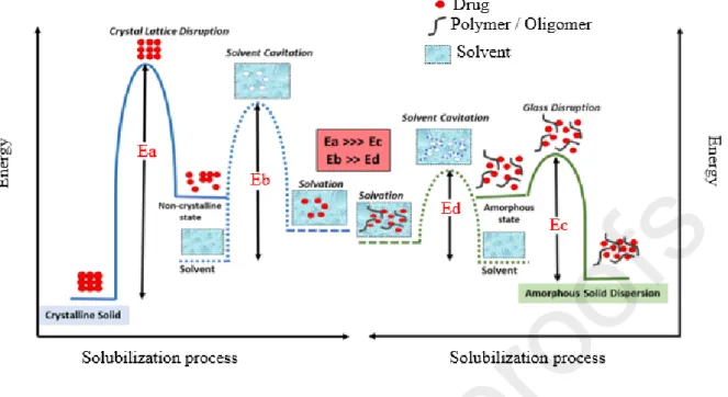
The amorphous form of API is used to improve their solubility and dissolution rate [50]. Indeed, due to the improvement of thermodynamic properties and the absence of crystalline lattice to break (fig.3), the amorphous form has a greater solubility (1.1 to 1000 fold) than that of the crystalline one [45]. The amorphous form can be obtained through various ways, but the most common of them are fast cooling and precipitation from solutions [51]. However, the amorphous form is very unstable and tends to recrystallize [52,53]. To overcome this instability, the formation of amorphous solid dispersions (ASD), the use of mesoporous systems and the co-amorphization are often carried out. The polymer-based solid dispersion formation is one of the most promising techniques because of its universal potential to improve the bioavailability of any API. This technique is based on the use of several methods of which the main ones are melt-based methods like hot melt extrusion and those based on solvent evaporation such as spray drying, coprecipitation or rotary evaporation [54,55].
Fig. 3: Activation energy diagram for the solubilization of a drug from a crystalline form (left)
and from an ASD (right). Ea is the energy required to disrupt crystalline lattice of a drug in a conventional formulation; Ec is the energy required to disrupt the glass solution of a drug in an ASD formulation, Eb is the energy required for solvent cavitation in the solubilization of a crystalline solid and Ed is the energy required for solvent cavitation in the case of ASD solubilization. Reprint with permission from [56].
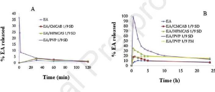
By using the solvent-evaporation methods, Li et al. (2013) evaluated the ability of cellulose derivatives such as carboxymethylcellulose acetate butyrate (CMCAB), cellulose adipate propionate (CAAdP) and hydroxypropyl methylcellulose acetate succinate (HPMCAS) to form ASD with EA, in order to improve its solubility. The release profiles of these resulting solid dispersions (SD) were carried out by dissolution tests and were compared to those of pure EA and SD of EA in polyvinyl pyrrolidinone (PVP), a widely and historically known ASD polymer. They observed that the release of EA from PVP SD was more rapid and complete (≈100%) at pH 6.8, followed by those from HPMCAS and CMCAB (fig.4 B). At pH 1.2. However, EA was also released from PVP (≈ 40%) and then recrystallizes rapidly (fig.4 A), arising the
problem of rational choice of nature and quantity of polymer used for API stabilization in ASD. Indeed, a good affinity and a reasonable proportions of API and polymer are necessary to slow down the recrystallization of the dissolved molecule [57]. The X-ray diffraction and Fourier transform infrared spectroscopy (FTIR) results indicated that EA was amorphous in HPMCAS and PVP SD at the EA proportion up to 25 wt%, and in CMCAB and CAAdP SDs at the EA concentration up to 10 wt% [32]. The maximum EA concentration from amorphous dispersion obtained by dissolution test at pH 6.8 was 1500 µg/mL with PVP and 280 µg/mL with HPMCAS corresponding to increases of more than 45 and 8 times the actual EA solubility.
Fig. 4 :Release profiles of an equivalent of 5 mg of EA from solid dispersions (SD) produced by solvent evaporation methods as a function of time. EA/CMCAB 1/9 SD, EA/HPMCAS 1/9 SD and EA/PVP 1/9 SD correspond to EA/polymer solid dispersions in a ratio of 1/9 w/w. EA/PVP 1/9 PM is the physical mixture in the same ratio. These dissolution tests were carried out at pH 1.2 (A) and pH 6.8 (B).Reprint with permission from [32].
The high crystal lattice force of EA (melting point ˃ 360°C) is at the origin of its poor solubility. Therefore, the realization of ASD is one of the best ways to circumvent the problem of poor aqueous solubility [58]. Indeed, the solubility of the drug substance in the ASD is improved by disrupting its crystalline lattice to produce a higher energy amorphous form [59,60]. This
concept was proved by Li et al. showing the increase of solubility from the ASD. However, Figure 4 shows a decrease in the EA concentration from the maximum values, presumably due to incomplete stabilization of supersaturated EA concentrations by the polymers. Indeed, a rational choice of the polymer could allow it to inhibit the crystallization of the amorphous EA from the solid carrier matrix and the supersaturated solution that is created upon dissolution. Authors chose cellulose derivatives for their study mainly because they are effective ASD polymers and PVP because it is a widely and historically known ASD polymer. Their choice was not based on possible strong attractive EA-polymer interactions, which could explain the instability of the supersaturated EA solution. Instability is the major drawback of amorphous forms, if not controlled, it can lose the advantage of these forms in terms of improved solubility compared to crystalline forms [61].
3.2 Complexation with cyclodextrins
Cyclodextrins (CD) are cyclic oligosaccharides formed from α-D-glucopyranose units, linked by α- (1-4) glycosidic bonds [62]. Natural cyclodextrins differ according to the number of α-D-glucopyranose units which can range from six to twelve [63], the most common being α, β, γ-cyclodextrin with 6, 7 and 8 α-D-glucopyranose units, respectively. The volume and diameter of the internal cavity of native cyclodextrins (Fig.5), which are 174 Ä 3 and 5.7Ä, 262 Ä 3 and 7.8 Ä and 427 Ä3 and 9.5 Ä, respectively for the αCD, βCD and γCD, increases with the number of glucopyranose units. The aqueous solubility does not follow this logic since at 25 ° C, it is 145 mg/mL, 18.5 mg/mL and 232 mg/mL for αCD, β-CD and γCD [64,65].
Cyclodextrins have a relatively hydrophobic central cavity furnished with carbon and hydrogen atoms and a relatively hydrophilic exterior furnished with primary hydroxyl groups (attached to C6 carbons) and secondary (attached to C2 carbons and C3). This configuration allows them to form inclusion complexes with a wide range of solid, liquid or gaseous substances [66]. In 1953, Freudenberg et al. demonstrated that inclusion complexes in CDs increase the solubility
of poorly soluble drugs, reduce the loss of volatile substances and protect oxidizable substances from oxidation [67]. Brewster and Loftsson (2007) estimated that inclusion complexes increase the bioavailability of poorly soluble drugs by 1.1 to 49 times, compared to that of crystalline and lyophilized drugs used as control formulations [68]. Thus, the use of CDs by the pharmaceutical industry nowadays is estimated at more than 3000 tons per year, i.e. 30% of annual production [69]. CDs and their derivatives, mainly the methylated, hydroxypropyl and sulfobutyl ethers, are currently found in over 50 marketed pharmaceutical products. They are used as excipient to protect APIs from specific and non-specific interactions in physiological media [70], to improve the permeability of drugs by interacting and destabilizing biological membranes [71], to modulate the rate and site of drug release [72] and finally, to increase their aqueous solubility [73–75].
The different methods developed to prepare inclusion compounds are liquid phase method, co-precipitation, kneading or slurry method, extrusion, damp mixing, dry mixture and co-grinding, spray drying and lyophilization [63].
Fig. 5 :Modelisation of the structures of the three main natural cyclodextrins (A), diagram of a α-D-glucopyranose unit (B), and annotated diagram of a cyclodextrin (C). Adapted with permission from [76].
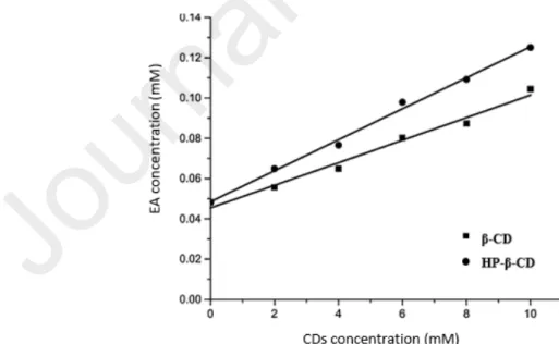
In order to evaluate the antimicrobial and antioxidant activity of EA, Savic et al. prepared inclusion complexes, using β-CD and hydroxypropyl β-CD (HP β-CD). The inclusion complexes were prepared by suspending EA and CDs in distilled water. The suspensions were stirred on a magnetic stirrer at 600 rpm and room temperature for 24 h, then evaporated on a rotary vacuum evaporator at 60°C and dried in a desiccator until a constant mass. The obtained complexes were further characterized by FTIR, XRD and nuclear magnetic resonance methods. The aqueous phase solubility study carried out by adding an excess of EA to 5 mL CD solutions (0-10 mM), showed that the EA solubility in 10 mM solutions of β-CD and HP- β-CD was respectively 2.2 and 2.6 times higher, than that observed in water without CDs. An AL-type diagram (fig.6) was obtained with both CDs and the stability constants were 161 dm3 mol-1 and 117 dm3 mol-1 for the complex with HP- β-CD and β-CD respectively [77].
Fig. 6 : Diagram of the phase solubility of EA in the presence of increasing concentrations of β-CD and HP-β-CD (0 -10 mM) in water at 25° C for a 24 h equilibration time. Reprint with permission from [77].
Other authors prepared complexes of EA and HPβ-CD by freeze-drying and studied the mechanism of complexation, using different analytical techniques including FTIR, powder X-ray diffraction, nuclear magnetic resonance, scanning electron microscopy and molecular modeling.The phase solubility study carried out with concentrations ranging from 0 to 24 mM of HPβ-CD in the presence of an excess of EA showed an AN type diagram, according to the classification of Higuchi and Connors (fig.7 A). In addition, the results of the dissolution tests carried out with pure EA, physical mixture (PM) and inclusion complex (EA - HPβ-CD) showed that the inclusion complex EA/HP-β-CD released up to 55% and 60 % of the drug into 15 and 30 min respectively, whereas pure EA showed a release of 10% after 15 min and up to 13% after 60 min (fig.7 C) [78]. Moreover, inclusion complexes of EA with β-CD prepared by freeze-drying significantly enhanced the aqueous solubility of EA (fig. 7 B) since it passed from 10.37 µg/ml to 39.14 µg/ml, which corresponds to an increase of 3.77 times of the intrinsic solubility.The results of the dissolution tests (fig.7 D) also showed a release rate of 27% in 30 min with the complex while that of pure EA only reached 13% after 60 min [79].
Fig. 7 : Diagram of the phase-solubility of EA with increasing concentrations (0-24 mM) of
HP-β-CD (A) and β-CD (B) in water at 30° C for a 72 h equilibration time. Dissolution rate profiles of pure EA (EA), physical mixture (PM) and EA-CD inclusion complex of EA-HP- β-CD inclusion complex (C) and EA-β-β-CD inclusion complex (D). These assays have been performed with equivalent amounts of 100 mg of EA. Reprint with permission from [67]for picture A and C, and [68] for picture B and D.
Chudasama et al. evaluated the bioavailability of EA complexed with methyl β-cyclodextrin (Me-β-CD) in rats. Thirty minutes after administration by gavage of a single dose of either EA (0.4 g/kg in 0.5% carboxymethylcellulose) or EA complexed with Me-β-CD (1:2 molar ratio), plasma levels of EA were 7-fold higher in EA/Me-β-CD treated rats than in EA treated rats, i.e. 36.61 + 14.12 ng/ml and 5.63 + 9.8 ng/ml, respectively. However, the authors did not specify the nature of the Me-β-CD used or the method of complexation [80]. Mady et al. (2018) estimated the effect of EA / β-CD inclusion complexes on solubility and bioavailability of EA.
The team found solubility of 49.79 µg/ml and 9.73 µg/ml, respectively for the inclusion complexes and pure EA. The bioavailability in animals was 1345.49 ng.hr.ml-1 for the inclusion complex versus 598.94 ng.hr.ml-1 for pure EA [81].
To perform inclusion, the substrate size has to be inferior to that of the cyclodextrin interior cavity and the driving energy for the various actors of inclusion, namely the cyclodextrin, the API and the solvent have to be favorable. Indeed, water molecules located in the cyclodextrin cavity have to be more easily replaced by less polar molecules [77]. Despite the size of the EA molecule (fig. 1), its low lipophilicity (log P=1.05), its planar structure which may limit its inclusion in the CD cavity due to steric effects, β-CD and derivatives have been successfully used as EA solubilizers. This could be explained by the formation of partial inclusion complexes involving carboxyl groups of EA and by the formation of association complexes involving hydroxyl groups. Based on the results of phase solubility studies, a linear increase of the EA solubility is observed in presence of increasing concentrations of β-CD and HP-β-CD up to 12 mM. Beyond that, the relationship is no longer linear. However, compared to the ASD formation approach, EA complexing with cyclodextrins seems to be less successful in terms of solubility improvement. In addition, developing a cyclodextrin-based drug is relatively expensive and is often limited to powerful drugs that can be used at low doses [31].
3.3 Particle size reduction
Reducing the particle size is one of the most interesting approaches to improve the bioavailability of substances poorly soluble in water. Indeed, it significantly increases the specific surface which, according to the modified Noyes and Whitney equation, increases the dissolution rate and consequently the bioavailability [82]. In addition, this approach also increases the saturation solubility when the particle size is less than one micrometer, while
reducing the thickness of the diffusion layer [83]. Also, small substances are more likely to cross the intestinal barrier thereby increasing their permeability [84].
The reduction in particle size has two components, namely micronization giving less than one millimeter size particles and the formation of nanoparticles with less than one micrometer size particles. The most common micronization technique consists of mechanical dry pulverization of larger drug particles, mainly using jet, ball and pin mills. The lowest particle size that can be achieved is about 2–3 µm. Also, the agglomeration of drug particles sometimes increases, resulting in the decrease of the surface available for dissolution. In such a case, wetting agents like surfactants would play a major role in increasing the effective area [45]. Nanoparticles are commonly produced by controlled precipitation, crystallization, high pressure homogenization, wet-milling with beads and use of supercritical fluids [85]. These particles are very cohesive with a strong tendency for aggregation, hence the need to disperse them in hydrophilic polymers and/or surfactants for more stability. Drug nanoparticles have many benefits, compared to the simple micronized drug powders. In fact, the increase in surface area to mass ratio for nanoparticles is much more drastic than that of microparticles, sometimes covering several orders of magnitude [86]. A review of the literature on the bioavailability of poorly soluble substances showed that the nanoparticle formulations allow 1.7–60-fold and 2–30-fold enhancements in Cmax and AUC respectively, compared to those of crystalline microparticles [45].
3.3.1 Ellagic acid microparticles formation
Various authors used micronization techniques to improve EA biopharmaceutical properties. Indeed, Li et al. (2015) used antisolvent precipitation and freeze-drying to prepare micronized EA (m-EA) powder for oral delivery. N-methyl pyrrolidone (NMP) and deionized water were utilized as solvent and antisolvent. The resulting precipitate was centrifuged and lyophilized to give m-EA powder. This powder was then characterized by scanning electron microscopy,
FTIR, liquid chromatography tandem mass spectrometry, X-ray diffraction, differential scanning calorimetry, thermogravimetric analysis and by in vitro dissolution and solubility tests. The results indicated a strong reduction in the crystallinity of m-EA compared to the raw EA. In addition, the apparent solubility obtained with m-EA was approximately 6.5 times higher than that of the EA. More interesting, bioavailability following oral administration in rats was about 2 times higher with m-EA (fig. 8) [87].
Fig. 8 : Bioavailability results obtained after oral administration in rats of equivalent amounts
of 2 mg EA. (a) raw EA, (b) physical mixture of maltodextrin and raw EA, (c) pomegranate herbal supplement, (d) m-EA freeze-dried powder. Reprint with permission from [87]. Montes et al. (2016), using the supercritical anti-solvent process (SAS) to generate m-EA and m-EA / Eudragit L100 coprecipitates, observed that the crystallinity and stability of SAS-processed EA remained unchanged and the coprecipitates released EA more rapidly than EA microparticles. This faster release would be explained by the fact that smaller EA microparticles were obtained in the presence of the polymer [88]. Jeong et al (2001) used Eudragit P-4135F, a pH-sensitive polymer, to deliver EA microspheres into the lower small intestine in rats.They obtained an effective release of EA at the desired site. Also, dissolution tests showed a release
of more than 95% of EA after 30 min from the microspheres in pH 7.4 and pH 8 buffer, and less than 40% from EA raw powder [89]. Alfei et al. (2019) prepared by spray drying a micro-dispersion form of EA in weakly methoxylated pectin, which was 30 times more soluble than pure EA [90].
3.3.2 Ellagic acid nanoparticles formation
Nanotechnology, especially made with stabilizer polymers, seems to promote oral availability of EA. Bala et al. (2005, 2006) produced EA loaded in poly (D,L-lactide-co-glycolide) nanoparticles, using polyvinyl alcohol (PVA), didodecyldimethylammonium bromide (DMAB) and PVA in combination with chitosan (80:20) as stabilizer. The intestinal permeability test performed in situ in the rat showed 66%, 75%, 73% and 87% permeation, respectively for pure drug and the drug encapsulated in nanoparticles prepared using PVA, PVA– chitosan blend and DMAB as stabilizer [36,91]. Mady and Shaker (2017) showed that the in vivo bioavailability of EA obtained in the rabbits was 3.6 times higher from EA / Poly ε-caprolactone nanoparticles produced by emulsion-diffusion-evaporation technique, compared to that of pure EA (Fig. 9) [92].
Fig. 9 : Plasma concentration - time curves of EA after oral administration to NZW rabbits at
50 mg/kg equivalent dose of free EA and EA / Poly ε-caprolactone nanoparticles made by emulsion-diffusion-evaporation technique. Reprint with permission from [92].
Biodegradable hollow zein nanoparticles with a mean diameter of about 70 nm for oral administration of EA have also been developed. The inner core, which consists of EA / sodium carbonate (EA / Na2CO3) obtained by coprecipitation, was then encapsulated in hollow zein nanoparticles (HTZN) with triethyl citrate as a plasticizer. The bioavailability in the rat was 3.6 and 2.6 times higher than that of pure EA and non-encapsulated nanoparticles of EA [93]. Alfei et al. (2019) have, for their part, produced nanoparticles of hydrophilic and amphiphilic non-polyamidoamine dendrimers of size between 60 and 70 nm and incorporating 46% and 53% (w / w) of EA. The resulting products have shown a water solubility of 300 to 1000 times higher than that of free EA [90].
Considering the high degree of crystallinity of EA, particle size reduction and more specifically nanonization, could be one of the best approaches to improve its solubility. Indeed, EA nanonization may reduce this crystal lattice force, increase the surface to volume ratio and improve solubility and dissolution rate, as evidenced by the significant results obtained by scientists in terms of improvement in solubility and bioavailability. However nanoparticles present various drawbacks such as their high cohesivity requiring stabilization to avoid aggregation and complex manufacturing issues [94].
3.4 Lipid-based formulations containing ellagic acid
Lipid-based formulations or Lipid-Based Drug Delivery Systems (LBDDS) are another formulation strategy used to overcome the low bioavailability of poorly water-soluble drugs [95]. Sustivas®, Fortovases®, Nor-virs®, Lamprenes®, SandimmuneTM and NeoralTM are examples of marketed LBDDS. Indeed, for many of these drugs, it is recognized that administration with fatty foods improves bioavailability [89,90]. The presence of exogenous fatty foods in the small intestine stimulates the endogenous secretion of bile salt and cholesterol, which increases the solubilization and absorption of lipid digestion products and drugs [98]. To successfully develop LBDDS, several complex biological processes, such as the digestion of
lipid excipients, the formation of different colloidal phases during lipid digestion and the release of the drug from these colloidal phases must be taken into account [99].
Pouton developed a Lipid-based Formulation Classification System (LFCS) and categorized the lipid-based formulations into four different types (Table II) according to their compositions [96,97]. They range from simple oil solutions to complex mixtures of oils, surfactants, co-surfactants and cosolvents [100]. From a biopharmaceutical point of view, any compound from the four BCS classes can be used in lipid-based formulations [101,102]. However, poorly water-soluble drugs with a Log P about 2 and high melting point (above 200°C), known as brisk dust, are not good candidates, unlike the so-called grease balls (log P greater than 2) [99]. Similarly, according to some authors, only drugs having an aqueous solubility inferior to 10 µg/mL, a Log P superior to 5, a solubility in oils and lipids above 25 mg/mL, a relatively low melting point and a good chemical stability, can be good candidates for lipid formulations [103]. Savla et al., on the other hand, consider that types IIIB and IV lipid formulations are more suitable for brick dust compounds and those of types I, II and IIIA, for grease balls [104].
Table II : Characteristics and compositions of the four types of formulations, according to the
lipid formulation classification system (LFCS) [97,104].
Nature and content (%w/w) of the excipients used in the formulation
Formu lation type Characteristics Oils: triglycerides or mixed mono and
diglycerides Water-insoluble Surfactants (HLB <12) Water-soluble Surfactants (HLB >12) Hydrophilic cosolvents (e.g. PEG, propylene glycol, transcutol) I Pure oils Limited or no dispersion Digestion required 100 - -
-II SEDDS Moderate dispersion needed to form an emulsion Likely to require digestion 40-80 20-60 - -III A SMEDDS Rapid dispersion to form micro- or nano-emulsion
May need digestion
40-80 - 20-40 0-40
III B SMEDDS
Rapid dispersion to form micro- or nano-emulsion
Digestion likely not needed <20 - 20-50 20-50 IV Oil free Rapid dispersion results in micellar solution No digestion needed - 0-20 30-80 0-50
SEDDS: Self-emulsifying drug delivery system; SMEDDS: Self-microemulsifying drug delivery system.
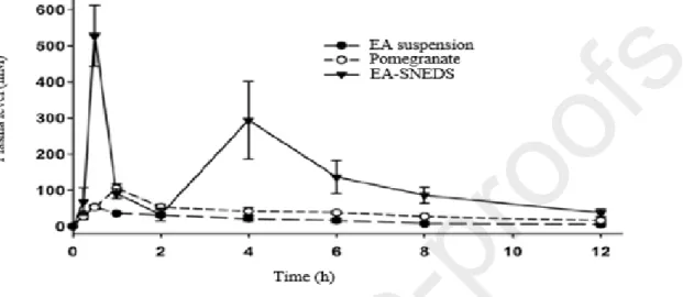
Wang et al. (2017)produced a self-nanoemulsifying drug delivery system (SNEDDS) intended to enhance the solubility and absorption of EA and therefore, its oral bioavailability.With an
optimal formulation consisting of PEG, polysorbate, caprylic/capric triacylglycerol, they obtained a fine nanoemulsion with a mean droplet size of about 120 nm. The pharmacokinetic study in rats (fig.10) showed that EA was 6.6 and 3.2 times more bioavailable with this formulation than with aqueous suspensions and pomegranate extract, respectively [105].
Fig. 10 : Mean plasma concentration/time profiles of EA after oral administration to rats of
EA-SNEDS, pomegranate extract or aqueous suspension, at 17.6 µmol/kg body weight EA equivalent. Reprint with permission from [105].
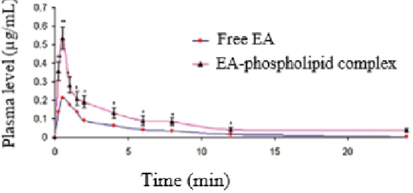
In order to improve EA solubility and permeability, SNEDDS formulation EA–phospholipid complex (EAPL complex) of 106 ± 0.198 nm vesicle size has been developed. For this purpose, EAPL complex prepared by the antisolvent precipitation method with soy lecithin, was then used to produce SNEDDS. The average vesicle size of the EAPL complex SNEDDS obtained was 106 ± 0.198 nm and its aqueous solubility was three times that of free EA. Ex vivo permeability studies performed in rats showed 84% and 94% of absorption rate in the stomach and small intestine and 23% and 30%, respectively for EAPL complex SNEDDS and EA aqueous suspension [106]. Hydrogenated soy phosphatidylcholine (HSPC) was also used to prepare EA - Phospholipid complex by the antisolvent precipitation method. An oral administration in rats of an equivalent dose of 80 mg/kg of complex and free EA, resulted in Cmax of 0.54 µg/mL and 0.21 µg/mL (fig.11), respectively [107].
Fig. 11 : Plasma EA concentration obtained after oral administration in rats of a dose of 80
mg/kg of free EA and EA-phospholipid complex. Reprint with permission from [107].
With an optimal formulation consisting of 10% ethyl oleate, 67.5% Tween 80, 22.5% polyethylene glycol 400, 0.5% polyvinylpyrrolidone K30 and 4 mg g-1 EA, Zheng et al. (2019) developed a self-microemulsifying supersaturated drug delivery system (S-SMEDDS) to improve the solubility of EA. The results of the in vitro dissolution study showed that EA could dissolve from the SMEDDS within a short time and exhibited more rapid release from S-SMEDDS than from S-SMEDDS and raw material [108].
Despite the low lipophilicity (log P =1.05) and high crystallinity (melting point ˃ 360°C) of EA, LBDDS, especially SNEDDS, have led to interesting improvements in the solubility and bioavailability of EA. This could be explained by the combined effects of particle size reduction and the use of formulation adjuvants. However, the complexity of LBDDS with numerous possible combinations of excipients, makes formulation development resource- and time-consuming, and also requires a significant amount of API [109].
As described above, several strategies have been used in numerous preclinical studies to bring up EA, a low soluble and low permeable drug, to a bioavailable and effective therapeutic one. They are summarized in table III.
Table III : Summary of the formulation approaches used by the different authors to improve solubility, bioavailability and/or therapeutic efficacy
of EA
Formulatio n approach
Methods Specific adjuvants used Main result obtained References
Amorphizat ion Spray drying Co-precipitation Rotary evaporation PVP, HPMCAS, CMCAB, CAAdP
Maximum EA concentration from amorphous dispersion obtained by dissolution test was 1500 µg/mL with PVP, 280 µg/mL with HPMCAS and 30 µg/mL with CMCAB.
[32]
Liquid phase method
β-CD, HP- β-CD Formation of 1:1 stoichiometric complex, 2.2 times and 2.6 times increase in solubility of EA with β-CD and HP-β-CD respectively, compared with the water solubility of EA,
[77]
Freeze-drying; Physical mixture
HP- β-CD Formation of EA-HP-B-CD complex of 1: 2 stoichiometry;
AN-type solubility curve obtained with cyclodextrin concentrations ranging from 0-24mM;
An improvement of 5.5 times the dissolution rate after 15 min compared to that of EA alone
[78] Inclusion
complexes
Freeze-drying Physical mixture
β-CD The aqueous solubility of EA drops increases from 10.37 µg / ml to 39.14 µg / ml (3.77 times) after interaction with β-CD in a stoichiometric ratio of 1: 2.
Formulatio n approach
Methods Specific adjuvants used Main result obtained References
- methyl β-CD 7-fold increase in bioavailability after 30 min; [80]
Liquid phase method
β-CD, cross-linked by dimethyl carbonate (DMC)
More than 5-fold and twice increase in solubility and bioavailability, respectively [81] Antisolvent precipitation followed by freeze drying N-methyl pyrrolidone, deionized water
The water solubility of free EA and m-EA at 37±0.5 C was 1.80 ± 0.9 µg/mL and 11.67 ± 0.46 g/mL, respectively;
Dissolution rate and the bioavailability of m-EA were both 2 times of free EA. [87] Supercritical antisolvent process; Supercritical CO2, NMP, Eudragit L 100
Faster release of coprecipitates EA/Eudragit L100 than the EA microparticles, which dissolved more rapidly than free EA.
[88] Micronizati
on
Spray drying Water, low methoxylated pectin
EA solid microdispersion aqueous solubility was 300 µg/mL and that of free EA was 9,73 µg/mL.
Formulatio n approach
Methods Specific adjuvants used Main result obtained References
Co-precipitation Ethanol, deionized water, zein,Na2CO3
Cmax of EA-HTZN was 0.34 μg·L−1 versus 0.085 μg·L−1 for pure EA. Increase in relative bioavailability of EA by 3.6 times.
[93]
Emulsion– diffusion– evaporation
Poly ε-caprolactone, PEG 200, ethyl acetate, water
3.6-time fold increasing of the EA relative bioavailability [92]
Physical encapsulation
non-polyamidoamine hydrophilic and amphiphilic dendrimers
Two EA nanodispersions were achieved (60–70 nm) with 46% and 53% (w/w) DL;
Water solubility was 300 to 1000 times higher than that of free EA;
[90] Emulsion– diffusion– evaporation Poly (D,L-lactide-co-glycolide), PEG 400, DMAB, PVA, chitosan
The permeation of free EA through the jejunum was 66% and that of the drug encapsulated in poly (D,L-lactide-co-glycolide) nanoparticles using PVA, VA-chitosan mixture and DMAB as stabilizer was 75%, 73% and 87%, respectively.
[36] Nanonizatio n Inverse emulsion (water-in-oil (w/o)) Schizophyllan, chitin, Span 80 and Tween 80
EA loading capacity of schizophyllan and chitin were 30.08% and 79.52%, respectively; [92] Lipid based formulation Mixing using a vortex mixer PEG 400- Tween 80-caprylic/ capric triacylglycerol (45/45/10 wt.%)
Increase of the EA bioavailability by 6.6 and 3.2 times, compared to that of EA aqueous suspension and pomegranate extract, respectively.
Formulatio n approach
Methods Specific adjuvants used Main result obtained References
Anti-solvent precipitation for complex and mixing under agitation for SNEDDS
soy lecithin/EA complex (3%), captex (40%), cremophor RH 40 (40%), PEG 400 (20%) and tocopherol (0.15%)
Tripling of EA aqueous solubility;
Increase of the ex vivo absorption rate of EA in the rat stomach and small intestine, from 23 and 30% for EA aqueous suspension to 84% and 94% for EAPL complex SNEDDS
[106]
anti-solvent precipitation
Hydrogenated soy phosphatidylcholine
2.5-fold increase of the bioavailability;
Better protection of the liver by the complex and maintains an effective concentration for a longer period in the serum.
[107]
Mixing using a vortex mixer
Ethyl oleate10%, Tween 80 67.5%, PEG 400 22.5%,
polyvinylpyrrolidone K30 0.5%
Particles were spherical with a size of about 40 nm; dissolution of EA from the S-SMEDDS within a short time and exhibited more rapid release from S-SMEDDS than from SMEDDS.
4. Optimization perspectives
The low absorption of EA could be attributed mainly to its very low solubility (less than 10µg/mL) but also to its low lipophilicity (log P=1.05) and its degradation at the intestinal level by the bacterial flora. Also, as a weak acid ionized at more than 50% in the intestine with a pH of 6.8, its absorption is limited since only the un-ionized fraction is available to cross the cell membrane because of the lipid nature of the membrane. Its low solubility could be explained by its high degree of crystallinity. Indeed, with its estimated melting temperature of more than 360°C, EA belongs to the group of molecules called "brick dust" which are soluble neither in water nor in oils. According to the decision tree for preclinical formulations of APIs with solubility limiting bioavailability (fig. 12), the use of co-solvents, nanoparticles or SD are the recommended formulation approaches for drugs with low log P and high melting point like EA [110].
Fig. 12 : Decision tree for preclinical formulation based on the assessment of melting point and
Being high cohesive crystal and weak acid, one of the best approaches to improve solubility, absorption in stomach and bioavailability of EA would consist in trapping its amorphous metastable form in acid-soluble polymers (amorphization by SD formation). A successful example of this type of formulation has indeed allowed to multiply the solubility and the bioavailability of atorvastatin by 74 and more than three, respectively. In this study, ASD of atorvastatin, a weak acid drug in BCS class II, and Eudragit® EPO, an acrylic derivative soluble in water at pH less than 5, were formed by co-precipitation [111]. Similar results have been obtained with ASD of curcumin, a polyphenolic compound in BCS class IV, and Eudragit® EPO [112]. The authors explain these good results by the existence of cation / anion interactions between Eudragit® EPO and the weak acids used. As such interactions can also occur, the ASD of EA and Eudragit® EPO could be successfully developed. Based on the empirical guide for selecting solid dispersion technology (fig.13) and with regard to the physicochemical properties of EA, only the KinetiSol® method, a recent melt-based processing technique capable of imparting high shear rates to the materials, appears to be more suitable for producing these ASD. This technique has already allowed the formation of ASD of meloxicam, another high melting point API (270°C) [113].
Fig.13 : Empirical guide to select a solid dispersion technology based on physicochemical properties of the API. Reprint with permission from [114].
Another approach that may be appropriate to improve the solubility of brick dust compounds such as EA is the formation of nanoparticles. Indeed, a crystalline material is depicted as having three-dimensional long-range symmetry operators over a domain of at least 1000 individual molecules. As a consequence, a dramatic reduction in crystal size leads to a decrease in crystallinity and melting temperature, that can increase the solubility [115]. This probably explains the fact that most drastic increases in EA solubility reported in the literature have been observed with the nanonization approach. The same way, some authors have increased the solubility and dissolution rate of griseofulvin, another high-melting point compound [116,117]. Beyond the traditional methods of particle size reduction mentioned above (3.3), hot melt extrusion can also increase the solubility of high-melting compounds, by considerably reducing their particle size and dispersing them on amorphous polymers [118].
Finally, being soluble in a solvent of pharmaceutical interest such as PEG 400, solution formulations for oral administration can be considered with EA.
5. Conclusion
Despite its many interesting therapeutic activities demonstrated in preclinical studies, EA fails to dissolve adequately in gastrointestinal fluids, to pass through the gastrointestinal membrane to the circulatory system and to reach the target in sufficient quantities after oral administration. Indeed, limited solubility of API may result in insufficient and variable absorption, which lead to unacceptable bioavailability and inadequate clinical efficacy. In order to overcome the biopharmaceutical challenge of EA, many physical formulation approaches to improve its solubility and therefore its bioavailability, have been employed by scientists. Among these approaches, cyclodextrin complexation, nanoparticle formation and lipid-based formulation of EA, have been widely studied. However, based on its physicochemical properties (high melting point, low log P), the use of co-solvents, nanoparticles or SD seem to be the most appropriate formulation approaches for EA. The use of amorphization, through the formation of SD, has received very little consideration in spite its high potential for improving EA solubility and the current success of ASD in improving solubility and dissolution rate of BCS II and BCS IV molecules. The reason could be its very high melting point and low solubility in organic solvents, making the use of conventional techniques to produce EA - ASD difficult. The recent KinetiSol® technology, a high-energy, solvent-free, thermokinetic method for manufacturing ASD, could provide a solution to this problem. It could thus allow the preparation of amorphous form of EA which can drastically increase its real solubility.However, the results obtained from some EA formulations, which allowed a solubility increase up to 1000 times, suggest that the development of effective oral formulations for EA can be speed-up.
The authors declare that they have no competing interests.
Acknowledgments
The authors thank the ARES-Research and Higher Education Academy of Wallonia and Brussels for financial support to Isaïe NYAMBA.
References
Pezzini, Challenges to improve the biopharmaceutical properties of poorly water-soluble drugs and the application of the solid dispersion technology, Rev. Mater. 23 (2018). https://doi.org/10.1590/s1517-707620180004.0558.
[2] L.F.B. Malaquias, H.L. Schulte, J.A. Chaker, K. Karan, T. Durig, R.N. Marreto, T. Gratieri, G.M. Gelfuso, M. Cunha-Filho, Hot Melt Extrudates Formulated Using Design Space: One Simple Process for Both Palatability and Dissolution Rate Improvement, J. Pharm. Sci. 107 (2018) 286–296. https://doi.org/10.1016/j.xphs.2017.08.014.
[3] N. Koch, O. Jennotte, B. Grignard, A. Lechanteur, B. Evrard, Impregnation of mesoporous silica with poor aqueous soluble molecule using pressurized carbon dioxide: is the solubility in the supercritical and subcritical phase a critical parameter?, Eur. J. Pharm. Sci. (2020) 105332. https://doi.org/10.1016/j.ejps.2020.105332.
[4] R. Ghadi, N. Dand, BCS class IV drugs: Highly notorious candidates for formulation development, J. Control. Release. 248 (2017) 71–95. https://doi.org/10.1016/j.jconrel.2017.01.014.
[5] S. Alfei, F. Turrini, S. Catena, P. Zunin, M. Grilli, A.M. Pittaluga, R. Boggia, Ellagic acid a multi-target bioactive compound for drug discovery in CNS? A narrative review, Eur. J. Med. Chem. 183 (2019) 111724. https://doi.org/10.1016/j.ejmech.2019.111724. [6] W.R. García-Niño, C. Zazueta, Ellagic acid: Pharmacological activities and molecular
mechanisms involved in liver protection, Pharmacol. Res. 97 (2015) 84–103. https://doi.org/10.1016/j.phrs.2015.04.008.
[7] M. Larrosa, M.T. García-Conesa, J.C. Espín, F.A. Tomás-Barberán, Ellagitannins, ellagic acid and vascular health, Mol. Aspects Med. 31 (2010) 513–539. https://doi.org/10.1016/j.mam.2010.09.005.
[8] L. Dhooghe, H. Meert, R.K. Cimanga, A.J. Vlietinck, L. Pieters, S. Apers, The quantification of ellagic acid in the crude extract of Phyllanthus amarus Schum. & Thonn. (Euphorbiaceae), Phytochem. Anal. 22 (2011) 361–366. https://doi.org/10.1002/pca.1288.
[9] A. Zeb, Ellagic acid in suppressing in vivo and in vitro oxidative stresses, Mol. Cell. Biochem. 448 (2018) 27–41. https://doi.org/10.1007/s11010-018-3310-3.
[10] A.S. Polonorum, L. Lipinska, E. Klewicka, Ellagitannins : a General Review *, Acta Sci. Pol., Technol. Aliment. 13 (2014) 289–299. https://doi.org/10.17306/J.AFS.2014.3.7. [11] Y. Fukushima, T. Ohie, Y. Yonekawa, K. Yonemoto, H. Aizawa, Y. Mori, M. Watanabe,
M. Takeuchi, M. Hasegawa, C. Taguchi, K. Kondo, Coffee and green tea as a large source of antioxidant polyphenols in the Japanese population, J. Agric. Food Chem. 57 (2009) 1253–1259. https://doi.org/10.1021/jf802418j.
[12] T. Ahmed, W. N. Setzer, S. Fazel Nabavi, I. Erdogan Orhan, N. Braidy, E. Sobarzo-Sanchez, S. Mohammad Nabavi, Insights Into Effects of Ellagic Acid on the Nervous System: A Mini Review, Curr. Pharm. Des. 22 (2016) 1350–1360. https://doi.org/10.2174/1381612822666160125114503.
[13] I. Kang, T. Buckner, N.F. Shay, L. Gu, S. Chung, Improvements in Metabolic Health with Consumption of Ellagic Acid and Subsequent Conversion into Urolithins : Evidence and, (2016) 961–972. https://doi.org/10.3945/an.116.012575.961.
[14] J.M. Landete, Ellagitannins, ellagic acid and their derived metabolites: A review about source, metabolism, functions and health, Food Res. Int. 44 (2011) 1150–1160. https://doi.org/10.1016/j.foodres.2011.04.027.
with Consumption of Ellagic Acid and Subsequent Conversion into Urolithins: Evidence and Mechanisms, Adv. Nutr. 7 (2016) 961–972. https://doi.org/10.3945/an.116.012575. [16] J.L. Ríos, R.M. Giner, M. Marín, M.C. Recio, A Pharmacological Update of Ellagic
Acid, Planta Med. 84 (2018) 1068–1093. https://doi.org/10.1055/a-0633-9492.
[17] P.N. Soh, B. Witkowski, D. Olagnier, M.L. Nicolau, M.C. Garcia-Alvarez, A. Berry, F. Benoit-Vical, In vitro and in vivo properties of ellagic acid in malaria treatment,
Antimicrob. Agents Chemother. 53 (2009) 1100–1106.
https://doi.org/10.1128/AAC.01175-08.
[18] M. Goudarzi, M.A. Mombeini, I. Fatemi, A. Aminzadeh, H. Kalantari, A. Nesari, H. Najafzadehvarzi, S. Mehrzadi, Neuroprotective effects of Ellagic acid against acrylamide-induced neurotoxicity in rats, Neurol. Res. 41 (2019) 419–428. https://doi.org/10.1080/01616412.2019.1576319.
[19] Y. Wang, Z. Qiu, B. Zhou, C. Liu, J. Ruan, Q. Yan, J. Liao, F. Zhu, In vitro antiproliferative and antioxidant effects of urolithin A, the colonic metabolite of ellagic acid, on hepatocellular carcinomas HepG2 cells, Toxicol. Vitr. 29 (2015) 1107–1115. https://doi.org/10.1016/j.tiv.2015.04.008.
[20] H. Kaneto, N. Katakami, M. Matsuhisa, T.A. Matsuoka, Role of reactive oxygen species in the progression of type 2 diabetes and atherosclerosis, Mediators Inflamm. 2010 (2010). https://doi.org/10.1155/2010/453892.
[21] X. Yang, F.A. Toma, Tea Is a Signi fi cant Dietary Source of Ellagitannins and Ellagic Acid ́, (2019). https://doi.org/10.1021/acs.jafc.8b05010.
[22] S. Alfei, S. Alfei, F. Turrini, S. Catena, P. Zunin, G. Zuccari, A.M. Pittaluga, Preparation of ellagic acid micro and nano formulations with amazingly increased water solubility
by its entrapment in pectin or non-PAMAM dendrimers suitable for clinical applications, New J. Chem. 43 (2019) 2438–2448. https://doi.org/10.1039/C8NJ05657A.
[23] J. Fotie, ChemInform Abstract: The Potential of Ellagic Acid as a Possible Antimalarial
Drug Candidate, ChemInform. 42 (2011) no-no.
https://doi.org/10.1002/chin.201105244.
[24] I. Kilic, Y. Yeşiloǧlu, Y. Bayrak, Spectroscopic studies on the antioxidant activity of ellagic acid, Spectrochim. Acta - Part A Mol. Biomol. Spectrosc. 130 (2014) 447–452. https://doi.org/10.1016/j.saa.2014.04.052.
[25] J.-P. SALMINEN, Seasonal Variation in the Bacterial Content of Oysters, Am. J. Public Health. 2 (2004) 24–27. https://doi.org/10.2105/ajph.2.1.24.
[26] G. Celik, A. Semiz, S. Karakurt, S. Arslan, O. Adali, A. Sen, Erratum to “A comparative study for the evaluation of two doses of ellagic acid on hepatic drug metabolizing and antioxidant enzymes in the rat,” Biomed Res. Int. 2014 (2014). https://doi.org/10.1155/2014/476290.
[27] Y.C. Hseu, C.W. Chou, K.J. Senthil Kumar, K.T. Fu, H.M. Wang, L.S. Hsu, Y.H. Kuo, C.R. Wu, S.C. Chen, H.L. Yang, Ellagic acid protects human keratinocyte (HaCaT) cells against UVA-induced oxidative stress and apoptosis through the upregulation of the HO-1 and Nrf-2 antioxidant genes, Food Chem. Toxicol. 50 (20HO-12) HO-1245–HO-1255. https://doi.org/10.1016/j.fct.2012.02.020.
[28] M.Z. Hussein, S.H. Al Ali, Z. Zainal, M.N. Hakim, Development of antiproliferative nanohybrid compound with controlled release property using ellagic acid as the active agent, Int. J. Nanomedicine. 6 (2011) 1373–1383. https://doi.org/10.2147/ijn.s21567. [29] D.V. Ratnam, D.D. Ankola, V. Bhardwaj, D.K. Sahana, M.N.V.R. Kumar, Role of
antioxidants in prophylaxis and therapy : A pharmaceutical perspective, 113 (2006) 189– 207. https://doi.org/10.1016/j.jconrel.2006.04.015.
[30] I. Bala, V. Bhardwaj, S. Hariharan, M.N.V.R. Kumar, Analytical methods for assay of ellagic acid and its solubility studies, J. Pharm. Biomed. Anal. 40 (2006) 206–210. https://doi.org/10.1016/j.jpba.2005.07.006.
[31] A. Maleki, H. Kettiger, A. Schoubben, J.M. Rosenholm, V. Ambrogi, M. Hamidi, Mesoporous silica materials: From physico-chemical properties to enhanced dissolution of poorly water-soluble drugs, J. Control. Release. 262 (2017) 329–347. https://doi.org/10.1016/j.jconrel.2017.07.047.
[32] B. Li, K. Harich, L. Wegiel, L.S. Taylor, K.J. Edgar, Stability and solubility enhancement of ellagic acid in cellulose ester solid dispersions, Carbohydr. Polym. 92 (2013) 1443–1450. https://doi.org/10.1016/j.carbpol.2012.10.051.
[33] A. Galano, M. Francisco Marquez, A. Pérez-González, Ellagic acid: An unusually versatile protector against oxidative stress, Chem. Res. Toxicol. 27 (2014) 904–918. https://doi.org/10.1021/tx500065y.
[34] J.C. Thermodynamics, J.Z. Dávalos, C.F.R.A.C. Lima, L.M.N.B.F. Santos, V.L. Romero, J.F. Liebman, Thermochemical and structural studies of gallic and ellagic acids, 129 (2019) 108–113. https://doi.org/10.1016/j.jct.2018.09.027.
[35] A. González-Sarrías, R. García-Villalba, M.Á. Núñez-Sánchez, J. Tomé-Carneiro, P. Zafrilla, J. Mulero, F.A. Tomás-Barberán, J.C. Espín, Identifying the limits for ellagic acid bioavailability: A crossover pharmacokinetic study in healthy volunteers after consumption of pomegranate extracts, J. Funct. Foods. 19 (2015) 225–235. https://doi.org/10.1016/j.jff.2015.09.019.
[36] I. Bala, V. Bhardwaj, S. Hariharan, S. V. Kharade, N. Roy, M.N.V.R. Kumar, Sustained release nanoparticulate formulation containing antioxidant-ellagic acid as potential prophylaxis system for oral administration, J. Drug Target. 14 (2006) 27–34. https://doi.org/10.1080/10611860600565987.
[37] I. Hubatsch, E.G.E. Ragnarsson, P. Artursson, Determination of drug permeability and prediction of drug absorption in Caco-2 monolayers, Nat. Protoc. 2 (2007) 2111–2119. https://doi.org/10.1038/nprot.2007.303.
[38] L. Lipińska, E. Klewicka, M. Sójka, Structure, occurrence and biological activity of ellagitannins: A general review, Acta Sci. Pol. Technol. Aliment. 13 (2014) 289–299. https://doi.org/10.17306/J.AFS.2014.3.7.
[39] J.C. Espín, M. Larrosa, M.T. García-Conesa, F. Tomás-Barberán, Biological significance of urolithins, the gut microbial ellagic acid-derived metabolites: The evidence so far, Evidence-Based Complement. Altern. Med. 2013 (2013). https://doi.org/10.1155/2013/270418.
[40] J.A. Giménez-Bastida, A. González-Sarrías, M. Larrosa, F. Tomás-Barberán, J.C. Espín, M.T. García-Conesa, Ellagitannin metabolites, urolithin A glucuronide and its aglycone urolithin A, ameliorate TNF-α-induced inflammation and associated molecular markers in human aortic endothelial cells, Mol. Nutr. Food Res. 56 (2012) 784–796. https://doi.org/10.1002/mnfr.201100677.
[41] L. Yan, P. Yin, C. Ma, Y. Liu, Method development and validation for pharmacokinetic and tissue distributions of ellagic acid using ultrahigh performance liquid chromatography-tandem mass spectrometry (UPLC-MS/MS), Molecules. 19 (2014) 18923–18935. https://doi.org/10.3390/molecules191118923.
as a promising tool in bioavailability enhancement of poorly water-soluble molecules: A
review, Int. J. Pharm. 580 (2020) 119200.
https://doi.org/10.1016/j.ijpharm.2020.119200.
[43] Biopharm Class. sytem.pdf, (n.d.). https://doi.org/10.1023/A:1016212804288.
[44] R. Kumar, Journal of Drug Delivery Science and Technology Nanotechnology based approaches to enhance aqueous solubility and bioavailability of griseofulvin : A literature survey, J. Drug Deliv. Sci. Technol. 53 (2019) 101221. https://doi.org/10.1016/j.jddst.2019.101221.
[45] Y. Kawabata, K. Wada, M. Nakatani, S. Yamada, S. Onoue, Formulation design for poorly water-soluble drugs based on biopharmaceutics classification system: Basic approaches and practical applications, Int. J. Pharm. 420 (2011) 1–10. https://doi.org/10.1016/j.ijpharm.2011.08.032.
[46] Y.S.. Krishnaiah, Pharmaceutical Technologies for Enhancing Oral Bioavailability of Poorly Soluble Drugs, J. Bioequiv. Availab. 02 (2010) 28–36. https://doi.org/10.4172/jbb.1000027.
[47] S. Marques, B. Sarmento, Amorphous solid dispersions : Rational selection of a
manufacturing process ☆, 100 (2016) 85–101.
https://doi.org/10.1016/j.addr.2016.01.012.
[48] H. Jain, N. Chella, Solubility Enhancement Techniques for Natural Product Delivery, 2020. https://doi.org/10.1007/978-3-030-41838-0.
[49] P. Kanaujia, P. Poovizhi, W.K. Ng, R.B.H. Tan, Amorphous formulations for dissolution and bioavailability enhancement of poorly soluble APIs, Powder Technol. 285 (2015) 2–15. https://doi.org/10.1016/j.powtec.2015.05.012.
[50] X. Ma, R.O.W. Iii, Journal of Drug Delivery Science and Technology Characterization of amorphous solid dispersions : An update, 50 (2019) 113–124. https://doi.org/10.1016/j.jddst.2019.01.017.
[51] K. Nagapudi, J. Jona, Amorphous Active Pharmaceutical Ingredients in Preclinical Studies : Amorphous Active Pharmaceutical Ingredients in Preclinical Studies : Preparation , Characterization , and Formulation, (2014). https://doi.org/10.2174/157340708786847852.
[52] Y. He, C. Ho, Amorphous Solid Dispersions: Utilization and Challenges in Drug Discovery and Development, J. Pharm. Sci. 104 (2015) 3237–3258. https://doi.org/10.1002/jps.24541.
[53] Y. Tian, J. Booth, E. Meehan, D.S. Jones, S. Li, G.P. Andrews, Construction of drug-polymer thermodynamic phase diagrams using flory-huggins interaction theory: Identifying the relevance of temperature and drug weight fraction to phase separation within solid dispersions, Mol. Pharm. 10 (2013) 236–248. https://doi.org/10.1021/mp300386v.
[54] B. Hens, J. Brouwers, M. Corsetti, P. Augustijns, Supersaturation and Precipitation of Posaconazole Upon Entry in the Upper Small Intestine in Humans, J. Pharm. Sci. 105 (2016) 2677–2684. https://doi.org/10.1002/jps.24690.
[55] J. Thiry, P. Lebrun, C. Vinassa, M. Adam, L. Netchacovitch, E. Ziemons, P. Hubert, F. Krier, B. Evrard, Continuous production of itraconazole-based solid dispersions by hot melt extrusion: Preformulation, optimization and design space determination, Int. J. Pharm. 515 (2016) 114–124. https://doi.org/10.1016/j.ijpharm.2016.10.003.
[56] U. Gala, D. Miller, R.O. Williams, Improved dissolution and pharmacokinetics of abiraterone through kinetisol® enabled amorphous solid dispersions, Pharmaceutics. 12
(2020) 1–24. https://doi.org/10.3390/pharmaceutics12040357.
[57] S. Baghel, H. Cathcart, N.J. O’Reilly, Polymeric Amorphous Solid Dispersions: A Review of Amorphization, Crystallization, Stabilization, Solid-State Characterization, and Aqueous Solubilization of Biopharmaceutical Classification System Class II Drugs, J. Pharm. Sci. 105 (2016) 2527–2544. https://doi.org/10.1016/j.xphs.2015.10.008. [58] A. Paudel, Z.A. Worku, J. Meeus, S. Guns, G. Van Den Mooter, Manufacturing of solid
dispersions of poorly water soluble drugs by spray drying: Formulation and process
considerations, Int. J. Pharm. 453 (2013) 253–284.
https://doi.org/10.1016/j.ijpharm.2012.07.015.
[59] Y. Liu, C. Sun, Y. Hao, T. Jiang, L. Zheng, S. Wang, Mechanism of dissolution enhancement and bioavailability of poorly water soluble celecoxib by preparing stable amorphous nanoparticles, J. Pharm. Pharm. Sci. 13 (2010) 589–606. https://doi.org/10.18433/j3530j.
[60] D. Psimadas, P. Georgoulias, V. Valotassiou, G. Loudos, Molecular Nanomedicine Towards Cancer :, J. Pharm. Sci. 101 (2012) 2271–2280. https://doi.org/10.1002/jps. [61] A. Haser, S. Huang, T. Listro, D. White, F. Zhang, An approach for chemical stability
during melt extrusion of a drug substance with a high melting point, Elsevier B.V., 2017. https://doi.org/10.1016/j.ijpharm.2017.03.070.
[62] S.S. Jambhekar, P. Breen, Cyclodextrins in pharmaceutical formulations I : structure and physicochemical properties , formation of complexes , and types of complex O OH OH OH OH OH HO HO HO O HO OH HO HO HO, 21 (2016).
[63] E.M.M. Del Valle, Cyclodextrins and their uses: A review, Process Biochem. 39 (2004) 1033–1046. https://doi.org/10.1016/S0032-9592(03)00258-9.
[64] J. Szejtli, Introduction and general overview of cyclodextrin chemistry, Chem. Rev. 98 (1998) 1743–1753. https://doi.org/10.1021/cr970022c.
[65] S. Jacob, A.B. Nair, Cyclodextrin complexes: Perspective from drug delivery and formulation, Drug Dev. Res. 79 (2018) 201–217. https://doi.org/10.1002/ddr.21452. [66] J. Xu, Y. Zhang, X. Li, Y. Zheng, Inclusion complex of nateglinide with sulfobutyl ether
β-cyclodextrin: Preparation, characterization and water solubility, J. Mol. Struct. 1141 (2017) 328–334. https://doi.org/10.1016/j.molstruc.2017.03.116.
[67] R. Maazaoui, R. Abderrahim, Applications of cyclodextrins : formation of inclusion complexes and their characterization Radhouan MAAZAOUI and Raoudha ABDERRAHIM * Laboratory of Physics of Lamellaires Materials and Hybrids Nanomaterials , University of Carthage , Faculty of Sciences, Int. J. Adv. Res. 3 (2015) 1030–1030.
[68] M.E. Brewster, T. Loftsson, Cyclodextrins as pharmaceutical solubilizers, Adv. Drug Deliv. Rev. 59 (2007) 645–666. https://doi.org/10.1016/j.addr.2007.05.012.
[69] P. Jansook, N. Ogawa, T. Loftsson, Cyclodextrins: structure, physicochemical properties and pharmaceutical applications, Int. J. Pharm. 535 (2018) 272–284. https://doi.org/10.1016/j.ijpharm.2017.11.018.
[70] E. Pinho, M. Grootveld, M. Henriques, Cyclodextrins as encapsulation agents for plant
bioactive compounds, 101 (2014) 121–135.
https://doi.org/10.1016/j.carbpol.2013.08.078.
[71] C. Aranda, K. Urbiola, A. Méndez Ardoy, J.M. García Fernández, C. Ortiz Mellet, C.T. De Ilarduya, Targeted gene delivery by new folate-polycationic amphiphilic cyclodextrin-DNA nanocomplexes in vitro and in vivo, Eur. J. Pharm. Biopharm. 85
![Fig. 1: Chemical structure (A) and three-dimensional molecular size (B) of EA. Adapted with permission from [28].](https://thumb-eu.123doks.com/thumbv2/123doknet/6858943.191905/8.892.109.781.273.625/fig-chemical-structure-dimensional-molecular-size-adapted-permission.webp)


![Table II : Characteristics and compositions of the four types of formulations, according to the lipid formulation classification system (LFCS) [97,104].](https://thumb-eu.123doks.com/thumbv2/123doknet/6858943.191905/25.892.115.838.774.972/table-characteristics-compositions-types-formulations-according-formulation-classification.webp)